Embed Size (px)
Citation preview
Hindawi Publishing CorporationCase Reports in Veterinary MedicineVolume 2011, Article ID 436752, 5 pagesdoi:10.1155/2011/436752
Case Report
Poorly Differentiated Uterine or Cervical Sarcoma in a Young Dog
Michelle C. Cora1 and Jennifer A. Neel2
1 Cellular and Molecular Pathology Branch, NIEHS/NIH, 111 Alexander Drive, MD B3-06, Research Triangle Park, NC 27709, USA2 Department of Population Health and Pathobiology, College of Veterinary Medicine, North Carolina State University,1060 William Moore Drive, Raleigh, NC 27607, USA
Correspondence should be addressed to Jennifer A. Neel, jennifer [email protected]
Received 10 May 2011; Accepted 31 May 2011
Academic Editors: M. Bugno-Poniewierska, K. Hittmair, and J. S. Munday
Copyright © 2011 M. C. Cora and J. A. Neel. This is an open access article distributed under the Creative Commons AttributionLicense, which permits unrestricted use, distribution, and reproduction in any medium, provided the original work is properlycited.
A 1.5 year old, female, spayed, Labrador retriever with a history of three abdominal surgeries within the previous two monthspresented to the North Carolina State University Veterinary Teaching Hospital for evaluation of a pelvic inlet mass causing fecaltenesmus, obstipation, and dysuria. Abdominal ultrasound revealed a caudal abdominal mass extending into the pelvic cavity.Cytologic evaluation of the mass showed a pleomorphic round to fusiform cell population with histiocytic and suppurativeinflammation. The primary differential was neoplasia, but inflammation with cellular pleomorphism could not be excluded. Viahistopathology and immunohistochemistry, a diagnosis of poorly differentiated sarcoma originating from the uterus or cervixwith widespread intra-abdominal dissemination and metastasis was made. Sarcomas of any type are rare in young dogs withonly sporadic cases of poorly or undifferentiated sarcomas reported. This case is a unique presentation of an aggressive, poorlydifferentiated sarcoma arising from the cervix or uterus in a young dog and illustrates the importance of histologic evaluation ofsurgically resected tissues that are abnormal in appearance.
1. Introduction
Sarcomas are rare in young dogs with few reported cases ofpoorly differentiated sarcomas. This paper describes a youngdog with an aggressive, pleomorphic, poorly differentiatedsarcoma, with widespread intra-abdominal disseminationand multiorgan metastasis.
2. Case Presentation
A 1.5 year old female spayed Labrador retriever presentedfor evaluation of a pelvic inlet mass causing fecal tenes-mus, obstipation, and dysuria. Two months prior, she hadpresented to the referring veterinarian for bloody vaginaldischarge. An open pyometra was diagnosed and an ovar-iohysterectomy performed. One week later, the dischargecontinued, and dysuria and tenesmus were also present.Vulvar swelling and serosanguinous discharge were noted,and rectal palpation revealed a painful swelling at theventral aspect of the colon. Radiographs showed a softtissue mass in the caudal mid-abdomen. Uterine stump
pyometra was suspected. During an exploratory laparotomy,an enlarged, inflamed uterine stump was found; tissue wasremoved to the point of the cervix, but it was not submittedfor histopathology. A urinary catheter was passed withoutdifficulty. After surgery, serosanguinous vulvar discharge,dysuria, and tenesmus continued and a ventral colon masswas still palpable. Over the next 6 days, a urinary catheterwas intermittingly placed, and enemas were administeredas needed. On day 7, abdominal radiographs revealed amarkedly impacted colon, and an enema was done underanesthesia. On day 9, repeat radiographs showed a largeradiopaque structure in the distal colon. During a secondexploratory laparotomy, a large, hard fecolith was removedfrom the colon. Her clinical signs continued postoperativelyand referral to the North Carolina State University, Veteri-nary Teaching Hospital (NCSU-VTH) was recommended.
At presentation at the NCSU-VTH, she was found tobe 5%–7% dehydrated based on physical exam, febrile(103.5◦F), and dysuric with a painful distended abdomenand large bladder. Red-brown mucoid vulvar dischargewas observed. A CBC revealed a moderate leukocytosis
2 Case Reports in Veterinary Medicine
NC stateSS
UB
Ventral
s
MassColon
Dors−
8 cm18 fps
Figure 1: Abdominal ultrasound image from a young dog with alarge caudal abdominal mass between the colon and the urinarybladder (UB). Primary differential diagnosis was a granuloma orneoplasia.
(27.01×10³/µL; reference interval 4.88–12.74×10³/µL) witha mature neutrophilia (21.07 × 10³/µL; reference interval2.53–12.88 × 10³/µL) and a moderate monocytosis (4.86 ×10³/µL; reference interval 0.21–1.05 × 10³/µL), consistentwith inflammation, and mild hypoproteinemia (5.8 g/dL;reference interval 6.1–7.5 g/dL).
A serum chemistry panel showed the following abnor-malities: Mild hyperphosphatemia (6.6 mg/dL; referenceinterval 2.5–5.6 mg/dL), slight hypomagnesemia (1.5 mg/dL;reference interval 1.8–2.5 mg/dL), and mild hypoproteine-mia (4.6 g/dL; reference interval 5.2–7.3 g/dL) due to hypoal-buminemia (2.3 g/dL reference interval 3–3.9 g/dL). Resultsof a urinalysis were unremarkable and urine culture wasnegative. The hypoproteinemia and hyoalbuminemia wereattributed to the severe peritoneal fluid effusion. Potentialcauses for the hyperphosphatemia include young age, hemol-ysis and decreased glomerular filtration rate (prerenal),although urea nitrogen and creatinine were within thereference interval. The hypomagnesemia was attributed tothe hypoproteinemia.
Abdominal ultrasound showed a caudal abdominal massextending into the pelvic cavity between the colon andurinary bladder (Figure 1). Differential diagnosis includeda granuloma or neoplasia. The medial iliac and hypogas-tric lymph nodes were enlarged and differentials includedinfection/inflammation, reactivity and, less likely, neopla-sia. Severe peritoneal fluid accumulation and a heteroe-choic mesentery were also seen. The peritoneal fluid wasserosanguinous with a cell count of 12.93 × 10³/µL and aprotein of 3 g/dL (exudate). Cytological examination of acytocentrifuged preparation of the fluid revealed 2% smallmononuclear cells, 11% large mononuclear cells, 84% non-degenerate neutrophils, and 3% eosinophils together witha reactive mesothelial cell population. No etiologic agentsor neoplastic cells were identified. Aspirates of the caudalabdominal mass were taken and submitted for cytologicalinterpretation (Figures 3 and 4).
The smears had good cellularity with focal areas ofan eosinophilic stippled background. A mildly to moder-ately pleomorphic mononuclear cell population was seenindividually and in loose clusters. Cells exhibited mild-to-moderate anisocytosis and anisokaryosis with a variable N/Cratio. They were round to oblong to occasionally fusiformin shape with variably distinct borders. Cytoplasm wasmoderate, and basophilic and occasionally contained discretevacuoles and/or blue pigment. Nuclei were round to oblongto reniform and eccentric with finely stippled chromatin and0–3 indistinct round to oval nucleoli each. Occasional unre-markable mitotic figures and activated macrophages werealso present; macrophages often contained a small amountof similar basophilic pigment. Neutrophils were mildlyincreased. The sample was interpreted as an atypical roundto fusiform cell population with evidence of histiocytic andsuppurative inflammation. The primary differential diagno-sis was neoplasia (histiocytic sarcoma, other sarcomas, oratypical lymphoma); however, given the patient’s age andthree recent surgeries, a chronic inflammatory lesion, suchas a granuloma, with marked cellular pleomorphism couldnot be excluded.
Due to a poor prognosis, she was humanely euthanized.On gross postmortem examination, a 6 cm diameter cranialvaginal mass was palpable. Greater than 2 liters of red-brown abdominal fluid was present. A 20 cm diameter,multilobulated, white-tan mass almost entirely effaced thecervix and was adherent to, and enveloped part of, the distalcolon, both ureters, the caudal poles of both kidneys andthe dorsal aspect of the urinary bladder (Figure 5). Themucosal surfaces of the ureters and bladder were normal.Many pinpoint to 1 cm diameter, white-tan plaques werepresent on the omentum, mesentery, abdominal wall, rightlateral liver lobe, and diaphragm. The serosal surface of theentire gastrointestinal tract was granular, and mottled darkred, yellow, and tan, consistent with peritonitis. Scatteredthroughout all lung lobes were numerous white, soft nodulesup to 1 cm in diameter. Marked enlargement of the iliac andsublumbar lymph nodes and moderate enlargement of thetracheobronchial and mediastinal lymph nodes were noted.
Histologically, the mass was an unencapsulated, infiltra-tive, multilobulated neoplasm composed of sheets, cords,and interlacing streams of pleomorphic spindle to roundcells separated by a variably thick, fibrous stroma with mul-tiple areas of necrosis and hemorrhage (Figure 2). Cells haddistinct borders with round to fusiform nuclei, finely stippledto densely packed chromatin and up to three magentanucleoli each. Cytoplasm ranged from elongated and fibrillarto scant and eosinophilic. Neoplastic foci were present withinthe colon, heart, lungs, mesentery, diaphragm, unilateraladrenal gland, and multiple lymph nodes. Immunohisto-chemical staining was performed. The neoplasm was stronglypositive for vimentin, and negative for cytokeratin, desmin,smooth muscle actin, myoglobin, CD18, and lysozyme.Based on histopathology and immunohistochemistry results,a diagnosis of a poorly differentiated sarcoma originatingfrom the uterus or cervix with widespread intra-abdominaldissemination and metastasis was made.
Case Reports in Veterinary Medicine 3
100 µm
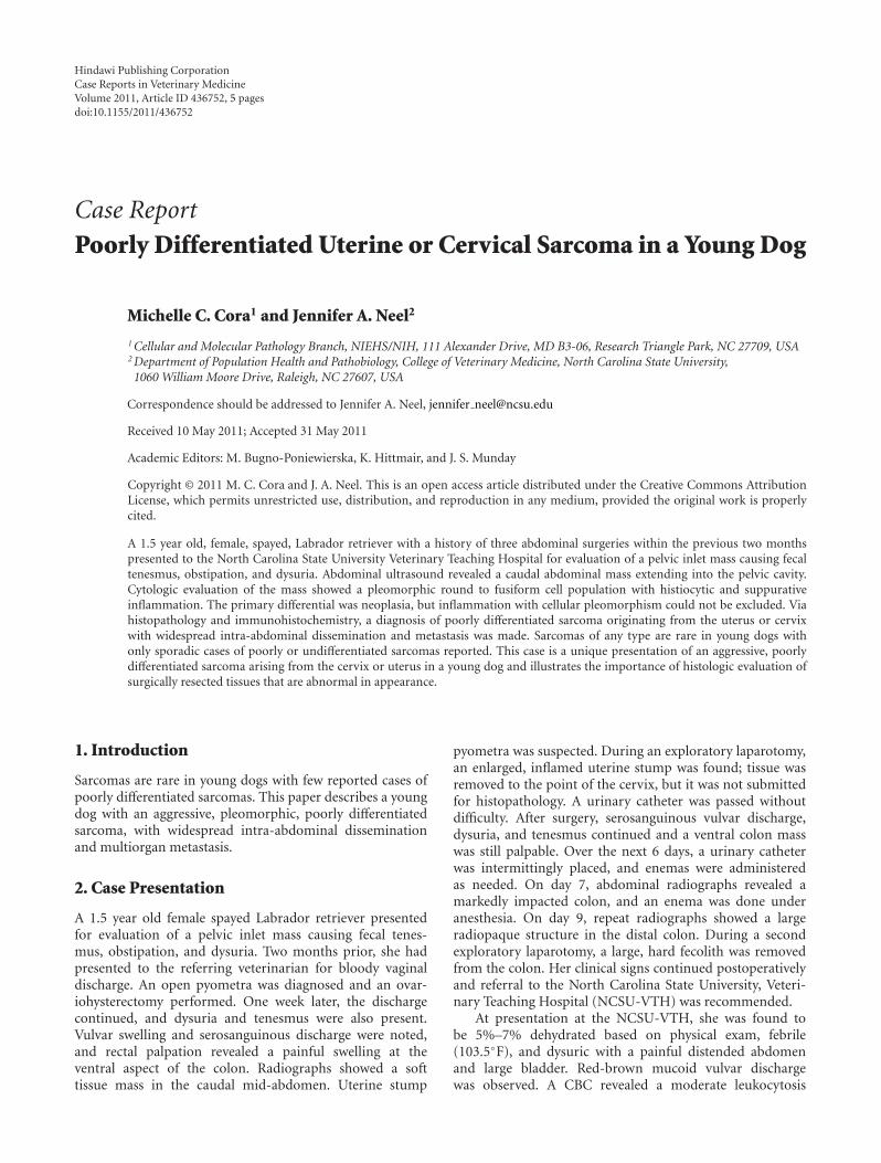
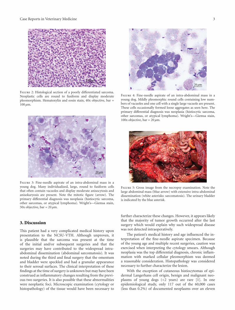
Figure 2: Histological section of a poorly differentiated sarcoma.Neoplastic cells are round to fusiform and display moderatepleomorphism. Hematoxylin and eosin stain, 40x objective, bar =100 µm.
20 µm
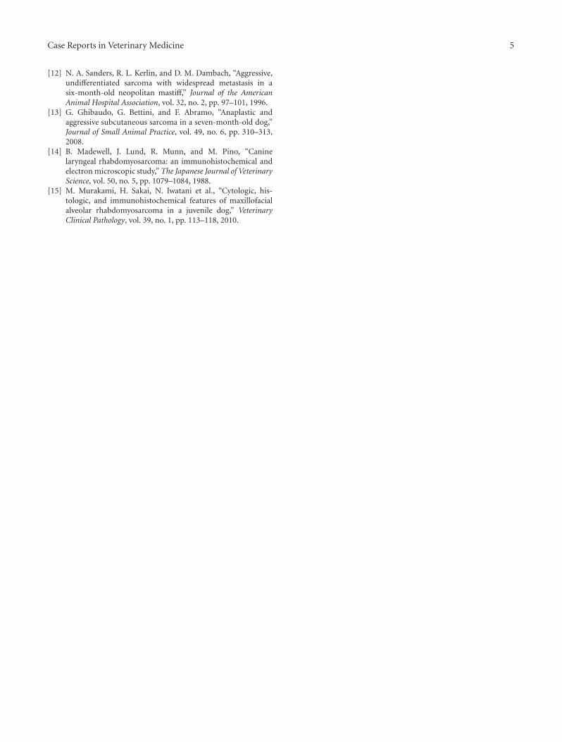
Figure 3: Fine-needle aspirate of an intra-abdominal mass in ayoung dog. Many individualized, large, round to fusiform cellsthat often contain vacuoles and display moderate anisocytosis andanisokaryosis are present. Note the mitotic figure (arrow). Theprimary differential diagnosis was neoplasia (histiocytic sarcoma,other sarcomas, or atypical lymphoma). Wright’s—Giemsa stain,50x objective, bar = 20 µm.
3. Discussion
This patient had a very complicated medical history uponpresentation to the NCSU-VTH. Although unproven, itis plausible that the sarcoma was present at the timeof the initial and/or subsequent surgeries and that thesurgeries may have contributed to the widespread intra-abdominal dissemination (abdominal sarcomatosis). It wasnoted during the third and final surgery that the omentumand bladder were speckled and had a granular appearanceto their serosal surfaces. The clinical interpretation of thesefindings at the time of surgery is unknown but may have beenconstrued as inflammatory changes resulting from the previ-ous two surgeries. It is also possible that these abnormalitieswere neoplastic foci. Microscopic examination (cytology orhistopathology) of the tissue would have been necessary to
20 µm
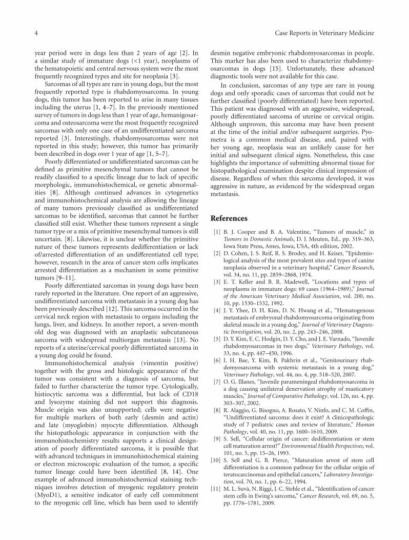
Figure 4: Fine-needle aspirate of an intra-abdominal mass in ayoung dog. Mildly pleomorphic round cells containing low num-bers of vacuoles and one cell with a single large vacuole are present.These cells occasionally formed loose aggregates as seen here. Theprimary differential diagnosis was neoplasia (histiocytic sarcoma,other sarcomas, or atypical lymphoma). Wright’s—Giemsa stain,100x objective, bar = 20 µm.
∗∗
∗∗
Figure 5: Gross image from the necropsy examination. Note thelarge abdominal mass (blue arrow) with extensive intra-abdominaldissemination (white asterisks: sarcomatosis). The urinary bladderis indicated by the blue asterisk.
further characterize these changes. However, it appears likelythat the majority of tumor growth occurred after the lastsurgery which would explain why such widespread diseasewas not detected intraoperatively.
The patient’s medical history and age influenced the in-terpretation of the fine-needle aspirate specimen. Becauseof the young age and multiple recent surgeries, caution wasexercised when interpreting the cytology smears. Althoughneoplasia was the top differential diagnosis, chronic inflam-mation with marked cellular pleomorphism was deemeda reasonable consideration. Histopathology was considerednecessary to further characterize the lesion.
With the exception of cutaneous histiocytomas of epi-dermal Langerhans cell origin, benign and malignant neo-plasms of young dogs (<2 years) are rare [1]. In oneepidemiological study, only 117 out of the 60,000 cases(less than 0.2%) of documented neoplasms over an eleven
4 Case Reports in Veterinary Medicine
year period were in dogs less than 2 years of age [2]. Ina similar study of immature dogs (<1 year), neoplasms ofthe hematopoietic and central nervous system were the mostfrequently recognized types and site for neoplasia [3].
Sarcomas of all types are rare in young dogs, but the mostfrequently reported type is rhabdomyosarcoma. In youngdogs, this tumor has been reported to arise in many tissuesincluding the uterus [1, 4–7]. In the previously mentionedsurvey of tumors in dogs less than 1 year of age, hemanigosar-coma and osteosarcoma were the most frequently recognizedsarcomas with only one case of an undifferentiated sarcomareported [3]. Interestingly, rhabdomyosarcomas were notreported in this study; however, this tumor has primarilybeen described in dogs over 1 year of age [1, 5–7].
Poorly differentiated or undifferentiated sarcomas can bedefined as primitive mesenchymal tumors that cannot bereadily classified to a specific lineage due to lack of specificmorphologic, immunohistochemical, or genetic abnormal-ities [8]. Although continued advances in cytogeneticsand immunohistochemical analysis are allowing the lineageof many tumors previously classified as undifferentiatedsarcomas to be identified, sarcomas that cannot be furtherclassified still exist. Whether these tumors represent a singletumor type or a mix of primitive mesenchymal tumors is stilluncertain. [8]. Likewise, it is unclear whether the primitivenature of these tumors represents dedifferentiation or lackof/arrested differentiation of an undifferentiated cell type;however, research in the area of cancer stem cells implicatesarrested differentiation as a mechanism in some primitivetumors [9–11].
Poorly differentiated sarcomas in young dogs have beenrarely reported in the literature. One report of an aggressive,undifferentiated sarcoma with metastasis in a young dog hasbeen previously described [12]. This sarcoma occurred in thecervical neck region with metastasis to organs including thelungs, liver, and kidneys. In another report, a seven-monthold dog was diagnosed with an anaplastic subcutaneoussarcoma with widespread multiorgan metastasis [13]. Noreports of a uterine/cervical poorly differentiated sarcoma ina young dog could be found.
Immunohistochemical analysis (vimentin positive)together with the gross and histologic appearance of thetumor was consistent with a diagnosis of sarcoma, butfailed to further characterize the tumor type. Cytologically,histiocytic sarcoma was a differential, but lack of CD18and lysozyme staining did not support this diagnosis.Muscle origin was also unsupported; cells were negativefor multiple markers of both early (desmin and actin)and late (myoglobin) myocyte differentiation. Althoughthe histopathologic appearance in conjunction with theimmunohistochemistry results supports a clinical design-ation of poorly differentiated sarcoma, it is possible thatwith advanced techniques in immunohistochemical stainingor electron microscopic evaluation of the tumor, a specifictumor lineage could have been identified [8, 14]. Oneexample of advanced immunohistochemical staining tech-niques involves detection of myogenic regulatory protein(MyoD1), a sensitive indicator of early cell commitmentto the myogenic cell line, which has been used to identify
desmin negative embryonic rhabdomyosarcomas in people.This marker has also been used to characterize rhabdomy-osarcomas in dogs [15]. Unfortunately, these advanceddiagnostic tools were not available for this case.
In conclusion, sarcomas of any type are rare in youngdogs and only sporadic cases of sarcomas that could not befurther classified (poorly differentiated) have been reported.This patient was diagnosed with an aggressive, widespread,poorly differentiated sarcoma of uterine or cervical origin.Although unproven, this sarcoma may have been presentat the time of the initial and/or subsequent surgeries. Pyo-metra is a common medical disease, and, paired withher young age, neoplasia was an unlikely cause for herinitial and subsequent clinical signs. Nonetheless, this casehighlights the importance of submitting abnormal tissue forhistopathological examination despite clinical impression ofdisease. Regardless of when this sarcoma developed, it wasaggressive in nature, as evidenced by the widespread organmetastasis.
References
[1] B. J. Cooper and B. A. Valentine, “Tumors of muscle,” inTumors in Domestic Animals, D. J. Meuten, Ed., pp. 319–363,Iowa State Press, Ames, Iowa, USA, 4th edition, 2002.
[2] D. Cohen, J. S. Reif, R. S. Brodey, and H. Keiser, “Epidemio-logical analysis of the most prevalent sites and types of canineneoplasia observed in a veterinary hospital,” Cancer Research,vol. 34, no. 11, pp. 2859–2868, 1974.
[3] E. T. Keller and B. R. Madewell, “Locations and types ofneoplasms in immature dogs: 69 cases (1964–1989),” Journalof the American Veterinary Medical Association, vol. 200, no.10, pp. 1530–1532, 1992.
[4] J. Y. Yhee, D. H. Kim, D. N. Hwang et al., “Hematogenousmetastasis of embryonal rhabdomyosarcoma originating fromskeletal muscle in a young dog,” Journal of Veterinary Diagnos-tic Investigation, vol. 20, no. 2, pp. 243–246, 2008.
[5] D. Y. Kim, E. C. Hodgin, D. Y. Cho, and J. E. Varnado, “Juvenilerhabdomyosarcomas in two dogs,” Veterinary Pathology, vol.33, no. 4, pp. 447–450, 1996.
[6] I. H. Bae, Y. Kim, B. Pakhrin et al., “Genitourinary rhab-domyosarcoma with systemic metastasis in a young dog,”Veterinary Pathology, vol. 44, no. 4, pp. 518–520, 2007.
[7] O. G. Illanes, “Juvenile parameningeal rhabdomyosarcoma ina dog causing unilateral denervation atrophy of masticatorymuscles,” Journal of Comparative Pathology, vol. 126, no. 4, pp.303–307, 2002.
[8] R. Alaggio, G. Bisogno, A. Rosato, V. Ninfo, and C. M. Coffin,“Undifferentiated sarcoma: does it exist? A clinicopathologicstudy of 7 pediatric cases and review of literature,” HumanPathology, vol. 40, no. 11, pp. 1600–1610, 2009.
[9] S. Sell, “Cellular origin of cancer: dedifferentiation or stemcell maturation arrest?” Environmental Health Perspectives, vol.101, no. 5, pp. 15–26, 1993.
[10] S. Sell and G. B. Pierce, “Maturation arrest of stem celldifferentiation is a common pathway for the cellular origin ofteratocarcinomas and epithelial cancers,” Laboratory Investiga-tion, vol. 70, no. 1, pp. 6–22, 1994.
[11] M. L. Suva, N. Riggi, J. C. Stehle et al., “Identification of cancerstem cells in Ewing’s sarcoma,” Cancer Research, vol. 69, no. 5,pp. 1776–1781, 2009.
Case Reports in Veterinary Medicine 5
[12] N. A. Sanders, R. L. Kerlin, and D. M. Dambach, “Aggressive,undifferentiated sarcoma with widespread metastasis in asix-month-old neopolitan mastiff,” Journal of the AmericanAnimal Hospital Association, vol. 32, no. 2, pp. 97–101, 1996.
[13] G. Ghibaudo, G. Bettini, and F. Abramo, “Anaplastic andaggressive subcutaneous sarcoma in a seven-month-old dog,”Journal of Small Animal Practice, vol. 49, no. 6, pp. 310–313,2008.
[14] B. Madewell, J. Lund, R. Munn, and M. Pino, “Caninelaryngeal rhabdomyosarcoma: an immunohistochemical andelectron microscopic study,” The Japanese Journal of VeterinaryScience, vol. 50, no. 5, pp. 1079–1084, 1988.
[15] M. Murakami, H. Sakai, N. Iwatani et al., “Cytologic, his-tologic, and immunohistochemical features of maxillofacialalveolar rhabdomyosarcoma in a juvenile dog,” VeterinaryClinical Pathology, vol. 39, no. 1, pp. 113–118, 2010.


![Review Article Potential Therapeutic Targets in Uterine ...downloads.hindawi.com/journals/sarcoma/2015/243298.pdf · Sarcoma treatment option [ ]. Interestingly, a randomized phase](https://img.pdfslide.net/doc/110x75/5f8cf06148d0b77022443ee4/review-article-potential-therapeutic-targets-in-uterine-sarcoma-treatment-option.jpg)