Embed Size (px)
Citation preview

www.bio-protocol.org/e2167 Vol 7, Iss 05, Mar 05, 2017 DOI:10.21769/BioProtoc.2167
Copyright © 2017 The Authors; exclusive licensee Bio-protocol LLC. 1
Surface Inoculation and Quantification of Pseudomonas syringae
Population in the Arabidopsis Leaf Apoplast Cristián Jacob1, #, *, Shweta Panchal2, #, * and Maeli Melotto1, *
1Department of Plant Sciences, University of California, Davis, USA; 2Centre for Genome Research,
Faculty of Science, the Maharaja Sayajirao University of Baroda, Baroda, India
*For correspondence: [email protected]; [email protected]; [email protected] #Contributed equally to this work
[Abstract] Bacterial pathogens must enter the plant tissue in order to cause a successful infection.
Foliar bacterial pathogens that are not able to directly penetrate the plant epidermis rely on wounds or
natural openings to internalize leaves. This protocol describes a procedure to estimate the population
size of Pseudomonas syringae in the leaf apoplast after surface inoculation of Arabidopsis rosettes.
Keywords: Leaf inoculation, Stomatal defense, Pseudomonas syringae, Foliar internalization,
Apoplastic bacterial population
[Background] Plant pathogenic bacteria causing foliar diseases may penetrate the leaf epidermis
through wounds and natural openings such as stomata. Stomata are microscopic pores that mediate
the regulation of transpiration and the exchange of gases between the plant and the atmosphere.
Interestingly, we have demonstrated that bacteria can induce stomatal closure. This phenomenon is
now recognized as stomatal defense, which hampers bacterial internalization into the leaf decreasing
disease development (reviewed by Melotto et al., 2017, in press). Here, we describe a method adapted
from Katagiri et al. (2002) and Panchal et al. (2016a and 2016b) to measure the total endophytic
bacterial population of Pseudomonas syringae within Arabidopsis leaf tissue after surface inoculation.
This procedure is useful to estimate bacterial penetration of leaves through stomata in a laboratory
setting.
Materials and Reagents
1. 3.5-inch square pots with holes (Hummert International, catalog number: 12-1300-1)
2. Soil mix, SunGro Sunshine® #1 Mix (Crop Production Services, catalog number: 1000590701)
or equivalent
3. Fine vermiculite (Growers Solution, catalog number: Vermiculite4cf)
4. 48 inch x 100 ft Charcoal fiberglass screen (The Home Depot, catalog number: 3000016)
5. 15 ml and 50 ml centrifuge tubes
6. Plastic domes (Hummert International, catalog number: 65-6964-1)
7. Sharpie markers, paper towels, Kimwipes, disposable gloves, rubber bands, forceps
8. Square Petri dishes (Thermo Fisher Scientific, Thermo ScientificTM, catalog number: 240835)
www.bio-protocol.org/e2167 Vol 7, Iss 05, Mar 05, 2017 DOI:10.21769/BioProtoc.2167
Copyright © 2017 The Authors; exclusive licensee Bio-protocol LLC. 2
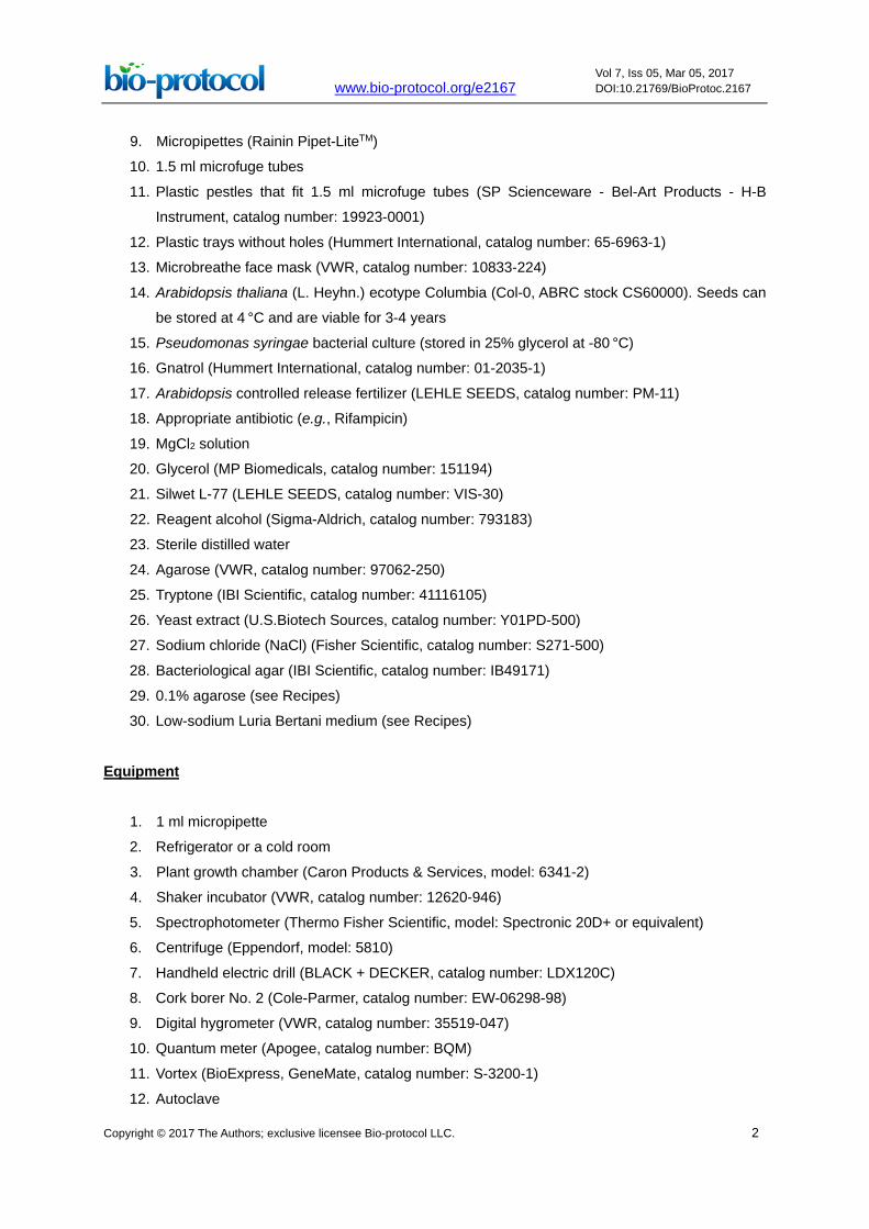
9. Micropipettes (Rainin Pipet-LiteTM)
10. 1.5 ml microfuge tubes
11. Plastic pestles that fit 1.5 ml microfuge tubes (SP Scienceware - Bel-Art Products - H-B
Instrument, catalog number: 19923-0001)
12. Plastic trays without holes (Hummert International, catalog number: 65-6963-1) 13. Microbreathe face mask (VWR, catalog number: 10833-224)
14. Arabidopsis thaliana (L. Heyhn.) ecotype Columbia (Col-0, ABRC stock CS60000). Seeds can
be stored at 4 °C and are viable for 3-4 years
15. Pseudomonas syringae bacterial culture (stored in 25% glycerol at -80 °C)
16. Gnatrol (Hummert International, catalog number: 01-2035-1)
17. Arabidopsis controlled release fertilizer (LEHLE SEEDS, catalog number: PM-11)
18. Appropriate antibiotic (e.g., Rifampicin)
19. MgCl2 solution
20. Glycerol (MP Biomedicals, catalog number: 151194)
21. Silwet L-77 (LEHLE SEEDS, catalog number: VIS-30)
22. Reagent alcohol (Sigma-Aldrich, catalog number: 793183)
23. Sterile distilled water
24. Agarose (VWR, catalog number: 97062-250)
25. Tryptone (IBI Scientific, catalog number: 41116105)
26. Yeast extract (U.S.Biotech Sources, catalog number: Y01PD-500)
27. Sodium chloride (NaCl) (Fisher Scientific, catalog number: S271-500)
28. Bacteriological agar (IBI Scientific, catalog number: IB49171)
29. 0.1% agarose (see Recipes)
30. Low-sodium Luria Bertani medium (see Recipes)
Equipment
1. 1 ml micropipette
2. Refrigerator or a cold room
3. Plant growth chamber (Caron Products & Services, model: 6341-2)
4. Shaker incubator (VWR, catalog number: 12620-946)
5. Spectrophotometer (Thermo Fisher Scientific, model: Spectronic 20D+ or equivalent)
6. Centrifuge (Eppendorf, model: 5810)
7. Handheld electric drill (BLACK + DECKER, catalog number: LDX120C)
8. Cork borer No. 2 (Cole-Parmer, catalog number: EW-06298-98)
9. Digital hygrometer (VWR, catalog number: 35519-047)
10. Quantum meter (Apogee, catalog number: BQM)
11. Vortex (BioExpress, GeneMate, catalog number: S-3200-1)
12. Autoclave

www.bio-protocol.org/e2167 Vol 7, Iss 05, Mar 05, 2017 DOI:10.21769/BioProtoc.2167
Copyright © 2017 The Authors; exclusive licensee Bio-protocol LLC. 3
13. laminar flow hood
Procedure A. Growing Arabidopsis plants
1. Fill pots with the soil mix and top it off with a 2-cm layer of vermiculite (do not pack too tightly).
Cover the pot with 7 x 7-inch fiberglass mesh screen (which prevents the leaves from contacting
soil) and hold it in place with a rubber band.
2. Place the pots in a tray and soak soil with an aqueous solution of Gnatrol (1 g L-1). Let it sit for
one day.
Note: This step is used to avoid development of fungus gnats.
3. Mix Arabidopsis seeds in 5-10 ml of 0.1% agarose in a 15-ml centrifuge tube. Vortex the
suspension for 10 sec at maximum speed to dispense the seeds evenly in agarose prior to
sowing.
4. Using a 1 ml micropipette, spot seeds (~5 seeds in one spot) in the four corners and the center
of the pot. Label each pot with name, date, and genotype of seeds.
5. Cover the tray with a plastic dome, leaving an opening of about 5-10 cm. Keep the tray in a cold
room (4-8 °C) for 2 days to allow for efficient and synchronous germination (Figure 1A).
Figure 1. Dip-inoculation procedure. A. Arabidopsis seeds are sown in a mesh-covered pot
and kept in a cold room (4-8 °C) for 2 days to allow for efficient and synchronous germination.
B. Arabidopsis 4-5 weeks old plants are used for inoculation. C. The rosette is dipped and gently
www.bio-protocol.org/e2167 Vol 7, Iss 05, Mar 05, 2017 DOI:10.21769/BioProtoc.2167
Copyright © 2017 The Authors; exclusive licensee Bio-protocol LLC. 4
swirled in the inoculum. D. Inoculated plants are placed in a tray and incubated in an
environmental chamber.
Note: If a growth chamber without relative humidity control is used, cover the tray with a plastic
dome leaving a 5-10 cm gap open to keep air relative humidity between 60-70%.
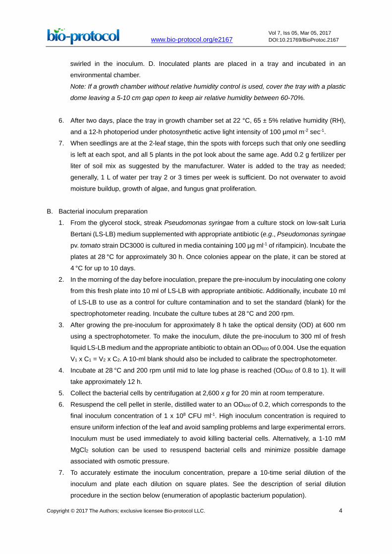
6. After two days, place the tray in growth chamber set at 22 °C, 65 ± 5% relative humidity (RH),
and a 12-h photoperiod under photosynthetic active light intensity of 100 µmol m-2 sec-1.
7. When seedlings are at the 2-leaf stage, thin the spots with forceps such that only one seedling
is left at each spot, and all 5 plants in the pot look about the same age. Add 0.2 g fertilizer per
liter of soil mix as suggested by the manufacturer. Water is added to the tray as needed;
generally, 1 L of water per tray 2 or 3 times per week is sufficient. Do not overwater to avoid
moisture buildup, growth of algae, and fungus gnat proliferation.
B. Bacterial inoculum preparation
1. From the glycerol stock, streak Pseudomonas syringae from a culture stock on low-salt Luria
Bertani (LS-LB) medium supplemented with appropriate antibiotic (e.g., Pseudomonas syringae
pv. tomato strain DC3000 is cultured in media containing 100 µg ml-1 of rifampicin). Incubate the
plates at 28 °C for approximately 30 h. Once colonies appear on the plate, it can be stored at
4 °C for up to 10 days.
2. In the morning of the day before inoculation, prepare the pre-inoculum by inoculating one colony
from this fresh plate into 10 ml of LS-LB with appropriate antibiotic. Additionally, incubate 10 ml
of LS-LB to use as a control for culture contamination and to set the standard (blank) for the
spectrophotometer reading. Incubate the culture tubes at 28 °C and 200 rpm.
3. After growing the pre-inoculum for approximately 8 h take the optical density (OD) at 600 nm
using a spectrophotometer. To make the inoculum, dilute the pre-inoculum to 300 ml of fresh
liquid LS-LB medium and the appropriate antibiotic to obtain an OD600 of 0.004. Use the equation
V1 x C1 = V2 x C2. A 10-ml blank should also be included to calibrate the spectrophotometer.
4. Incubate at 28 °C and 200 rpm until mid to late log phase is reached (OD600 of 0.8 to 1). It will
take approximately 12 h.
5. Collect the bacterial cells by centrifugation at 2,600 x g for 20 min at room temperature.
6. Resuspend the cell pellet in sterile, distilled water to an OD600 of 0.2, which corresponds to the
final inoculum concentration of 1 x 10⁸ CFU ml-1. High inoculum concentration is required to
ensure uniform infection of the leaf and avoid sampling problems and large experimental errors.
Inoculum must be used immediately to avoid killing bacterial cells. Alternatively, a 1-10 mM
MgCl2 solution can be used to resuspend bacterial cells and minimize possible damage
associated with osmotic pressure.
7. To accurately estimate the inoculum concentration, prepare a 10-time serial dilution of the
inoculum and plate each dilution on square plates. See the description of serial dilution
procedure in the section below (enumeration of apoplastic bacterium population).
www.bio-protocol.org/e2167 Vol 7, Iss 05, Mar 05, 2017 DOI:10.21769/BioProtoc.2167
Copyright © 2017 The Authors; exclusive licensee Bio-protocol LLC. 5
8. Add 0.02% Silwet L-77 to the inoculum to ensure that leaves are evenly covered with the
inoculum; otherwise, inoculum will run off and infection will be spotty. This step is required to
guarantee reproducibility of the results.
C. Leaf surface inoculation by dipping
1. Use 4-5-week-old Arabidopsis plants with fully expanded leaves for infection (Figure 1B).
2. Acclimate plants at 25 °C for approximately 12 h before inoculation. Environmental condition,
other than temperature, must be the same as the used for plant growth.
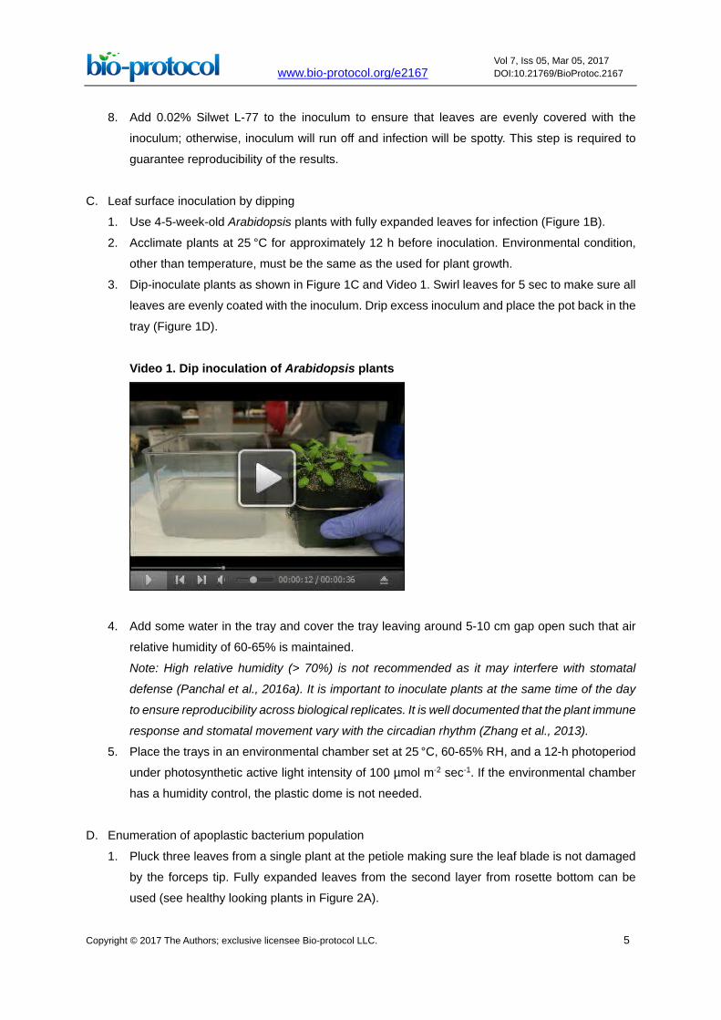
3. Dip-inoculate plants as shown in Figure 1C and Video 1. Swirl leaves for 5 sec to make sure all
leaves are evenly coated with the inoculum. Drip excess inoculum and place the pot back in the
tray (Figure 1D).
Video 1. Dip inoculation of Arabidopsis plants
4. Add some water in the tray and cover the tray leaving around 5-10 cm gap open such that air
relative humidity of 60-65% is maintained.
Note: High relative humidity (> 70%) is not recommended as it may interfere with stomatal
defense (Panchal et al., 2016a). It is important to inoculate plants at the same time of the day
to ensure reproducibility across biological replicates. It is well documented that the plant immune
response and stomatal movement vary with the circadian rhythm (Zhang et al., 2013).
5. Place the trays in an environmental chamber set at 25 °C, 60-65% RH, and a 12-h photoperiod
under photosynthetic active light intensity of 100 µmol m-2 sec-1. If the environmental chamber
has a humidity control, the plastic dome is not needed.
D. Enumeration of apoplastic bacterium population
1. Pluck three leaves from a single plant at the petiole making sure the leaf blade is not damaged
by the forceps tip. Fully expanded leaves from the second layer from rosette bottom can be
used (see healthy looking plants in Figure 2A).

www.bio-protocol.org/e2167 Vol 7, Iss 05, Mar 05, 2017 DOI:10.21769/BioProtoc.2167
Copyright © 2017 The Authors; exclusive licensee Bio-protocol LLC. 6
Note: It is highly recommended to do a time course experiment up to 3 days after inoculation.
Grow all plants in the same batch for each experiment and use different plants from the same
batch for each time point. The time course must include sampling right after inoculation (day 0)
to obtain the initial level of total live bacterial population on the plant. For this first time point,
leaves should not be sterilized as described in step D2. Do not use the younger (top) or older
(bottom) leaves of the rosette, or stressed leaves with curved edges or purpling. For description
of Arabidopsis growth stages, please see Boyes et al. (2001).
Figure 2. Enumeration of the bacterial population in the leaf. A. Three equal sized infected
leaves are plucked and surface sterilized with 70% ethanol. B. Fours leaf disks are cut from
each leaf using a cork borer. C and D. Four leaf disks diluted and 10 μl from each dilution is
spotted twice (technical replicates) on square plates. Repeat B-D for each of the three leaves.
2. Place the leaves in 70% ethanol for 2 min. Next, put the leaves in sterile water for 1 min while
rinsing it lightly from time to time in a way that the leaf blade is not damaged.
3. Dry the leaves by blotting on paper towel.
4. Sterilize the cork borer and forceps at every step from here on by rinsing in 70% ethanol and
then in sterile water. Remove water from the cork borer by blotting on the paper towel.
5. Using the cork borer number 2, which has an area of 0.125 cm2, punch out four leaf discs (total
area of 0.5 cm2), two on either side of the midrib (Figure 2B).
6. Use surface sterilized forceps to collect the punched-out leaf discs and put them in 100 µl sterile
water in a microfuge tube (represented by the first tube in Figure 2C). Grind the tissue sample
using a plastic pestle attached to a hand-held electric drill. No broken leaf pieces should be
visible (see Video 2).

www.bio-protocol.org/e2167 Vol 7, Iss 05, Mar 05, 2017 DOI:10.21769/BioProtoc.2167
Copyright © 2017 The Authors; exclusive licensee Bio-protocol LLC. 7
Video 2. Leaf tissue sampling and homogenization
7. Take 10 µl of this first solution and add to 90 µl of sterile water in another microfuge tube making
a 1:10 dilution. Similarly, serially dilute until 10-6 for day 0 (Figure 2C) or until 10-8 for subsequent
sampling time points.
8. Pipet 10 µl from each tube and spot in one square of a Petri dish containing LS-LB medium with
appropriate antibiotic (see Figure 2D). Each dilution should be spotted twice as technical
replicates (first and second row on the plate shown in Figure 2D). (see Video 3)
9. Repeat steps D5 to D8 (Figures 2B-2D) for each of the three leaves (Figure 2A).
10. After plating all dilutions, let the plate air dry and incubate it at 28 °C for approximately 30 h.
Video 3. Bacterial enumeration using a serial dilution plating method
Data analysis
Count single colonies forming units (CFU) from one of the dilutions (e.g., the fourth or fifth column
on the plate illustrated in Figure 2D). Choose the dilution that yields a 10-100 CFU range. Estimate
the bacterial population by multiplying the CFU by the dilution factor. To express the value as

www.bio-protocol.org/e2167 Vol 7, Iss 05, Mar 05, 2017 DOI:10.21769/BioProtoc.2167
Copyright © 2017 The Authors; exclusive licensee Bio-protocol LLC. 8
CFU/cm2, multiply the total CFU count by 2 as the total area of four leaf discs (Figure 2B) is 0.5 cm2.
For example, if you count 25 colonies in the dilution lane of 10-5 (5th column), then the bacterial
population will be 25 x 105 x 2 = 5 x 10⁶ CFU/cm2. For each sample, there should be three biological
replicates (3 leaves; Figure 2A) with 2 technical replicates (2 spots on the medium for each dilution;
Figure 2D). Statistical analysis should be done by calculating the average (n = 6) and standard error
using Microsoft Excel or any other statistical analysis software. Significance of the difference
between two samples can be obtained by performing the Student’s t-test. Additional biological
replicates must be performed by repeating the whole experiment with other plants to assess the
robustness of the analysis. For scientific rigor, this experimental procedure should be repeated three
times and each time should yield similar bacterial growth trends. See examples of data graphs in
Panchal et al. (2016a).
Notes
1. Be careful not to wound the leaves while picking them with forceps, otherwise the leaves will be
squishy and hard to punch holes. Also, wounding will potentially allow 70% ethanol to enter
inside the leaf tissue, killing the internal bacterial population and thus giving skewed results.
2. For reducing variability in biological replicates, it is extremely crucial that plants are not stressed
while growing and during the infection period. Light, temperature, and relative humidity should
be kept constant. Acclimatize plants for at least 12 h in the same growth chamber where infected
plants are going to be placed.
3. Increasing the number of leaves sampled can reduce standard error.
4. Growing bacterial cultures beyond OD600 of 1.0 does not give ideal infection results.
5. Always use bacterial pathogen from fresh plate streaked from glycerol stock. Multiple sub-
culturing may lead to loss of virulence.
Recipes
1. 0.1% agarose
Dissolve 0.1 g agarose in 100 ml sterile distilled water by heating
Keep swirling the solution intermittently while it cools
Store and use at room temperature
2. Low-sodium Luria Bertani medium
10 g/L tryptone
5 g/L yeast extract
5 g/L NaCl
2.5% agar (only for solid medium)
Adjust pH to 7.0
Autoclave medium at 15 psi, 120 °C for 15 min

www.bio-protocol.org/e2167 Vol 7, Iss 05, Mar 05, 2017 DOI:10.21769/BioProtoc.2167
Copyright © 2017 The Authors; exclusive licensee Bio-protocol LLC. 9
Allow medium to cool down to about 55 °C and add appropriate antibiotic if needed
Pour medium into plates in a laminar flow hood
Store plates in plastic bags at 4 °C to avoid medium dehydration
Acknowledgments
This work was supported by a grant from the US National Institute of Allergy and Infectious Disease
(5R01AI068718) to Dr. Maeli Melotto.
References
1. Boyes, D. C., Zayed, A. M., Ascenzi, R., McCaskill, A. J., Hoffman, N. E., Davis, K. R. and
Görlach, J. (2001). Growth stage-based phenotypic analysis of Arabidopsis: A model for high
throughput functional genomics in plants. Plant Cell 13: 1499-1510.
2. Katagiri, F., Thilmony, R. and He, S. Y. (2002). The Arabidopsis thaliana-Pseudomonas syringae
interaction. Arabidopsis Book 1: e0039.
3. Melotto, M., Zhang, L., Oblessuc, P. R. and He, S. Y. (2017). Stomatal defense a decade later.
Plant Physiol (in press).
4. Panchal, S., Chitrakar, R., Thompson, B. K., Obulareddy, N., Roy, D., Hambright, W. S. and
Melotto, M. (2016a). Regulation of stomatal defense by air relative humidity. Plant Physiol
172(3): 2021-2032.
5. Panchal, S., Roy, D., Chitrakar, R., Price, L., Breitbach, Z. S., Armstrong, D. W. and Melotto, M.
(2016b). Coronatine facilitates Pseudomonas syringae infection of Arabidopsis leaves at night.
Front Plant Sci 7: 880.
6. Zhang, C., Xie, Q., Anderson, R. G. Ng, G., Seitz, N. C., Peterson, T., McClung, C. R., McDowell,
J. M., Kong, D., Kwak, J. M. and Lu, H. (2013). Crosstalk between the circadian clock and innate
immunity in Arabidopsis. PLoS Pathogens 9(6): e1003370.


![Extracellular Vesicles Isolated from the Leaf Apoplast ... · Extracellular Vesicles Isolated from the Leaf Apoplast Carry Stress-Response Proteins1[OPEN] Brian D. Rutter and Roger](https://img.pdfslide.net/doc/110x75/5f0b26077e708231d42f1669/extracellular-vesicles-isolated-from-the-leaf-apoplast-extracellular-vesicles.jpg)


![Soak It Up! [#052 Special]](https://img.pdfslide.net/doc/110x75/568c52a41a28ab4916b77dde/soak-it-up-052-special.jpg)