Embed Size (px)
Citation preview
Print Close Window
Note: Large images and tables on this page may necessitate printing in landscape mode.
Copyright © The McGraw-Hill Companies. All rights reserved. Basic and Clinical Pharmacology > Chapter 33. Agents Used in Anemias; Hematopoietic Growth Factors >
CASE STUDY
A 65-year-old woman with a long-standing history of poorly controlled type 2 diabetes mellitus presents with
increasing numbness and paresthesias in her extremities, generalized weakness, a sore tongue, and gastrointestinal
discomfort. Physical examination reveals a frail-looking, pale woman with diminished vibration sensation, diminished
spinal reflexes, and a positive Babinski sign. Examination of her oral cavity reveals Hunter's glossitis, in which the
tongue appears deep red in color and abnormally smooth and shiny due to atrophy of the lingual papillae. Laboratory
testing reveals a macrocytic anemia based on a hematocrit of 30% (normal for women, 37–48%), a hemoglobin
concentration of 9.4 g/dL (normal for elderly women, 11.7–13.8 g/dL), an erythrocyte mean cell volume (MCV) of
113 fL (normal, 84–99 fL), an erythrocyte mean cell hemoglobin concentration (MCHC) of 34% (normal, 31–36%),
and a low reticulocyte count. Further laboratory testing reveals a normal serum folate concentration and a serum
vitamin B12 (cobalamin) concentration of 98 pg/mL (normal, 250–1100 pg/mL). Results of a Schilling test indicate a
diagnosis of pernicious anemia. Once megaloblastic anemia was identified, why was it important to measure serum concentrations of both folic acid and cobalamin? Should this patient be treated with oral or parenteral vitamin B12?
AGENTS USED IN ANEMIAS; HEMATOPOIETIC GROWTH FACTORS:
INTRODUCTION
Hematopoiesis, the production from undifferentiated stem cells of circulating erythrocytes, platelets, and leukocytes,
is a remarkable process that produces over 200 billion new blood cells per day in the normal person and even greater
numbers of cells in people with conditions that cause loss or destruction of blood cells. The hematopoietic machinery
resides primarily in the bone marrow in adults and requires a constant supply of three essential nutrients—iron, vitamin B12, and folic acid—as well as the presence of hematopoietic growth factors, proteins that regulate the
proliferation and differentiation of hematopoietic cells. Inadequate supplies of either the essential nutrients or the
growth factors result in deficiency of functional blood cells. Anemia, a deficiency in oxygen-carrying erythrocytes, is
the most common and several forms are easily treated. Sickle cell anemia, a condition resulting from a genetic
alteration in the hemoglobin molecule, is common but is not easily treated. It is discussed in the Box: Sickle Cell
Disease and Hydroxyurea. Thrombocytopenia and neutropenia are not rare and some forms are amenable to drug
therapy. In this chapter, we first consider treatment of anemia due to deficiency of iron, vitamin B12 , or folic acid
and then turn to the medical use of hematopoietic growth factors to combat anemia, thrombocytopenia, and
neutropenia, and to support stem cell transplantation.
AGENTS USED IN ANEMIAS
IRON
Basic Pharmacology
Iron deficiency is the most common cause of chronic anemia. Like other forms of chronic anemia, iron deficiency
anemia leads to pallor, fatigue, dizziness, exertional dyspnea, and other generalized symptoms of tissue hypoxia. The
cardiovascular adaptations to chronic anemia—tachycardia, increased cardiac output, vasodilation—can worsen the
condition of patients with underlying cardiovascular disease.
Iron forms the nucleus of the iron-porphyrin heme ring, which together with globin chains forms hemoglobin.
Hemoglobin reversibly binds oxygen and provides the critical mechanism for oxygen delivery from the lungs to other
tissues. In the absence of adequate iron, small erythrocytes with insufficient hemoglobin are formed, giving rise to
microcytic hypochromic anemia.
Pharmacokinetics
Free inorganic iron is extremely toxic, but iron is required for essential proteins such as hemoglobin; therefore,
Page 1 of 23AccessMedicine | Print: Chapter 33. Agents Used in Anemias; Hematopoietic Growth Fac...
2/8/2010http://www.accessmedicine.com.ezp-prod1.hul.harvard.edu/popup.aspx?aID=4513326&pri...

evolution has provided an elaborate system for regulating iron absorption, transport, and storage (Figure 33–1). The
system uses specialized transport, storage, ferroreductase, and ferroxidase proteins whose concentrations are
controlled by the body's demand for hemoglobin synthesis and adequate iron stores (Table 33–1). Nearly all of the
iron used to support hematopoiesis is reclaimed from catalysis of the hemoglobin in senescent or damaged
erythrocytes. Normally, only a small amount of iron is lost from the body each day, so dietary requirements are small
and easily fulfilled by the iron available in a wide variety of foods. However, in special populations with either
increased iron requirements (eg, growing children, pregnant women) or increased losses of iron (eg, menstruating
women), iron requirements can exceed normal dietary supplies and iron deficiency can develop.
Figure 33–1
Absorption, transport, and storage of iron. Intestinal epithelial cells actively absorb inorganic iron and heme iron (H). Ferrous iron that is absorbed or released from absorbed heme iron in the intestine (1) is actively transported into the blood or complexed with apoferritin (AF) and stored as ferritin (F). In the blood, iron is transported by transferrin (Tf) to erythroid precursors in the bone marrow for synthesis of hemoglobin (Hgb) (2) or to hepatocytes for storage as ferritin (3). The transferrin-iron complexes bind to transferrin receptors (TfR) in erythroid precursors and hepatocytes and are internalized. After release of the iron, the TfR-Tf complex is recycled to the plasma membrane and Tf is released. Macrophages that phagocytize senescent erythrocytes (RBC) reclaim the iron from the RBC hemoglobin and either export it or store it as ferritin (4). Hepatocytes use several mechanisms to take up iron and store the iron as ferritin. DMT1, divalent metal transporter; FP, ferroportin; FR, ferrireductase; HCP1, heme carrier protein 1. See text.
Table 33–1 Iron Distribution in Normal Adults.1
Iron Content (mg)
Men Women
Page 2 of 23AccessMedicine | Print: Chapter 33. Agents Used in Anemias; Hematopoietic Growth Fac...
2/8/2010http://www.accessmedicine.com.ezp-prod1.hul.harvard.edu/popup.aspx?aID=4513326&pri...
ABSORPTION
The average diet in the USA contains 10–15 mg of elemental iron daily. A normal individual absorbs 5–10% of this
iron, or about 0.5–1 mg daily. Iron is absorbed in the duodenum and proximal jejunum, although the more distal
small intestine can absorb iron if necessary. Iron absorption increases in response to low iron stores or increased iron
requirements. Total iron absorption increases to 1–2 mg/d in menstruating women and may be as high as 3–4 mg/d
in pregnant women.
Iron is available in a wide variety of foods but is especially abundant in meat. The iron in meat protein can be
efficiently absorbed, because heme iron in meat hemoglobin and myoglobin can be absorbed intact without first
having to be dissociated into elemental iron (Figure 33–1). Iron in other foods, especially vegetables and grains, is
often tightly bound to organic compounds and is much less available for absorption. Nonheme iron in foods and iron
in inorganic iron salts and complexes must be reduced by a ferroreductase to ferrous iron (Fe2+) before it can be absorbed by intestinal mucosal cells.
Iron crosses the luminal membrane of the intestinal mucosal cell by two mechanisms: active transport of ferrous iron
and absorption of iron complexed with heme (Figure 33–1). The divalent metal transporter, DMT1, efficiently
transports ferrous iron across the luminal membrane of the intestinal enterocyte. The rate of iron uptake is regulated
by mucosal cell iron stores such that more iron is transported when stores are low. Together with iron split from
absorbed heme, the newly absorbed iron can be actively transported into the blood across the basolateral membrane
by a transporter known as ferroportin and oxidized to ferric iron (Fe3+) by a ferroxidase. Excess iron can be stored in intestinal epithelial cells as ferritin, a water-soluble complex consisting of a core of ferric hydroxide covered by a shell
of a specialized storage protein called apoferritin. In general, when total body iron stores are high and iron
requirements by the body are low, newly absorbed iron is diverted into ferritin in the intestinal mucosal cells. When
iron stores are low or iron requirements are high, newly absorbed iron is immediately transported from the mucosal
cells to the bone marrow to support hemoglobin production.
TRANSPORT
Iron is transported in the plasma bound to transferrin, a -globulin that specifically binds two molecules of ferric iron (Figure 33–1). The transferrin-iron complex enters maturing erythroid cells by a specific receptor mechanism.
Transferrin receptors—integral membrane glycoproteins present in large numbers on proliferating erythroid cells—
bind and internalize the transferrin-iron complex through the process of receptor-mediated endocytosis. In
endosomes, the ferric iron is released, reduced to ferrous iron, and transported by DMT1 into the cell, where it is
funneled into hemoglobin synthesis or stored as ferritin. The transferrin-transferrin receptor complex is recycled to
the plasma membrane, where the transferrin dissociates and returns to the plasma. This process provides an efficient
mechanism for supplying the iron required by developing red blood cells.
Increased erythropoiesis is associated with an increase in the number of transferrin receptors on developing erythroid
cells. Iron store depletion and iron deficiency anemia are associated with an increased concentration of serum
transferrin.
STORAGE
In addition to the storage of iron in intestinal mucosal cells, iron is also stored, primarily as ferritin, in macrophages
in the liver, spleen, and bone, and in parenchymal liver cells (Figure 33–1). Apoferritin synthesis is regulated by the
levels of free iron. When these levels are low, apoferritin synthesis is inhibited and the balance of iron binding shifts
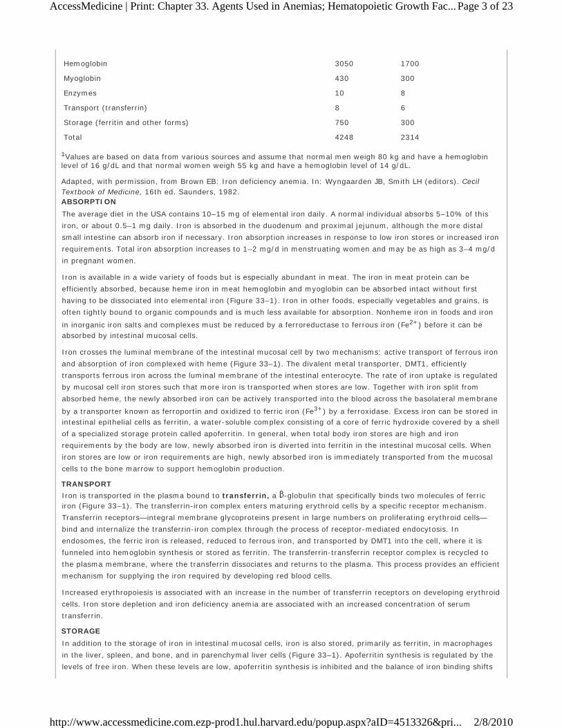
Hemoglobin 3050 1700
Myoglobin 430 300
Enzymes 10 8
Transport (transferrin) 8 6
Storage (ferritin and other forms) 750 300
Total 4248 2314 1Values are based on data from various sources and assume that normal men weigh 80 kg and have a hemoglobin level of 16 g/dL and that normal women weigh 55 kg and have a hemoglobin level of 14 g/dL.
Adapted, with permission, from Brown EB: Iron deficiency anemia. In: Wyngaarden JB, Smith LH (editors). Cecil Textbook of Medicine, 16th ed. Saunders, 1982.
Page 3 of 23AccessMedicine | Print: Chapter 33. Agents Used in Anemias; Hematopoietic Growth Fac...
2/8/2010http://www.accessmedicine.com.ezp-prod1.hul.harvard.edu/popup.aspx?aID=4513326&pri...

toward transferrin. When free iron levels are high, more apoferritin is produced to sequester more iron and protect
organs from the toxic effects of excess free iron.
Ferritin is detectable in serum. Since the ferritin present in serum is in equilibrium with storage ferritin in
reticuloendothelial tissues, the serum ferritin level can be used to estimate total body iron stores.
ELIMINATION
There is no mechanism for excretion of iron. Small amounts are lost in the feces by exfoliation of intestinal mucosal
cells, and trace amounts are excreted in bile, urine, and sweat. These losses account for no more than 1 mg of iron
per day. Because the body's ability to excrete iron is so limited, regulation of iron balance must be achieved by
changing intestinal absorption and storage of iron, in response to the body's needs. As noted below, impaired
regulation of iron absorption leads to serious pathology.
Clinical Pharmacology INDICATIONS FOR THE USE OF IRON
The only clinical indication for the use of iron preparations is the treatment or prevention of iron deficiency anemia.
This manifests as a hypochromic, microcytic anemia in which the erythrocyte mean cell volume (MCV) and the mean
cell hemoglobin concentration are low (Table 33–2). Iron deficiency is commonly seen in populations with increased
iron requirements. These include infants, especially premature infants; children during rapid growth periods;
pregnant and lactating women; and patients with chronic kidney disease who lose erythrocytes at a relatively high
rate during hemodialysis and also form them at a high rate as a result of treatment with the erythrocyte growth
factor erythropoietin (see below). Inadequate iron absorption can also cause iron deficiency. This is seen frequently
after gastrectomy and in patients with severe small bowel disease that results in generalized malabsorption. Iron
deficiency in these gastrointestinal conditions is due to inadequate iron absorption.
The most common cause of iron deficiency in adults is blood loss. Menstruating women lose about 30 mg of iron with
each menstrual period; women with heavy menstrual bleeding may lose much more. Thus, many premenopausal
women have low iron stores or even iron deficiency. In men and postmenopausal women, the most common site of
blood loss is the gastrointestinal tract. Patients with unexplained iron deficiency anemia should be evaluated for
occult gastrointestinal bleeding.
TREATMENT
Iron deficiency anemia is treated with oral or parenteral iron preparations. Oral iron corrects the anemia just as
rapidly and completely as parenteral iron in most cases if iron absorption from the gastrointestinal tract is normal. An
exception is the high requirement for iron of patients with advanced chronic kidney disease who are undergoing
hemodialysis and treatment with erythropoietin; for these patients, parenteral iron administration is preferred.
Oral Iron Therapy
A wide variety of oral iron preparations is available. Because ferrous iron is most efficiently absorbed, only ferrous
salts should be used. Ferrous sulfate, ferrous gluconate, and ferrous fumarate are all effective and inexpensive and
are recommended for the treatment of most patients.
Different iron salts provide different amounts of elemental iron, as shown in Table 33–3. In an iron-deficient
Table 33–2 Distinguishing Features of the Nutritional Anemias.
Nutritional Deficiency
Type of Anemia Laboratory Abnormalities
Iron Microcytic, hypochromic with MCV < 80 fL and MCHC < 30%
Low SI < 30 mcg/dL with increased TIBC, resulting in a % transferrin saturation (SI/TIBC) of < 10%; low serum ferritin level (< 20 mcg/L)
Folic acid Macrocytic, normochromic with MCV > 100 fL and normal or elevated MCHC
Low serum folic acid (< 4 ng/mL)
Vitamin B12 Low serum cobalamin (< 150 pmol/L) accompanied by increased serum homocysteine (> 13 mol/L), and increased serum (> 0.4 mol/L) and urine (> 3.6 mmol/mol creatinine) methylmalonic acid
MCV, mean cell volume; MCHC, mean cell hemoglobin concentration; SI, serum iron; TIBC, transferrin iron-binding capacity.
Page 4 of 23AccessMedicine | Print: Chapter 33. Agents Used in Anemias; Hematopoietic Growth Fac...
2/8/2010http://www.accessmedicine.com.ezp-prod1.hul.harvard.edu/popup.aspx?aID=4513326&pri...

individual, about 50–100 mg of iron can be incorporated into hemoglobin daily, and about 25% of oral iron given as
ferrous salt can be absorbed. Therefore, 200–400 mg of elemental iron should be given daily to correct iron
deficiency most rapidly. Patients unable to tolerate such large doses of iron can be given lower daily doses of iron,
which results in slower but still complete correction of iron deficiency. Treatment with oral iron should be continued
for 3–6 months after correction of the cause of the iron loss. This corrects the anemia and replenishes iron stores.
Common adverse effects of oral iron therapy include nausea, epigastric discomfort, abdominal cramps, constipation,
and diarrhea. These effects are usually dose-related and can often be overcome by lowering the daily dose of iron or
by taking the tablets immediately after or with meals. Some patients have less severe gastrointestinal adverse effects
with one iron salt than another and benefit from changing preparations. Patients taking oral iron develop black stools;
this has no clinical significance in itself but may obscure the diagnosis of continued gastrointestinal blood loss.
Parenteral Iron Therapy
Parenteral therapy should be reserved for patients with documented iron deficiency who are unable to tolerate or
absorb oral iron and for patients with extensive chronic anemia who cannot be maintained with oral iron alone. This
includes patients with advanced chronic renal disease requiring hemodialysis and treatment with erythropoietin,
various postgastrectomy conditions and previous small bowel resection, inflammatory bowel disease involving the
proximal small bowel, and malabsorption syndromes.
The challenge with parenteral iron therapy is that parenteral administration of inorganic free ferric iron produces
serious dose-dependent toxicity, which severely limits the dose of that can be administered. However, when the ferric
iron is formulated as a colloid containing particles with a core of iron oxyhydroxide surrounded by a core of
carbohydrate, bioactive iron is released slowly from the stable colloid particles. In the USA, the three available forms
of parenteral iron are iron dextran, sodium ferric gluconate complex, and iron sucrose.
Iron dextran is a stable complex of ferric oxyhydroxide and dextran polymers containing 50 mg of elemental iron
per milliliter of solution. It can be given by deep intramuscular injection or by intravenous infusion, although the
intravenous route is used most commonly. Intravenous administration eliminates the local pain and tissue staining
that often occur with the intramuscular route and allows delivery of the entire dose of iron necessary to correct the
iron deficiency at one time. Adverse effects of intravenous iron dextran therapy include headache, light-headedness,
fever, arthralgias, nausea and vomiting, back pain, flushing, urticaria, bronchospasm, and, rarely, anaphylaxis and
death. Owing to the risk of a hypersensitivity reaction, a small test dose of iron dextran should always be given
before full intramuscular or intravenous doses are given. Patients with a strong history of allergy and patients who
have previously received parenteral iron dextran are more likely to have hypersensitivity reactions after treatment
with parenteral iron dextran. The iron dextran formulations used clinically are distinguishable as high-molecular-
weight and low-molecular-weight forms. In the USA, the InFeD preparation is a low-molecular-weight form while
DexFerrum is a high-molecular-weight form. Clinical data—primarily from observational studies—indicate that the risk
of anaphylaxis is largely associated with high-molecular-weight iron dextran formulations.
Sodium ferric gluconate complex and iron-sucrose complex are alternative parenteral iron preparations. These
agents can be given only by the intravenous route. They appear to be less likely than high-molecular-weight iron
dextran to cause hypersensitivity reactions.
For patients who are treated chronically with parenteral iron, it is important to monitor iron storage levels to avoid
the serious toxicity associated with iron overload. Unlike oral iron therapy, which is subject to the regulatory
mechanism provided by the intestinal uptake system, parenteral administration, which bypasses this regulatory
Table 33–3 Some Commonly Used Oral Iron Preparations.
Preparation Tablet Size Elemental Iron per Tablet Usual Adult Dosage (Tablets per Day)
Ferrous sulfate, hydrated 325 mg 65 mg 3–4
Ferrous sulfate, desiccated 200 mg 65 mg 3–4
Ferrous gluconate 325 mg 36 mg 3–4
Ferrous fumarate 100 mg 33 mg 6–8
325 mg 106 mg 2–3
Page 5 of 23AccessMedicine | Print: Chapter 33. Agents Used in Anemias; Hematopoietic Growth Fac...
2/8/2010http://www.accessmedicine.com.ezp-prod1.hul.harvard.edu/popup.aspx?aID=4513326&pri...

system, can deliver more iron than can be safely stored. Iron stores can be estimated on the basis of serum
concentrations of ferritin and the transferrin saturation, which is the ratio of the total serum iron concentration to the
total iron-binding capacity (TIBC).
Clinical Toxicity ACUTE IRON TOXICITY
Acute iron toxicity is seen almost exclusively in young children who accidentally ingest iron tablets. Although adults
are able to tolerate large doses of oral iron without serious consequences, as few as 10 tablets of any of the
commonly available oral iron preparations can be lethal in young children. Adult patients taking oral iron preparations
should be instructed to store tablets in child-proof containers out of the reach of children. Children who are poisoned
with oral iron experience necrotizing gastroenteritis, with vomiting, abdominal pain, and bloody diarrhea followed by
shock, lethargy, and dyspnea. Subsequently, improvement is often noted, but this may be followed by severe
metabolic acidosis, coma, and death. Urgent treatment is necessary. Whole bowel irrigation (see Chapter 58)
should be performed to flush out unabsorbed pills. Deferoxamine, a potent iron-chelating compound, can be given
systemically to bind iron that has already been absorbed and to promote its excretion in urine and feces. Activated
charcoal, a highly effective adsorbent for most toxins, does not bind iron and thus is ineffective. Appropriate
supportive therapy for gastrointestinal bleeding, metabolic acidosis, and shock must also be provided.
CHRONIC IRON TOXICITY
Chronic iron toxicity (iron overload), also known as hemochromatosis, results when excess iron is deposited in the
heart, liver, pancreas, and other organs. It can lead to organ failure and death. It most commonly occurs in patients
with inherited hemochromatosis, a disorder characterized by excessive iron absorption, and in patients who receive
many red cell transfusions over a long period of time (eg, patients with thalassemia major).
Chronic iron overload in the absence of anemia is most efficiently treated by intermittent phlebotomy. One unit of
blood can be removed every week or so until all of the excess iron is removed. Iron chelation therapy using
parenteral deferoxamine is much less efficient as well as more complicated, expensive, and hazardous, but it may be
the only option for iron overload that cannot be managed by phlebotomy, such as the iron overload experienced by
patients with thalassemia major.
The oral iron chelator deferasirox is approved for treatment of iron overload. Deferasirox appears to be as effective
as deferoxamine at reducing liver iron concentrations and is much more convenient. However, it is not clear yet
whether deferasirox is as effective as deferoxamine at protecting the heart from iron overload.
VITAMIN B12
Vitamin B12 (cobalamin) serves as a cofactor for several essential biochemical reactions in humans. Deficiency of vitamin B12 leads to megaloblastic anemia (Table 33–2), gastrointestinal symptoms, and neurologic abnormalities.
Although deficiency of vitamin B12 due to an inadequate supply in the diet is unusual, deficiency of B12 in adults—
especially older adults—due to inadequate absorption of dietary vitamin B12 is a relatively common and easily treated
disorder.
Chemistry
Vitamin B12 consists of a porphyrin-like ring with a central cobalt atom attached to a nucleotide. Various organic
groups may be covalently bound to the cobalt atom, forming different cobalamins. Deoxyadenosylcobalamin and
methylcobalamin are the active forms of the vitamin in humans. Cyanocobalamin and hydroxocobalamin (both
available for therapeutic use) and other cobalamins found in food sources are converted to the active forms. The ultimate source of vitamin B12 is from microbial synthesis; the vitamin is not synthesized by animals or plants. The
chief dietary source of vitamin B12 is microbially derived vitamin B12 in meat (especially liver), eggs, and dairy
products. Vitamin B12 is sometimes called extrinsic factor to differentiate it from intrinsic factor, a protein
normally secreted by the stomach that is required for gastrointestinal uptake of dietary vitamin B12.
Pharmacokinetics
The average diet in the USA contains 5–30 mcg of vitamin B12 daily, 1–5 mcg of which is usually absorbed. The vitamin is avidly stored, primarily in the liver, with an average adult having a total vitamin B12 storage pool of 3000–5000 mcg. Only trace amounts of vitamin B12 are normally lost in urine and stool. Because the normal daily
Page 6 of 23AccessMedicine | Print: Chapter 33. Agents Used in Anemias; Hematopoietic Growth Fac...
2/8/2010http://www.accessmedicine.com.ezp-prod1.hul.harvard.edu/popup.aspx?aID=4513326&pri...

requirements of vitamin B12 are only about 2 mcg, it would take about 5 years for all of the stored vitamin B12 to be
exhausted and for megaloblastic anemia to develop if B12 absorption were stopped. Vitamin B12 in physiologic
amounts is absorbed only after it complexes with intrinsic factor, a glycoprotein secreted by the parietal cells of the gastric mucosa. Intrinsic factor combines with the vitamin B12 that is liberated from dietary sources in the stomach
and duodenum, and the intrinsic factor-vitamin B12 complex is subsequently absorbed in the distal ileum by a highly
selective receptor-mediated transport system. Vitamin B12 deficiency in humans most often results from
malabsorption of vitamin B12 due either to lack of intrinsic factor or to loss or malfunction of the specific absorptive
mechanism in the distal ileum. Nutritional deficiency is rare but may be seen in strict vegetarians after many years
without meat, eggs, or dairy products.
Once absorbed, vitamin B12 is transported to the various cells of the body bound to a family of specialized glycoproteins, transcobalamin I, II, and III. Excess vitamin B12 is transported to the liver for storage.
Pharmacodynamics
Two essential enzymatic reactions in humans require vitamin B12 (Figure 33–2). In one, methylcobalamin serves as
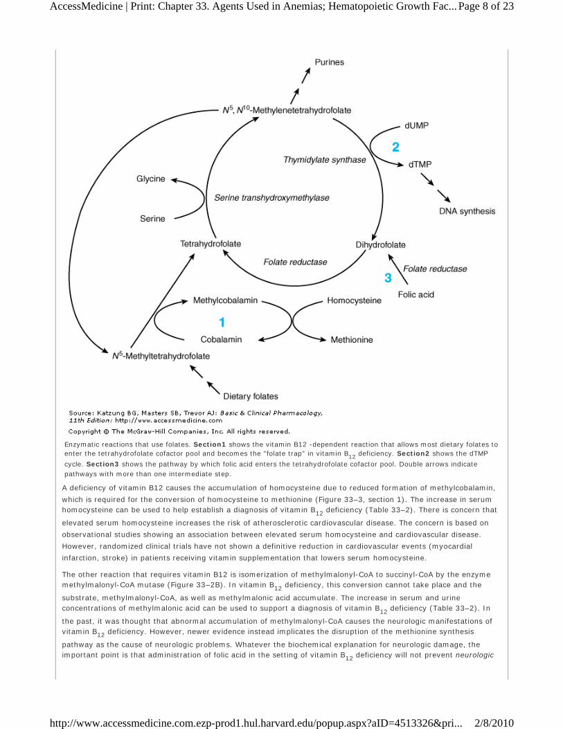
an intermediate in the transfer of a methyl group from N5-methyltetrahydrofolate to homocysteine, forming methionine (Figure 33–2A; Figure 33–3, section 1). Without vitamin B12, conversion of the major dietary and storage
folate, N5-methyltetrahydrofolate, to tetrahydrofolate, the precursor of folate cofactors, cannot occur. As a result, a deficiency of folate cofactors necessary for several biochemical reactions involving the transfer of one-carbon groups
develops. In particular, the depletion of tetrahydrofolate prevents synthesis of adequate supplies of the
deoxythymidylate (dTMP) and purines required for DNA synthesis in rapidly dividing cells, as shown in Figure 33–3,
section 2. The accumulation of folate as N5-methyltetrahydrofolate and the associated depletion of tetrahydrofolate cofactors in vitamin B12 deficiency have been referred to as the "methylfolate trap." This is the biochemical step
whereby vitamin B12 and folic acid metabolism are linked, and it explains why the megaloblastic anemia of vitamin
B12 deficiency can be partially corrected by ingestion of relatively large amounts of folic acid. Folic acid can be
reduced to dihydrofolate by the enzyme dihydrofolate reductase (Figure 33–3, section 3) and thus serve as a source
of the tetrahydrofolate required for synthesis of the purines and dTMP that are needed for DNA synthesis.
Figure 33–2
Enzymatic reactions that use vitamin B12 . See text for details.
Figure 33–3
Page 7 of 23AccessMedicine | Print: Chapter 33. Agents Used in Anemias; Hematopoietic Growth Fac...
2/8/2010http://www.accessmedicine.com.ezp-prod1.hul.harvard.edu/popup.aspx?aID=4513326&pri...
A deficiency of vitamin B12 causes the accumulation of homocysteine due to reduced formation of methylcobalamin,
which is required for the conversion of homocysteine to methionine (Figure 33–3, section 1). The increase in serum homocysteine can be used to help establish a diagnosis of vitamin B12 deficiency (Table 33–2). There is concern that
elevated serum homocysteine increases the risk of atherosclerotic cardiovascular disease. The concern is based on
observational studies showing an association between elevated serum homocysteine and cardiovascular disease.
However, randomized clinical trials have not shown a definitive reduction in cardiovascular events (myocardial
infarction, stroke) in patients receiving vitamin supplementation that lowers serum homocysteine.
The other reaction that requires vitamin B12 is isomerization of methylmalonyl-CoA to succinyl-CoA by the enzyme methylmalonyl-CoA mutase (Figure 33–2B). In vitamin B12 deficiency, this conversion cannot take place and the
substrate, methylmalonyl-CoA, as well as methylmalonic acid accumulate. The increase in serum and urine concentrations of methylmalonic acid can be used to support a diagnosis of vitamin B12 deficiency (Table 33–2). In
the past, it was thought that abnormal accumulation of methylmalonyl-CoA causes the neurologic manifestations of vitamin B12 deficiency. However, newer evidence instead implicates the disruption of the methionine synthesis
pathway as the cause of neurologic problems. Whatever the biochemical explanation for neurologic damage, the important point is that administration of folic acid in the setting of vitamin B12 deficiency will not prevent neurologic
Enzymatic reactions that use folates. Section1 shows the vitamin B12 -dependent reaction that allows most dietary folates to enter the tetrahydrofolate cofactor pool and becomes the "folate trap" in vitamin B12 deficiency. Section2 shows the dTMP
cycle. Section3 shows the pathway by which folic acid enters the tetrahydrofolate cofactor pool. Double arrows indicate pathways with more than one intermediate step.
Page 8 of 23AccessMedicine | Print: Chapter 33. Agents Used in Anemias; Hematopoietic Growth Fac...
2/8/2010http://www.accessmedicine.com.ezp-prod1.hul.harvard.edu/popup.aspx?aID=4513326&pri...

manifestations even though it will largely correct the anemia caused by the vitamin B12 deficiency.
Clinical Pharmacology
Vitamin B12 is used to treat or prevent deficiency. The most characteristic clinical manifestation of vitamin B12 deficiency is megaloblastic, macrocytic anemia (Table 33–2), often with associated mild or moderate leukopenia or
thrombocytopenia (or both), and a characteristic hypercellular bone marrow with an accumulation of megaloblastic erythroid and other precursor cells. The neurologic syndrome associated with vitamin B12 deficiency usually begins
with paresthesias in peripheral nerves and weakness and progresses to spasticity, ataxia, and other central nervous system dysfunctions. Correction of vitamin B12 deficiency arrests the progression of neurologic disease, but it may
not fully reverse neurologic symptoms that have been present for several months. Although most patients with neurologic abnormalities caused by vitamin B12 deficiency have megaloblastic anemia when first seen, occasional
patients have few if any hematologic abnormalities.
Once a diagnosis of megaloblastic anemia is made, it must be determined whether vitamin B12 or folic acid deficiency
is the cause. (Other causes of megaloblastic anemia are very rare.) This can usually be accomplished by measuring
serum levels of the vitamins. The Schilling test, which measures absorption and urinary excretion of radioactively labeled vitamin B12, can be used to further define the mechanism of vitamin B12 malabsorption when this is found to
be the cause of the megaloblastic anemia.
The most common causes of vitamin B12 deficiency are pernicious anemia, partial or total gastrectomy, and
conditions that affect the distal ileum, such as malabsorption syndromes, inflammatory bowel disease, or small bowel
resection.
Pernicious anemia results from defective secretion of intrinsic factor by the gastric mucosal cells. Patients with
pernicious anemia have gastric atrophy and fail to secrete intrinsic factor (as well as hydrochloric acid). The Schilling
test shows diminished absorption of radioactively labeled vitamin B12 , which is corrected when intrinsic factor is administered with radioactive B12, since the vitamin can then be normally absorbed.
Vitamin B12 deficiency also occurs when the region of the distal ileum that absorbs the vitamin B12-intrinsic factor complex is damaged, as when the ileum is involved with inflammatory bowel disease or when the ileum is surgically resected. In these situations, radioactively labeled vitamin B12 is not absorbed in the Schilling test, even when
intrinsic factor is added. Other rare causes of vitamin B12 deficiency include bacterial overgrowth of the small bowel,
chronic pancreatitis, and thyroid disease. Rare cases of vitamin B12 deficiency in children have been found to be
secondary to congenital deficiency of intrinsic factor or to defects of the receptor sites for vitamin B12-intrinsic factor
complex located in the distal ileum.
Almost all cases of vitamin B12 deficiency are caused by malabsorption of the vitamin; therefore, parenteral injections of vitamin B12 are required for therapy. For patients with potentially reversible diseases, the underlying
disease should be treated after initial treatment with parenteral vitamin B12. Most patients, however, do not have
curable deficiency syndromes and require lifelong treatment with vitamin B12.
Vitamin B12 for parenteral injection is available as cyanocobalamin or hydroxocobalamin. Hydroxocobalamin is
preferred because it is more highly protein-bound and therefore remains longer in the circulation. Initial therapy should consist of 100–1000 mcg of vitamin B12 intramuscularly daily or every other day for 1–2 weeks to replenish
body stores. Maintenance therapy consists of 100–1000 mcg intramuscularly once a month for life. If neurologic
abnormalities are present, maintenance therapy injections should be given every 1–2 weeks for 6 months before switching to monthly injections. Oral vitamin B12-intrinsic factor mixtures and liver extracts should not be used to
treat vitamin B12 deficiency; however, oral doses of 1000 mcg of vitamin B12 daily are usually sufficient to treat
patients with pernicious anemia who refuse or cannot tolerate the injections. After pernicious anemia is in remission following parenteral vitamin B12 therapy, the vitamin can be administered intranasally as a spray or gel.
FOLIC ACID
Reduced forms of folic acid are required for essential biochemical reactions that provide precursors for the synthesis
of amino acids, purines, and DNA. Folate deficiency is not uncommon, even though the deficiency is easily corrected
by administration of folic acid. The consequences of folate deficiency go beyond the problem of anemia because folate
deficiency is implicated as a cause of congenital malformations in newborns and may play a role in vascular disease
Page 9 of 23AccessMedicine | Print: Chapter 33. Agents Used in Anemias; Hematopoietic Growth Fac...
2/8/2010http://www.accessmedicine.com.ezp-prod1.hul.harvard.edu/popup.aspx?aID=4513326&pri...

(see Folic Acid Supplementation: A Public Health Dilemma).
Chemistry
Folic acid (pteroylglutamic acid) is composed of a heterocycle (pteridine), p-aminobenzoic acid, and glutamic acid
(Figure 33–4). Various numbers of glutamic acid moieties may be attached to the pteroyl portion of the molecule,
resulting in monoglutamates, triglutamates, or polyglutamates. Folic acid can undergo reduction, catalyzed by the
enzyme dihydrofolate reductase ("folate reductase"), to give dihydrofolic acid (Figure 33–3, section 3).
Tetrahydrofolate can subsequently be transformed to folate cofactors possessing one-carbon units attached to the 5-
nitrogen, to the 10-nitrogen, or to both positions (Figure 33–3). The folate cofactors are interconvertible by various
enzymatic reactions and serve the important biochemical function of donating one-carbon units at various levels of
oxidation. In most of these, tetrahydrofolate is regenerated and becomes available for reutilization.
Folic Acid Supplementation: A Public Health Dilemma
Starting in January 1998, all products made from enriched grains in the USA were required to be
supplemented with folic acid. This FDA ruling was issued to reduce the incidence of congenital neural tube
defects (NTDs). Epidemiologic studies show a strong correlation between maternal folic acid deficiency and
the incidence of NTDs such as spina bifida and anencephaly. The FDA requirement for folic acid
supplementation is a public health measure aimed at the significant number of women in the USA who do not
receive prenatal care and are not aware of the importance of adequate folic acid ingestion for preventing
birth defects in their infants. Observational studies from the USA and from other countries that supplement
grains with folic acid have found that supplementation is associated with a significant (30–75%) reduction in
NTD rates. These studies indicate that the reduction in NTDs is dose-dependent and that supplementation of
grains in the USA with higher levels of folic acid could result in an even greater reduction in the rate of NTDs.
Observational studies also suggest that rates of other types of congenital anomalies (heart and orofacial)
have fallen after supplementation began.
There may be an added benefit for adults. N5-Methyltetrahydrofolate is required for the conversion of
homocysteine to methionine (Figure 33–2; Figure 33–3, reaction 1). Impaired synthesis of N5-methyltetrahydrofolate results in elevated serum concentrations of homocysteine. Data from several sources
suggest a positive correlation between elevated serum homocysteine and occlusive vascular diseases such as
ischemic heart disease and stroke. Clinical data suggest that the folate supplementation program has
improved the folate status and reduced the prevalence of hyperhomocysteinemia in a population of middle-
aged and older adults who did not use vitamin supplements. It is possible, although the evidence thus far has
been negative, that the increased ingestion of folic acid will also reduce the risk of vascular disease in this
population.
Although the potential benefits of supplemental folic acid during pregnancy are compelling, the decision to
require folic acid in grains was controversial. As described in the text, ingestion of folic acid can partially or
totally correct the anemia caused by vitamin B12 deficiency. However, folic acid supplementation does not prevent the potentially irreversible neurologic damage caused by vitamin B12 deficiency. People with
pernicious anemia and other forms of vitamin B12 deficiency are usually identified because of signs and
symptoms of anemia, which typically occur before neurologic symptoms. The opponents of folic acid
supplementation were concerned that increased folic acid intake in the general population would mask vitamin B12 deficiency and increase the prevalence of neurologic disease in the elderly population. To put this
in perspective, approximately 4000 pregnancies, including 2500 live births, in the USA each year are affected
by neural tube defects. In contrast, it is estimated that over 10% of the elderly population in the USA, or several million people, are at risk for the neuropsychiatric complications of vitamin B12 deficiency. In
acknowledgment of this controversy, the FDA kept its requirements for folic acid supplementation at a
somewhat low level. There now is evidence that the current level of folic acid supplementation in the USA has not masked a significant amount of vitamin B12-associated anemia, and there is some discussion about
increasing the amount of folic acid supplementation of grains in an effort to further reduce the rates of NTDs.
Figure 33–4
Page 10 of 23AccessMedicine | Print: Chapter 33. Agents Used in Anemias; Hematopoietic Growth ...
2/8/2010http://www.accessmedicine.com.ezp-prod1.hul.harvard.edu/popup.aspx?aID=4513326&pri...

Pharmacokinetics
The average diet in the USA contains 500–700 mcg of folates daily, 50–200 mcg of which is usually absorbed,
depending on metabolic requirements. Pregnant women may absorb as much as 300–400 mcg of folic acid daily.
Various forms of folic acid are present in a wide variety of plant and animal tissues; the richest sources are yeast,
liver, kidney, and green vegetables. Normally, 5–20 mg of folates are stored in the liver and other tissues. Folates
are excreted in the urine and stool and are also destroyed by catabolism, so serum levels fall within a few days when
intake is diminished. Because body stores of folates are relatively low and daily requirements high, folic acid
deficiency and megaloblastic anemia can develop within 1–6 months after the intake of folic acid stops, depending on
the patient's nutritional status and the rate of folate utilization.
Unaltered folic acid is readily and completely absorbed in the proximal jejunum. Dietary folates, however, consist
primarily of polyglutamate forms of N5-methyltetrahydrofolate. Before absorption, all but one of the glutamyl residues of the polyglutamates must be hydrolyzed by the enzyme -1-glutamyl transferase ("conjugase") within the
brush border of the intestinal mucosa. The monoglutamate N5-methyltetrahydrofolate is subsequently transported into the bloodstream by both active and passive transport and is then widely distributed throughout the body. Inside
cells, N5-methyltetrahydrofolate is converted to tetrahydrofolate by the demethylation reaction that requires vitamin B12 (Figure 33–3, section 1).
Pharmacodynamics
Tetrahydrofolate cofactors participate in one-carbon transfer reactions. As described earlier in the discussion of
vitamin B12 , one of these essential reactions produces the dTMP needed for DNA synthesis. In this reaction, the
enzyme thymidylate synthase catalyzes the transfer of the one-carbon unit of N5,N10-methylenetetrahydrofolate to deoxyuridine monophosphate (dUMP) to form dTMP (Figure 33–3, section 2). Unlike all the other enzymatic reactions
that use folate cofactors, in this reaction the cofactor is oxidized to dihydrofolate, and for each mole of dTMP
produced, 1 mole of tetrahydrofolate is consumed. In rapidly proliferating tissues, considerable amounts of
tetrahydrofolate are consumed in this reaction, and continued DNA synthesis requires continued regeneration of
tetrahydrofolate by reduction of dihydrofolate, catalyzed by the enzyme dihydrofolate reductase. The tetrahydrofolate
thus produced can then reform the cofactor N5,N10-methylenetetrahydrofolate by the action of serine transhydroxymethylase and thus allow for the continued synthesis of dTMP. The combined catalytic activities of dTMP
synthase, dihydrofolate reductase, and serine transhydroxymethylase are referred to as the dTMP synthesis cycle.
Enzymes in the dTMP cycle are the targets of two anticancer drugs; methotrexate inhibits dihydrofolate reductase,
and a metabolite of 5-fluorouracil inhibits thymidylate synthase (see Chapter 54).
Cofactors of tetrahydrofolate participate in several other essential reactions. N5-Methylenetetrahydrofolate is required for the vitamin B12 -dependent reaction that generates methionine from homocysteine (Figure 33–2A; Figure 33–3,
The structure of folic acid.
(Reproduced, with permission, from Murray RK et al: Harper's Biochemistry, 24th ed. McGraw-Hill, 1996.)
Page 11 of 23AccessMedicine | Print: Chapter 33. Agents Used in Anemias; Hematopoietic Growth ...
2/8/2010http://www.accessmedicine.com.ezp-prod1.hul.harvard.edu/popup.aspx?aID=4513326&pri...

section 1). In addition, tetrahydrofolate cofactors donate one-carbon units during the de novo synthesis of essential
purines. In these reactions, tetrahydrofolate is regenerated and can reenter the tetrahydrofolate cofactor pool.
Clinical Pharmacology
Folate deficiency results in a megaloblastic anemia that is microscopically indistinguishable from the anemia caused
by vitamin B12 deficiency (see above). However, folate deficiency does not cause the characteristic neurologic syndrome seen in vitamin B12 deficiency. In patients with megaloblastic anemia, folate status is assessed with assays
for serum folate or for red blood cell folate. Red blood cell folate levels are often of greater diagnostic value than
serum levels, because serum folate levels tend to be labile and do not necessarily reflect tissue levels.
Folic acid deficiency, unlike vitamin B12 deficiency, is often caused by inadequate dietary intake of folates. Patients
with alcohol dependence and patients with liver disease can develop folic acid deficiency because of poor diet and
diminished hepatic storage of folates. Pregnant women and patients with hemolytic anemia have increased folate
requirements and may become folic acid-deficient, especially if their diets are marginal. Evidence implicates maternal
folic acid deficiency in the occurrence of fetal neural tube defects, eg, spina bifida. (See Folic Acid Supplementation: A
Public Health Dilemma.) Patients with malabsorption syndromes also frequently develop folic acid deficiency. Patients
who require renal dialysis develop folic acid deficiency because folates are removed from the plasma during the
dialysis procedure.
Folic acid deficiency can be caused by drugs. Methotrexate and, to a lesser extent, trimethoprim and pyrimethamine,
inhibit dihydrofolate reductase and may result in a deficiency of folate cofactors and ultimately in megaloblastic
anemia. Long-term therapy with phenytoin can also cause folate deficiency, but only rarely causes megaloblastic
anemia.
Parenteral administration of folic acid is rarely necessary, since oral folic acid is well absorbed even in patients with
malabsorption syndromes. A dose of 1 mg folic acid orally daily is sufficient to reverse megaloblastic anemia, restore
normal serum folate levels, and replenish body stores of folates in almost all patients. Therapy should be continued
until the underlying cause of the deficiency is removed or corrected. Therapy may be required indefinitely for patients
with malabsorption or dietary inadequacy. Folic acid supplementation to prevent folic acid deficiency should be
considered in high-risk patients, including pregnant women, patients with alcohol dependence, hemolytic anemia,
liver disease, or certain skin diseases, and patients on renal dialysis.
Sickle Cell Disease and Hydroxyurea
Sickle cell disease is an important genetic cause of hemolytic anemia, a form of anemia due to increased
erythrocyte destruction, instead of the reduced mature erythrocyte production seen with iron, folic acid, and vitamin B12 deficiency. Patients with sickle cell disease are homozygous for the aberrant -hemoglobin S (HbS) allele or heterozygous for HbS and a second mutated -hemoglobin gene such as hemoglobin C (HbC) or -thalassemia. Sickle cell disease has an increased prevalence in individuals of African descent presumably because the heterozygous trait confers resistance to malaria.
In the majority of patients with sickle cell disease, anemia is not the major problem; the anemia is generally
well compensated even though such individuals have a chronically low hematocrit (20–30%), a low serum
hemoglobin level (7–10 g/dL), and an elevated reticulocyte count. Instead, the primary problem is that
deoxygenated HbS chains form polymeric structures that dramatically change erythrocyte shape, reduce
deformability, and elicit membrane permeability changes that further promote hemoglobin polymerization.
Abnormal erythrocytes aggregate in the microvasculature—where oxygen tension is low and hemoglobin is
deoxygenated—and cause veno-occlusive damage. The clinical manifestations of sickle cell disease reflect
organ damage by veno-occlusive events. In the musculoskeletal system, this results in characteristic,
extremely painful bone and joint pain. In the cerebral vascular system, it causes ischemic stroke. Damage to
the spleen increases the risk of infection, particularly by encapsulated bacteria such as Streptococcus
pneumoniae. In the pulmonary system, there is an increased risk of infection and, in adults, an increase in
embolism and pulmonary hypertension. In the male genitourinary system, priapism can occur. Supportive
treatment includes analgesics, antibiotics, pneumococcal vaccination, and blood transfusions. In addition, the
cancer chemotherapeutic drug hydroxyurea (hydroxycarbamide) reduces veno-occlusive events. It is
approved in the USA for treatment of adults with recurrent sickle cell crises and approved in Europe in adults
and children with recurrent vaso-occlusive events. As an anticancer drug used in the treatment of chronic
Page 12 of 23AccessMedicine | Print: Chapter 33. Agents Used in Anemias; Hematopoietic Growth ...
2/8/2010http://www.accessmedicine.com.ezp-prod1.hul.harvard.edu/popup.aspx?aID=4513326&pri...

and acute myelogenous leukemia, hydroxyurea inhibits ribonucleotide reductase and thereby depletes
deoxynucleoside triphosphate and arrests cells in the S phase of the cell cycle (see Chapter 54). In the
treatment of sickle cell disease, hydroxyurea acts through poorly defined pathways to increase the production of fetal hemoglobin (HbF), which interferes with the polymerization of HbS. Clinical trials have shown that hydroxyurea decreases painful crises in adults and children with severe sickle cell disease. Its adverse effects
include hematopoietic depression, gastrointestinal effects, and teratogenicity in pregnant women.
HEMATOPOIETIC GROWTH FACTORS
The hematopoietic growth factors are glycoprotein hormones that regulate the proliferation and differentiation of
hematopoietic progenitor cells in the bone marrow. The first growth factors to be identified were called colony-
stimulating factors because they could stimulate the growth of colonies of various bone marrow progenitor cells in
vitro. Many of these growth factors have been purified and cloned, and their effects on hematopoiesis have been
extensively studied. Quantities of these growth factors sufficient for clinical use are produced by recombinant DNA
technology.
Of the known hematopoietic growth factors, erythropoietin (epoetin alfa and epoetin beta), granulocyte
colony-stimulating factor (G-CSF), granulocyte-macrophage colony-stimulating factor (GM-CSF), and
interleukin-11 (IL-11) are currently in clinical use. Romiplostim (AMG-531) is a novel biologic agent that
activates the thrombopoietin receptor.
The hematopoietic growth factors and drugs that mimic their action have complex effects on the function of a wide
variety of cell types, including nonhematologic cells. Their usefulness in other areas of medicine, particularly as
potential anticancer and anti-inflammatory drugs, is being investigated.
ERYTHROPOIETIN
Chemistry & Pharmacokinetics
Erythropoietin, a 34–39 kDa glycoprotein, was the first human hematopoietic growth factor to be isolated. It was
originally purified from the urine of patients with severe anemia. Recombinant human erythropoietin (rHuEPO,
epoetin alfa) is produced in a mammalian cell expression system. After intravenous administration, erythropoietin has
a serum half-life of 4–13 hours in patients with chronic renal failure. It is not cleared by dialysis. It is measured in
international units (IU). Darbepoetin alfa is a modified form of erythropoietin that is more heavily glycosylated as a
result of changes in amino acids. Darbepoetin alfa has a twofold to threefold longer half-life than epoetin alfa.
Methoxy polyethylene glycol epoetin beta is an isoform of erythropoietin covalently attached to a long polyethylene
glycol polymer. This long-lived recombinant product is administered as a single intravenous or subcutaneous dose at
2-week or monthly intervals whereas epoetin alfa is generally administered three times a week and darbepoetin is
administered weekly.
Pharmacodynamics
Erythropoietin stimulates erythroid proliferation and differentiation by interacting with erythropoietin receptors on red
cell progenitors. The erythropoietin receptor is a member of the JAK/STAT superfamily of cytokine receptors that use
protein phosphorylation and transcription factor activation to regulate cellular function (see Chapter 2). Erythropoietin
also induces release of reticulocytes from the bone marrow. Endogenous erythropoietin is primarily produced in the
kidney. In response to tissue hypoxia, more erythropoietin is produced through an increased rate of transcription of
the erythropoietin gene. This results in correction of the anemia, provided that the bone marrow response is not
impaired by red cell nutritional deficiency (especially iron deficiency), primary bone marrow disorders (see below), or
bone marrow suppression from drugs or chronic diseases.
Normally, an inverse relationship exists between the hematocrit or hemoglobin level and the serum erythropoietin
level. Nonanemic individuals have serum erythropoietin levels of less than 20 IU/L. As the hematocrit and hemoglobin
levels fall and anemia becomes more severe, the serum erythropoietin level rises exponentially. Patients with
moderately severe anemia usually have erythropoietin levels in the 100–500 IU/L range, and patients with severe
anemia may have levels of thousands of IU/L. The most important exception to this inverse relationship is in the
anemia of chronic renal failure. In patients with renal disease, erythropoietin levels are usually low because the
kidneys cannot produce the growth factor. These are the patients most likely to respond to treatment with exogenous
erythropoietin. In most primary bone marrow disorders (aplastic anemia, leukemias, myeloproliferative and
Page 13 of 23AccessMedicine | Print: Chapter 33. Agents Used in Anemias; Hematopoietic Growth ...
2/8/2010http://www.accessmedicine.com.ezp-prod1.hul.harvard.edu/popup.aspx?aID=4513326&pri...

myelodysplastic disorders, etc) and most nutritional and secondary anemias, endogenous erythropoietin levels are
high, so there is less likelihood of a response to exogenous erythropoietin (but see below).
Clinical Pharmacology
The availability of erythropoiesis-stimulating agents (ESAs) has had a significant positive impact for patients with
several different types of anemia (Table 33–4). The ESAs consistently improve the hematocrit and hemoglobin level,
often eliminate the need for transfusions, and reliably improve quality of life indices. The ESAs are used routinely in
patients with anemia secondary to chronic kidney disease. In patients treated with an ESA, an increase in reticulocyte
count is usually observed in about 10 days and an increase in hematocrit and hemoglobin levels in 2–6 weeks.
Dosages of ESAs are adjusted to maintain a target hemoglobin up to, but not exceeding, 10–12 g/dL. To support the
increased erythropoiesis, nearly all patients with chronic kidney disease will require oral or parenteral iron
supplementation. Folate supplementation may also be necessary in some patients.
In selected patients, erythropoietin is also useful for the treatment of anemia due to primary bone marrow
disorders and secondary anemias. This includes patients with aplastic anemia and other bone marrow failure
states, myeloproliferative and myelodysplastic disorders, multiple myeloma and perhaps other chronic bone marrow
malignancies, and the anemias associated with chronic inflammation, AIDS, and myelosuppressive cancer
chemotherapy. Patients with these disorders who have disproportionately low serum erythropoietin levels for their
degree of anemia are most likely to respond to treatment with this growth factor. Patients with endogenous
erythropoietin levels of less than 100 IU/L have the best chance of response, although patients with erythropoietin
levels between 100 and 500 IU/L respond occasionally. These patients generally require higher erythropoietin doses
to achieve a response, and responses are often incomplete. Methoxy polyethylene glycol epoetin beta should not be
used for treatment of anemia caused by cancer chemotherapy because a clinical trial found significantly more deaths
among patients receiving this form of erythropoietin.
Table 33–4 Clinical Uses of Hematopoietic Growth Factors and Agents That Mimic Their Actions.
Hematopoietic Growth Factor Clinical Condition Being Treated or Prevented
Recipients
Erythropoietin, darbepoetin alfa Anemia Patients with chronic renal failure
HIV-infected patients treated with zidovudine
Cancer patients treated with myelosuppressive cancer chemotherapy
Patients scheduled to undergo elective, noncardiac, nonvascular surgery
Granulocyte colony-stimulating factor (G-CSF; filgrastim)
Neutropenia Cancer patients treated with myelosuppressive cancer chemotherapy
Granulocyte-macrophage colony-stimulating factor (GM-CSF; sargramostim)
Patients with severe chronic neutropenia
Stem cell transplantation Patients with nonmyeloid malignancies treated with stem cell transplantation
Mobilization of peripheral blood progenitor cells (PBPCs)
Mobilization of peripheral blood progenitor cells (PBPCs)
Patients with nonmyeloid malignancies
Donors of stem cells for allogeneic or autologous transplantation
Interleukin-11 (IL-11, oprelvekin) Thrombocytopenia Patients with nonmyeloid malignancies who receive myelosuppressive cancer chemotherapy
Romiplostim Thrombocytopenia Patients with idiopathic thrombocytopenic purpura
Page 14 of 23AccessMedicine | Print: Chapter 33. Agents Used in Anemias; Hematopoietic Growth ...
2/8/2010http://www.accessmedicine.com.ezp-prod1.hul.harvard.edu/popup.aspx?aID=4513326&pri...

Erythropoietin has been used successfully to offset the anemia produced by zidovudine treatment in patients with HIV
infection and in the treatment of the anemia of prematurity. It can also be used to reduce the need for transfusion in
high-risk patients undergoing elective, noncardiac, nonvascular surgery; to accelerate erythropoiesis after
phlebotomies for autologous transfusion for elective surgery; or for treatment of iron overload (hemochromatosis).
Erythropoietin is one of the drugs banned by the International Olympic Committee. The use of erythropoietin by
athletes is based on their hope that increased red blood cell concentration will increase oxygen delivery to muscles
and improve performance.
Toxicity
The most common adverse effects of erythropoietin are hypertension and thrombotic complications. In March 2007,
the FDA issued a warning that patients with chronic renal failure or cancer whose serum hemoglobin is raised to more
than 12 g/dL with an ESA face a greater risk of a thrombotic event or, in patients with advanced head and neck
cancers, faster tumor growth. The warning was primarily based on clinical trial data from patients with chronic kidney
disease indicating an increased rate of mortality and cardiovascular events (stroke, myocardial infarction, worsening
congestive heart failure, and hypertension) in patients dosed with an ESA to a target hemoglobin level of 12–16 g/dL
or dosed to maintain a normal hematocrit (42%) versus a lower target hematocrit of 30%. In addition, a meta-
analysis of 51 placebo-controlled trials of ESAs in cancer patients reported an increased rate of all-cause mortality
and venous thrombosis in those receiving an ESA. Based on the accumulated evidence, it is recommended that the
hemoglobin level not exceed 12 g/dL in patients with chronic kidney disease receiving an ESA, and that ESAs be used
conservatively in cancer patients (eg, when hemoglobin levels are < 10 g/dL) and with the lowest dose needed to
avoid transfusion.
Allergic reactions to ESAs have been infrequent. There have been a small number of cases of pure red cell aplasia
(PRCA) accompanied by neutralizing antibodies to erythropoietin. PRCA was most commonly seen in dialysis patients
treated subcutaneously for a long period with a particular form of epoetin alfa (Eprex with a polysorbate 80 stabilizer
rather than human serum albumin) that is not available in the USA. After regulatory agencies required that Eprex be
administered intravenously rather than subcutaneously, the rate of ESA-associated PRCA diminished. However, rare
cases have still been seen with all ESAs administered subcutaneously for long periods to patients with chronic kidney
disease.
MYELOID GROWTH FACTORS
Chemistry & Pharmacokinetics
G-CSF and GM-CSF, the two myeloid growth factors currently available for clinical use, were originally purified from
cultured human cell lines (Table 33–4). Recombinant human G-CSF (rHuG-CSF; filgrastim ) is produced in a
bacterial expression system. It is a nonglycosylated peptide of 175 amino acids, with a molecular weight of 18 kDa.
Recombinant human GM-CSF ( rHuGM-CSF; sargramostim ) is produced in a yeast expression system. It is a
partially glycosylated peptide of 127 amino acids, with three molecular species with molecular weights of 15,500;
15,800; and 19,500. These preparations have serum half-lives of 2–7 hours after intravenous or subcutaneous
administration. Pegfilgrastim, a covalent conjugation product of filgrastim and a form of polyethylene glycol, has a
much longer serum half-life than recombinant G-CSF, and it can be injected once per myelosuppressive
chemotherapy cycle instead of daily for several days.
Pharmacodynamics
The myeloid growth factors stimulate proliferation and differentiation by interacting with specific receptors found on
various myeloid progenitor cells. Like the erythropoietin receptor, these receptors are members of the JAK/STAT
superfamily (see Chapter 2). G-CSF stimulates proliferation and differentiation of progenitors already committed to
the neutrophil lineage. It also activates the phagocytic activity of mature neutrophils and prolongs their survival in
the circulation. G-CSF also has a remarkable ability to mobilize hematopoietic stem cells, ie, to increase their
concentration in peripheral blood. This biologic effect underlies a major advance in transplantation—the use of
peripheral blood stem cells (PBSCs) rather than bone marrow stem cells for autologous and allogeneic
hematopoietic stem cell transplantation (see below).
GM-CSF has broader biologic actions than G-CSF. It is a multipotential hematopoietic growth factor that stimulates
proliferation and differentiation of early and late granulocytic progenitor cells as well as erythroid and megakaryocyte
Page 15 of 23AccessMedicine | Print: Chapter 33. Agents Used in Anemias; Hematopoietic Growth ...
2/8/2010http://www.accessmedicine.com.ezp-prod1.hul.harvard.edu/popup.aspx?aID=4513326&pri...

progenitors. Like G-CSF, GM-CSF also stimulates the function of mature neutrophils. GM-CSF acts together with
interleukin-2 to stimulate T-cell proliferation and appears to be a locally active factor at the site of inflammation. GM-
CSF mobilizes peripheral blood stem cells, but it is significantly less efficacious than G-CSF in this regard.
Clinical Pharmacology CANCER CHEMOTHERAPY-INDUCED NEUTROPENIA
Neutropenia is a common adverse effect of the cytotoxic drugs used to treat cancer and increases the risk of serious
infection in patients receiving chemotherapy. Unlike the treatment of anemia and thrombocytopenia, transfusion of
neutropenic patients with granulocytes collected from donors is performed rarely and with limited success. The
introduction of G-CSF in 1991 represented a milestone in the treatment of chemotherapy-induced neutropenia. This
growth factor dramatically accelerates the rate of neutrophil recovery after dose-intensive myelosuppressive
chemotherapy (Figure 33–5). It reduces the duration of neutropenia and usually raises the nadir count, the lowest
neutrophil count seen following a cycle of chemotherapy.
The ability of G-CSF to increase neutrophil counts after myelosuppressive chemotherapy is nearly universal, but its
impact on clinical outcomes is more variable. Many, but not all, clinical trials and meta-analyses have shown that G-
CSF reduces episodes of febrile neutropenia, requirements for broad-spectrum antibiotics, infections, and days of
hospitalization. Clinical trials have not shown improved survival in cancer patients treated with G-CSF. Clinical
guidelines for the use of G-CSF after cytotoxic chemotherapy recommend reserving G-CSF for patients at high risk for
febrile neutropenia based on age, medical history, and disease characteristics; patients receiving dose-intensive
chemotherapy regimens that carry a greater than 40% risk of causing febrile neutropenia; patients with a prior
episode of febrile neutropenia after cytotoxic chemotherapy; patients at high risk for febrile neutropenia; and patients
who are unlikely to survive an episode of febrile neutropenia. Pegfilgrastim is an alternative to G-CSF for prevention
of chemotherapy-induced febrile neutropenia. Pegfilgrastim can be administered less frequently, and it may shorten
the period of severe neutropenia slightly more than G-CSF.
Like G-CSF and pegfilgrastim, GM-CSF also reduces the duration of neutropenia after cytotoxic chemotherapy. It has
been more difficult to show that GM-CSF reduces the incidence of febrile neutropenia, probably because GM-CSF itself
can induce fever. In the treatment of chemotherapy-induced neutropenia, G-CSF, 5 mcg/kg/d, or GM-CSF, 250
mcg/m2/d, is usually started within 24–72 hours after completing chemotherapy and is continued until the absolute neutrophil count is greater than 10,000 cells/ L. Pegfilgrastim is given as a single dose instead of daily injections.
The utility and safety of the myeloid growth factors in the postchemotherapy supportive care of patients with acute
myeloid leukemia (AML) have been the subject of a number of clinical trials. Because leukemic cells arise from
Figure 33–5
Effects of granulocyte colony-stimulating factor (G-CSF; red line) or placebo (green line) on absolute neutrophil count (ANC) after cytotoxic chemotherapy for lung cancer. Doses of chemotherapeutic drugs were administered on days 1 and 3. G-CSF or placebo injections were started on day 4 and continued daily through day 12 or 16. The first peak in ANC reflects the recruitment of mature cells by G-CSF. The second peak reflects a marked increase in new neutrophil production by the bone
marrow under stimulation by G-CSF. (Normal ANC is 2.2–8.6 x 10⁹/L.)
(Modified and reproduced, with permission, from Crawford et al: Reduction by granulocyte colony-stimulating factor of fever and neutropenia induced by chemotherapy in patients with small-cell lung cancer. N Engl J Med 1991;325:164.)
Page 16 of 23AccessMedicine | Print: Chapter 33. Agents Used in Anemias; Hematopoietic Growth ...
2/8/2010http://www.accessmedicine.com.ezp-prod1.hul.harvard.edu/popup.aspx?aID=4513326&pri...

progenitors whose proliferation and differentiation are normally regulated by hematopoietic growth factors, including
GM-CSF and G-CSF, there was concern that myeloid growth factors could stimulate leukemic cell growth and increase
the rate of relapse. The results of randomized clinical trials suggest that both G-CSF and GM-CSF are safe following
induction and consolidation treatment of myeloid and lymphoblastic leukemia. There has been no evidence that these
growth factors reduce the rate of remission or increase relapse rate. On the contrary, the growth factors accelerate
neutrophil recovery and reduce infection rates and days of hospitalization. Both G-CSF and GM-CSF have FDA
approval for treatment of patients with AML.
OTHER APPLICATIONS
G-CSF and GM-CSF have also proved to be effective in treating the neutropenia associated with congenital
neutropenia, cyclic neutropenia, myelodysplasia, and aplastic anemia. Many patients with these disorders
respond with a prompt and sometimes dramatic increase in neutrophil count. In some cases, this results in a
decrease in the frequency of infections. Because neither G-CSF nor GM-CSF stimulates the formation of erythrocytes
and platelets, they are sometimes combined with other growth factors for treatment of pancytopenia.
The myeloid growth factors play an important role in autologous stem cell transplantation for patients
undergoing high-dose chemotherapy. High-dose chemotherapy with autologous stem cell support is increasingly used
to treat patients with tumors that are resistant to standard doses of chemotherapeutic drugs. The high-dose regimens
produce extreme myelosuppression; the myelosuppression is then counteracted by reinfusion of the patient's own
hematopoietic stem cells (which are collected prior to chemotherapy). The administration of G-CSF or GM-CSF early
after autologous stem cell transplantation has been shown to reduce the time to engraftment and to recovery from
neutropenia in patients receiving stem cells obtained either from bone marrow or from peripheral blood. These effects
are seen in patients being treated for lymphoma or for solid tumors. G-CSF and GM-CSF are also used to support
patients who have received allogeneic bone marrow transplantation for treatment of hematologicmalignancies or
bone marrow failure states. In this setting, the growth factors speed the recovery from neutropenia without
increasing the incidence of acute graft-versus-host disease.
Perhaps the most important role of the myeloid growth factors in transplantation is for mobilization of PBSCs. Stem
cells collected from peripheral blood have nearly replaced bone marrow as the hematopoietic preparation used for
autologous transplantation, and the use of PBSCs for allogeneic transplantation is also being investigated. The cells
can be collected in an outpatient setting with a procedure that avoids much of the risk and discomfort of bone
marrow collection, including the need for general anesthesia. In addition, there is evidence that PBSC transplantation
results in more rapid engraftment of all hematopoietic cell lineages and in reduced rates of graft failure or delayed
platelet recovery.
G-CSF is the cytokine most commonly used for PBSC mobilization because of its increased efficacy and reduced
toxicity compared with GM-CSF. To mobilize stem cells, patients or donors are given 5–10 mcg/kg/d subcutaneously
for 4 days. On the fifth day, they undergo leukapheresis. The success of PBSC transplantation depends on transfusion
of adequate numbers of stem cells. CD34, an antigen present on early progenitor cells and absent from later,
committed, cells, is used as a marker for the requisite stem cells. The goal is to reinfuse at least 5 x 10⁶ CD34 cells/kg; this number of CD34 cells usually results in prompt and durable engraftment of all cell lineages. It can take
several separate leukaphereses to collect enough CD34 cells, especially from older patients and patients who have
been exposed to radiation therapy or chemotherapy.
Toxicity
Although the three growth factors have similar effects on neutrophil counts, G-CSF and pegfilgrastim are used more
frequently than GM-CSF because they are is better tolerated. G-CSF and pegfilgrastim can cause bone pain, which
clears when the drugs are discontinued. GM-CSF can cause more severe side effects, particularly at higher doses.
These include fever, malaise, arthralgias, myalgias, and a capillary leak syndrome characterized by peripheral edema
and pleural or pericardial effusions. Allergic reactions may occur but are infrequent. Splenic rupture is a rare but
serious complication of the use of G-CSF for PBSC.
MEGAKARYOCYTE GROWTH FACTORS
Patients with thrombocytopenia have a high risk of hemorrhage. Although platelet transfusion is commonly used to
treat thrombocytopenia, this procedure can cause adverse reactions in the recipient; furthermore, a significant
Page 17 of 23AccessMedicine | Print: Chapter 33. Agents Used in Anemias; Hematopoietic Growth ...
2/8/2010http://www.accessmedicine.com.ezp-prod1.hul.harvard.edu/popup.aspx?aID=4513326&pri...

number of patients fail to exhibit the expected increase in platelet count. Thrombopoietin and IL-11 both appear to
be key endogenous regulators of platelet production. A recombinant form of IL-11 was the first agent to gain FDA
approval for treatment of thrombocytopenia. Recombinant human thrombopoietin and a pegylated form of a
shortened human thrombopoietin protein underwent extensive clinical investigation in the 1990s. However, further
development was abandoned after autoantibodies to the native thrombopoietin formed in healthy human subjects
and caused thrombocytopenia. Efforts shifted to investigation of novel, nonimmunogenic peptide agonists of the
thrombopoietin receptor, which is known as Mpl. The first of these—romiplostim—was approved by the FDA for
idiopathic thrombocytopenic purpura in 2008.
Chemistry & Pharmacokinetics
Interleukin-11 is a 65–85 kDa protein produced by fibroblasts and stromal cells in the bone marrow. Oprelvekin,
the recombinant form of IL-11 approved for clinical use (Table 33–4), is produced by expression in Escherichia coli.
The half-life of IL-11 is 7–8 hours when the drug is injected subcutaneously.
Romiplostim (AMG 531) is a member of new class of therapeutics called "peptibodies," which are peptides with key
biologic activities covalently linked to antibody fragments that serve to extend the peptide's half-life. Romiplostim contains two disulfide-bonded human Fc fragments, each covalently attached through a polyglycine sequence to a
peptide chain containing two Mpl-binding peptides that are linked to one another by a second polyglycine sequence.
The Mpl-binding peptide was selected from a peptide library based on its ability in cell assays to activate the
thrombopoietin receptor. The Mpl-binding peptide has no sequence homology with human thrombopoietin and there
is no evidence in animal or human studies that the Mpl-binding peptide or romiplostim induces antibodies to
thrombopoietin. After subcutaneous administration, romiplostim is eliminated by the reticuloendothelial system with
an average half-life of 3–4 days. Its half-life is inversely related to the serum platelet count; it has a longer half-life in
patients with thrombocytopenia and a shorter half-life in patients whose platelet counts have recovered to normal
levels.
Eltrombopag is a new orally active small molecule agonist at the thrombopoietin receptor licensed for use in
idiopathic thrombocytopenia. Because of toxicity concerns, eltrombopag is restricted to use by registered physicians
and patients.
Pharmacodynamics
Interleukin-11 acts through a specific cell surface cytokine receptor to stimulate the growth of multiple lymphoid and
myeloid cells. It acts synergistically with other growth factors to stimulate the growth of primitive megakaryocytic
progenitors and, most importantly, increases the number of peripheral platelets and neutrophils.
Romiplostim has high affinity for the human Mpl receptor. It causes a dose-dependent increase in platelet count that
begins on day 5 after subcutaneous administration and peaks at days 12–15.
Clinical Pharmacology
Interleukin-11 is approved for the secondary prevention of thrombocytopenia in patients receiving cytotoxic
chemotherapy for treatment of nonmyeloid cancers. Clinical trials show that it reduces the number of platelet
transfusions required by patients who experience severe thrombocytopenia after a previous cycle of chemotherapy.
Although IL-11 has broad stimulatory effects on hematopoietic cell lineages in vitro, it does not appear to have
significant effects on the leukopenia caused by myelosuppressive chemotherapy. Interleukin-11 is given by
subcutaneous injection at a dose of 50 mcg/kg/d. It is started 6–24 hours after completion of chemotherapy and continued for 14–21 days or until the platelet count passes the nadir and rises to more than 50,000 cells/ L.
In patients with chronic idiopathic thrombocytopenia (ITP) who failed to respond adequately to previous treatment
with steroids, immunoglobulins, or splenectomy, romiplostim significantly increased platelet count in most patients.
In a 6-week placebo-controlled study in which patients were treated weekly with 1 or 3 mcg/kg, 12 of 16 patients
reached the targeted platelet range of 50,000–450,000 platelets/mL. Romiplostim does not appear to decrease the
rate of platelet destruction in ITP as platelet counts returned to pretreatment levels after the drug's discontinuation.
An open label trial found that many patients maintained a platelet count of 100,000 platelets/mL or higher over a 48-
week period and that over half of the patients were able to discontinue other therapies. Romiplostim is initiated as a
weekly subcutaneous dose of 1 mcg/kg and then continued at the lowest dose required to maintain a platelet count
of at least 50,000 platelets/mL.
Page 18 of 23AccessMedicine | Print: Chapter 33. Agents Used in Anemias; Hematopoietic Growth ...
2/8/2010http://www.accessmedicine.com.ezp-prod1.hul.harvard.edu/popup.aspx?aID=4513326&pri...

Toxicity
The most common adverse effects of IL-11 are fatigue, headache, dizziness, and cardiovascular effects. The
cardiovascular effects include anemia (due to hemodilution), dyspnea (due to fluid accumulation in the lungs), and
transient atrial arrhythmias. Hypokalemia has also been seen in some patients. All of these adverse effects appear to
be reversible.
Romiplostim appears to be well tolerated except for a mild headache on the day of administration. A potential long-
term concern is that two patients treated with romiplostim had an increase in bone marrow reticulin, a possible
marker of myelodysplastic or myeloproliferative processes. However, neither patient had evidence of increased
collagenfibrosis or of abnormal bone marrow cytogenetics.
SUMMARY: AGENTS USED IN ANEMIAS AND HEMATOPOIETIC GROWTH
FACTORS
Agents Used in Anemias and Hematopoietic Growth Factors
Subclass Mechanism of Action
Effects Clinical Applications Pharmacokinetics, Toxicities, Interactions
Iron
Ferrous sulfate Required for the biosynthesis of heme and heme-containing proteins, including hemoglobin and myoglobin
Adequate supplies are required for normal heme synthesis deficiency results in inadequate heme production
Treatment of iron deficiency, which manifests as microcytic anemia
Complicated endogenous system for absorbing, storing, and transporting iron no mechanism for iron excretion other than cell and blood loss Toxicity: Acute overdose
results in necrotizing gastroenteritis, abdominal pain, bloody diarrhea, shock, lethargy, and dyspnea chronic iron overload results in hemochromatosis, with damage to the heart, liver, pancreas, and other organs organ failure and death can ensue
Ferrous gluconate and ferrous fumarate: Oral iron preparations
Iron dextran, iron sucrose complex, and sodium ferric gluconate complex: Parenteral preparations; can cause hypersensitivity reactions
Iron chelators
Deferoxamine (see also Chapters 58 and 59)
Chelates excess iron
Reduces the toxicity associated with acute or chronic iron overload
Treatment of acute iron poisoning and for inherited or acquired hemochromatosis that is not adequately treated by phlebotomy
Preferred route of administration is IM or SC Toxicity: Rapid IV administration may cause hypotension acute respiratory distress has been observed with long infusions neurotoxicity and increased susceptibility to certain infections has occurred with long-term use
Deferasirox: Orally administered iron chelator for treatment of hemochromatosis that is not adequately treated by phlebotomy
Vitamin B12
Cyanocobalamin A cofactor required for essential
Adequate supplies are required for
Treatment of vitamin B12 deficiency, which
Parenteral vitamin B12 is required for pernicious
Page 19 of 23AccessMedicine | Print: Chapter 33. Agents Used in Anemias; Hematopoietic Growth ...
2/8/2010http://www.accessmedicine.com.ezp-prod1.hul.harvard.edu/popup.aspx?aID=4513326&pri...
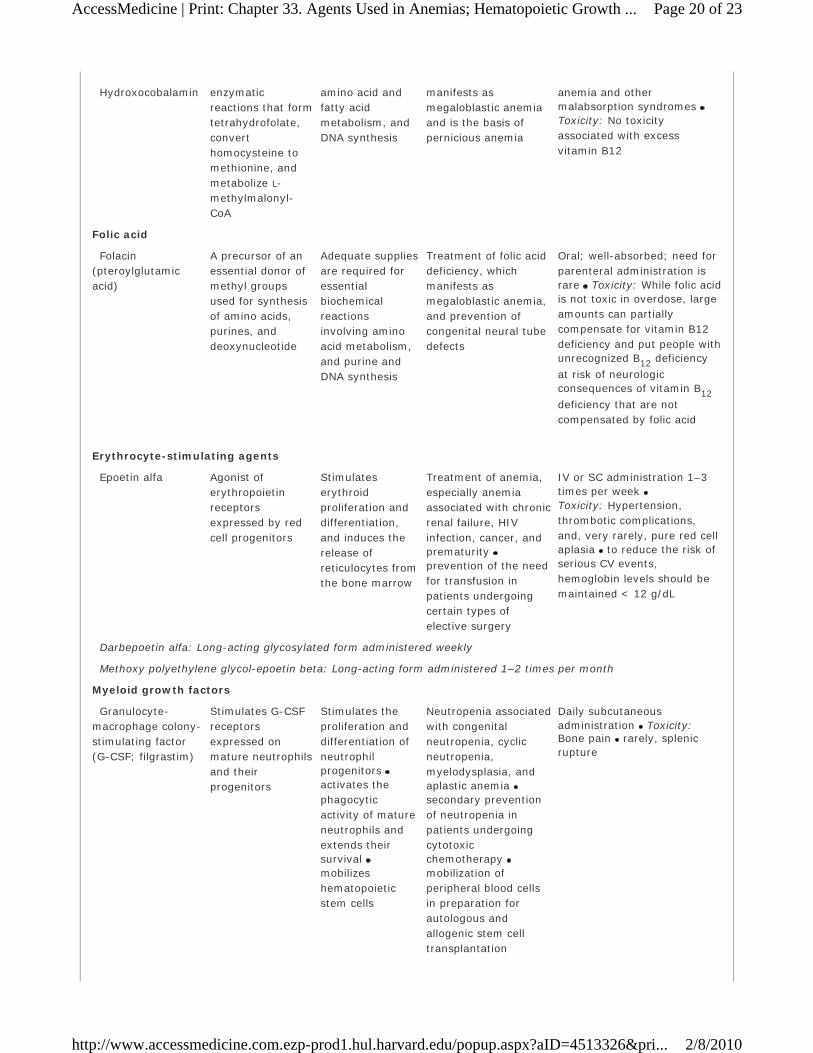
Hydroxocobalamin enzymatic reactions that form tetrahydrofolate, convert homocysteine to methionine, and metabolize L-methylmalonyl-CoA
amino acid and fatty acid metabolism, and DNA synthesis
manifests as megaloblastic anemia and is the basis of pernicious anemia
anemia and other malabsorption syndromes Toxicity: No toxicity associated with excess vitamin B12
Folic acid
Folacin (pteroylglutamic acid)
A precursor of an essential donor of methyl groups used for synthesis of amino acids, purines, and deoxynucleotide
Adequate supplies are required for essential biochemical reactions involving amino acid metabolism, and purine and DNA synthesis
Treatment of folic acid deficiency, which manifests as megaloblastic anemia, and prevention of congenital neural tube defects
Oral; well-absorbed; need for parenteral administration is rare Toxicity: While folic acid is not toxic in overdose, large amounts can partially compensate for vitamin B12 deficiency and put people with unrecognized B12 deficiency
at risk of neurologic consequences of vitamin B12
deficiency that are not compensated by folic acid
Erythrocyte-stimulating agents
Epoetin alfa Agonist of erythropoietin receptors expressed by red cell progenitors
Stimulates erythroid proliferation and differentiation, and induces the release of reticulocytes from the bone marrow
Treatment of anemia, especially anemia associated with chronic renal failure, HIV infection, cancer, and prematurity prevention of the need for transfusion in patients undergoing certain types of elective surgery
IV or SC administration 1–3 times per week Toxicity: Hypertension, thrombotic complications, and, very rarely, pure red cell aplasia to reduce the risk of serious CV events, hemoglobin levels should be maintained < 12 g/dL
Darbepoetin alfa: Long-acting glycosylated form administered weekly
Methoxy polyethylene glycol-epoetin beta: Long-acting form administered 1–2 times per month
Myeloid growth factors
Granulocyte-macrophage colony-stimulating factor (G-CSF; filgrastim)
Stimulates G-CSF receptors expressed on mature neutrophils and their progenitors
Stimulates the proliferation and differentiation of neutrophil progenitors activates the phagocytic activity of mature neutrophils and extends their survival mobilizes hematopoietic stem cells
Neutropenia associated with congenital neutropenia, cyclic neutropenia, myelodysplasia, and aplastic anemia secondary prevention of neutropenia in patients undergoing cytotoxic chemotherapy mobilization of peripheral blood cells in preparation for autologous and allogenic stem cell transplantation
Daily subcutaneous administration Toxicity: Bone pain rarely, splenic rupture
Page 20 of 23AccessMedicine | Print: Chapter 33. Agents Used in Anemias; Hematopoietic Growth ...
2/8/2010http://www.accessmedicine.com.ezp-prod1.hul.harvard.edu/popup.aspx?aID=4513326&pri...

Pegfilgrastim: Long-acting form of filgrastim that is covalently linked to a type of polyethylene glycol
GM-CSF (sargramostim): Myeloid growth factor that acts through a distinct GM-CSF receptor to stimulate proliferation and differentiation of early and late granulocytic progenitor cells, and erythroid and megakaryocyte progenitors; clinical uses are similar to those of G-CSF but it is more likely than G-CSF to cause fever, arthralgia, myalgia, and capillary leak syndrome
Megakaryocyte growth factors
Oprelvekin (interleukin-11; IL-11)
Recombinant form of an endogenous cytokine activates IL-11 receptors
Stimulates the growth of multiple lymphoid and myeloid cells, including megakaryocyte progenitors increases the number of circulating platelets and neutrophils
Secondary prevention of thrombocytopenia in patients undergoing cytotoxic chemotherapy for nonmyeloid cancers
Administered daily by SC injection Toxicity: Fatigue, headache, dizziness, anemia, fluid accumulation in the lungs, and transient atrial arrhythmias
Romiplostim: Genetically engineered protein in which the Fc component of a human antibody is fused to two copies
of a peptide that stimulates the thrombopoietin receptors; approved for treatment of idiopathic thrombocytopenic purpura
PREPARATIONS AVAILABLE
Darbepoetin alfa (Aranesp)
Parenteral: 25, 40, 60, 100, 200, 300, 500 mcg/mL for IV or SC injection Deferasirox (Exjade)
Oral: 125, 250, 500 mg tablets Deferoxamine (generic, Desferal)
Parenteral: 500, 2000 mg vials for IM, SC, or IV injection Epoetin alfa (erythropoietin, EPO) (Epogen, Procrit)
Parenteral: 2000, 3000, 4000, 10000, 20000, 40000 IU/mL vials for IV or SC injection Epoetin beta (Methoxy polyethylene glycol-epoetin beta) (Mircera)
Parenteral: 50, 100, 200, 300, 400, 600, 1000 mcg/mL in single-dose vials and prefilled syringes for IV or SC injection
Filgrastim (G-CSF) (Neupogen)
Parenteral: 300 mcg vials and prefilled syringes for IV or SC injection Folic acid (folacin, pteroylglutamic acid) (generic)
Oral: 0.4, 0.8, 1 mg tablets
Parenteral: 5 mg/mL for injection Iron (generic)
Oral: See Table 33–3.
Parenteral (Iron dextran) (InFeD, DexFerrum): 50 mg elemental iron/mL
Parenteral (Sodium ferric gluconate complex) (Ferrlecit): 12.5 mg elemental iron/mL
Page 21 of 23AccessMedicine | Print: Chapter 33. Agents Used in Anemias; Hematopoietic Growth ...
2/8/2010http://www.accessmedicine.com.ezp-prod1.hul.harvard.edu/popup.aspx?aID=4513326&pri...

Parenteral (Iron sucrose) (Venofer): 20 mg elemental iron/mL
Oprelvekin (IL-11) (Neumega)
Parenteral: 5 mg single-dose vials for SC injection Pegfilgrastim (Neulasta)
Parenteral: 10 mg/mL solution in single-dose syringe Romiplostim (Nplate)
Parenteral: 250, 500 mcg in single-dose vials for SC injection Sargramostim (GM-CSF) (Leukine)
Parenteral: 250, 500 mcg vials for IV infusion Vitamin B12 (generic cyanocobalamin or hydroxocobalamin)
Oral (cyanocobalamin): 100, 500, 1000, 5000 mcg tablets, 50, 100, 250, 500 mcg lozenges
Nasal (Nascobal, CaloMist): 2500, 5000 mcg/mL (250, 500 mcg/spray)
Parenteral (cyanocobalamin): 100, 1000 mcg/mL for IM or SC injection
Parenteral (hydroxocobalamin): 1000 mcg/mL for IM injection only
REFERENCES
Anderson GJ et al: Mechanisms of haem and non-haem iron absorption: Lessons from inherited disorders of iron
metabolism. Biometals 2005;18:339. [PMID: 16158226]
American Society of Clinical Oncology Supportive Care and Quality of Life Practice Guidelines Website:
http://www.asco.org/ASCO
Auerbach M, Al Talib K: Low-molecular weight iron dextran and iron sucrose have similar comparative safety profiles
in chronic kidney disease. Kidney Int 2008;73:528. [PMID: 18274543]
Bhana N: Granulocyte colony-stimulating factors in the management of chemotherapy-induced neutropenia: Evidence
based review. Curr Opin Oncol 2007;19:328. [PMID: 17545795]
Cook JD: Diagnosis and management of iron-deficiency anaemia. Best Pract Res Clin Haematol 2005;18:309.
Crawford J et al: Reduction by granulocyte colony-stimulating factor of fever and neutropenia induced by
chemotherapy in patients with small-cell lung cancer. N Engl J Med 1991;325:164. [PMID: 1711156]
De Domenico I, McVey Ward D, Kaplan J: Regulation of iron acquisition and storage: Consequences for iron-linked
disorders. Nat Rev Mol Cell Biol 2008;9:72.
de Montalembert M: Management of sickle cell disease. BMJ 2008;8:337.
Du X, Williams DA: Interleukin-11: Review of molecular, cell biology and clinical use. Blood 1997;89:3897. [PMID:
9166826]
Fisher JW: Erythropoietin: Physiology and pharmacology update. Exp Biol Med 2003;228:1. [PMID: 12524467]
Johnston RB Jr: Will increasing folic acid in fortified grain products further reduce neural tube defects without causing
harm? Consideration of the evidence. Pediatr Res 2008;63:2. [PMID: 18043498]
Kuter DJ: New thrombopoietic growth factors. Blood 2007;109:4607. [PMID: 17289815]
Kwiatkowski JL: Oral iron chelators. Pediatr Clin North Am 2008;55:461. [PMID: 18381096]
McKoy JM et al: Epoetin-associated pure red cell aplasia: Past, present, and future considerations. Transfusion
Page 22 of 23AccessMedicine | Print: Chapter 33. Agents Used in Anemias; Hematopoietic Growth ...
2/8/2010http://www.accessmedicine.com.ezp-prod1.hul.harvard.edu/popup.aspx?aID=4513326&pri...

2008;48:1754. [PMID: 18482185]
Novak JE, Szczech LA: Triumph and tragedy: Anemia management in chronic kidney disease. Curr Opin Nephrol
Hypertens 2008;17:580. [PMID: 18941350]
Pelus LM: Peripheral blood stem cell mobilization: New regimens, new cells, where do we stand. Curr Opin Hematol
2008;15:285. [PMID: 18536564]
Copyright © The McGraw-Hill Companies. All rights reserved. Privacy Notice. Any use is subject to the Terms of Use and Notice.
Page 23 of 23AccessMedicine | Print: Chapter 33. Agents Used in Anemias; Hematopoietic Growth ...
2/8/2010http://www.accessmedicine.com.ezp-prod1.hul.harvard.edu/popup.aspx?aID=4513326&pri...