Embed Size (px)
Citation preview

BIOCOMPATIBILITY STUDIES
Porous bioactive scaffolds: characterization and biologicalperformance in a model of tibial bone defect in rats
Hueliton Wilian Kido • Carla Roberta Tim • Paulo Sergio Bossini •
Nivaldo Antonio Parizotto • Cynthia Aparecida de Castro • Murilo Camuri Crovace •
Ana Candida Martins Rodrigues • Edgar Dutra Zanotto • Oscar Peitl Filho •
Fernanda de Freitas Anibal • Ana Claudia Muniz Renno
Received: 5 May 2014 / Accepted: 6 November 2014
� Springer Science+Business Media New York 2015
Abstract The aim of this study was to evaluate the
effects of highly porous Biosilicate� scaffolds on bone
healing in a tibial bone defect model in rats by means of
histological evaluation (histopathological and immunohis-
tochemistry analysis) of the bone callus and the systemic
inflammatory response (immunoenzymatic assay). Eighty
Wistar rats (12 weeks-old, weighing ±300 g) were ran-
domly divided into 2 groups (n = 10 per experimental
group, per time point): control group and Biosilicate�
group (BG). Each group was euthanized 3, 7, 14 and
21 days post-surgery. Histological findings revealed a
similar inflammatory response in both experimental
groups, 3 and 7 days post-surgery. During the experimental
periods (3–21 days post-surgery), it was observed that the
biomaterial degradation, mainly in the periphery region,
provided the development of the newly formed bone into
the scaffolds. Immunohistochemistry analysis demon-
strated that the Biosilicate� scaffolds stimulated cycloox-
ygenase-2, vascular endothelial growth factor and runt-
related transcription factor 2 expression. Furthermore, in
the immunoenzymatic assay, BG presented no difference in
the level of tumor necrosis factor alpha in all experimental
periods. Still, BG showed a higher level of interleukin 4
after 14 days post-implantation and a lower level of
interleukin 10 in 21 days post-surgery. Our results dem-
onstrated that Biosilicate� scaffolds can contribute for
bone formation through a suitable architecture and by
stimulating the synthesis of markers related to the bone
repair.
1 Introduction
Although bone tissues have the ability of healing them-
selves, multiple factors may impair fracture consolidation,
including fractures beyond critical size dimension, bone
loss caused by diseases, infections or tumor resections,
which may lead to the development of pseudoarthosis or
even non-union fractures [1]. In this context, several sur-
gical procedures are required to treat such clinical condi-
tions, which are related to considerable morbidity and
increased health care needs [2]. Bone grafts to enhance
bone repair have been emerging as a promising alternative
and include the use of autografts, allografts and synthetic
bone substitutes [3–5].
Nevertheless, the limited availability of autogenous
bone implants and the possibility of infectious diseases or
tissue rejection associated to the use of allogenous implants
are pivotal restrictions related to bone healing therapies [6].
H. W. Kido � C. R. Tim � N. A. Parizotto
Department of Physiotherapy, Post-Graduate Program of
Biotechnology, Federal University of Sao Carlos (UFSCar),
Sao Carlos, SP, Brazil
e-mail: [email protected]
P. S. Bossini � A. C. M. Renno (&)
Department of Biosciences, Federal University of Sao Paulo
(UNIFESP), Ana Costa, 95, Santos, SP, Brazil
e-mail: [email protected]
C. A. de Castro
Department of Physiological Sciences, Federal University of Sao
Carlos (UFSCar), Sao Carlos, SP, Brazil
M. C. Crovace � A. C. M. Rodrigues � E. D. Zanotto �O. P. Filho
Department of Materials Engineering, Vitreous Materials
Laboratory (LaMaV), Federal University of Sao Carlos
(UFSCar), Sao Carlos, SP, Brazil
F. de Freitas Anibal
Department of Morphology and Pathology, Federal University of
Sao Carlos (UFSCar), Sao Carlos, SP, Brazil
123
J Mater Sci: Mater Med (2015) 26:74
DOI 10.1007/s10856-015-5411-9
As an alternative, synthetic bone substitutes such as cal-
cium phosphate (CaP) ceramics [7], polymer-based mate-
rials [8], bioactive glass and glass–ceramics [9] have been
developed in order to overcome these limitations [10–12].
Bioactive glasses are a well-known class of materials,
with a markedly osteogenic potential, able of stimulating
bone metabolism and accelerating bone healing [13–15].
These materials when immersed in body fluids promote
release of ions in the medium, leading to the formation of a
porous layer which is rich in silica, followed by the for-
mation of hydroxy carbonate-apatite (HCA) layer on the
surface of the material [16]. The formation of the HCA
layer may contribute to the development of bone tissue,
once the HCA is equivalent to inorganic mineral phase of
bone [16].
Despite the osteogenic potential of the bioactive glasses,
their use has been limited because of their poor mechanical
properties and very high crystallization tendency when
heated [17]. As an alternative, some glass–ceramics
obtained by controlled crystallization of certain glasses
based on the quaternary Na2O–CaO–SiO2–P2O5 system
having improved mechanical properties, including Biosi-
licate�, have been developed [17]. It was demonstrated that
Biosilicate� is biocompatible with bone tissues and pre-
sents non-cytotoxicity [18]. Furthermore, its osteogenic
effects have already been demonstrated by using both
in vitro and in vivo studies [19–21]. Granito et al. [21]
found that Biosilicate� presented higher bone volume
when compared to Bioglass 45S5 in a tibial bone defect
model in rats 20 days post-surgery.
The current availability of glass ceramics for the treat-
ment of bone defects, including Biosilicate�, is still mainly
in solid pieces or in the form of granules. One of the main
disadvantages of those forms is that they may not have the
proper porosity to allow tissue ingrowth and may not
degrade according to the rate of bone tissue formation [22].
In this context, many efforts have been made to develop
improved bone graft substitutes that interact more appro-
priately with the complex biological environment of bone
tissue [23]. Biosilicate� porous scaffolds offer a three-
dimensional structure which mimics the structure of the
extracellular matrix of natural bone, allowing bone cell
attachment, proliferation and differentiation at the region
of the defect [24].
An initial in vivo study demonstrated that a porous Bi-
osilicate� scaffold (total porosity of 44 %) was able to
support bone ingrowth in the region of the tibial bone
defect, thus highlighting the osteogenic potential of the
material. However, the amount of newly formed bone was
not significantly different from the control group which
may be related to its relatively low porosity [25].
In order to obtain more appropriate bone substitutes to
be used as grafts, highly porous scaffolds may be an
interesting alternative with useful properties for biomedical
applications, i.e. biodegradability and more appropriate
structure to allow tissue ingrowth [26].
In this context, a new Biosilicate� scaffold, with
increased porosity (total porosity of 82 %), was developed
[18]. It was hypothesized that this innovative osteogenic
scaffold would offer a more suitable template for bone cell
attraction and tissue ingrowth. Consequently, the present
study aimed to evaluate the in vivo orthotopic response of
this new porous bioactive scaffold, during different
experimental set points (3, 7, 14 and 21 days after
implantation) in a tibial bone defect model in rats. His-
tology and immunohistochemistry analyses of the factors
involved in osteogenesis (COX-2, VEGF, Runx2) were
used to evaluate the effects of the porous bioactive scaffold
in the bone callus. Furthermore, an immunoenzymatic
assay was performed to evaluate the action of the material
on the systemic inflammatory response by quantifying the
inflammatory cytokines levels (IL-4, IL-10 and TNF-a) in
rat serum.
2 Materials and methods
2.1 Fabrication and characterization of the Biosilicate�
scaffolds
Biosilicate� was obtained by melting reagent grade raw
materials (Na2CO3—JT Baker, CaCO3—JT Baker, Na2-
HPO4—JT Baker, and SiO2—Zetasil 2) in a platinum
crucible at 1250 �C for 4 h. The glass was poured in a
stainless steel mould and heat treated until it reached full
crystallization in an electric furnace. More details of the
synthesis of Biosilicate� are described in the WO
2004/074199 patent [27]. Glass pieces were crushed in a
porcelain mortar and milled in a planetary ball mill at
550 rpm for 240 min. In this study, the Biosilicate� scaf-
folds were manufactured by a method based in the addition
of a porogen agent. This method was described with details
in a previous work [18, 28]. This method is therefore only
shortly described here: initially, 100 ml of a suspension
containing 67 vol% of isopropyl alcohol anhydrous
(QHEMIS), 3 vol% of polyvinyl butiral (Butvar B-98), 24
vol% of carbon black (CABOT BP-120), and 6 vol% of
Biosilicate� was prepared. Then isopropyl alcohol, PVB,
and Biosilicate� were mixed in an agate jar and milled in a
planetary ball mill (Pulverisette 6—FRITSCH) at 550 rpm
for 1 h. The agate spheres were removed from the sus-
pension and the pre-sieved carbon black (300–600 lm)
was added and then mixed for 5 min at 150 rpm. The
suspension was poured into a plastic container and dried
with a heat gun (DEKEL DK1210). The resulting granu-
lated powder was pressed in two steps, the first unixial
74 Page 2 of 13 J Mater Sci: Mater Med (2015) 26:74
123

using a cylindrical steel mould and the second isostatical.
Finally, the cylindrical samples were heat treated for
organics burn-out and to promote Biosilicate� sintering.
Scaffolds of approximately 3 mm (diameter) by 2 mm
(thickness) were obtained. Sterilization was performed in
an electric oven at 130 �C for 14 h.
For microstructural observation, six scaffolds were
embedded in epoxy resin (EpoThin�—BUEHLER) under
vacuum. The embedded samples were ground in silicon
carbide paper until grit size 1200 and polished with cerium
oxide. Then, they were coated with a thin layer of gold by
sputtering (Quorum Q150R ES) and analyzed in SEM
(Philips FEG XL-30). Both transversal and longitudinal
sections of the scaffolds were analyzed. The average pore
size and total porosity were determined by analysis of SEM
images using the software Image-J (version 1.46i).
2.2 Experimental design
This study was conducted according to the Guiding Prin-
ciples for the Use of Laboratory Animals and it was
approved by the Animal Care Committee guidelines at
Federal University of Sao Carlos (protocol 046/2012).
In this investigation, 80 male Wistar rats were used
(12 weeks old and weighing 300 g), and were maintained
under controlled conditions of temperature (24 ± 2 �C)
with light–dark periods of 12 h, with free access to water
and commercial diet. The experimental animals were ran-
domly distributed into 2 groups: Control group (CG) and
Biosilicate� group (BG). Each group was divided into 4
four subgroups (n = 10 animals) euthanized 3, 7, 14 and
21 days post-surgery.
2.3 Surgical procedures
Before surgery, all the animals were anesthetized by
intraperitoneal injection of ketamine (40 mg/kg, Agener�,
Brasılia, Brazil) and xylazine (20 mg/kg, Syntec�, Cotia,
Brazil). After exposing the right proximal tibia of each
animal, a standardized 3.0 mm diameter non-critical bone
defect was created by using a motorized drill under irri-
gation with saline solution [21, 25, 29]. The porous bio-
active scaffolds were implanted, in the created defects in a
randomization scheme. The skin was closed and sutured
with 4–0 nylon monofilament (Shalon�, Sao Luis de
Montes Belos, GO, Brazil), and disinfected with povidone
iodine. The health status of the animals was monitored on a
daily basis.
The animals were housed in pairs and the intake of water
and food was monitored in the initial postoperative period.
Moreover, the animals were observed for signs of pain,
infection and activity. According to each experimental
period, animals were euthanized by CO2 asphyxiation.
The blood and the right tibia of each animal were col-
lected for analysis. The blood samples were used for the
quantification of inflammatory factors and the right tibia
was taken to histological analyzes.
2.4 Histopathological analysis
The right tibias were fixed in 10 % buffered formalin
(Merck, Darmstadt, Germany) for 24 h. Afterwards, the
specimens were decalcified in 10 % EDTA solution (ethy-
lenediaminetetraacetic acid, Labsynth�, Diadema, Brazil)
for 40 days, dehydrated and embedded in paraffin blocks.
Three sections (5 lm) of each specimen were longitudi-
nally sectioned (Microtome Leica Microsystems SP 1600,
Nussloch, Germany) and stained with hematoxylin and
eosin (H.E. stain, Merck, Darmstadt, Germany). The mor-
phological description of the bone defect was performed
with an optical microscopy (Olympus Optical Co., Tokyo,
Japan) according to the following parameters: granulation
tissue, inflammatory process, area of fibrosis, necrotic tis-
sue, bone formation and biomaterial degradation.
2.5 Immunohistochemistry
Histological sections (5 lm) were deparaffinized using
xylene and rehydrated in graded ethanol. After, each
specimen was pre-treated in a Steamer with buffer Diva
Decloaker (Biocare Medical, CA, USA) for 5 min for
antigen retrieval. The material was pre-incubated with
0.3 % hydrogen peroxide (Labsynth�, Diadema, Brazil) in
phosphate-buffered saline (PBS) solution for 30 min in
order to inactivate endogenous peroxidase and then block
with 5 % normal goat serum in PBS solution for 20 min.
Three sections of each specimen were incubated for 2 h
with polyclonal primary antibody anti-Cyclooxygenase-2,
anti-Vascular endothelial growth factor and anti-Runt-
related transcription factor 2, all at a concentration of 1:200
(Santa Cruz Biotechnology, Santa Cruz, USA). Afterwards,
the sections were incubated with biotin conjugated sec-
ondary antibody anti-rabbit IgG (Vector Laboratories,
Burlingame, CA, USA) at a concentration of 1:200 in PBS
for 30 min, followed by the application of preformed avi-
din biotin complex conjugated to peroxidase (Vector
Laboratories, Burlingame, CA, USA) for 30 min. A solu-
tion of 3-30-diaminobenzidine solution (0.05 %) and Harris
hematoxylin were applied.
The expression of cyclooxygenase-2 (COX-2), vascular
endothelial growth factor (VEGF) and runt-related tran-
scription factor 2 (Runx2) were assessed qualitatively
(presence and location of the immunomarkers) in five pre-
determined fields using an optical light microscope (Leica
Microsystems AG, Wetzlar, Germany). The analysis was
performed by 2 observers (CRT and HWK) in a blinded way.
J Mater Sci: Mater Med (2015) 26:74 Page 3 of 13 74
123

2.6 Immunoenzymatic assay
Quantification of plasma cytokines was performed
using the immunoenzymatic assay (ELIZA). In this
study, the cytokines interleukin 4 (IL-4), interleukin 10
(IL-10) and tumor necrosis factor alpha (TNF-a) were
evaluated by their influences on the inflammatory
process [30].
For this purpose, the collected blood from each animal
(5 ml) was placed in tubes without anticoagulant for about
2 h until its coagulation. Then, the samples were centri-
fuged at 1500 rpm for 15 min. The serum that resulted
from this centrifugation was aliquoted into microtube and
frozen at -80 �C. Cytokines were measured using Duo Set
kits (R&D Systems�, Minnesota, USA), following the
manufacturer’s recommendations. The serum samples were
used to measure IL-4, IL-10 and TNF-a. The high affinity
microplates were sensitized with monoclonal anti-cyto-
kines and remained ‘‘overnight’’ at room temperature.
Afterwards, the plates were blocked (with PBS) and
washed. Supernatants and standard curves (made with
recombinant cytokines) were added. The plates were
maintained at room temperature for 2 h and then another
washing was performed. Subsequently, biotinylated anti-
cytokine antibodies were added and maintained for 1 h at
room temperature. The results were expressed in pg/ml for
all cytokines evaluated.
2.7 Statistical analysis
Data were expressed as mean values and standard devia-
tions (SD) for each sample group. The normal distribution
of all variables was checked using the Shapiro–Wilk’s W
test. Two-way ANOVA with Tukey post hoc tests were
used to evaluate the variance between groups. All analyses
were performed using Excel (2007) and STATISTICA 7.0.
For all the tests, the significance level of 5 % (p \ 0.05)
was considered.
3 Results
3.1 Material characterization
The porous bioactive scaffolds which were obtained via
addition of carbon black as a porogen agent are highly por-
ous, as can be seen in the images captured via stereomi-
croscopy (Fig. 1a, b). SEM photomicrographies revealed
that the macroporosity was 72 ± 6 %, with an average pore
size of 275 lm (Fig. 1c, d). The mechanical strength of the
material was sufficient for handling and placing it inside the
surgical site.
3.2 General findings
In this study, no animal of CG died and all tibia samples
were used for analysis. Only two animals of BG were lost
due to a respiratory depression induced by the anesthesia.
The other animals rapidly returned to their normal diet and
no post-operative complications were observed during the
experimental period. At the end of the experiment, 38 tibial
implants were retrieved, of which 35 were used for analysis
(3 porous bioative scaffolds were lost due to implant
fracturing during the histological processing). An overview
of the number of implants placed, retrieved and used for
analysis is presented in Table 1.
3.3 Histopathological analysis
Representative histological sections of all experimental
groups are depicted in Figs. 2 and 3.
3.3.1 3 days
Three days after surgery, histological evaluation of CG
revealed that the bone defect area was mostly filled with
inflammatory cells and granulation tissue (Fig. 2b). In BG,
the integrity of the implant was affected, with material
degradation, especially in the borders. The presence of
inflammatory cells around the material particles was
observed, with ingrowth of granulation tissue (Fig. 2d).
3.3.2 7 days
Seven days after implantation, bone defect area of control
animals was filled mostly by granulation tissue, accompa-
nied by some inflammatory cells (Fig. 2f). Furthermore,
immature newly formed bone was observed in the periphery
of the defect (Fig. 2f). For BG, the degradation of the
material continued, leaving lower amounts of material
compared to the previous experimental set point (Fig. 2h).
Furthermore, in the spaces previously occupied by the
material, a discrete inflammatory process and granulation
tissue was noticed (Fig. 2h). Newly formed bone was noticed
in the contact area between the edges of the bone defect and
the remained implant (Fig. 2h).
3.3.3 14 days
For CG, the amount of granulation tissue increased in the
bone defect area and some inflammatory cells still could be
observed in some specimens (Fig. 3b). In addition, newly
formed bone was observed into the area of the defect,
mainly at the periphery (Fig. 3b). The degradation of the
scaffold had continued, allowing the ingrowth of granula-
tion tissue and newly formed bone (Fig. 3d).
74 Page 4 of 13 J Mater Sci: Mater Med (2015) 26:74
123

3.3.4 21 days
After 21 days of implantation, for both experimental
groups, bone defect was mostly filled with newly formed
bone in both experimental groups (Fig. 3f, h). Some par-
ticles of the material could still be noticed the bone defect,
mainly in the center of the defect (Fig. 3h).
3.4 Immunohistochemistry
3.4.1 COX-2
COX-2 immunoexpression was observed mainly in the
granulation tissue for both experimental groups, 3 and
7 days post-surgery (Fig. 4a, b, c, d). Fourteen days after
surgery, for CG and BG, COX-2 expression was observed
in the granulation tissue and in the osteoblast cells (Fig. 4e,
f). At day 21 after surgery, CG showed COX-2 immuno-
expression mainly in the osteoblasts (Fig. 4g). For BG,
COX-2 immunoreactivity was detected in the granulation
tissue and in the osteocytes (Fig. 4h).
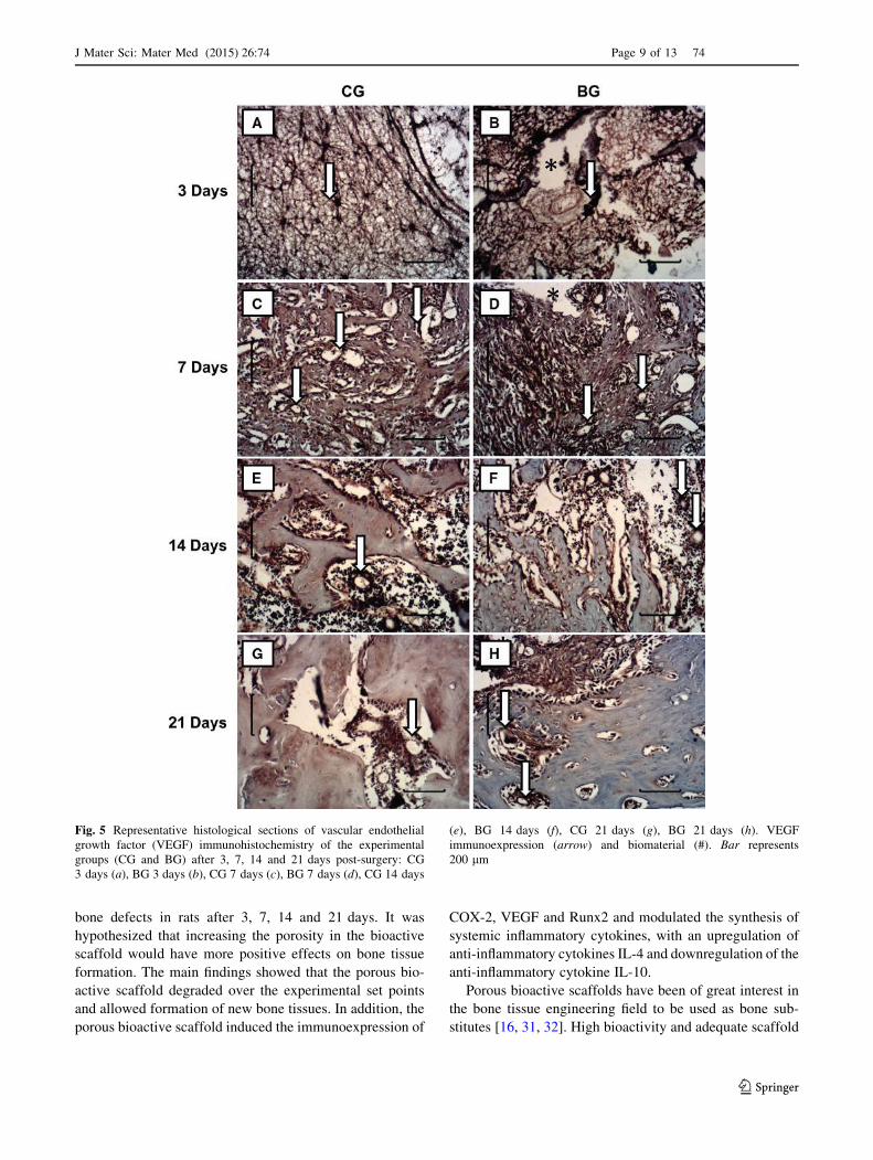
3.4.2 VEGF
Three days after surgery, VEGF immunoreactivity was
observed in the granulation tissue in CG and BG (Fig. 5a,
b). In this period, for BG, VEGF expression was more
evident in the granulation tissue located around the mate-
rial (Fig. 5b). In the other experimental periods (7, 14 and
21 days), VEGF expression was predominantly detected in
the cells involving capillary walls for both groups CG and
BG (Fig. 5c, d, e, f, g, h).
Fig. 1 Images of the Biosilicate� scaffolds obtained with the stereomicroscope Leica MZ75 (a, b) and SEM images of two scaffolds embedded
in epoxy resin under vacuum: longitudinal section (c) and transversal section (d)
Table 1 Number of implants placed, retrieved and used for histological analyses for the tibial defect implants
Implants placed Implants retrieved Implants used for analysis
Day 3 Day 7 Day 14 Day 21 Day 3 Day 7 Day 14 Day 21 Day 3 Day 7 Day 14 Day 21
Tibial implants
Biosilicate� scaffolds 10 10 10 10 9a 9a 10 10 8b 8b 9b 10
a Deviation from number of implants placed due to animal deadb Deviation from number of implants retrieved due to fracturing of implants during to the histological processing
J Mater Sci: Mater Med (2015) 26:74 Page 5 of 13 74
123

3.4.3 Runx2
Similar to COX-2 and VEGF expression, Runx2 was
predominantly detected in the granulation tissue for
both CG and BG on day 3 after the surgery (Fig. 6a,
b). In the same period, for BG, Runx2 immunoreac-
tivity was mainly observed in the granulation tissue
around the material (Fig. 6b). At day 7 after surgery,
Fig. 2 Representative histological sections of tibial bone defects of
the control (CG) and Biosilicate� Group (BG) 3–7 days after surgery:
CG 3 days (a, b), BG 3 days (c, d), CG 7 days (e, f), BG 7 days (g,
h). Newly formed bone (asterisk), granulation tissue (black arrow),
infiltrate of inflammatory cells (filled inverse triangle) and biomate-
rial (#). Bar represents 500 lm (a, c, e, g) and 200 lm (b, d, f, h).
Hematoxylin and eosin staining
74 Page 6 of 13 J Mater Sci: Mater Med (2015) 26:74
123
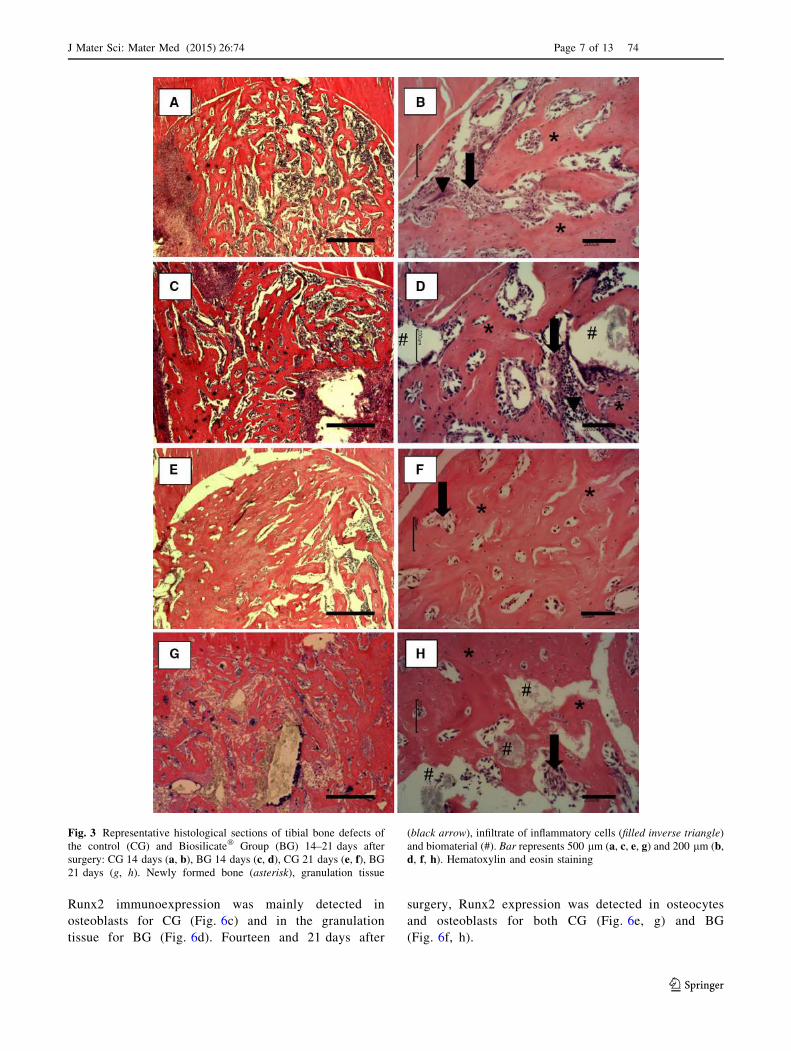
Runx2 immunoexpression was mainly detected in
osteoblasts for CG (Fig. 6c) and in the granulation
tissue for BG (Fig. 6d). Fourteen and 21 days after
surgery, Runx2 expression was detected in osteocytes
and osteoblasts for both CG (Fig. 6e, g) and BG
(Fig. 6f, h).
Fig. 3 Representative histological sections of tibial bone defects of
the control (CG) and Biosilicate� Group (BG) 14–21 days after
surgery: CG 14 days (a, b), BG 14 days (c, d), CG 21 days (e, f), BG
21 days (g, h). Newly formed bone (asterisk), granulation tissue
(black arrow), infiltrate of inflammatory cells (filled inverse triangle)
and biomaterial (#). Bar represents 500 lm (a, c, e, g) and 200 lm (b,
d, f, h). Hematoxylin and eosin staining
J Mater Sci: Mater Med (2015) 26:74 Page 7 of 13 74
123

3.5 Immunoenzymatic assessment
The immunoenzymatic assessment showed no statistic
difference in the levels of TNF-a comparing CG and BG in
the experimental periods (Fig. 7). For IL-4, a significantly
higher level of this cytokine was observed in BG when
compared to CG, 14 days after implantation (Fig. 8).
Moreover, the immunoenzymatic evaluation indicated a
lower level of IL-10 (Fig. 9) in BG compared to CG,
21 days after the surgery.
4 Discussion
This study aimed to evaluate the biological in vivo response
after the implantation of porous bioactive scaffolds in tibial
Fig. 4 Representative histological sections of cyclooxygenase-2
(COX-2) immunohistochemistry of the experimental groups (CG
and BG) after 3, 7, 14 and 21 days post-surgery: CG 3 days (a), BG
3 days (b), CG 7 days (c), BG 7 days (d), CG 14 days (e), BG
14 days (f), CG 21 days (g), BG 21 days (h). COX-2 immunoex-
pression (arrow) and biomaterial (#). Bar represents 200 lm
74 Page 8 of 13 J Mater Sci: Mater Med (2015) 26:74
123
bone defects in rats after 3, 7, 14 and 21 days. It was
hypothesized that increasing the porosity in the bioactive
scaffold would have more positive effects on bone tissue
formation. The main findings showed that the porous bio-
active scaffold degraded over the experimental set points
and allowed formation of new bone tissues. In addition, the
porous bioactive scaffold induced the immunoexpression of
COX-2, VEGF and Runx2 and modulated the synthesis of
systemic inflammatory cytokines, with an upregulation of
anti-inflammatory cytokines IL-4 and downregulation of the
anti-inflammatory cytokine IL-10.
Porous bioactive scaffolds have been of great interest in
the bone tissue engineering field to be used as bone sub-
stitutes [16, 31, 32]. High bioactivity and adequate scaffold
Fig. 5 Representative histological sections of vascular endothelial
growth factor (VEGF) immunohistochemistry of the experimental
groups (CG and BG) after 3, 7, 14 and 21 days post-surgery: CG
3 days (a), BG 3 days (b), CG 7 days (c), BG 7 days (d), CG 14 days
(e), BG 14 days (f), CG 21 days (g), BG 21 days (h). VEGF
immunoexpression (arrow) and biomaterial (#). Bar represents
200 lm
J Mater Sci: Mater Med (2015) 26:74 Page 9 of 13 74
123

porosity are essential characteristics to stimulate osteo-
progenitor cells and to support bone ingrowth [3, 16, 33].
Furthermore, resorption of the material with the same rate
of the bone formation is required [34]. Several in vivo
studies demonstrated that Biosilicate�, used in powder or
scaffolds, was able to stimulate bone metabolism and
accelerate the process of bone healing in different animal
models, thus highlighting the osteogenic potential of the
glass ceramic [25, 35, 36]. These findings are in line with
the results of the current study which revealed a continuous
newly bone tissue ingrowth at the defect area and in the
spaces left by the degraded material. Many studies dem-
onstrated that Biosilicate� scaffolds have bioactive prop-
erties [19–21]. Immediately upon the implantation, ions
Fig. 6 Representative histological sections of runt-related transcrip-
tion factor-2 (Runx2) immunohistochemistry of the experimental
groups (CG and BG) after 3, 7, 14 and 21 days post-surgery: CG
3 days (a), BG 3 days (b), CG 7 days (c), BG 7 days (d), CG 14 days
(e), BG 14 days (f), CG 21 days (g), BG 21 days (h). Runx2
immunoexpression (arrow) and biomaterial (#). Bar represents
200 lm
74 Page 10 of 13 J Mater Sci: Mater Med (2015) 26:74
123

dissolution from the scaffold to bone tissue stimulates the
formation of a hydroxyapatite layer, which acts as a tem-
plate for osteoblast growth, which can affect osteogenesis
[16]. Furthermore, high porosity and adequate pore sizes
are essential factors for an effective bone substitute mate-
rial [26]. Scaffolds with pores between of 100–400 lm are
of optimal size to allow bone ingrowth and to support
neovascularization [37]. The pore size and porosity of the
bioactive scaffold used in the present study indicate that it
has morphological characteristics which make them suit-
able to be used as a bone graft.
Moreover, the histological findings demonstrated that
the scaffold degraded over time and the degradation hap-
pened according to the rate of tissue ingrowth. Besides
adequate porosity, proper scaffold degradation is also
essential for the process to happen, since formation of new
bone tissue needs space to grow in [38].
COX-2, VEGF and Runx2 immunoexpressions were
increased in the porous bioactive scaffolds implanted ani-
mals. COX-2 and Runx2 have regulatory effects on the
proliferation and differentiation of osteoblasts [39, 40],
while the VEGF is the most important signal protein pro-
duced by cells that stimulates vasculogenesis and angio-
genesis [41]. In the current study, the ions released from
the scaffolds, such as silicon (Si) and calcium (Ca) may
have provided the necessary stimuli to increase the
expression of COX-2 and Runx2, and consequently lead to
the proliferation of osteoblastic cells. Xynos et al. [33]
observed that inorganic particles of Bioglass 45S5�,
mainly Si and Ca, may carry specific morphogenic clues
that stimulate the proliferation of osteoblastic cells. Fur-
thermore, the increased VEGF expression may be also
related to the ions dissolution of the porous bioactive
scaffolds. These findings corroborate those of Matsumoto
et al. [36] who demonstrated an increased VEGF immu-
noexpression in the calvaria defects in rabbits after Biosi-
licate� implantation.
Additionally, severe local and systemic inflammatory
responses caused by the implantation of biomaterials may
result in delay of the bone healing [34]. The organic
response is mainly related to the composition of the
material, which may stimulate the expression of inflam-
matory factors such as interleukins and TNF-a. In the
present study, the ELISA assay was used to measure the
systemic reaction caused by the porous bioactive scaffold
tibial implantation and demonstrated that no significant
increase in TNF-a was observed in any experimental
group. TNF-a is a factor which is involved in sys-
temic inflammation and is mainly produced by acti-
vated macrophages [42]. The fact that the expression of
this cytokine did not increase is an indicative that the
porous bioactive scaffolds implantation did not induce any
systemic inflammatory process.
In addition, porous bioactive scaffolds induced a higher
expression of IL-4 on day 14 after implantation and a lower
expression of IL-10 on day 21 after surgery. IL-4 and IL-10
are anti-inflammatory cytokines that can regulate the
effects of the TNF-a [29]. Cytokines such as IL-4, indi-
rectly promote the bone formation by increasing the
Fig. 7 Levels of TNF-a cytokines evaluated in the serum of rats
undergoing implantation of the Biosilicate� scaffolds in different
experimental periods
Fig. 8 Levels of IL-4 cytokines evaluated in the serum of rats
undergoing implantation of the Biosilicate� scaffolds in different
experimental periods. Significant differences of p \ 0.05 are repre-
sented by an asterisk
Fig. 9 Levels of IL-10 cytokines evaluated in the serum of rats
undergoing implantation of the Biosilicate� scaffolds in different
experimental periods. Significant differences of p \ 0.05 are repre-
sented by an asterisk
J Mater Sci: Mater Med (2015) 26:74 Page 11 of 13 74
123

expression of osteoprotegerin (OPG), inhibiting osteo-
clastogenesis [43]. In this context, the increase of the
synthesis of IL-4 cytokines in the scaffold treated animals
may have contributed to bone formation.
The results of this initial investigation confirmed our
hypothesis that the high porous bioactive scaffold has an
adequate porosity structure and is able to support bone
tissue ingrowth, thus constituting a promising alternative to
be used as bone grafts for tissue engineering. However, in
the present study the comparison of the performance of the
material was made using an empty control defect model.
Future investigations should be performed using standard
materials such as calcium phosphate or 45S5 Bioglass.
Additionally, the biological performance of the scaffold
should be investigated in different bone defect models such
as those of critical-size or compromised situations (e.g.
osteoporosis).
5 Conclusions
In summary, the results indicated that the porous bioactive
scaffold has good adequate porosity and proper degrada-
bility and bone-forming properties. The innovative scaffold
enhanced the expression of vascular and osteogenic factors
and did not induce any systemic inflammatory response.
Further long-term studies should be carried out to provide
additional information concerning the late stages of mate-
rial degradation and the bone regeneration induced by the
porous scaffold. Moreover, further researches are required
to evaluate the biological performance of this new bio-
material in compromised situations to support the use of
this promising scaffold for bone engineering applications.
Acknowledgments The authors thank FAPESP (Fundacao de
Amparo a Pesquisa do Estado de Sao Paulo) for their financial support.
References
1. Phieffer LS, Goulet JA. Delayed unions of the Tibia. J Bone Joint
Surg Am. 2006;88:205–16.
2. Axelrad TW, Kakar S, Einhorn TA. New technologies for the
enhancement of skeletal repair. Injury. 2007;38:S49–62.
3. Valimaki V, Yrjans JJ, Vuorio E, Aro HT. Combined effect of
bone morphogenetic protein-2 gene therapy and bioactive glass
microspheres in enhancement of new bone formation. J Biomed
Mater Res. 2005;75:501–9.
4. Drosse I, Volkmer E, Seitz S, Seitz H, Penzkofer R, Zahn K,
Matis U, Mutschler W, Augat P, Schieker M. Validation of a
femoral critical size defect model for orthotopic evaluation of
bone healing: a biomechanical, veterinary and trauma surgical
perspective. Tissue Eng Part C Methods. 2008;14:79–88.
5. Bhatt RA, Rozental TD. Bone graft substitutes. Hand Clin.
2012;28:457–68.
6. Nandi SK, Roy S, Mukherjee P, Kundu B, De DK, Basu D.
Orthopaedic applications of bone graft and graft substitutes: a
review. Indian J Med Res. 2010;132:15–30.
7. Dorozhkin S. Calcium orthophosphate-based biocomposites and
hybrid biomaterials. J Mater Sci. 2009;44:2343–87.
8. Hutmacher DW, Schantz JT, Lam CX, Tan KC, Lim TC. State of
the art and future directions of scaffold-based bone engineering
from a biomaterials perspective. J Tissue Eng Regen Med.
2007;1:245–60.
9. Renno AC, Bossini PS, Crovace MC, Rodrigues AC, Zanotto ED,
Parizotto NA. Characterization and in vivo biological perfor-
mance of biosilicate. Biomed Res Int. 2013;. doi:10.1155/2013/
141427.
10. De Long WG Jr, Einhorn TA, Koval K, McKee M, Smith W,
Sanders R, Watson T. Bone grafts and bone graft substitutes in
orthopaedic trauma surgery. A critical analysis. J Bone Joint Surg
Am. 2007;89:649–58.
11. Hak DJ. The use of osteoconductive bone graft substitutes in
orthopaedic trauma. J Am Acad Ortho Surg. 2007;15:525–36.
12. Ohtsuki C, Kamitakahara M, Miyazaki T. Bioactive ceramic-
based materials with designed reactivity for bone tissue regen-
eration. J R Soc Interface. 2009;6:S349–60.
13. Hench LL, Xynos ID, Polak JM. Bioactive glasses for
in situ tissue regeneration. J Biomater Sci Polym Ed. 2004;15:
543–62.
14. Hu YC, Zhong JP. Osteostimulation of bioglass. Chin Med J.
2009;122:2386–9.
15. Xin R, Zhang Q, Gao J. Identification of the wollastonite phase in
sintered 45S5 bioglass and its effect on in vitro bioactivity. J Non
Cryst Solids. 2010;356:1180–4.
16. Hench LL, Polak JM. Third-generation biomedical materials.
Science. 2002;295:1014–7.
17. Peitl Filho O, LaTorre GP, Hench LL. Effect of crystallization on
apatite-layer formation of bioactive glass 45S5. J Biomed Mater
Res. 1996;30:509–14.
18. Kido HW, Oliveira P, Parizotto NA, Crovace MC, Zanotto ED,
Peitl-Filho O, Fernandes KP, Mesquita-Ferrari RA, Ribeiro DA,
Renno AC. Histopathological, cytotoxicity and genotoxicity
evaluation of Biosilicate� glass-ceramic scaffolds. J Biomed
Mater Res A. 2013;101:667–73.
19. Moura J, Teixeira LN, Ravagnani C, Peitl O, Zanotto ED, Beloti
MM, Panzeri H, Rosa AL, De Oliveira PT. In vitro osteogenesis
on a highly bioactive glass–ceramic (Biosilicate�). J Biomed
Mater Res A. 2007;82:545–57.
20. Bossini PS, Renno AC, Ribeiro DA, Fangel R, Peitl O, Zanotto
ED, Parizotto NA. Biosilicate� and low-level laser therapy
improve bone repair in osteoporotic rats. J Tissue Eng Regen
Med. 2011;5:229–37.
21. Granito RN, Renno AC, Ravagnani C, Bossini PS, Mochiuti D,
Jorgetti V, Driusso P, Peitl O, Zanotto ED, Parizotto NA, Oishi J.
In vivo biological performance of a novel highly bioactive glass-
ceramic (Biosilicate�): a biomechanical and histomorphometric
study in rat tibial defects. J Biomed Mater Res B Appl Biomater.
2011;97:139–47.
22. Wu C, Zhu Y, Chang J, Zhang Y, Xiao Y. Bioactive inorganic-
materials/alginate composite microspheres with controllable
drug-delivery ability. J Biomed Mater Res B Appl Biomater.
2010;94:32–43.
23. Sachot N, Castano O, Mateos-Timoneda MA, Engel E, Planell
JA. Hierarchically engineered fibrous scaffolds for bone regen-
eration. J R Soc Interface. 2013;10:20130684.
24. Pascu EI, Stokes J, McGuinness GB. Electrospun composites of
PHBV, silk fibroin and nano-hydroxyapatite for bone tissue
engineering. Mater Sci Eng C Mater Biol Appl. 2013;33:
4905–16.
74 Page 12 of 13 J Mater Sci: Mater Med (2015) 26:74
123

25. Pinto KN, Tim CR, Crovace MC, Matsumoto MA, Parizotto NA,
Zanotto ED, Peitl O, Renno AC. Effects of Biosilicate� scaffolds
and low-level laser therapy on the process of bone healing.
Photomed Laser Surg. 2013;31:252–60.
26. Schieker M, Seitz H, Drosse I, Seitz S, Mutschler W. Biomate-
rials as scaffold for bone tissue engineering. Eur J Trau ma.
2006;32:114–24.
27. Zanotto ED, Ravagnani C, Peitl O, Panzeri H, Lara EH. Process
and compositions for preparing particulate, bioactive or resorb-
able biosilicates for use in the treatment of oral ailments. Sao
Carlos: Universidade Federal de Sao Carlos, Universidade de Sao
Paulo; 2004. International Classification C03C10/00, WO
2004/074199 (INPI 03006441).
28. Crovace MC. Obtencao de estruturas porosas altamente bioativas
via sinterizacao do Biosilicate�. Dissertation (MSc in Materials
Engineering), Post-Graduate Program in Science and Materials
Engineering, Federal University of Sao Carlos, Sao Carlos; 2009.
29. Oliveira P, Ribeiro DA, Pipi EF, Driusso P, Parizotto NA, Renno
AC. Low-level laser therapy does not modulate the outcomes of a
highly bioactive glassceramic (Biosilicate�) on bone consolida-
tion in rats. J Mater Sci Mater Med. 2010;21:1379–84.
30. Pape HC, Marcucio R, Humphrey C, Colnot C, Knobe M, Harvey
EJ. Trauma-induced inflammation and fracture healing. J Orthop
Trauma. 2010;24:522–5.
31. Gauthier O, Muller R, von Stechow D, Lamy B, Weiss P, Bouler
JM, Aguado E, Daculsi G. In vivo bone regeneration with inject-
able calcium phosphate biomaterial: a three-dimensional micro-
computed tomographic, biomechanical and SEM study. Biomate-
rials. 2005;26:5444–53.
32. Link DP, Van den dolder J, Van den Beucken JJJP, Cuijpers VM,
Wolke JGC, Mikos AG, Jansen JA. Evaluation of the biocom-
patibility of calcium phosphate cement/PLGA microparticle
composites. J Biomed Mater Res A. 2008;87:760–9.
33. Xynos ID, Edgar AJ, Buttery LDK, Hench LL, Polak JM. Ionic
products of bioactive glass dissolution increase proliferation of
human osteoblasts and induce insulin-like growth factor II
mRNA expression and protein synthesis. Biochem Biophys Res
Commun. 2000;276:461–5.
34. Anderson JM, Mcnally AK. Biocompatibility of implants: lym-
phocyte/macrophage interactions. Semin Immunopathol. 2011;
33:221–33.
35. Granito RN, Ribeiro DA, Renno AC, Ravagnani C, Bossini PS,
Peitl-Filho O, Zanotto ED, Parizotto NA, Oishi J. Effects of bi-
osilicate and bioglass 45S5 on tibial bone consolidation on rats: a
biomechanical and a histological study. J Mater Sci Mater Med.
2009;20:2521–6.
36. Matsumoto MA, Holgado LA, Renno ACM, Caviquioli G, Big-
uetti CC, Saraiva PP, Kawakami RY. A novel bioactive vitr-
oceramic presents similar biological responses as autogenous
bone grafts. J Mater Sci Mater Med. 2012;23:1447–56.
37. Karageorgiou V, Kaplan D. Porosity of 3D biomaterial scaffolds
and osteogenesis. Biomaterials. 2005;26:5474–91.
38. Salgado AJ, Coutinho OP, Reis RL. Bone tissue engineering:
state of the art and future trends. Macromol Biosci. 2004;4:
743–65.
39. Zhang X, Schwarz EM, Young DA, Puzas E, Rosier RN, O’keefe
RJ. Cyclo-oxygenase-2 regulates mesenchymal cell differentia-
tion into the osteoblast lineage and is critically involved in bone
repair. J. Clin. Invest. 2002;109:1405–15.
40. Komori T. Regulation of skeletal development by the Runx
family of transcription factors. J Cell Biochem. 2005;95:445–53.
41. Keramarisa NC, Calorib GM, Nikolaoua VS, Schemitschc EH,
Giannoudisa PV. Fracture vascularity and bone healing: a sys-
tematic review of the role of VEGF. Injury. 2008;39:S45–57.
42. Hallab NJ, Jacobs JJ. Biologic Effects of Implant Debris. Bull
NYU Joint Dis. 2009;67:182–8.
43. Shioi A, Teitelbaum SL, Ross FP, Welgus HG, Suzuki H, Ohara
J, Lacey DL. Interleukin 4 inhibits murine osteoclast formation
in vitro. J Cell Biochem. 1991;47:272–7.
J Mater Sci: Mater Med (2015) 26:74 Page 13 of 13 74
123