Embed Size (px)
Citation preview

CASE REPORT Open Access
Portal hypertension as a result of theincomplete surgically treated advancedalveolar echinococcosis: a case descriptionŁ. Pielok1* , M. Karczewski2, W. Cierach2, P. Zmora3, E. Lenartowicz3 and J. Stefaniak1
Abstract
Background: Infection of Echinococcus multilocularis causes in humans the alveolar echinococcosis. Although theinfection has world-wide distribution it is rarely detected. Diagnosis of alveococcosis is difficult because of nottypical clinical picture and irregular results of radiological examinations suggesting neoplasmatic process whichbegins in the liver tissue or in the biliary tracts. The parasitic growth is slow, so the illness is quite often establishedin late invasion period. Treatment of long-lasting and late diagnosed infection is difficult and requires cooperationof parasitologists together with surgeons to avoid life-threatening organ dysfunction.
Case presentation: We describe a young male patient, diagnosed, according to the radiological, immunologicaland histological examination results, infection of Echinococcus multilocularis, who was treated with not radicalresection of pathologic mass together with persistent albendazole intake. The right hepatectomy was performed. Inaddition, visible cysts were removed from the left lobe of the liver in nonanatomical resection and suspiciouscalcified lesions in hepatoduodenal ligament were also removed. After the operation portal hypertension, withsplenomegaly and symptoms of the liver cirrhosis occurred (thrombocytopenia, collateral venous circulation, firstdegree varices oesophagii). The portal hypertension probably could be a result of incomplete surgery due toextended parasitic infection and liver anathomical changes due to performed procedures, because the portalhypertension and it’s further complications had not been observed before the operation.
Conclusions: Echinococcus multilocularis should be taken under consideration in differential diagnosis of irregularlesions within the liver. Lon-lasting invasion could be responsible for the irreversible secondary liver changes suchas cirrhosis and portal hypertension. The surgery treatment (treatment of choice) is difficult and it’s results dependson the invasion period the patient is operated on. After the surgery the patient requires careful follow – up, todetect early complications.
Keywords: Echinococcus multilocularis, Human alveococcosis, Portal hypertension, Liver, Joidance
BackgroundAlveolar echinococcosis, caused by the metacestode ofthe fox tapeworm Echinococcus multilocularis, is themost pathogenic zoonosis in temperate and arctic re-gions of the northern hemisphere [1]. Parasite
transmission occurs when eggs of the tapeworm, ex-creted by the final hosts (usually foxes but also dogs,wolves and cats), are ingested accidentally by humans [2,3]. In humans E.mulitlocularis infection is one of thereasons of liver lesions. For many years the illness is notdetected because is asymptomatic. Because of increase offoxes population in Poland the risk of parasite transmis-sion to humans is mounting [4]. Diagnosis of alveococ-cosis is difficult because of not typical clinical picture
© The Author(s). 2020 Open Access This article is licensed under a Creative Commons Attribution 4.0 International License,which permits use, sharing, adaptation, distribution and reproduction in any medium or format, as long as you giveappropriate credit to the original author(s) and the source, provide a link to the Creative Commons licence, and indicate ifchanges were made. The images or other third party material in this article are included in the article's Creative Commonslicence, unless indicated otherwise in a credit line to the material. If material is not included in the article's Creative Commonslicence and your intended use is not permitted by statutory regulation or exceeds the permitted use, you will need to obtainpermission directly from the copyright holder. To view a copy of this licence, visit http://creativecommons.org/licenses/by/4.0/.The Creative Commons Public Domain Dedication waiver (http://creativecommons.org/publicdomain/zero/1.0/) applies to thedata made available in this article, unless otherwise stated in a credit line to the data.
* Correspondence: [email protected] and Clinic of Tropical and Parasitic Diseases, Poznan Universityof Medical Sciences, Przybyszewskiego Street 49, 60-355 Poznań, PolandFull list of author information is available at the end of the article
Pielok et al. BMC Gastroenterology (2020) 20:176 https://doi.org/10.1186/s12876-020-01320-0

and irregular results of radiological examinations (ultra-sound of the abdomen cavity -USG, computedtomography-CT, magnetic resonance imaging-MRI) sug-gesting neoplasmatic process which begins in the livertissue or in the biliary tracts [5–7]. According to thepathologic lesions localization the PNM (primary liverlocation, involvement of neighbouring organs and meta-static changes) classification is used to evaluate the dis-ease advanced [8]. Helpful in diagnostics are serologytools performed by screening ELISA- enzyme-linked im-munosorbent assay method, which detects non-specificanty-Echinococcus IgG. Western-blot confirms the diag-nosis, and EM2-EM18 ELISA detects very specific anty-multilocularis antibodies [9–12]. In controversial casesthe diagnosis can also be confirmed after histopathologysection of the liver tissue or after performing polymerasechain reaction- PCR, which detects parasite DNA frag-ments [13, 14]. Long asymptomatic parasite’s develop-ment causes that diagnosis is often established inadvanced infection period, which delay initiation of spe-cific treatment . All these leads to progressive organ dys-function with full symptomatic liver cirrhosis [15].After only several years, when the patient is not
treated, cholestasis develops, thrombotic disturbancesappears and changes in other distant organs [16]. Theseall pathologic processes as well as presence of the para-site is responsible for the full symptomatic liver fibrosiswith ascites, collateral venous circulation with oesophagivarices. In such cases the patient requires combinedmultidrug therapy together with paliative surgical proce-dures (hemihepatectomy, gastroscopy, biliary tract artifi-cial) [17] and frequent, both parasitological and surgical,follow-up. Sometimes the patient requires the livertransplantation [18, 19].Echinococcus multilocularis infection should be taking
under consideration in differential diagnosis in pa-tients with non –specific liver focuses, specially sus-pected of neoplasmatic disorders with normal liverfunction tests-LFTs (GGTP, ALP, ALT, AST) [20].Early alveococosis diagnosis and suitable treatmentinitiation, could protect the patient from life –threat-ening complications, which correlates with longer sur-vival and better quality of life [21].In this work we present a case of a young man with a
huge pathological mass within the liver, who was diag-nosed alveococcosis and treated with the not-radical op-eration theater together with albendazole (Zentel, GSK)intake in whom the portal hypertension occurred as apostsurgical complication.
Case presentationA 31-year old male patient admitted to the Tropical andParasitic Disease Department of Poznań University ofMedical Sciences, Poland, because of the presence of a
tumor-like lesion within the liver. The patient had beenliving in a small village surrounded by forests in which abig foxes population has been detected.Prior to the admission the patient had suffered from
influenza like syndromes, pain in the right subcostal re-gion and suddenly joidance.He was admitted to the local Surgery Department with
suspicion of biliary tract pathology. CT scan gave theevidence of irregular mass with disseminated calcifica-tions. He was diagnosed undifferentiated hepatitis withcholestasis.Because of atypical radiology results suspicion of Echi-
nococus infection was done. ELISA serology test waspositive (2.9 Units; positive above 1.0). The patient wasmoved to the Tropical and Parasitic Clinic in Poznań forfurther investigations.On admission day the physical examination was unre-
markable. Blood tests showed elevated levels of biliru-bine (2 mg%), alkaline phosphatase (172-248 U/l) gammaglutamylo trans peptidase- GGTP (135-262 U/l).USG of the abdominal cavity revealed presence of a
huge calcified lesion in the VII-th liver segment with thediameter of 12.3 × 2.8 cm and in the II-nd liver segmenta solid hyperechogenic focus with calcifications inside aswell as disseminated calcifications in the interhepatic bil-iary tracts neighborhood. MRI showed the liver enlarge-ment, with irregular tissue. In the VII, VI and Vsegments polycyclic fluid lesion and disseminated insidethe right lobe smaller fluid foci as well as biliary tractwidening (Fig. 1).According to the picturesque data suspicion of alveco-
coccosis was done.ELISA test (Echinococcus IgG) was positive – 50 NTU
(positive above 11NTU) and confirmed with positive
Fig. 1 MRI of the abdomen cavity - fluid lesions and disseminatedcalcifications within V, VI and VII liver segments and widening of theintrahepatic biliary tracts
Pielok et al. BMC Gastroenterology (2020) 20:176 Page 2 of 6

Western-blot which revealed presence of specific forEchinococcus multilocularis IgG (7,16,18, 26–28 kDa).ELISA EM2-plus (anty-E.multilocularis) was also highpositive (> 3.0ABS). The patient was finally diagnosedthe liver alveococcosis with P2M0N0 stage.When the diagnosis was established the albendazol
therapy (2x400mg/day) was initiated together with urso-deoksycholic acid (2x250mg/day) in order to loweredbilirubine level and protect from apoptosis healthy livertissue . The patient was qualified to the surgery treat-ment and was moved to the General and TransplantSurgery Department, Poznan University of Medical Sci-ences. MELD score of the patient was 10 (creatinine1.13 mg/dl; bilirubin 1,12 mg/dl, INR 1,15, not dialysed).The right hepatectomy was performed. Access to the
liver was achieved by bilateral subcostal incision andthen mobilizing the liver from its ligamentous attach-ments, including the coronary ligament, and left andright triangular ligaments, then anatomic resection ofthe fifth, sixth, seventh and eighth liver segments wasperformed. Right portal vein, right hepatic artery andright hepatic duct was ligated and cutted. In addition,visible cysts were removed from the left lobe of the liverin nonanatomical resection and suspicious calcified le-sions in hepatoduodenal ligament were also removed(Fig. 2). Postoperative course complicated by lymphor-rhea, conservative treatment was initiated, obtaining im-provement. In ultrasound on the 8th postoperative day,spleen enlargement occurs (141x55mm).The histopathology examination of the all 3 speci-
ments revealed presence of chronic inflammatorychanges with thick wall calcified granulomas and accelu-lar homogeny infiltrates with necrosis cavities.Additionally, the biological material after surgery was
used for molecular genotyping of Echinococcus sp. Forthis reason, the DNA was extracted from the removed
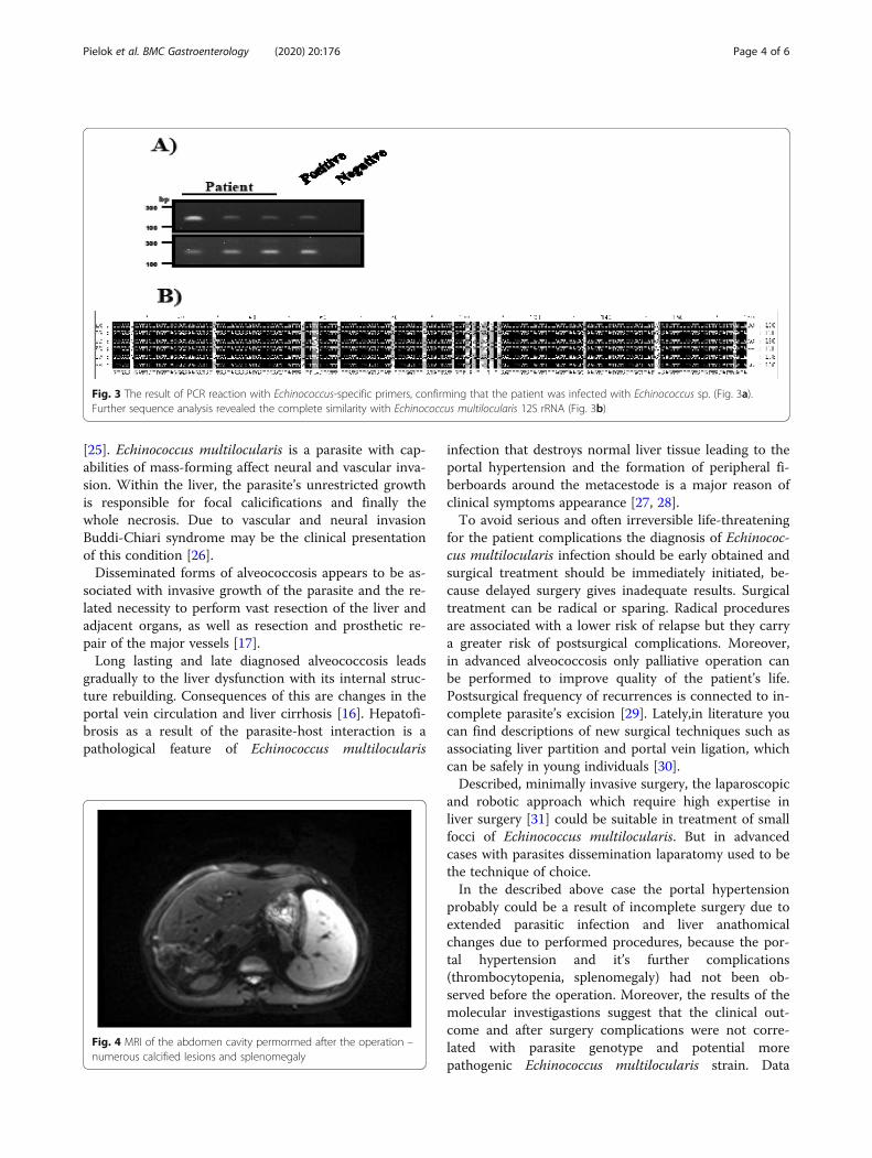
liver tissue with lesions, using the commercially availablekit (NucleoSpin Tissue, Macherey Nagel, Dueren,Germany). Next, the isolated DNA was used as a tem-plate in the PCR with Echinococcus sp. 12S rRNA-specific primers (EM-H15 and EM-H17), according toStieger et al. (2007) [22]. Moreover, as a control of DNAquality and presence of PCR inhibitors, we used isolatedgenetic material to amplify human GAPDH, accordingto Xiang et al. (2012) [23]. Finally, the PCR product wassequenced and the obtained sequences were alignedusing BioEdit software.Based on the PCR reactions with Echinococcus-specific
primers, we confirmed that the patient was infected withEchinococcus sp. (Fig. 3a). Further sequence analysis re-vealed the complete similarity with Echinococcus multi-locularis 12S rRNA (Fig. 3b), suggesting that the clinicaloutcome and after surgery complications were not corre-lated with parasite genotype and potential more patho-genic Echinococcus multilocularis strain.After the operation albendazol treeatmet was contin-
ued. With no other major or minor complication patientwith normal level of bilirubin, alanine transaminase andaspartate transaminase was discharge on 17th postopera-tive day.The patient’s follow –up, performed 10 months after
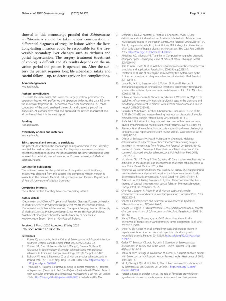
the operation gave the evidence of thrombocytopenia(80G/l), leucopenia (3.1G/l) and syderopenic anemia.MR of the abdomen cavity showed presence of numer-ous hypodensic partially calcified lesions within theremaining liver segments. Moreover critical portal veinconstriction (the diameter 3-4 mm), collateral venouscirculation in the liver hil were detected. The examin-ation revealed also splenomegaly as a result of the portalhypertension (Fig. 4). Performed endoscopy of uppergastrointestinal tract revealed presence of first degreevarices oesophagi as well as gastritis and duodenitis.Recently, patient had control MRI (06.03.2020) and
there are no active outbreaks of alveococosis in the liverparenchyma, compared to previous studies, the image isstable. Collateral vessels are visible in the liver cavity.The spleen is enlarged, by a maximum length of about16 cm (before operation spleen length was 14 cm). Pa-tient is clinically asymptomatic. But he still requiresregular, every 6 months follow-up in the Tropical andParasitic Clinic.
Discussion and ConclussionsInfection caused by Echinococcus multilocularis is themost dangerous parasitic zoonosis in Europe. Left un-treated, it has a very high mortality rate after located invital organs such as liver, lungs, brain [24]. The alveolarcyst causes a maling tumor-like lesions with infiltrative,proliferative and destructive character, which locates inthe liver primarily, then metastasizes to the other organs
Fig. 2 Removed enlarged liver with parasitic (E.multilocularis) masses
Pielok et al. BMC Gastroenterology (2020) 20:176 Page 3 of 6
[25]. Echinococcus multilocularis is a parasite with cap-abilities of mass-forming affect neural and vascular inva-sion. Within the liver, the parasite’s unrestricted growthis responsible for focal calicifications and finally thewhole necrosis. Due to vascular and neural invasionBuddi-Chiari syndrome may be the clinical presentationof this condition [26].Disseminated forms of alveococcosis appears to be as-
sociated with invasive growth of the parasite and the re-lated necessity to perform vast resection of the liver andadjacent organs, as well as resection and prosthetic re-pair of the major vessels [17].Long lasting and late diagnosed alveococcosis leads
gradually to the liver dysfunction with its internal struc-ture rebuilding. Consequences of this are changes in theportal vein circulation and liver cirrhosis [16]. Hepatofi-brosis as a result of the parasite-host interaction is apathological feature of Echinococcus multilocularis
infection that destroys normal liver tissue leading to theportal hypertension and the formation of peripheral fi-berboards around the metacestode is a major reason ofclinical symptoms appearance [27, 28].To avoid serious and often irreversible life-threatening
for the patient complications the diagnosis of Echinococ-cus multilocularis infection should be early obtained andsurgical treatment should be immediately initiated, be-cause delayed surgery gives inadequate results. Surgicaltreatment can be radical or sparing. Radical proceduresare associated with a lower risk of relapse but they carrya greater risk of postsurgical complications. Moreover,in advanced alveococcosis only palliative operation canbe performed to improve quality of the patient’s life.Postsurgical frequency of recurrences is connected to in-complete parasite’s excision [29]. Lately,in literature youcan find descriptions of new surgical techniques such asassociating liver partition and portal vein ligation, whichcan be safely in young individuals [30].Described, minimally invasive surgery, the laparoscopic
and robotic approach which require high expertise inliver surgery [31] could be suitable in treatment of smallfocci of Echinococcus multilocularis. But in advancedcases with parasites dissemination laparatomy used to bethe technique of choice.In the described above case the portal hypertension
probably could be a result of incomplete surgery due toextended parasitic infection and liver anathomicalchanges due to performed procedures, because the por-tal hypertension and it’s further complications(thrombocytopenia, splenomegaly) had not been ob-served before the operation. Moreover, the results of themolecular investigastions suggest that the clinical out-come and after surgery complications were not corre-lated with parasite genotype and potential morepathogenic Echinococcus multilocularis strain. Data
Fig. 3 The result of PCR reaction with Echinococcus-specific primers, confirming that the patient was infected with Echinococcus sp. (Fig. 3a).Further sequence analysis revealed the complete similarity with Echinococcus multilocularis 12S rRNA (Fig. 3b)
Fig. 4 MRI of the abdomen cavity permormed after the operation –numerous calcified lesions and splenomegaly
Pielok et al. BMC Gastroenterology (2020) 20:176 Page 4 of 6
showed in this manuscript proofed that Echinococcusmultilocularis should be taken under consideration indifferential diagnosis of irregular lesions within the liver.Long-lasting invasion could be responsible for the irre-versible secondary liver changes such as cirrhosis andportal hypertension. The surgery treatment (treatmentof choice) is difficult and it’s results depends on the in-vasion period the patient is operated on. After the sur-gery the patient requires long life albendazol intake andcareful follow – up, to detect early or late complications.
AcknowledgementsNot applicable.
Authors’ contributionsŁP – write the manuscript, WC- write the surgery section, performed theoperation theater, MK- performed the operation, collected the data, PZ writethe molecular fragment, EL –performed molecular examination, JS –madeconception of the work, analyzed the results and created a part of thediscussion. All authors have read and approved the revised manuscript andall confirmed that it is the case report.
FundingNot applicable.
Availability of data and materialsNot applicable.
Ethics approval and consent to participatethe patient, described in the manuscript, during admission to the UniversityHospital, had written the permission for diagnostics, treatment and datacollections performed during the hospitalization. No other declarations wererequired from ethical point of view in our Poznań University of MedicalSciences, Poland.
Consent for publicationThe written consent for the publication of the patient and identifyingimages was obtained from the patient. The completed written version isavailable in the Patient’s Medical History (Tropical and Parasitic Departmentof Poznań, University of Medical Sciences).
Competing interestsThe authors declare that they have no competing interest.
Author details1Department and Clinic of Tropical and Parasitic Diseases, Poznan Universityof Medical Sciences, Przybyszewskiego Street 49, 60-355 Poznań, Poland.2Department and Clinic of General and Transplant Surgery, Poznan Universityof Medical Sciences, Przybyszewskiego Street 49, 60-355 Poznań, Poland.3Institute of Bioorganic Chemistry Polish Academy of Sciences, Z.Noskowskiego Street 12/14, 61-704 Poznań, Poland.
Received: 2 March 2020 Accepted: 27 May 2020
References1. Kotwa JD, Isaksson M, Jardine CM, et al. Echinococcus multilocularis infection,
southern Ontario, Canada. Emerg Infect Dis. 2019;25(2):265–72.2. Vuitton DA, Zhon H, Bresson-Hadini S, Wang Q, Piarroux M, Raoul R,
Giraudoux P. Epidemiology of alveolar echinococcosis with particularreference to China and Europa. Parasitology. 2003;127(Suppl):S87–107.
3. Nahorski W, Knap J, Pawłowski Z, et al. Human alveolar echinococcosis inPoland: 1990–2011. PLoS Negl Trop Dis. 2013;7(1):e1986. https://doi.org/10.1371/journal.pntd.0001986.
4. Tylkowska A, Pilarczyk B, Pilarczyk R, Zysko M, Tomza-Marciniak A. Presenceof tapeworms (Cestoda) in Red Fox (Vulpes vulpes) in North-Western Polandwith particular emphasis on Echinococcus Multilocularis. J Vet Res. 2019;63(1):71–8. https://doi.org/10.2478/jvetres-2019-0005 e.Collection.2019 Mar.
5. Stefaniak J, Paul M, Kacprzak E, Polański J, Chomicz L, Myjak P. Casedefinitions and clinical evaluation of patients infected with Echinococcusmultilocularis treated in the Poznań Center. Ann Parasitol. 2004;50(3):447–54.
6. Aoki T, Hagiwara M, Yabuki H, Ito A. Unique MRI findings for differentiationof an early stage of hepatic alveolar echinococcosis. BMJ Case Rep. 2015;19:2015. https://doi.org/10.1136/bcr-2014-208123.
7. Abdullaev AG, Milonova VB, Tsarenko IA. Computed tomography diagnosisof hepatic space –occupying lesion of different nature. Khirurgiia (Mosk).2005;(6):61–5.
8. Kern P, Wen H, Sato N, et al. WHO classificication of alveolar echinococcosis:principles and application. Parasitol Int. 2006;55(suppl):S283–7.
9. Poletaeva, et al. Use of an enzyme immunoassay test system with cysticEchinococcus antigen to diagnose echinococosi alveolaris. Med Parazitol.2011;(2):44–5.
10. Liance M, Janin V, Bresson-Hadni S, Vuitton D, Houin R, Piarroux R.Immunonodiagnosis of Echinococcus infections: confirmatory testing andspecies differentiation by a new commercial western blot. J Clin Microbiol.2000;38:3718–21.
11. Sulima M, Szostakowska B, Nahorski W, Sikorska K, Wołyniec W, Wąż P. Theusefulness of commercially available serological tests in the diagnosis andmonitoring of treatment in patients with alveolar echinococcosis. Clin ExpHepatol. 2019;5(4):327–33.
12. Demirkazik M, Koltas IS, Inceboz T, Korkmaz M, Gumurdulu D. Use of theELISA (Em2-Em18) and western-blotting methods on diagnosis of alveolarechinococcosis. Turkiye Parazitol Derq. 2019;43(Suppl-1):13–7.
13. Stefaniak J. Guidelines for diagnosis and treatment of liver alveococcosiscaused by Echinococcus multilocularis. Wiad Parazytol. 2007;53(3):189–94.
14. Atanasov G, et al. Alveolar echinococcosis – spreading disease challengingclinicians: a case report and literature review. World J Gastroenterol. 2013;19(26):4257–61.
15. Dybicz M, Borkowski PK, Padzik M, Baltaza W, Chomicz L. Moleculardetermination of suspected alveolar echinococcosis requiring surgicaltreatment in human cases from Poland. Ann Parasitol. 2018;64(4):339–42.
16. Nowak SP, Pielok Ł, Stefaniak J. Thrombosis of inferior vena cava in thecourse of advanced alveolar echinococcosis. Pol Arch Inter Med. 2019;129(3):204–5.
17. Mc Manus DP, Li Z, Yang S, Grey DJ, Yang YK. Case studiem emphesising hedifficulties in the diagnosis and management of alveolar echinococcosis inrural China. Parasit Vectors. 2001;4:196.
18. Vishnevski VA, Zotikov AE, Efanov MG, Ikramov RZ, Ziadov VK. Enlarged sinistralhemihepatectomy and prosthetic repair of the inferior vene cava in locallydisseminated hepatic alveococcosis. Angiol Sosud Khir. 2009;15(4):114–8.
19. Patkowski W, Kotulski M, Remiszewski P, et al. Alveococcosis of the liver –strategy of surgical treatment with special fokus on liver transplantation.Transpl Infect Dis. 2016;18(5):661–6.
20. Chomicz L, Szubert P, Fiedor P, et al. Human cystic and alveolarechinococcosses as indicator to liver transplantation. Transplant Proc. 2003;35(6):2260–1.
21. Vanista J. Clinical picture and treatment of alveococcosis. EpidemiolMikrobiol Immunol. 1997;46(4):166–7.
22. Stieger C, Hegglin D, Schwarzenbach G, et al. Spatial and temporal aspectsof urban transmission of Echinococcus multilocularis. Parasitology. 2002;124:631–40.
23. Xiang X, Deng Z, Zhuang X, et al. Grhl2 determines the epithelialphenotype of breast cancers and promotes tumor progression. PLoS One.2012;7(12):e50781.
24. Engler A, Ski R, Beer M, et al. Simple liver cysts and cystoids lesions inhepatic alveolar echinococcosis: a retrospective cohort study withHounsfield analysis. Parasite. 2019;26:54. https://doi.org/10.1051/parasite/2019057.
25. Gurler AT, Bolukbas CS, Acici M, Umir S. Overview of Echinocococcusmultilocularis in Turkey and in the world. Turkiye Parazitol Derg. 2018;43(Suppl 1):18–35.
26. Bansal N, Vij V, Restogi M, Wadkowan M, Kumar A. A report on three patientwith Echinococcus multilocularis: lessons learned. Indian Gastroenterol. 2018;37(41):353–8.
27. Niu F, Chong S, Qin M, Li S, Wei P, Zhao J. Mechanism of fibrosis inducedby Echinococcus spp. Diseases. 2019;7(3):E51. https://doi.org/10.3390/diseases/030051.
28. Forster S, Kozial U, Schafer T, et al. The role of fibroblast growth factorsignalin in Echinococcus multilocularis development and host-parasite
Pielok et al. BMC Gastroenterology (2020) 20:176 Page 5 of 6

interaction. PLoS Negl Trop Dis. 2019;13(3):e0006959. https://doi.org/10.1371/journal.pntd.0006959.
29. Kowalczyk M, Kurpiewski W, Zieliński E, et al. A rare case of the simultaneoulocation of Echinococcus multilocularis in the liver and the head of thepancreas case report and review of literature. BMC Infect Dis. 2019;19(1):66.https://doi.org/10.1186/s12879-019-4274-y.
30. Akbulut S, Cicek E, Udu M, Sahin TT, Yilman S. Associating liver partition anportal vein ligation for staged hepatectomy for extensive alveolarechinococcosis first case report in the literature. World J GastrointestinalSurg. 2018;10(1):1–5.
31. Ruzzente A, Alaimo L, Conci S, Bagante F, Compagnoro T, Ciangherotti A,Guglierii A. Laparoscoic treatment of Carolis disease. Laparosc Surg. 2020.https://doi.org/10.21037/ls.2019.11.01.
Publisher’s NoteSpringer Nature remains neutral with regard to jurisdictional claims inpublished maps and institutional affiliations.
Pielok et al. BMC Gastroenterology (2020) 20:176 Page 6 of 6


![Safe use of medication in patients with cirrhosis ......by portal hypertension can lead to fluid accumulation in the abdomen (i.e. ascites) [5,7]. Portal hypertension can further result](https://img.pdfslide.net/doc/110x75/5feafbb23acda067bb72703e/safe-use-of-medication-in-patients-with-cirrhosis-by-portal-hypertension.jpg)