Embed Size (px)
Citation preview
1
Post-Operative Physical Therapy Management of a
Reverse Total Shoulder Arthroplasty (rTSA)
Steve Volpe PT, MBA, OCS, CHT, CSCS Regional Director of Rehabilitation Services,
ProMedica Health System – South Region,
Address: Total Rehab 610 Plaza Dr.
Fostoria, OH 44830
e-mail: [email protected]
transitional Doctorate of Physical Therapy candidate, Marymount University, Arlington, VA
Dr Jason A Craig MCSP, DPhil Department of Physical Therapy,
Marymount University,
Arlington, VA 22207
e-mail: [email protected]
2
Abstract
Study Design: Case Report
Background/purpose: The reverse shoulder total arthroplasty (rTSA) has been gaining
frequency in usage in the United States since its FDA approval in 2004. The rTSA is typically
used as a salvage procedure for patients 70 years or older who have disabling shoulder
pathologies and associated rotator cuff deficiency. The purpose of this case report is to describe
the physical therapy management of a patient who underwent this procedure.
Case description: A 76 year old female with the medical diagnosis of severe left shoulder
osteoarthritis and rotator cuff deficiency underwent rTSA. A post operative protocol was
developed by the author which guided the physical therapy post operative management. The
patient started physical therapy 3 weeks post op. She attended physical therapy for a total of 30
visits, 3 times a week for 10 weeks.
Outcome: At the time of discharge the patient had experienced improvements in pain scores,
impairments and functional limitations. At discharge the patient recorded pain as 2/10, from
initial pain of 6/10. The patient’s active shoulder flexion and abduction had increased from 70º to
145º and 140º, respectively. The patient was able to reach above her head, and control weights
up to 5 pounds overhead using her left upper extremity.
Discussion: This case demonstrated good outcomes following the physical therapy protocol
developed to manage the patient post rTSA. This case report provides a detailed protocol which
may potentially guide future post operative physical therapy care for patients who undergo this
procedure. Future research using a broader population could establish the validity and
effectiveness of this protocol for a wider patient base.
Key words: Physical Therapy, Reverse Total Shoulder Arthroplasty
3
Background
The reverse total shoulder arthroplasty (rTSA) is a relatively new total joint arthroplasty
procedure that has recently been gaining acceptance in the United States of America. The rTSA,
as designed by Paul Grammont1, has been in clinical use in Europe since 1985. This technique
was approved by the FDA in March of 2004, for use in the USA1. In the normal shoulder joint,
the articulation follows that of a ball and socket alignment with the humeral head representing
the convex ball and the glenoid fossa representing the concave socket. Traditional total shoulder
arthroplasty attempted to replace the gleno-humeral (GH) joint following the same bony
configuration of a normal shoulder. The conventional shoulder hemiarthroplasty replaces only
the humeral head of the GH joint with a convex humeral component. The rTSA essentially
inverts this typical convex-concave relationship of the normal GH joint by replacing the glenoid
fossa with a glenospherical ball component and the humeral head with a concave cup. The rTSA
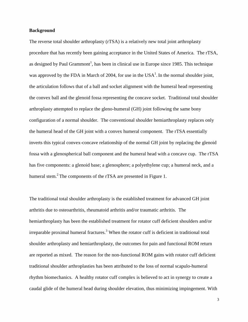
has five components: a glenoid base; a glenosphere; a polyethylene cup; a humeral neck, and a
humeral stem.2
The components of the rTSA are presented in Figure 1.
The traditional total shoulder arthroplasty is the established treatment for advanced GH joint
arthritis due to osteoarthritis, rheumatoid arthritis and/or traumatic arthritis. The
hemiarthroplasty has been the established treatment for rotator cuff deficient shoulders and/or
irreparable proximal humeral fractures.3 When the rotator cuff is deficient in traditional total
shoulder arthroplasty and hemiarthroplasty, the outcomes for pain and functional ROM return
are reported as mixed. The reason for the non-functional ROM gains with rotator cuff deficient
traditional shoulder arthroplasties has been attributed to the loss of normal scapulo-humeral
rhythm biomechanics. A healthy rotator cuff complex is believed to act in synergy to create a
caudal glide of the humeral head during shoulder elevation, thus minimizing impingement. With
4
the unopposed superior migration of the humeral head in a rotator cuff and deficient shoulder,
the deltoid muscle is unable to produce the proper arthrokinematics during shoulder elevation.
The resultant effect is the classic shoulder shrugging due to scapular elevation and upward
rotation with no significant elevation of the upper extremity away from the torso.
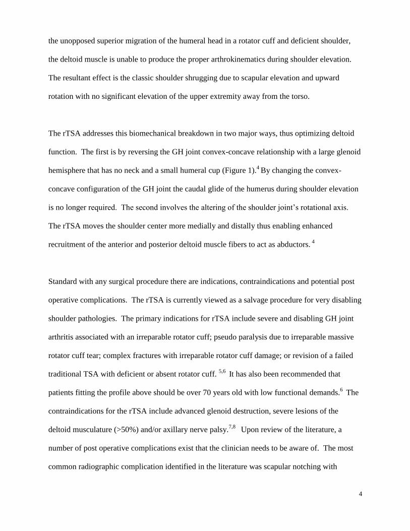
The rTSA addresses this biomechanical breakdown in two major ways, thus optimizing deltoid
function. The first is by reversing the GH joint convex-concave relationship with a large glenoid
hemisphere that has no neck and a small humeral cup (Figure 1).4
By changing the convex-
concave configuration of the GH joint the caudal glide of the humerus during shoulder elevation
is no longer required. The second involves the altering of the shoulder joint’s rotational axis.
The rTSA moves the shoulder center more medially and distally thus enabling enhanced
recruitment of the anterior and posterior deltoid muscle fibers to act as abductors. 4
Standard with any surgical procedure there are indications, contraindications and potential post
operative complications. The rTSA is currently viewed as a salvage procedure for very disabling
shoulder pathologies. The primary indications for rTSA include severe and disabling GH joint
arthritis associated with an irreparable rotator cuff; pseudo paralysis due to irreparable massive
rotator cuff tear; complex fractures with irreparable rotator cuff damage; or revision of a failed
traditional TSA with deficient or absent rotator cuff. 5,6
It has also been recommended that
patients fitting the profile above should be over 70 years old with low functional demands.6
The
contraindications for the rTSA include advanced glenoid destruction, severe lesions of the
deltoid musculature (>50%) and/or axillary nerve palsy.7,8
Upon review of the literature, a
number of post operative complications exist that the clinician needs to be aware of. The most
common radiographic complication identified in the literature was scapular notching with
5
inferior scapular notching being most common as compared to either posterior or anterior
notching.9
It appears that the scapular notching is mainly due to less than optimal positioning of
the glenoid component. Other complications that have been documented post operatively
included dislocation, infection, aseptic humeral loosening, peri-prosthetic fracture, late acromial
fracture and axillary nerve palsy.10,11
Outcomes for patients who underwent the rTSA procedure
tended to be better for etiologies of primary rotator cuff tear, primary osteoarthritis with rotator
cuff tear and massive rotator cuff tear versus etiologies of post traumatic arthritis and revisional
arthroplasty.12
For this particular new surgical procedure, the post operative therapy management research is
lagging behind. In a recent search, little to no peer reviewed literature was found regarding
therapy management for patients who underwent rTSA. The purpose of this case report
therefore is to describe the overall physical therapy management of a patient who underwent an
rTSA procedure. The intervention received by the patient entailed a post operative protocol
developed by the author in collaboration with the orthopedic surgeon who performed the surgery.
In developing the protocol, soft tissue healing principles and clinical decision making regarding
risk elimination for shoulder dislocation post arthroplasty were relied upon heavily.
6
Case Description
The patient was asked if her case could be used as a research project due to its relative
uniqueness and newness. The patient agreed and a research consent form was signed by the
patient.
History:
The patient was a 76 year old Caucasian female who presented with the medical diagnosis of left
shoulder OA/DJD, three weeks status post reverse total shoulder arthroplasty (rTSA). The patient
was seen by the physical therapist for initial evaluation three weeks post op per the orthopedic
surgeon’s request. During the initial three weeks post-op the patient was instructed to remain in
the sling and swathe for 24 hours except for hygiene and performance of pendulum swing
exercises 3-4 times per day. The patient was instructed by the physician that she could begin to
decrease sling usage after three weeks, per tolerance.
The surgical report was obtained by the evaluating physical therapist for review. The surgical
report indicated that the preoperative diagnoses were left shoulder large cyst and left shoulder
marked chronic rotator cuff deficiency. The surgical procedures performed were listed as:
excision of cyst and rTSA using a Zimmer anatomic shoulder inverse/reverse humeral cup; zero
degrees retro with 36mm glenoid head; an un-cemented anatomic shoulder inverse/reverse
glenoid component with two glenoid screws and a size 7 humeral cemented stem with the PE
insert for a 36mm polyethylene. Further review of the procedure description revealed that the
long head of the biceps was resected due to attenuation and the subscapularis muscle was taken
down off of the lesser tuberosity with later reattachment. The latter finding would have definite
effect on the protocol development as the complete rotator cuff was not deficient and
7
subscapularis protection during the post op management would need to be taken into
consideration.
Review of relevant past medical history revealed no previous left shoulder surgeries. The patient
reported that after the surgery she had to remain in the hospital for 4 days due to heart and blood
pressure related issues. The patient indicated on medical history intake and systems review that
she had a history of tachycardia and high blood pressure. At the time of the evaluation she
indicated that both were under control via medications. Heart rate was measured as 64 bpm and
blood pressure as 125/80 prior to commencement of treatment. Social history review revealed
that the patient was a retired homemaker whose husband had been providing home ADL support
as needed. The patient was a non-smoker and non-drinker. No other medical issues were
identified.
The patient reported current pain of 6/10 that was described as being dull and achy in nature.
The patient denied any left upper extremity paraesthesia. The patient reported her goal for
physical therapy was to regain maximum mobility and usage of her left shoulder.
Initial Examination:
Observation: The patient had a slightly endomorphic body type. The sling/swathe was removed,
the patient donned a gown and the involved shoulder girdle was observed. At the time of the
initial evaluation the sutures had already been removed and the surgical incision was closed with
signs of continued healing with no complication. The majority of the ecchymosis had also
resolved. Skin temperature was normal to touch.
8
ROM: At this juncture (3 weeks post-op) active and passive motion guidelines were established.
Since the subscapularis muscle was taken down and reattached, as seen with conventional total
shoulder replacements, and due to a lack of published protocol information available for the
rTSA, Brotzman’s Clinical Orthopedic Rehabilitation 2nd
edition13
was consulted to review their
protocol for a typical TSA. In order to remain consistent with their TSA protocol, the following
protocol parameters were adopted: no active internal rotation from 0-6 weeks post-op, active
internal rotation commencing between 6-12 weeks and progressing to resistive internal rotation
from 12 weeks post-op onwards.13
The next task was to determine the parameters that would indicate progression from passive
ROM activities to AROM for other movement planes. This was a difficult task as little to no
clearly stated post operative protocols were located in the literature search. In pioneering work
by Baulot, Charbernaud and Grammont from 1995, unrestricted AROM was commenced as early
as 2 weeks postoperative.14
The lack of soft tissue insult to the deltoid/periscapular musculature
and the long head of the bicep during resection in the present case, was a consideration in
following this proposed unrestricted AROM protocol. Therefore, abiding by the subscapularis
precautions listed above and the fact that this patient was three weeks post op, AROM was
assessed.
The final ROM task was to determine boundaries and progression. This also proved difficult due
to the paucity of established protocols and the fact that those that were located did not provide
detailed ROM boundaries. As has been standard rehabilitation considerations with conventional
TSA, shoulder flexion, abduction, external rotation and extension were restricted. Based upon
understanding of soft tissue healing principles and understanding of dislocation risks for an
9
rTSA, the following ROM guidelines were established for the Protective phase of 0-6 weeks and
are summarized in Table 1: 0-140 degrees shoulder flexion, 0-90 degrees shoulder abduction in
neutral rotation, 0-40 degrees external rotation, no shoulder extension past midline and no
combination shoulder extension/adduction/internal rotation i.e. reaching behind the back. All
progression would be gradual and done with proper neuromuscular control.
At the time of initial evaluation the patient’s ROM measurements were as follows: active
shoulder flexion 70º, passive shoulder flexion 130º, active shoulder abduction 70º, passive
shoulder abduction 90º, unable to perform active shoulder external rotation, passive shoulder
external rotation 40º, active shoulder internal rotation deferred and passive shoulder internal
rotation 50º (summarized in Table 4).
Muscle Performance: Isolated manual muscle testing against resistance was deferred at this
juncture as the patient was only 3 weeks post op. Grading active ROM attempts revealed the
patient’s ability to actively lift the arm against gravity as follows: shoulder flexion and abduction
were all graded as 3-/5, shoulder external rotation was graded as 2-/5 and shoulder internal
rotation grading was deferred. Elbow flexion, extension and general periscapular motion were all
graded as 3/5.
ADLs: The patient demonstrated functional limitations of being unable to work above the head,
being unable to work at shoulder level, restricted in lifting objects from any positional height and
restricted in reaching behind her back.
10
Barriers: The patient was orientated x 3. No emotional, language or learning barriers were
identified.
Evaluation: According to the Guide to Physical Therapy Practice, 2nd
edition, the patient was
classified as falling within pattern 4I.15
Pattern 4I is defined as ‘impaired joint mobility, motor
function, muscle performance, and range of motion associated with bony or soft tissue surgery’.
The patient had a good prognosis. An excellent prognosis was not given due to the fact that
rTSA is considered a salvage procedure and full ROM and strength return is not a typical
expectation following this procedure.16
Intervention:
The patient attended Physical Therapy three times a week for a total of 10 weeks. The patient
completed a total of 30 visits. The treatment consisted of progression through the protocol
presented in Tables 1-3 which clearly show the breakdown of each of the three phases with
regard to precautions, goals, therapeutic interventions and progression. The protocol was sent to
the referring orthopedic surgeon for review. The surgeon reviewed and agreed to the guidelines
and intervention progression.
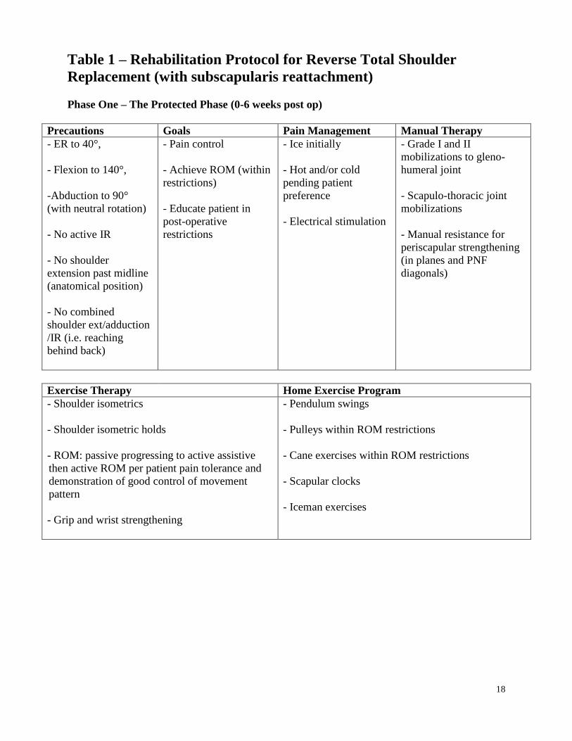
During the Protected phase of 0-6 weeks, the patient was educated on the previously stated ROM
restrictions (Table 1). The patient was also instructed that she was able to wean herself from the
sling per tolerance, however the patient was instructed to wear the sling in public for the next
three weeks for protection. Since the patient at the initial evaluation ROM assessment
demonstrated passive ROM near the restricted level, PROM was done gently and with caution as
not to create instability. Grade I and II mobilizations were not performed due to significant early
11
PROM ability. The exercises at this point focused on creating stability and enhanced
neuromuscular control. The modalities of cold pack and interferential stimulation were used for
pain management. The goals of the Protective phase were to decrease pain, increase ROM to
restricted range, and educate patient on post op restrictions.
Phase II, the Restoration phase, was begun at 6 weeks and extended to approximately 12 weeks
post op. Criteria for progression included minimal pain and achievement of proper
biomechanical controlled ROM. Precautions included no heavy lifting, no strenuous ADLs and
no resistive exercise to the subscapularis i.e. no resistive internal rotation. This was to ensure
sufficient healing of the reattached subscapularis muscle and to allow the therapist to gradually
increase the activity of subscapularis in a more controlled manor. The goals of phase II were to
maximize ROM without creating instability and progressively regain strength and neuromuscular
control with movement. Once proper biomechanics were achieved with AROM, light resistance
was initiated to the upper extremity. Initially isotonic exercise in short lever positions that
isolated muscle groups were implemented then progression to isotonic exercises that used long
lever arms with coupled muscle groups were executed.
Phase III, the Functional Activity phase began at 12 weeks post op. If pain was minimal and
sound strength allowing proper biomechanical controlled movement was seen, the patient was
allowed to resume functional activities with the upper extremity. Gradual return to leisure ADLs
was permitted with avoidance of high impact activity that required sudden lifting or
pushing/pulling motions. Exercises were progressed to include resistive exercise to the
subscapularis (i.e. resistive internal rotation) and more advanced resistive exercise were
implemented, including PNF high and low D1 and D2 thera-band resistance patterns. Exercises
12
from Phase II were continued with increase in volume and intensity. A home maintenance
program was established and adjusted appropriately as progress plateaued.
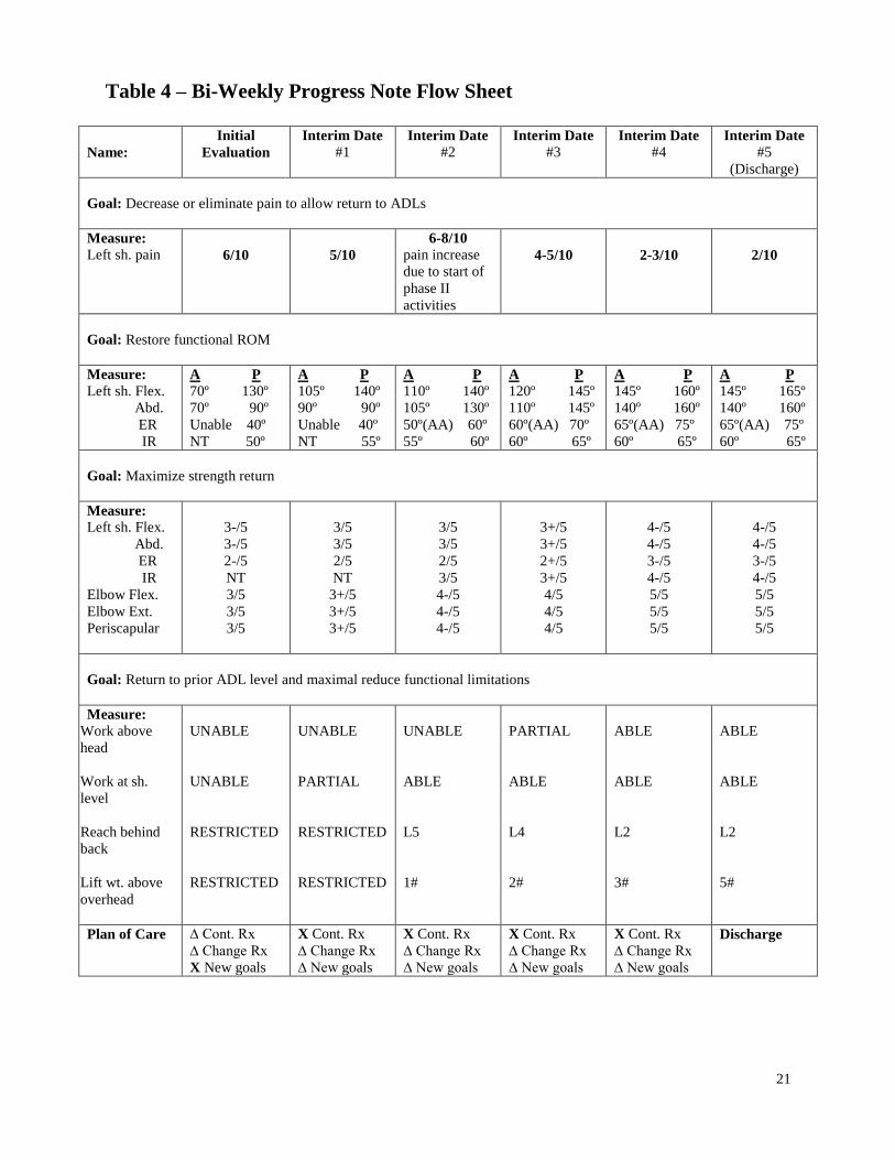
Outcomes:
The patient was reassessed every two weeks toward the established goals set at the time of the
initial evaluation. A total of five interim reassessments were completed, including the final
discharge. The goals established were impairment goals of pain, ROM and strength and
functional limitation goals of material handling and non-material handling. The patient status at
the time of initial evaluation was that of a patient with moderate pain in the left shoulder
accompanied by significant loss of active and passive range of motion. Full assessment was
carried out at 5 interim time points, roughly 2 weeks apart and the results of each of these interim
evaluations are presented in Table 4. The patient showed steady gains from each interim
assessment to the next, except between assessment 4 and 5, when ROM and strength did not
change. At that juncture it was felt that the patient had reached her near maximum medical
improvement and was ready for commencement of a home maintenance program. The patient
status at the time of discharge, as clearly presented in Table 4 and Figures 2, 3 and 4 was
minimal pain in the left shoulder with minimal loss of both active and passive ranges of motion
in all movements. Muscle strength had improved and was generally just below that of the
opposite limb and elbow range of motion and strength were normal. With regard to general
mobility of the upper extremity, the patient was able to reach overhead, to reach behind her back
to the level of L2, and able to lift 5lbs overhead and lift 10lbs from floor to waist and waist to
shoulder with the affected arm.
Discussion
13
The intent of this case report was to report on a post operative management protocol for a
relatively new surgical procedure, rTSA, which is gaining popularity, for the surgical
management of rotator cuff deficient shoulder arthrosis pathologies. Despite the current
insufficiency in the literature regarding post operative physical therapy management, the
protocol developed here appeared to have provided the patient good outcomes without
complication. The literature did provide data regarding shoulder elevation changes pre and post
rTSA. Indeed, Sirveaux et al, in a study of 80 patients who underwent a rTSA, showed a mean
active forward elevation increase from 73º to 138º17
, however little information was given as to
the exact protocol used to achieve these results. In another study by Frankle et al, 60 patients
who underwent rTSA demonstrated a mean increase of shoulder flexion from 55º to 105.1º and
increased shoulder abduction from 41.4º to 101.8º.18
Once again there was little detail provided
as to the rehabilitation protocol used to achieve these results. The patient in this present case
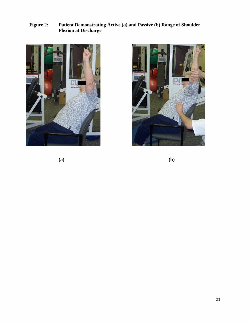
report demonstrated increases in both active shoulder flexion and abduction, as seen in Figures 2
and 3, from 70º to 145º and from 70º to 140º, respectively. The patient presented here did not
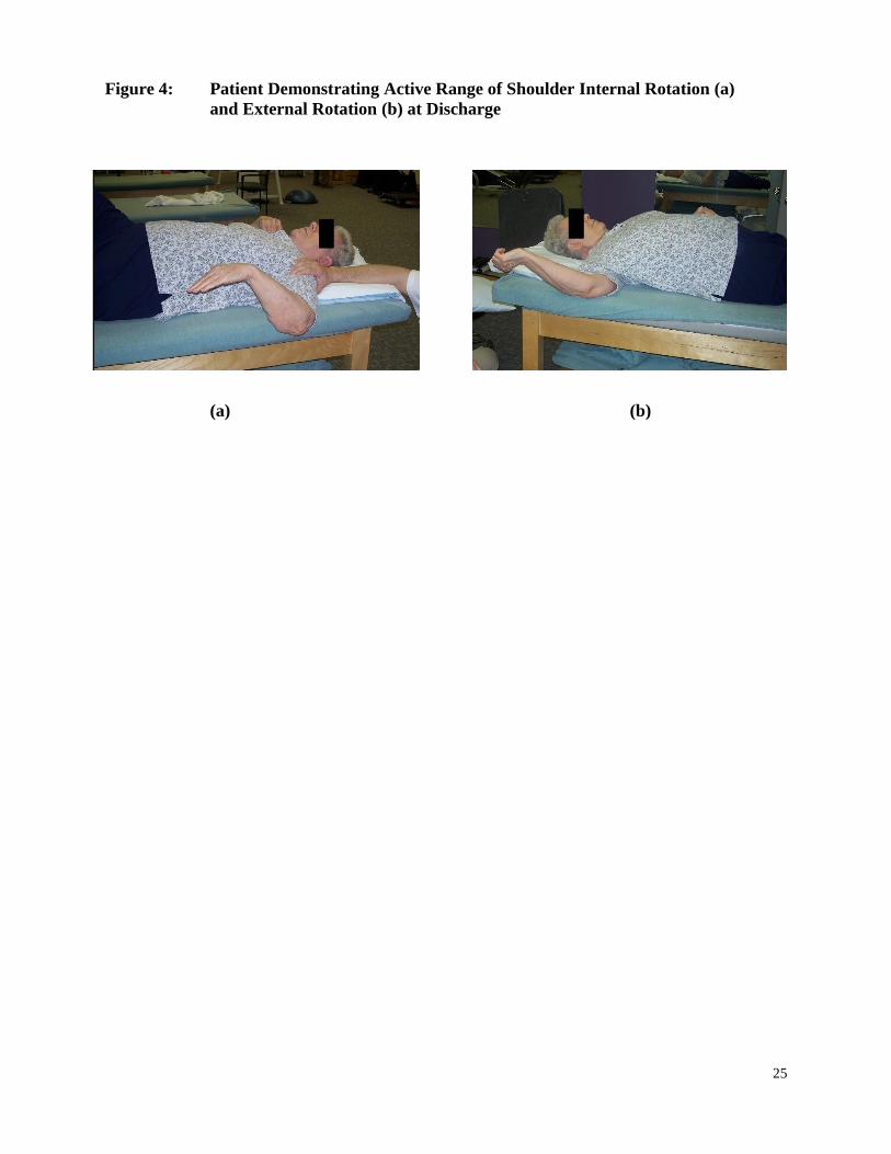
regain full active shoulder external rotation, as seen in Figure 4, and unfortunately developed a
positive hornblower’s sign. The hornblower’s sign is where the examiner supports the patient’s
arm at 90º abduction in the plane of the scapula, the elbow is flexed to 90º and the patient is
asked to externally rotate the forearm against the resistance of the examiner’s hand.19
If the
patient cannot externally rotate the shoulder then the patient is deemed to have a positive
hornblower’s sign, indicating a deficient teres minor muscle. The relevance of this finding
suggests that although the rTSA was successful in restoring shoulder elevation via inverting the
bony configuration of the joint and changing the joint axis of the shoulder, it may not have been
successful in allowing the deltoid muscle to function as an effective external rotator. Gerber et
14
al reiterated this finding with the suggestion of combining a latissimus dorsi transfer and rTSA to
address the lack of return of external rotation and subsequent positive hornblower’s sign.20
Limitations of this case report, as with many case reports, is lack of applicability to a greater
patient population and cause-effect relationship via randomized controls. Another limitation
would be not using standardized shoulder score tables such as the American Shoulder and Elbow
Surgeons Score or Simple Shoulder Test to further objectify functional outcomes with this
protocol. A final limitation would be not assessing the contribution of upward rotation of the
scapula in contributing to shoulder elevation. The scapulohumeral rhythm of a normal shoulder
has roughly a 2:1 ratio of full shoulder elevation occurring at the GH joint and scapula,
respectively. A few of the available studies on this subject have indicated that with the rTSA the
scapulohumeral rhythm changes and upward rotation of the scapula contributes to a greater
degree of shoulder elevation than seen with a normal shoulder.21,22
A future research possibility would be the ongoing application of the established protocol to a
larger patient population to determine its value. Another future research possibility would be
using standardized shoulder tests with this protocol to assess its expanded applicability to
patient’s functional needs
Conclusion
The rTSA has been gaining acceptance and increased use since its FDA approval in 2004.
Establishing best practice post operative therapy management of this surgical procedure is
critical to the success of this procedure and the welfare of the patients who undergo this surgery.
This case report established a protocol for post operative management for the rTSA. The patient
15
who underwent the physical therapy treatment following this protocol had successful outcomes
without iatrogenic complications. Therefore, the author believes that the protocol established
could be a viable guideline for future post operative therapy management of patients who have
undergone an rTSA procedure. Further studies applying this protocol to a larger patient
population would be beneficial to assist in determining its wider effectiveness.
16
Reference List
1. McFarland EG, Sanguanjit P, et al. The reverse shoulder prosthesis: A review of imaging
features and complications. Skeletal Radiol. 2006 Jul;35(7):488-96.
2. Matsen FA 3rd
, Boileau P, Walch G, Gerber C, Bicknell RT. The reverse total shoulder
arthroplasty. J Bone Joint Surg Am. 2007 Mar;89(3):660-7
3. Levy J, Frankie M, Mighell M, Pupello D. The use of the reverse shoulder prosthesis for
the treatment of failed hemiarthroplasty for proximal humeral fracture. J Bone Joint Surg
Am. 2007Feb;89(2):292-300
4. Boileau P, Watkinson DJ, Hatzidakis AM, Baig F. Grammont reverse prosthesis: design,
rationale, and biomechanics. J Shoulder Elbow Surg. 2005 Jan-Feb;14(1 Suppl S):147S-
1661S
5. Werner CM, Steinmann PA, Gilbart M, Gerber C. Treatment of painful pseudoparesis
due to irreparable rotator cuff dysfunction with the Delta III rreverse-ball-and-socket total
shoulder prosthesis. . J Bone Joint Surg Am. 2005 Jul;87(7):1476-86
6. Guery J, Favard L, Sirveaux F, Oudet D, Mole D, Walch G. Reverse total shoulder
arthroplasty. Survivorship analysis of eighty replacements followed for five to ten years. .
J Bone Joint Surg Am. 2006 Aug;88(8):1742-7
7. Gohlk F, Rolf O. Revision of failed fracture hemiarthroplasties to reverse total shoulder
prosthesis through the transhumeral approach: method incorporating a pectoralis-major-
pedicled bone window. Oper Orthop Traumatol. 2007 Jun:19(2):185-208
8. Seebauer L, Walter W, Keyl W. Reverse total shoulder arthroplasty for the treatment of
defect arthropathy. Oper Orthop Traumatol. 2005 Feb;17(1):1-24
9. Simovitch RW, Zumstein MA, Lohri E, Helmy N, Gerber C. Predictors of scapular
notching in patients managed with the Delta III reverse total shoulder replacement. . J
Bone Joint Surg Am. 2007 Mar;89(3):588-600
10. Boileau P, Watkinson D, Hatzidakis AM, Hovorka I. Neer Award 2005: The Grammont
reverse shoulder prosthesis: results in cuff tear arthritis, fracture sequelae, and revision
arthroplasty. J Shoulder Elbow Surg. 2006 Sept-Oct;15(5):527-40
11. Rittmeister M, Kerschbaumer F. Grammont reverse total shoulder arthroplasty in patients
with rheumatoid arthritis and nonreconstructible rotator cuff lesions. J Shoulder Elbow
Surg. 2001 Jan-Feb;10(1):17-22
12. Wall B, Nove-Josserand L, et al. Reverse total shoulder arthroplasty: a review of results
according to etiology. J Bone Joint Surg Am. 2007 Jul;89(7):1476-85
13. Brotzman SB, Wilk K. Clinical Orthopaedic Rehabilitation. 2nd
edition Philadelphia, PA:
Mosby; 2003.
17
14. Baulot E, Chabernaud D, Grammont PM. Results of Grammont’s inverted prosthesis in
omarthritis associated with major cuff destruction. Acta Orthop Belg. 1995;61 Suppl
1:112-9
15. Rothstein J. Guide to Physical Therapy Practice. 2nd
edition. Phys Ther. 2001;81:9-744.
16. Dines JS, Fealy s, et al. Outcomes analysis of revision total shoulder replacement. J
Bone Joint Surg Am. 2006 Jul;88(7):1494-500
17. Sirceaux F, Favard L, et al. Grammont inverted total shoulder arthroplasty in the
treatment of glneohumeral osteoarthritis with massive rupture of the cuff. Results of a
multicentre study of 80 shoulders. J Bone Joint Surg Br. 2004 Apr;86(3):388-95.
18. Frankle M, Siegal S, et al. The reverse shoulder prosthesis for glenohumeral arthritis
associated with sevre rotator cuff deficiency. A minimum two year follow-up study of
sixty patients. J Bone Joint Surg Am. 2005 Aug;87(8):1697-705.
19. Walch G, Boulahia A, et al. The ‘dropping’ and ‘hornblower’s sign in evaluation of
rotator cuff trears. J Bone Joint Surg Br. 1988 Jul;80(4):624-9
20. Gerber C, Pennington SD, Lingerfelter EJ, Sukhankar A. Reverse Delta III total shoulder
replacement combined with latissimus dorsi transfer. A preliminary report. J Bone Joint
Surg Am. 2007 May;89(5):940-7
21. Mahfouz M, Nicholon G, Komistek R, Kubo M. In vivo determination of the dynamics
of normal rotator cuff deficient total and reverse replacements shoulders. J Bone Joint
Surg Am. 2005;87 Suppl 2:107-13.
22. Kontaxis A, Johnson GR. Adaptation of scapula lateral rotation after reverse anatomy
shoulder replacement. Comput Methods Biomech Engin. 2007 Oct; 15(1)
18
Table 1 – Rehabilitation Protocol for Reverse Total Shoulder
Replacement (with subscapularis reattachment)
Phase One – The Protected Phase (0-6 weeks post op)
Precautions Goals Pain Management Manual Therapy
- ER to 40°,
- Flexion to 140°,
-Abduction to 90°
(with neutral rotation)
- No active IR
- No shoulder
extension past midline
(anatomical position)
- No combined
shoulder ext/adduction
/IR (i.e. reaching
behind back)
- Pain control
- Achieve ROM (within
restrictions)
- Educate patient in
post-operative
restrictions
- Ice initially
- Hot and/or cold
pending patient
preference
- Electrical stimulation
- Grade I and II
mobilizations to gleno-
humeral joint
- Scapulo-thoracic joint
mobilizations
- Manual resistance for
periscapular strengthening
(in planes and PNF
diagonals)
Exercise Therapy Home Exercise Program
- Shoulder isometrics
- Shoulder isometric holds
- ROM: passive progressing to active assistive
then active ROM per patient pain tolerance and
demonstration of good control of movement
pattern
- Grip and wrist strengthening
- Pendulum swings
- Pulleys within ROM restrictions
- Cane exercises within ROM restrictions
- Scapular clocks
- Iceman exercises
19
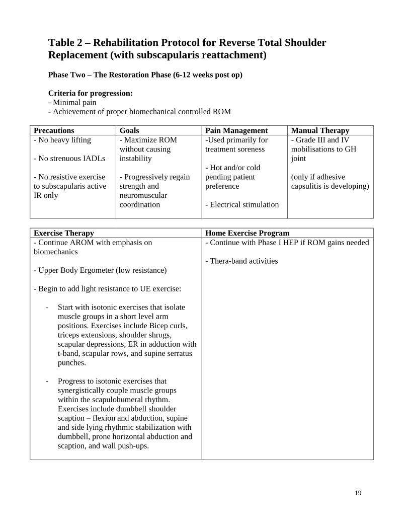
Table 2 – Rehabilitation Protocol for Reverse Total Shoulder
Replacement (with subscapularis reattachment)
Phase Two – The Restoration Phase (6-12 weeks post op)
Criteria for progression:
- Minimal pain
- Achievement of proper biomechanical controlled ROM
Precautions Goals Pain Management Manual Therapy
- No heavy lifting
- No strenuous IADLs
- No resistive exercise
to subscapularis active
IR only
- Maximize ROM
without causing
instability
- Progressively regain
strength and
neuromuscular
coordination
-Used primarily for
treatment soreness
- Hot and/or cold
pending patient
preference
- Electrical stimulation
- Grade III and IV
mobilisations to GH
joint
(only if adhesive
capsulitis is developing)
Exercise Therapy Home Exercise Program
- Continue AROM with emphasis on
biomechanics
- Upper Body Ergometer (low resistance)
- Begin to add light resistance to UE exercise:
- Start with isotonic exercises that isolate
muscle groups in a short level arm
positions. Exercises include Bicep curls,
triceps extensions, shoulder shrugs,
scapular depressions, ER in adduction with
t-band, scapular rows, and supine serratus
punches.
- Progress to isotonic exercises that
synergistically couple muscle groups
within the scapulohumeral rhythm.
Exercises include dumbbell shoulder
scaption – flexion and abduction, supine
and side lying rhythmic stabilization with
dumbbell, prone horizontal abduction and
scaption, and wall push-ups.
- Continue with Phase I HEP if ROM gains needed
- Thera-band activities
20
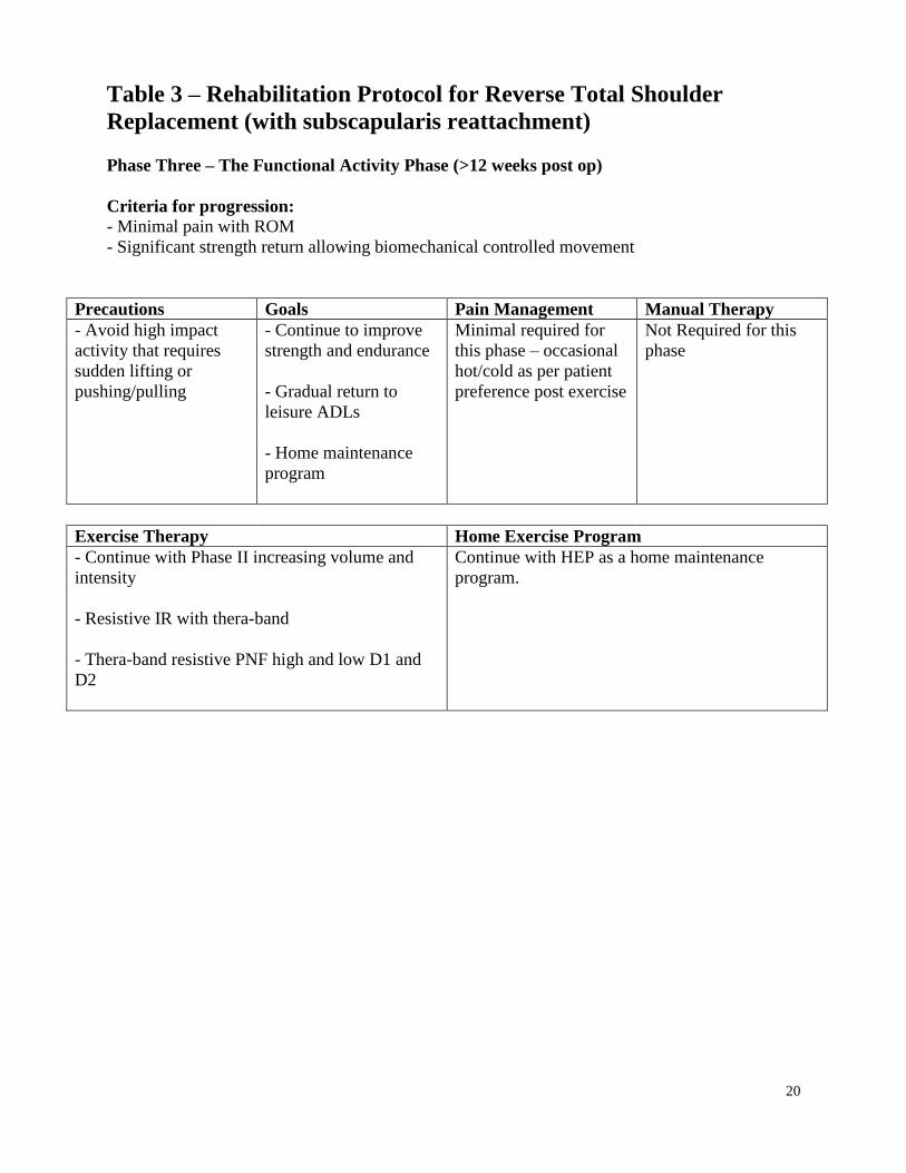
Table 3 – Rehabilitation Protocol for Reverse Total Shoulder
Replacement (with subscapularis reattachment)
Phase Three – The Functional Activity Phase (>12 weeks post op)
Criteria for progression:
- Minimal pain with ROM
- Significant strength return allowing biomechanical controlled movement
Precautions Goals Pain Management Manual Therapy
- Avoid high impact
activity that requires
sudden lifting or
pushing/pulling
- Continue to improve
strength and endurance
- Gradual return to
leisure ADLs
- Home maintenance
program
Minimal required for
this phase – occasional
hot/cold as per patient
preference post exercise
Not Required for this
phase
Exercise Therapy Home Exercise Program
- Continue with Phase II increasing volume and
intensity
- Resistive IR with thera-band
- Thera-band resistive PNF high and low D1 and
D2
Continue with HEP as a home maintenance
program.
21
Table 4 – Bi-Weekly Progress Note Flow Sheet
Name:
Initial
Evaluation
Interim Date
#1 Interim Date
#2 Interim Date
#3 Interim Date
#4 Interim Date
#5
(Discharge)
Goal: Decrease or eliminate pain to allow return to ADLs
Measure:
Left sh. pain
6/10
5/10
6-8/10
pain increase
due to start of
phase II
activities
4-5/10
2-3/10
2/10
Goal: Restore functional ROM
Measure:
Left sh. Flex.
Abd.
ER
IR
A P
70º 130º
70º 90º
Unable 40º
NT 50º
A P
105º 140º
90º 90º
Unable 40º
NT 55º
A P
110º 140º
105º 130º
50º(AA) 60º
55º 60º
A P
120º 145º
110º 145º
60º(AA) 70º
60º 65º
A P
145º 160º
140º 160º
65º(AA) 75º
60º 65º
A P
145º 165º
140º 160º
65º(AA) 75º
60º 65º
Goal: Maximize strength return
Measure:
Left sh. Flex.
Abd.
ER
IR
Elbow Flex.
Elbow Ext.
Periscapular
3-/5
3-/5
2-/5
NT
3/5
3/5
3/5
3/5
3/5
2/5
NT
3+/5
3+/5
3+/5
3/5
3/5
2/5
3/5
4-/5
4-/5
4-/5
3+/5
3+/5
2+/5
3+/5
4/5
4/5
4/5
4-/5
4-/5
3-/5
4-/5
5/5
5/5
5/5
4-/5
4-/5
3-/5
4-/5
5/5
5/5
5/5
Goal: Return to prior ADL level and maximal reduce functional limitations
Measure:
Work above
head
Work at sh.
level
Reach behind
back
Lift wt. above
overhead
UNABLE
UNABLE
RESTRICTED
RESTRICTED
UNABLE
PARTIAL
RESTRICTED
RESTRICTED
UNABLE
ABLE
L5
1#
PARTIAL
ABLE
L4
2#
ABLE
ABLE
L2
3#
ABLE
ABLE
L2
5#
Plan of Care
∆ Cont. Rx
∆ Change Rx
X New goals
X Cont. Rx
∆ Change Rx
∆ New goals
X Cont. Rx
∆ Change Rx
∆ New goals
X Cont. Rx
∆ Change Rx
∆ New goals
X Cont. Rx
∆ Change Rx
∆ New goals
Discharge

22
Figure 1: Surgical Component Parts of the typical Reverse Total Shoulder
Arthroplasty
Image used with permission from Richard J Friedman, MD, Charleston Orthopaedic Associates,
Charleston, SC
23
Figure 2: Patient Demonstrating Active (a) and Passive (b) Range of Shoulder
Flexion at Discharge
(a) (b)

24
Figure 3: Patient Demonstrating Active (a) and Passive (b) Range of Shoulder
Abduction at Discharge
(a) (b)
25
Figure 4: Patient Demonstrating Active Range of Shoulder Internal Rotation (a)
and External Rotation (b) at Discharge
(a) (b)