Embed Size (px)
Citation preview
1
Post-translational covalent assembly of CAR and synNotch receptors for programmable antigen targeting Jason Lohmueller1,2,3,*, Adam A. Butchy4, Yaniv Tivon5, Michael Kvorjak1,2, Natasa Miskov-Zivanov4,6,7, Alexander Deiters5, Olivera J. Finn2 Affiliations: 1University of Pittsburgh School of Medicine, Department of Surgery, Pittsburgh, PA USA
2University of Pittsburgh School of Medicine, Department of Immunology, Pittsburgh, PA USA
3University of Pittsburgh, Center for Systems Immunology, Pittsburgh, PA USA
4University of Pittsburgh, Department of Bioengineering, Pittsburgh, PA USA
5University of Pittsburgh, Department of Chemistry, Pittsburgh, PA USA
6University of Pittsburgh, Department of Electrical and Computer Engineering, Pittsburgh, PA
USA
7University of Pittsburgh, Department of Computational and Systems Biology, Pittsburgh, PA
USA
Classification: Biological Sciences/ Immunology and Inflammation
Keywords: synthetic biology, immunotherapy, adoptive cell therapy, bioconjugation
*To whom correspondence should be addressed:
Jason Lohmueller
UPMC Hillman Cancer Center
5117 Centre Ave
Pittsburgh, PA 15232, USA
Telephone: 603-819-9492
Email: [email protected]
(which was not certified by peer review) is the author/funder. All rights reserved. No reuse allowed without permission. The copyright holder for this preprintthis version posted January 17, 2020. . https://doi.org/10.1101/2020.01.17.909895doi: bioRxiv preprint
(which was not certified by peer review) is the author/funder. All rights reserved. No reuse allowed without permission. The copyright holder for this preprintthis version posted January 17, 2020. . https://doi.org/10.1101/2020.01.17.909895doi: bioRxiv preprint
(which was not certified by peer review) is the author/funder. All rights reserved. No reuse allowed without permission. The copyright holder for this preprintthis version posted January 17, 2020. . https://doi.org/10.1101/2020.01.17.909895doi: bioRxiv preprint
(which was not certified by peer review) is the author/funder. All rights reserved. No reuse allowed without permission. The copyright holder for this preprintthis version posted January 17, 2020. . https://doi.org/10.1101/2020.01.17.909895doi: bioRxiv preprint
(which was not certified by peer review) is the author/funder. All rights reserved. No reuse allowed without permission. The copyright holder for this preprintthis version posted January 17, 2020. . https://doi.org/10.1101/2020.01.17.909895doi: bioRxiv preprint
(which was not certified by peer review) is the author/funder. All rights reserved. No reuse allowed without permission. The copyright holder for this preprintthis version posted January 17, 2020. . https://doi.org/10.1101/2020.01.17.909895doi: bioRxiv preprint
2
Abstract
Chimeric antigen receptors (CARs) and synthetic Notch (synNotch) receptors are engineered
cell-surface receptors that sense a target antigen and respond by activating T cell receptor
signaling or a customized gene program, respectively. To expand the targeting capabilities of
these receptors, we have developed switchable adaptor receptor systems for which receptor
specificity can be directed post-translationally via covalent attachment of a co-administered
antibody. Instead of directly targeting an antigen, our receptors contain the SNAPtag self-
labeling enzyme, which reacts with benzylguanine (BG)-conjugated antibodies to assemble
covalently-associated antigen receptors. We demonstrate that activation of SNAP-CAR and
SNAP-synNotch receptors and their downstream effector functions can be successfully targeted
by several clinically-relevant BG-conjugated antibodies in an antigen-specific and antibody
dose-dependent manner. To better define parameters affecting receptor signaling, we developed
a mathematical model of switchable receptor systems. SNAP receptors provide a powerful new
strategy to post-translationally reprogram the targeting specificity of engineered cells.
(which was not certified by peer review) is the author/funder. All rights reserved. No reuse allowed without permission. The copyright holder for this preprintthis version posted January 17, 2020. . https://doi.org/10.1101/2020.01.17.909895doi: bioRxiv preprint
3
Introduction
Engineered antigen receptors are revolutionizing the treatment of blood cancers and show
promise in cell therapies treating a wide range of other diseases1. The most clinically advanced
of these technologies are chimeric antigen receptors (CARs), synthetic T cell receptors most
often comprised of an antigen-specific antibody single chain variable fragment (scFv) fused by
spacer and transmembrane domains to intracellular T cell signaling domains2-4. Upon binding to
a target antigen, CARs stimulate T cell activation and effector functions including cytokine
production, cell proliferation, and target cell lysis. Adoptively transferred CAR T cells targeting
the B cell antigen CD19 are now FDA-approved and have been highly successful in treating
refractory acute lymphoblastic leukemia5-7. Creating CARs against additional targets to treat
other types of cancer and immune-related diseases is a major research focus8,9. Another class of
highly versatile antigen receptors are synthetic Notch, “synNotch” receptors which consist of an
antigen binding domain, the Notch core protein from the Notch/Delta signaling pathway, and a
transcription factor 10-12. Instead of activating T cell signaling upon binding to the target antigen,
the Notch core protein is cleaved by endogenous cell proteases thus releasing the transcription
factor from the cell membrane. Subsequent nuclear translocalization leads to transcriptional
regulation of one or more target genes. These receptors are highly modular, they can be created
to target different cell surface antigens by changing the scFv, and can positively or negatively
regulate any gene of interest by either fusing different transcription factors as components of the
receptors or by changing the transgenes under their control. This versatile receptor type receives
increasing clinical interest in immunotherapies as well as applications to tissue engineering13,14.
To gain additional control over CAR function, we and others have developed
“switchable” adaptor CAR systems for which the CAR, instead of directly binding to an antigen
(which was not certified by peer review) is the author/funder. All rights reserved. No reuse allowed without permission. The copyright holder for this preprintthis version posted January 17, 2020. . https://doi.org/10.1101/2020.01.17.909895doi: bioRxiv preprint

4
on a target cell, binds to common tag molecule fused or conjugated to an antigen-specific
antibody15-21. These systems are designed such that a patient would be infused with both a
tagged, antigen-specific antibody that binds to target cells and CAR T cells that react with the
tagged antibody on the surface of target cells. Adaptor CARs are also referred to as “universal
CARs” as they have the potential to allow for one population of T cells to target multiple tumor
antigens by administering different antibodies sequentially or simultaneously. Additionally, the
activity of the adaptor CARs can be tuned by altering the concentration of tagged antibodies or
halting antibody administration for better control over potential toxicities resulting from over-
active CAR T cells. Adaptor CAR systems have been developed recognizing a variety of
peptides or small molecules conjugated to antibodies including biotin, fluorescein isothiocyanate
(FITC), peptide neo-epitopes (PNE), Fc�, and leucine zippers, and the first adaptor CAR system
is currently in clinical trials15-21.
Here, we describe key advances in antigen receptor design – the creation of a switchable
adaptor synNotch system and the creation of a novel universal CAR system that acts through
self-labeling enzyme chemistry. Our first attempt to create a switchable adaptor synNotch system
that functioned through transient binding of the receptor to an antibody was unsuccessful, and we
reasoned that a stronger antibody/receptor interaction would be necessary. Seeking to create a
self-labeling synNotch receptor that would perform a chemical reaction to covalently fuse with
the adaptor antibody, we generated a synNotch receptor containing the SNAPtag protein.
SNAPtag is a modified human O-6-methylguanine-DNA methyltransferase (MGMT) that was
engineered to react to benzylguanine, a bio-orthogonal tag molecule, and is known to be specific
and efficient at self-labeling in both cells and animals (Fig. 1a)22-24. As previous literature for
adaptor CARs has shown a positive correlation between CAR function and the CAR/adaptor
(which was not certified by peer review) is the author/funder. All rights reserved. No reuse allowed without permission. The copyright holder for this preprintthis version posted January 17, 2020. . https://doi.org/10.1101/2020.01.17.909895doi: bioRxiv preprint

5
binding affinity, we also created and characterized a CAR containing the SNAPtag15,18. The
SNAP-CAR and SNAP-synNotch systems are highly modular receptor platforms for diverse
programming of cell behaviors using covalent chemistry (Fig. 1b,c,d).
Results
Engineering a self-labeling SNAP switchable adaptor synNotch receptor. We first created
and tested a putative universal synNotch system with a biotin-avidin tag-receptor interaction
using the biotin-binding protein, ‘mSA2’ as the targeting domain. The goal of this system was to
target receptor specificity by combining mSA2-synNotch cells with biotinylated antibodies,
similar to our previously reported mSA2 CAR T cell system 17. We cloned the mSA2 synNotch
receptor into a lentiviral expression vector and transduced Jurkat cells with the receptor along
with a Gal4-driven TagBFP reporter gene (Supplementary Fig. 1a). In brief, the process of
synNotch receptor activation culminates in the release of the Gal4-VP64 transcription factor
from the membrane and transcriptional activation of the Gal4 target gene, for which we are using
the TagBFP reporter. Assaying the cells by flow cytometry, we found that the receptor was
efficiently expressed on the cell surface (Supplementary Fig. 1b). Furthermore, incubating
mSA2-synNotch cells with plate-immobilized biotin showed TagBFP response gene activation
(Supplementary Fig. 1c). However, the receptor was ultimately not functional at detecting cell-
surface antigen, as we saw no receptor activation when we incubated the cells with biotinylated
antibody-labeled tumor cells (Supplementary Fig. 1d). We posited that the lack of signaling by
the mSA2 synNotch receptor, in contrast to potent signaling by mSA2 CAR T cells, was the
result of the Notch receptor’s differing signaling mechanism that requires a pulling force25. We
(which was not certified by peer review) is the author/funder. All rights reserved. No reuse allowed without permission. The copyright holder for this preprintthis version posted January 17, 2020. . https://doi.org/10.1101/2020.01.17.909895doi: bioRxiv preprint

6
reasoned that a stronger receptor to tag binding interaction (mSA2-biotin Kd = 5.5x10⁻⁹ M) may
be required to create a functional, universally applicable synNotch system26.
Thus, we next created a synNotch receptor containing the SNAPtag self-labeling enzyme
which forms a covalent bond via a benzene ring with a BG-tagged molecule (Fig. 2a)23,24,27. The
goal of this system was to direct receptor activation by combining SNAP-synNotch cells with
BG-labeled antibodies (Fig. 1b,c). We generated a lentiviral vector encoding the SNAP-
synNotch receptor and transduced Jurkat cells (Fig. 2b). Antibody labeling of the myc epitope
tag and labeling with a fluorophore-conjugated BG reagent confirmed cell surface expression of
the receptor and SNAP-BG cell-surface labeling activity (Fig. 2c).
To generate the adaptor antibodies, we conjugated BG to lysines or N-termini of several
clinically-relevant antibodies using a synthetic BG-NHS ester (Fig. 1a). These antibodies
included Rituximab targeting CD20, FMC63 targeting CD19, Herceptin targeting HER2, and
Cetuximab targeting EGFR28-31. While the conjugation products were heterogenous, we
quantified the average number of BG molecules conjugated to each antibody by a SNAP protein-
labeling assay in which SNAP-conjugation led to a shift in the antibodies’ molecular weight that
could be resolved by SDS-PAGE (Supplementary Fig. S2). The frequency of BG molecules per
antibody ranged from 2.0-2.8 as summarized in Supplementary Table S1. We characterized the
antigen expression and BG-antibody staining of various target cell lines by flow cytometry in
which the antibodies displayed expected antigen specificities (Supplementary Fig. S3).
Next, we tested the BG-conjugated FMC63 antibody (FMC63-BG) for its ability to
activate synNotch signaling in response to CD19 positive tumor cells. We performed a co-
incubation assay of SNAP-synNotch cells and CD19 positive and negative tumor cells in the
presence of different levels of the FMC63-BG antibody conjugate, and after 48 hours we assayed
(which was not certified by peer review) is the author/funder. All rights reserved. No reuse allowed without permission. The copyright holder for this preprintthis version posted January 17, 2020. . https://doi.org/10.1101/2020.01.17.909895doi: bioRxiv preprint
7
for TagBFP response gene expression by flow cytometry. TagBFP expression was significantly
up-regulated in response to CD19 positive tumor cells for various concentrations of antibody.
Receptor activation was sensitive, with significant activation observed at an antibody
concentration as low as 0.04 μg/mL and increasing to a peak at 0.25 μg/mL. Response gene
activation then decreased with increasing antibody amounts before being completely inhibited at
a concentration of 10 μg/mL, indicative of a “hook effect”, in which case the antibody is in such
excess that different antibody molecules are saturating both the target cells and synNotch cells
without formation of ternary complexes. This behavior is commonly observed with chemical and
cell processes that involve ternary complex formation (Fig. 2d) 32.
We found that BG-conjugated antibodies targeting other antigens were also capable of
activating the SNAP-synNotch receptor in an antigen-specific manner (Fig. 2d). We performed
similar co-incubation assays of SNAP-synNotch cells and antigen positive and negative tumor
cells in the presence of different levels of BG-conjugated Cetuximab, Herceptin, and Rituximab
antibodies and assayed for TagBFP response gene expression by flow cytometry. Significant up-
regulation of TagBFP was again observed for each of the tested antibodies, tunable through
increasing antibody concentrations, and in a target antigen-specific manner. A similar activation
pattern was observed for each of the antibodies – an increase with the antibody dose until a peak
activation level followed by an antibody dose-dependent decrease. Receptor activation was also
target cell dose-dependent, having optimal activity at high target to SNAP-synNotch cell ratios
(Supplementary Fig. S4).
Next, we tested whether SNAP-synNotch receptor was capable of regulating expression
of the IL-7 response gene, a candidate therapeutic gene of interest for its ability to promote T cell
proliferation33. We generated an IL-7 response gene expression construct in which the TagBFP
(which was not certified by peer review) is the author/funder. All rights reserved. No reuse allowed without permission. The copyright holder for this preprintthis version posted January 17, 2020. . https://doi.org/10.1101/2020.01.17.909895doi: bioRxiv preprint

8
gene was replaced by the IL-7 coding region and placed under synNotch control33. This construct
was again transcriptionally activated by the Gal4-VP64 transcription factor upon receptor
activation. We transduced SNAP-synNotch cells with this response vector and co-incubated
them with several different antibodies and antigen positive and negative tumor cells for
evaluation of IL-7 response gene expression by ELISA (Fig. 2e). Similar to TagBFP response
gene activation, IL-7 was significantly up-regulated in an antigen-specific and antibody dose-
responsive manner demonstrating the modularity of the SNAP-synNotch system in controlling
different output genes.
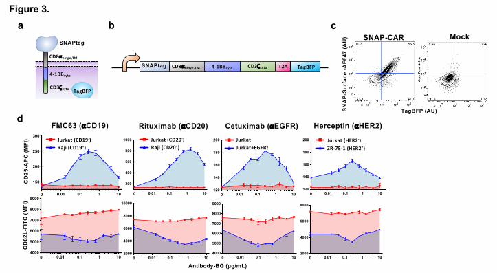
Engineering a self-labeling SNAP switchable adaptor CAR. We next aimed to create a
switchable adaptor CAR system using the SNAPtag protein domain to target T cell receptor
signaling when combined with BG-tagged antibodies (Fig. 1d). We cloned the SNAPtag domain
into a lentiviral vector containing the standard CAR components including the CD8� hinge and
transmembrane domains, the 4-1BB cytoplasmic co-signaling domain, the CD3zeta T cell
signaling cytoplasmic domain, and a TagBFP reporter gene co-expressed via a T2A co-
translational peptide (Fig. 3a,b). We packaged this vector into lentivirus particles and transduced
Jurkat cells. We found that the receptor was efficiently expressed and that the SNAPtag protein
was functional as indicated by TagBFP expression and staining with a BG-fluorophore reagent
and analysis by flow cytometry (Fig. 3c).
We next tested whether BG-conjugated antibodies could be combined with SNAP-CAR
Jurkat cells to target their T cell activation signaling. We co-incubated SNAP-CAR cells with
various antigen positive or negative tumor cell lines and various doses of BG-conjugated
antibodies. After 24 hours we assayed the cells for T cell activation by staining with antibodies
specific for CD25, which is up-regulated upon T cell activation, and CD62L, which is down-
(which was not certified by peer review) is the author/funder. All rights reserved. No reuse allowed without permission. The copyright holder for this preprintthis version posted January 17, 2020. . https://doi.org/10.1101/2020.01.17.909895doi: bioRxiv preprint
9
regulated. We assessed the expression levels of these markers in SNAP-CAR cells by
specifically gating on the TagBFP CAR+ cell population. We found that expression of these
markers was controlled in an antigen-specific and dose-responsive manner by the BG-conjugated
antibodies (Fig. 3d). Similar to SNAP-synNotch cells, SNAP-CAR activation signaling strength
peaked at an antibody dose level between 0.1-1.0 μg/mL before steadily decreasing, again
indicative of a hook effect.
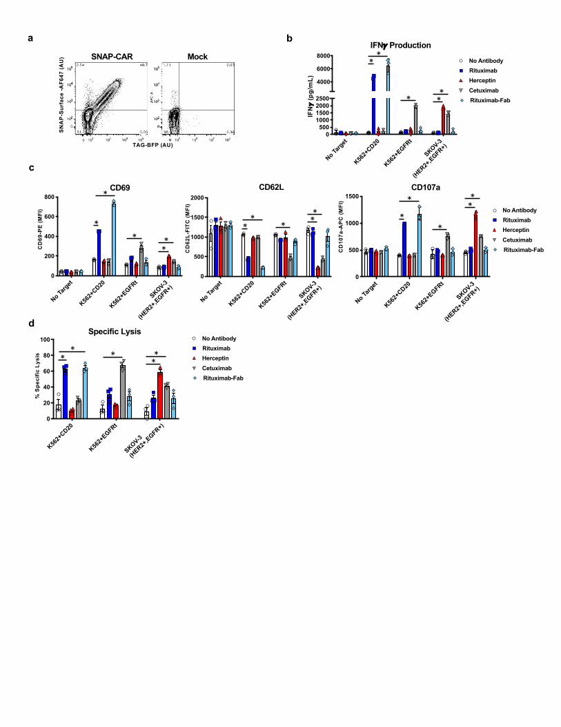
The SNAP-CAR is functional in primary human T cells. We then tested the expression level
and in vitro functional activity of the SNAP-CAR in primary human T cells transduced with the
SNAP-CAR lentivirus. Staining with a BG-fluorophore conjugate and assaying by flow
cytometry, we found that the SNAP receptor was efficiently expressed in ~40% of cells in a
manner that correlated well with the expression of the TagBFP marker gene (Fig. 4a). To test
CAR functionality, we co-incubated SNAP-CAR T cells with various antigen positive or
negative target tumor cell lines and 1.0 μg/mL of BG-conjugated antibodies for 24 hours.
Targeted antigens included CD20, EGFR, and HER2. Analyzing the supernatants of the co-
incubated cells by ELISA we found that the SNAP-CAR T cells could be directed by the
covalently attached BG-antibodies to produce significant amounts of IFN� in response to antigen
positive target cells (Fig. 4b). Our analysis of co-incubated cells by flow cytometry revealed that
the antibodies also led to induction of T cell activation markers, up-regulation of CD69 and
CD107a, and down-regulation of CD62L (Fig. 4c). CAR cells and target cells were also co-
cultured and evaluated for target cell lysis by flow cytometry and showed high levels of target
specific cell lysis (Fig. 4d). Again, T cell marker activation and target cell lysis were
significantly higher only when the co-administered antibody targeted an antigen expressed by the
co-administered cells, indicating antibody specificity. In addition to full length IgG antibodies,
(which was not certified by peer review) is the author/funder. All rights reserved. No reuse allowed without permission. The copyright holder for this preprintthis version posted January 17, 2020. . https://doi.org/10.1101/2020.01.17.909895doi: bioRxiv preprint
10
we also tested a BG-conjugated Fab fragment of Rituximab. This molecule, more similar to the
scFv antibody fragment found in traditional CARs, also showed potent activity for each of the
effector functions equal to or greater than that of the full-length Rituximab.
BG-antibody pre-loading experiments to interrogate the receptor signaling mechanism. To
further investigate the mechanism of SNAP-synNotch receptor activation, we performed co-
incubation assays in which we pre-labeled either the target cells or SNAP-synNotch cells with
BG-conjugated antibodies. We incubated CD19 positive and negative tumor cells with BG-
conjugated antibody, washed away residual unbound antibody, and co-incubated these cells with
SNAP-synNotch cells for 48 hours. Evaluating response gene activation, we found that CD19
positive tumor cells significantly up-regulated TagBFP gene expression to a level comparable to
the peak level of activation in the previous dose-response assay (Fig. 5a, 2c). We then pre-
labeled the SNAP-synNotch cells with BG-conjugated antibody and after washing away residual
antibody, we performed a similar co-incubation assay with CD19 positive or negative tumor
cells. No significant response gene activation was observed (Fig. 5b).
Performing the same pre-staining experiments with SNAP-CAR cells, we found that the
SNAP-CAR was functional both when the SNAP-CAR cells or tumor cells were pre-labeled
with BG-conjugated antibodies. We incubated antigen positive or negative tumor cells with BG-
conjugated antibody, washed away residual unbound antibody, and co-incubated these cells with
Jurkat SNAP-CAR cells for 24 hours. Assaying by flow cytometry for the CD25 T cell activation
marker, we found that the labeled tumor cells induced a significant up-regulation of CD25
expression to a level comparable to the peak level of activation in the previous dose-response
assay (Fig. 5a). We then pre-labeled the SNAP-CAR cells with BG-labeled antibody and after
washing away residual antibody, we performed a similar co-incubation assay with CD20 or
(which was not certified by peer review) is the author/funder. All rights reserved. No reuse allowed without permission. The copyright holder for this preprintthis version posted January 17, 2020. . https://doi.org/10.1101/2020.01.17.909895doi: bioRxiv preprint
11
CD19 positive and negative tumor cells. Significant up-regulation of T cell activation was
observed, however, to a lower magnitude than that with pre-labeled cancer cells (Fig. 5b).
Mathematical model of switchable adaptor complex formation. To gain a better
understanding of the switchable adaptor receptor signaling and the observed hook effect, we
generated a continuous mathematical model of the ternary complex formation between T cell,
adaptor antibody, and target cell. Using Python Jupyter Notebook, we created a generalizable
model of ordinary differential equations (ODEs) that describe the binding reactions. A system of
equations was defined to describe the accumulation and concentration of each of the six species
in the model: T cells, antibodies, tumor cells, T cells bound to antibody, tumor cells bound to
antibody, and T cell-antibody-tumor cell ternary complexes (Fig. 6a, Methods).
To validate the model, we ran simulations using kinetic parameters taken directly from
the literature and then through bounded parameter fitting (Supplementary Table S2)34-37. Using
direct literature values, the model was able to recapitulate the general features of our
experimental data, including the observed hook effect and a prediction accurate within an order
of magnitude of each antibody dose expected to yield maximum receptor signaling (Fig. 6b). We
used the sum of squared error (SSE) calculations to measure error in model simulations against
experimental results. With the exception of Rituximab, model simulations using literature values
alone, resulted in good recapitulations of experimental data (average SSE with literature values =
1.03) (Supplementary Table S3). Next, using SciPy, we minimized the sum of squared error to
optimize the kinetic parameters and better fit the model to the experimental data for each
antibody pair38. During parameter estimation, the literature values were used as initial estimates
and bounded within one order of magnitude. With these constraints, we were able to minimize
our model error in seven of the experimental results to (average SSE after fitting = 0.09)
(which was not certified by peer review) is the author/funder. All rights reserved. No reuse allowed without permission. The copyright holder for this preprintthis version posted January 17, 2020. . https://doi.org/10.1101/2020.01.17.909895doi: bioRxiv preprint

12
(Supplementary Table S3).
With a validated model, we next aimed to use the model to predict how different system
parameters would affect receptor signaling and conducted parameter scans with the model. We
first varied kf1, the forward reaction rate of the antibody binding to the T cell receptor, to
simulate the effects increasing the on-rate, which could also be experimentally varied by
changing the number of BG’s conjugated to an antibody. We found that the model predicted that
increasing the number of BG’s per antibody would lead to greater ternary body formation at
higher concentrations of antibody. Once the kf1 rate becomes greater than a threshold of 10-3 nm-
1sec-1, this effect was expected to plateau. (Fig. 6c). Next, we scanned different values for the
antibody to T cell affinity, the parameter maximized by the use of the covalent SNAP to
antibody interaction. We found that stronger affinity was predicted to lead to ternary body
formation over a wider range of antibody concentrations and to lead to a higher overall level of
ternary body formation. Finally, as target antigen concentration can vary based on the antigen
being targeted, and for cancer, expression levels can also significantly vary greatly between
patients and on cells within the same patient, we performed a parameter scan varying the level of
tumor antigen concentration. We found that greater antigen concentrations were expected to
broaden the effective antibody dosage window for successful ternary complex formation, while
lower antigen levels were predicted to require a higher amount of antibody to induce signaling,
and were predicted to be more susceptible to inhibition by the hook effect.
Discussion
The switchable adaptor SNAP-synNotch system further increases the versatility of the
synNotch receptor framework leading to post-translational control of receptor signaling by co-
administered antibody dose, as well as the ability to target multiple antigens using a single
(which was not certified by peer review) is the author/funder. All rights reserved. No reuse allowed without permission. The copyright holder for this preprintthis version posted January 17, 2020. . https://doi.org/10.1101/2020.01.17.909895doi: bioRxiv preprint

13
genetically-encoded receptor. While other adaptor CARs have been described, this is, to our
knowledge, the first adaptor synNotch system. Our unsuccessful initial attempts to create an
adaptor synNotch system using a non-covalent interaction (between mSA2 and biotin) suggest
that a very high affinity interaction (ideally covalent) between the synNotch receptor and the tag
would be required for the adaptor system to function; presumably since the signaling mechanism
for Notch is based on an actual pulling force25.
With the covalent bond generated by the self-labeling enzyme, the SNAP-CAR has
several beneficial characteristics over other adaptor CAR technologies15-21. The results of others,
and our results supported by our modeling analysis, suggest that the affinity of the interaction
between the CAR and the adaptor molecule is a key parameter for productive receptor
signaling15,18. The covalent bond produced by the SNAP enzyme and the benzylguanine moiety
provides the tightest theoretical bond – a covalent bond – and thus maximizes this critical
parameter. While many antibodies will be functional with a non-covalent, lower-affinity adaptor
CAR, our model predicts that covalent bond formation could enable use of antibodies that
otherwise have a binding affinity to the target antigen that is too low to elicit an effect.
Furthermore, the SNAP enzyme reacting to the bio-orthogonal benzylguanine grants the CAR
exquisite specificity, and, being an enzyme of human origin, the SNAP protein is likely to be
well-tolerated in a human host. This characteristic satisfies a key requirement for the persistence
of the adoptively transferred therapeutic cells and minimizes the possibility of toxicities resulting
from their immune rejection39-41.
The ability to create functional CARs by preloading the SNAP receptor, followed by
removal of excess BG-antibody, provides unique opportunities to test candidate antigen binding
regions as components of traditional CARs (Fig 5b). Compared to adaptor CARs binding to
(which was not certified by peer review) is the author/funder. All rights reserved. No reuse allowed without permission. The copyright holder for this preprintthis version posted January 17, 2020. . https://doi.org/10.1101/2020.01.17.909895doi: bioRxiv preprint

14
antigen through a transient interaction, the covalently assembled receptor will more closely
resemble that of a traditional CAR. We were originally surprised that the pre-assembled SNAP-
synNotch receptors were not functional and instead require pre-targeting of the cancer cells.
However, this result can potentially be explained by considering the mechanism of signaling, in
which the receptor is proteolytically cleaved and thus destroyed following activation, not
allowing for multiple signaling events from recycled receptors. This result suggests that multiple
bursts of receptor activation from distinct receptors over time may be needed to sufficiently
trigger synNotch signaling. As the pre-assembled CARs were capable of signaling, pre-loading
the SNAP-CAR T cells could be a potential clinical approach, however, upon T cell activation,
the cells would be induced to expand thus diluting out the assembled receptor, requiring
supplementation of additional antibody. Similarly, while the SNAP-CAR T cells showed
significant up-regulation of T cell activation markers for both pre-loading conditions, more
robust T cell activation was observed with pre-loaded tumor cells than pre-loaded SNAP-CAR
cells. That both the CAR and SynNotch receptors were maximally effective against tumor cells
pre-labeled with antibodies suggests that pre-dosing of a patient with tagged antibody prior to T
cell infusion could be the optimal treatment regimen.
Based on our results, additional self-labeling or covalent protein assembly systems could
also provide good frameworks for universal adaptor CARs. Such systems include candidates
such as: CLIPtag, Halotag, SpyTag, SnoopTag, Isopeptag, Sortase or split inteins 23,42-45. It is
possible that CARs made with other self-labeling enzymes, having different kinetic
binding/dissociation rates, molecular size, and could vary in expression level, could provide
more robust signaling depending on the characteristics of the antigen(s) being targeted.
(which was not certified by peer review) is the author/funder. All rights reserved. No reuse allowed without permission. The copyright holder for this preprintthis version posted January 17, 2020. . https://doi.org/10.1101/2020.01.17.909895doi: bioRxiv preprint
15
Additionally, other synthetic receptor platforms could be amenable to the switchable adaptor
format by creating receptors with the SNAPtag protein domain46,47.
Future developments of the SNAP adaptor systems to clinical applications will be
important in several key areas. Developing site-specific tagging approaches will help to lead to
more homogeneous antibody-BG conjugates and potentially identify optimal BG-conjugation
sites that could further maximize receptor signaling output. For any specific disease indication
there is the need to determine the optimal antibody or antibodies, to combine with the adaptor T
cells to provide disease-specific or at least disease-associated induction of receptor signaling.
While it is known that SNAP self-labeling is efficient in animal models, testing the SNAP T cells
with specific adaptor antibody combinations will be important to confirm potent function and to
optimize the therapeutic delivery strategy22.
Our molecular model of switchable receptor systems provided key insights into our
observed signaling behaviors and yielded predictions on how to potentially optimize receptor
function. The model results suggested that the binding strength between the CAR and the adaptor
is a critical parameter for signaling and that our SNAP receptors for which this interaction
strength is maximized via a covalent bond are expected to be desirable. Furthermore, the model
suggested that one way to improve activity would be to increase the forward reaction rate of the
CAR binding to the adaptor, which could potentially be accomplished by increasing the number
of BG molecules per antibody. Lastly the model predicted that using adaptor CARs to target
antigens that are expressed at high levels is preferable as these antigens would be expected to
induce receptor signaling at lower antibody concentrations and would be less susceptible to the
hook effect at higher antibody doses.
(which was not certified by peer review) is the author/funder. All rights reserved. No reuse allowed without permission. The copyright holder for this preprintthis version posted January 17, 2020. . https://doi.org/10.1101/2020.01.17.909895doi: bioRxiv preprint
16
SNAP-synNotch and SNAP-CAR T cells provide a powerful new adaptor strategy for
fully programmable targeting of engineered cells to multiple antigens using covalent chemistry.
These systems have great potential for clinical application and biotechnological utility by
providing researchers with the ability to rapidly screen CAR and synNotch antibody candidates
and to rewire and activate cellular programs in response to highly specific antibody-antigen
interactions.
Methods
Construction of lentiviral expression vectors. pHR_PGK_antiCD19_synNotch_Gal4VP64 and
pHR_Gal4UAS_tBFP_PGK_mCherry were gifts from Wendell Lim (Addgene plasmid# 79125;
http://n2t.net/addgene:79125; RRID:Addgene_79125 and Addgene plasmid# 79130;
http://n2t.net/addgene:79130; RRID:Addgene_79130, respectively). Sequences for all receptor
coding regions and response constructs are listed in Supplementary Table S4. To generate
pHR-PGK-SNAP-41BBζ, a DNA fragment encoding SNAP-41BBζ was codon-optimized,
synthesized (Integrated DNA Technologies) and cloned into the pHR-PGK vector backbone
using isothermal assembly. To generate pHR-PGK-SNAP-synNotch-Gal4VP64 and pHR-PGK-
mSA2-synNotch-Gal4VP64, DNA encoding the SNAP or mSA2 coding region was codon-
optimized and synthesized (Integrated DNA Technologies) and cloned in place of the anti-
CD19scFv in plasmid pHR-PGK-antiCD19-synNotch-Gal4VP64 (Addgene# 79125) using
isothermal assembly. To generate pHR-Gal4UAS-IL7-PGK-mCherry, a DNA fragment encoding
IL-7 was codon-optimized, synthesized (Integrated DNA Technologies) and cloned in place of
TagBFP in the pHR_Gal4UAS_tBFP_PGK_mCherry vector backbone using isothermal
assembly. Virus was generated using the above described transfer vectors following methods
(which was not certified by peer review) is the author/funder. All rights reserved. No reuse allowed without permission. The copyright holder for this preprintthis version posted January 17, 2020. . https://doi.org/10.1101/2020.01.17.909895doi: bioRxiv preprint
17
described previously in detail48.
Production of BG-antibody conjugates. Rituximab (Rituxan, Genentech), Cetuximab (Erbitux,
Eli Lily), and Herceptin (Traztuzumab, Genentech) and FMC63 (Novus Biologicals) underwent
buffer exchange into PBS using 2 mL 7K MWCO Zeba Spin Desalting Columns (ThermoFisher
Scientific). The Rituximab Fab fragment was generated using the Fab Preparation Kit (Pierce)
following the manufacturer’s protocol. Antibodies were then co-incubated with a 20-fold molar
excess of BG-GLA-NHS (NEB) for 30 minutes at room temperature, followed by buffer
exchange into PBS using 2 mL 7K MWCO Zeba Spin Desalting Columns.
Quantification of BGs on BG-conjugated antibodies. For in vitro conjugation of whole
antibodies with SNAPtag, BG-conjugated purified antibodies (0.5 µg) were incubated with
recombinant SNAPtag protein (2 µg). The solution was incubated in PBS (10 µL, pH 7.4)
containing DTT (1 mM) at 37°C for two hours. Conjugation solutions were then diluted with
Laemmli buffer, boiled for 5 minutes, and analyzed on an 8% SDS-PAGE (120 V, 1.5 h). Gels
were visualized using imidazole-SDS-Zn reverse staining.1 Briefly, gels were stained with a 200
mM imidazole aqueous solution containing 0.1% SDS for 15 minutes with light agitation. The
staining solution was decanted and replaced with water. After 30 seconds, the water was
decanted and the gel was developed for 45 seconds with a 200 mM ZnSO4 aqueous solution with
light agitation. The gel was then rinsed under running water for 10 seconds. Gels were imaged on
a ChemiDoc (Bio-Rad) using epi white light on a black background. Relative band intensities
were quantified with ImageJ. A correction factor of 1.5 was applied to the average number of
BG/antibody to account for the light chain. Light chains were conjugated to SNAPtag in the
(which was not certified by peer review) is the author/funder. All rights reserved. No reuse allowed without permission. The copyright holder for this preprintthis version posted January 17, 2020. . https://doi.org/10.1101/2020.01.17.909895doi: bioRxiv preprint

18
same manner except 3 µg of antibody was incubated with 6 µg SNAPtag, gels were analyzed on
a 10% SDS-PAGE (120 V, 1.2 h) and stained with Coomassie, and a correction factor was not
applied.
Cell line culture. Human tumor cell lines Jurkat Clone E6-1 (TIB-152), ZR-75-1(CRL-1500),
K562 (CCL-243), SKOV-3(HTB-77), and Raji (CCL-86) were obtained from American Type
Culture Collection (ATCC) and cultured at 37°C in RPMI medium supplemented with 1X MEM
amino acids solution, 10mM Sodium Pyruvate, 10% fetal bovine serum (FBS), and Penicillin-
Streptomycin (Life Technologies). K562+EGFRt, K562+CD20, and Jurkat+EGFRt cells that
stably express full-length CD20 and the EGFRt gene
https://www.ncbi.nlm.nih.gov/pmc/articles/PMC3152493/, were generated by transducing Jurkat
cells with the indicated tumor antigen expressing lentivirus and sorting for cells positive for
antigen expression. To create the SNAP-CAR stable cell line, Jurkat cells were transduced with
SNAP-41BBζ, and underwent fluorescence-activated cell sorting (FACS) for TagBFP
expression. and reporter (mCherry+) expression. To generate SNAP-synNotch lines, SNAP-
synNotch-Gal4VP64 was co-transduced with either pHR-Gal4UAS-tBFP-PGKmCherry or pHR-
Gal4UAS-IL7-PGKmCherry lentivirus, and receptor and response construct positive cells were
obtained by FACS for anti-Myc-Tag antibody staining (Cell signaling Technology) and mCherry
expression, respectively. HEK293T cells (ATTC, CRL-3216), used for lentivirus production
were cultured at 37°C in DMEM supplemented with 10% FBS, and Penicillin-Streptomycin.
Primary human T cell culture and transduction. All primary T cells for experiments were
sourced from deidentified human Buffy Coat samples purchased from the Pittsburgh Central
(which was not certified by peer review) is the author/funder. All rights reserved. No reuse allowed without permission. The copyright holder for this preprintthis version posted January 17, 2020. . https://doi.org/10.1101/2020.01.17.909895doi: bioRxiv preprint

19
Blood Bank fulfilling the basic exempt criteria 45 CFR 46.101(b)(4) in accordance with the
University of Pittsburgh IRB guidelines. PBMC were isolated from a Buffy Coat from healthy
volunteer donors using Ficoll gradient centrifugation and human T cells were isolated using the
Human Pan T cell isolation kit (Miltenyi Biotec). Human T cells were cultured in supplemented
RPMI media as described for cell lines above, however, 10% Human AB serum (Gemini Bio
Products) was used instead of FBS, and the media was further supplemented with 100 U/ml
human IL-2 IS (Miltenyi Biotec), 1 ng/ml IL-15 (Miltenyi Biotec), and 4mM L-Arginine (Sigma
Aldrich). T cells were stimulated and expanded using TransAct Human T cell activation reagent
(Miltenyi Biotec). For transduction, 48 hours after activation, lentivirus was added to cells at a
multiplicity of infection of 10–50 in the presence of 6 mg/ml of DEAE-dextran (Sigma Aldrich).
After 18 hours, cells were washed and resuspended in fresh T cell media containing 100 U/ml
IL-2 and 1 ng/ml IL-15. Cells were split to a concentration of 1M/mL and supplemented with
fresh IL-2 and IL-15 every 2-3 days. After 10-12 days of stimulation and expansion, transduced
cells were evaluated for CAR expression by flow cytometry and evaluated for activity in
subsequent functional assays.
Flow cytometry staining. Cells were washed and resuspended in flow cytometry buffer (PBS +
2% FBS) and then stained using the indicated antibodies for 30 minutes at 4°C followed by two
washes with flow cytometry buffer. Live cells and singlets were gated based on scatter. To
evaluate SNAP-CAR and SNAP-synNotch expression, 1M cells were labeled with SNAP-
Surface 647 (NEB) following manufacturer’s recommendation (50 μM concentration of SNAP-
Surface 647 in complete cell media) for 30 minutes at 37°C and washed three times in complete
culture media. SNAP-synNotch cells were additionally stained with anti-Myc-Tag-FITC
(which was not certified by peer review) is the author/funder. All rights reserved. No reuse allowed without permission. The copyright holder for this preprintthis version posted January 17, 2020. . https://doi.org/10.1101/2020.01.17.909895doi: bioRxiv preprint
20
antibody (Cell Signaling Technology) to stain the Myc-Tag on the N-terminus of the receptor.
SNAP-synNotch cell and target cell co-incubation assays for antibody-mediated activation.
100,000 synNotch Jurkat effector cells were co-cultured with 200,000 of the indicated target
cells and BG-conjugated antibody for 48 hours. For assays with SNAP-synNotch cells
engineered with the pHR_Gal4UAS_tBFP_PGK_mCherry response construct, co-incubated cells
were evaluated by flow cytometry, gating for synNotch cells by mCherry positivity, and then
quantifying TagBFP fluorescence for this mCherry+ population. For assays with SNAP-
synNotch cells engineered with the pHR_Gal4UAS_IL7_PGK_mCherry construct, following a
48 hour co-incubation, cells were spun down and supernatants were collected and analyzed by
ELISA for IL-7 following the manufacturer’s recommended protocol (Peprotech).
SNAP-CAR T cell and target cell co-incubation assays for antibody-mediated activation.
100,000 SNAP-CAR Jurkat or primary human T cell effector cells were co-incubated with
200,000 of the indicated target cells and antibody concentrations for 24 hours and assayed by
flow cytometry for T cell marker gene expression. For primary cell assays, cells were stained
with CD69-PE (BD Biosciences), CD62L-FITC (BD Biosciences), and CD107a-APC (BD
Biosciences) antibodies and for Jurkat effector assays, cells were stained with CD62L-FITC (BD
Biosciences) and CD25-APC (BD Biosciences) antibodies. For flow cytometry CAR+ cells
were analyzed by gating for the TagBFP+ population. Supernatants from primary cell assays
were also collected and analyzed for IFN� by ELISA (BioLegend). All assays were performed
in triplicate and average IFN� production was plotted with standard deviation.
(which was not certified by peer review) is the author/funder. All rights reserved. No reuse allowed without permission. The copyright holder for this preprintthis version posted January 17, 2020. . https://doi.org/10.1101/2020.01.17.909895doi: bioRxiv preprint
21
Target cell lysis assay. The indicated target cells were stained with Cell Trace Yellow following
the manufacturer’s recommended protocol (ThermoFisher), and 10,000 target cells per well were
co-cultured with 50,000 SNAP-CAR T cells (E:T=5:1) in a 96 well V-bottom plate with 1.0
μg/mL of the indicated BG-conjugated antibody. Plates underwent a quick-spin to collect cells at
the bottom of the wells and were then incubated at 37°C for 24 hours. To identify lysed cells, co-
incubated cells were stained with Ghost Dye Red Viability Dye (Tonbo Biosciences) and
analyzed by flow cytometry. Target cells were identified by Cell Trace Yellow and lysed target
cells were identified by positive Ghost Dye staining. Percent specific cytotoxicity was calculated
by the equation: 100*(% experimental lysis – % target-only lysis) / (100 – % target-only lysis).
Pre-labeling co-incubation assays. Pre-labeling co-incubation activation assays, were carried out
as above, except prior to co-incubation for pre-labeled SNAP effector cell assays, SNAP-CAR or
SNAP-synNotch Jurkat cells were first labeled with 1.0 μg/mL of the indicated BG-modified
antibody in complete media for 30 minutes at 37°C and then washed three times in complete
media, and for pre-labeled target cell assays, target cells were labeled with 5.0 μg/mL of the
indicated antibody for 30 minutes at 4°C and washed two times with flow buffer. No additional
antibody was added to these co-incubations.
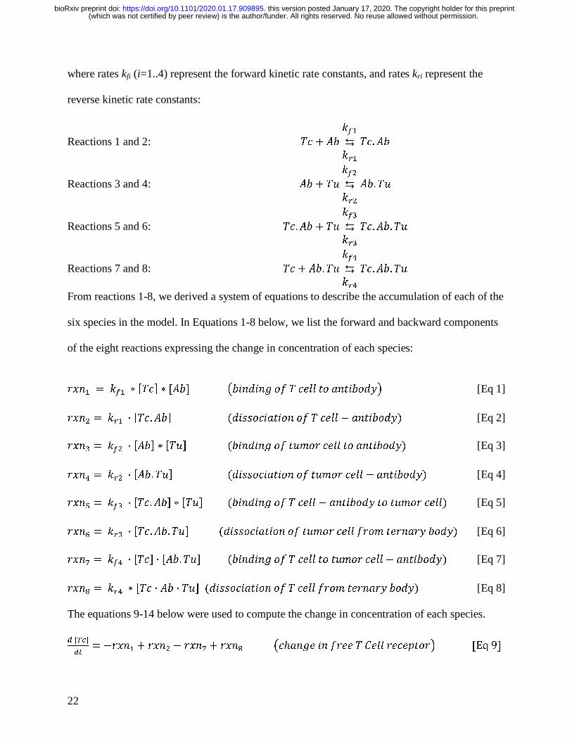
Mathematical model. The model for ternary body formation considered the following 8 binding
reactions between the tumor cells, T cells, and antibody with six different species: T cell (Tc),
antibody (Ab), tumor cell (Tu), T cell bound to antibody (Tc.Ab), tumor cell bound to antibody
(Ab.Tu), and a ternary body complex of a T cell bound to antibody and tumor cell (Tc.Ab.Tu) and
(which was not certified by peer review) is the author/funder. All rights reserved. No reuse allowed without permission. The copyright holder for this preprintthis version posted January 17, 2020. . https://doi.org/10.1101/2020.01.17.909895doi: bioRxiv preprint
22
where rates kfi (i=1..4) represent the forward kinetic rate constants, and rates kri represent the
reverse kinetic rate constants:
Reactions 1 and 2: �� � ������
���
��. ��
Reactions 3 and 4: �� � �����
���
��. �
Reactions 5 and 6: ��. �� � �����
���
��. ��. �
Reactions 7 and 8: �� � ��. �����
���
��. ��. �
From reactions 1-8, we derived a system of equations to describe the accumulation of each of the
six species in the model. In Equations 1-8 below, we list the forward and backward components
of the eight reactions expressing the change in concentration of each species:
�� � � ��� � ���� � ���� ��� �� � �� � ���� �� � ������� [Eq 1] �� � � ��� · ���. ��� ��� ������� �� � ���� ! � ������" [Eq 2] �� � � ��� · ���� � ��� ��� �� � �� �$�� ���� �� � ������" [Eq 3] �� � � ��� · ���. �� ��� ������� �� �$�� ���� ! � ������" [Eq 4] �� � � ��� · ���. ��� � ��� ��� �� � �� � ���� ! � ������ �� �$�� ����" [Eq 5] �� � � ��� · ���. ��. �� ��� ������� �� �$�� ���� ���$ ��� ��� ����" [Eq 6] �� � ��� · ���� · ���. �� ��� �� � �� � ���� �� �$�� ���� ! � ������" [Eq 7] �� � ��� � ��� · �� · �� ��� ������� �� � ���� ���$ ��� ��� ����" [Eq 8] The equations 9-14 below were used to compute the change in concentration of each species. � � ��
��� !�� � � �� � ! �� � �� ��%� �� � ���� � &��� ����'���� �Eq 9�
(which was not certified by peer review) is the author/funder. All rights reserved. No reuse allowed without permission. The copyright holder for this preprintthis version posted January 17, 2020. . https://doi.org/10.1101/2020.01.17.909895doi: bioRxiv preprint
23
� ����
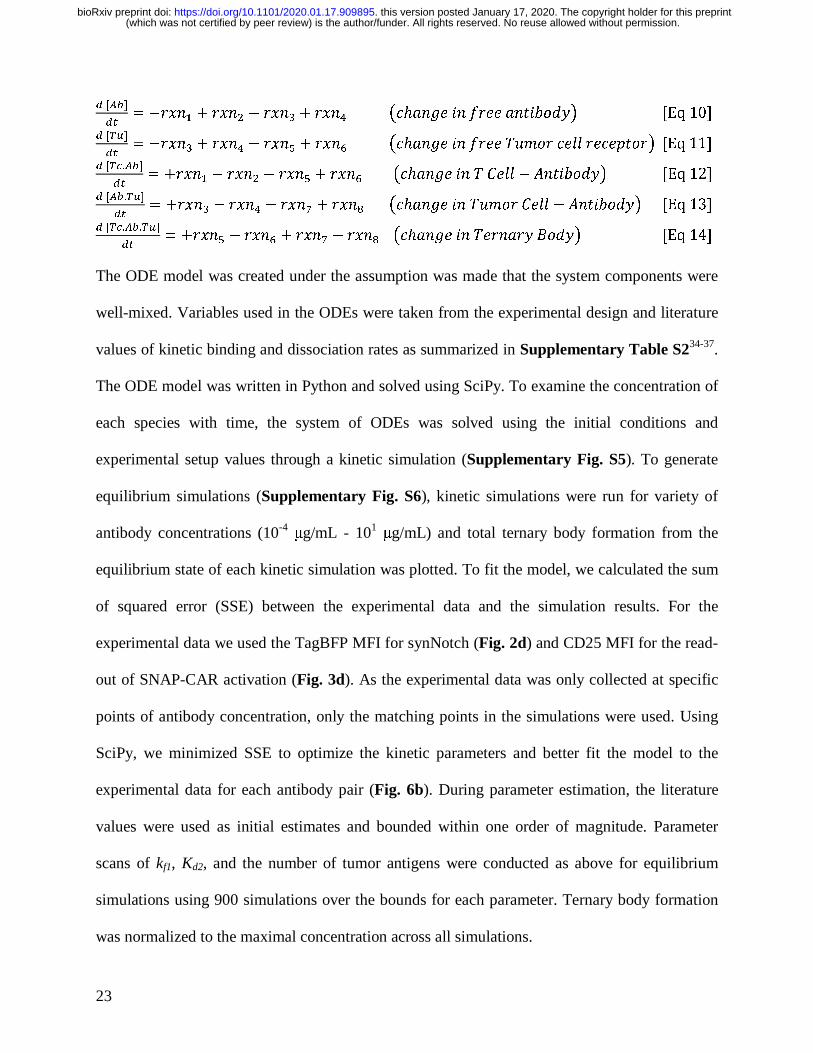
��� !�� � � �� � ! �� � � �� � ��%� �� � ���� � ������� �Eq 10�
� � ��
��� !�� � � �� � ! �� � � �� � ��%� �� � ���� �$�� ���� ����'���� �Eq 11�
� � �.���
��� ��� � ! �� � ! �� � � �� � ��%� �� � � &��� ! � ������� �Eq 12�
� ���. ��
��� ��� � ! �� � ! �� � �� ��%� �� � �$�� &��� ! � ������� �Eq 13�
� � �.��. ��
��� ��� � ! �� � � �� ! �� ��%� �� � ��� ��� /���� �Eq 14�
The ODE model was created under the assumption was made that the system components were
well-mixed. Variables used in the ODEs were taken from the experimental design and literature
values of kinetic binding and dissociation rates as summarized in Supplementary Table S234-37.
The ODE model was written in Python and solved using SciPy. To examine the concentration of
each species with time, the system of ODEs was solved using the initial conditions and
experimental setup values through a kinetic simulation (Supplementary Fig. S5). To generate
equilibrium simulations (Supplementary Fig. S6), kinetic simulations were run for variety of
antibody concentrations (10-4 μg/mL - 101 μg/mL) and total ternary body formation from the
equilibrium state of each kinetic simulation was plotted. To fit the model, we calculated the sum
of squared error (SSE) between the experimental data and the simulation results. For the
experimental data we used the TagBFP MFI for synNotch (Fig. 2d) and CD25 MFI for the read-
out of SNAP-CAR activation (Fig. 3d). As the experimental data was only collected at specific
points of antibody concentration, only the matching points in the simulations were used. Using
SciPy, we minimized SSE to optimize the kinetic parameters and better fit the model to the
experimental data for each antibody pair (Fig. 6b). During parameter estimation, the literature
values were used as initial estimates and bounded within one order of magnitude. Parameter
scans of kf1, Kd2, and the number of tumor antigens were conducted as above for equilibrium
simulations using 900 simulations over the bounds for each parameter. Ternary body formation
was normalized to the maximal concentration across all simulations.
(which was not certified by peer review) is the author/funder. All rights reserved. No reuse allowed without permission. The copyright holder for this preprintthis version posted January 17, 2020. . https://doi.org/10.1101/2020.01.17.909895doi: bioRxiv preprint
24
Statistical methods. The number of replicates, mean value, and error are described in the
respective figure legends and/or methods. Error bars are shown for all data points with replicates
as a measure of variation within a group.
Data availability
All data generated and analyzed during this study are included in this published article and its
Supplementary Information or are available from the corresponding author upon reasonable
request.
Acknowledgments
This work was supported by the NIH grant R35 CA210039 (O.J.F.); NIH grant R21 AI130815
(A.D.); DARPA award W911NF-17-1-0135 (N.M-Z.); and by the Michael G. Wells Prize (J.L.).
This work benefitted from using the SPECIAL BD LSRFORTESSA funded by NIH
1S10OD011925-01.
Author contributions
J.L. and A.D. designed the research. A.A.B. and N.M-Z. created the mathematical model, and
A.A.B. performed the model data fitting and parameter scans. J.L., Y.T., and M.K. carried out
the experiments and analyzed the data. J.L., N.M-Z., A.D., and O.J.F. supervised the work. J.L.,
A.B., Y.T., M.K., N.M-Z., A.D., and O.J.F. interpreted the data and wrote the manuscript.
Competing interests
(which was not certified by peer review) is the author/funder. All rights reserved. No reuse allowed without permission. The copyright holder for this preprintthis version posted January 17, 2020. . https://doi.org/10.1101/2020.01.17.909895doi: bioRxiv preprint
25
J.L. and A.D. have filed a provisional patent application on the universal SNAP receptor
technology described herein.
Materials & correspondence
Correspondence and requests for materials should be addressed to J.L.
Supplementary Information:
Figure S1 | mSA2 biotin-binding synNotch receptor is incapable of cell-bound antibody
activation.
Figure S2 | SDS-PAGE quantification of antibody-conjugated benzyl guanines by SNAP
conjugation reaction.
Figure S3 | Staining of target cell lines by benzyl guanine modified antibodies.
Figure S4 | Effector to target effect on SNAP-synNotch receptor activity.
Figure S5 | Representative kinetic simulation.
Figure S6 | Representative equilibrium simulation.
Table S1 | Quantification of the number of BG molecules conjugated per antibody.
Table S2 | Model parameters used in simulations.
Table S3 | Model simulation error.
Table S4 | DNA sequences of lentiviral receptor expression and response inserts.
References
1 Lim, W. A. & June, C. H. The Principles of Engineering Immune Cells to Treat Cancer. Cell 168, 724-740, doi:10.1016/j.cell.2017.01.016 (2017).
2 Gross, G., Waks, T. & Eshhar, Z. Expression of immunoglobulin-T-cell receptor chimeric molecules as functional receptors with antibody-type specificity. Proc Natl Acad Sci U S A 86, 10024-10028, doi:10.1073/pnas.86.24.10024 (1989).
(which was not certified by peer review) is the author/funder. All rights reserved. No reuse allowed without permission. The copyright holder for this preprintthis version posted January 17, 2020. . https://doi.org/10.1101/2020.01.17.909895doi: bioRxiv preprint
26
3 June, C. H. & Sadelain, M. Chimeric Antigen Receptor Therapy. N Engl J Med 379, 64-73, doi:10.1056/NEJMra1706169 (2018).
4 Sadelain, M., Brentjens, R. & Riviere, I. The basic principles of chimeric antigen receptor design. Cancer Discov 3, 388-398, doi:10.1158/2159-8290.CD-12-0548 (2013).
5 Lee, D. W. et al. T cells expressing CD19 chimeric antigen receptors for acute lymphoblastic leukaemia in children and young adults: a phase 1 dose-escalation trial. Lancet 385, 517-528, doi:10.1016/S0140-6736(14)61403-3 (2015).
6 Maude, S. L. et al. Chimeric antigen receptor T cells for sustained remissions in leukemia. N Engl J Med 371, 1507-1517, doi:10.1056/NEJMoa1407222 (2014).
7 Park, J. H. et al. Long-Term Follow-up of CD19 CAR Therapy in Acute Lymphoblastic Leukemia. N Engl J Med 378, 449-459, doi:10.1056/NEJMoa1709919 (2018).
8 Lohmueller, J. & Finn, O. J. Current modalities in cancer immunotherapy: Immunomodulatory antibodies, CARs and vaccines. Pharmacol Ther 178, 31-47, doi:10.1016/j.pharmthera.2017.03.008 (2017).
9 Maldini, C. R., Ellis, G. I. & Riley, J. L. CAR T cells for infection, autoimmunity and allotransplantation. Nat Rev Immunol 18, 605-616, doi:10.1038/s41577-018-0042-2 (2018).
10 Morsut, L. et al. Engineering Customized Cell Sensing and Response Behaviors Using Synthetic Notch Receptors. Cell 164, 780-791, doi:10.1016/j.cell.2016.01.012 (2016).
11 Roybal, K. T. et al. Engineering T Cells with Customized Therapeutic Response Programs Using Synthetic Notch Receptors. Cell 167, 419-432 e416, doi:10.1016/j.cell.2016.09.011 (2016).
12 Roybal, K. T. et al. Precision Tumor Recognition by T Cells With Combinatorial Antigen-Sensing Circuits. Cell 164, 770-779, doi:10.1016/j.cell.2016.01.011 (2016).
13 Esensten, J. H., Bluestone, J. A. & Lim, W. A. Engineering Therapeutic T Cells: From Synthetic Biology to Clinical Trials. Annu Rev Pathol 12, 305-330, doi:10.1146/annurev-pathol-052016-100304 (2017).
14 Toda, S., Blauch, L. R., Tang, S. K. Y., Morsut, L. & Lim, W. A. Programming self-organizing multicellular structures with synthetic cell-cell signaling. Science 361, 156-162, doi:10.1126/science.aat0271 (2018).
15 Cho, J. H., Collins, J. J. & Wong, W. W. Universal Chimeric Antigen Receptors for Multiplexed and Logical Control of T Cell Responses. Cell 173, 1426-1438 e1411, doi:10.1016/j.cell.2018.03.038 (2018).
16 Kudo, K. et al. T lymphocytes expressing a CD16 signaling receptor exert antibody-dependent cancer cell killing. Cancer research 74, 93-103, doi:10.1158/0008-5472.CAN-13-1365 (2014).
17 Lohmueller, J. J., Ham, J. D., Kvorjak, M. & Finn, O. J. mSA2 affinity-enhanced biotin-binding CAR T cells for universal tumor targeting. Oncoimmunology 7, e1368604, doi:10.1080/2162402X.2017.1368604 (2017).
18 Ma, J. S. et al. Versatile strategy for controlling the specificity and activity of engineered T cells. Proc Natl Acad Sci U S A 113, E450-458, doi:10.1073/pnas.1524193113 (2016).
19 Rodgers, D. T. et al. Switch-mediated activation and retargeting of CAR-T cells for B-cell malignancies. Proc Natl Acad Sci U S A 113, E459-468, doi:10.1073/pnas.1524155113 (2016).
20 Tamada, K. et al. Redirecting gene-modified T cells toward various cancer types using tagged antibodies. Clinical cancer research : an official journal of the American
(which was not certified by peer review) is the author/funder. All rights reserved. No reuse allowed without permission. The copyright holder for this preprintthis version posted January 17, 2020. . https://doi.org/10.1101/2020.01.17.909895doi: bioRxiv preprint
27
Association for Cancer Research 18, 6436-6445, doi:10.1158/1078-0432.CCR-12-1449 (2012).
21 Urbanska, K. et al. A universal strategy for adoptive immunotherapy of cancer through use of a novel T-cell antigen receptor. Cancer research 72, 1844-1852, doi:10.1158/0008-5472.CAN-11-3890 (2012).
22 Yang, G. et al. Genetic targeting of chemical indicators in vivo. Nat Methods 12, 137-139, doi:10.1038/nmeth.3207 (2015).
23 Gautier, A. et al. An engineered protein tag for multiprotein labeling in living cells. Chem Biol 15, 128-136, doi:10.1016/j.chembiol.2008.01.007 (2008).
24 Sun, X. et al. Development of SNAP-tag fluorogenic probes for wash-free fluorescence imaging. Chembiochem 12, 2217-2226, doi:10.1002/cbic.201100173 (2011).
25 Wang, X. & Ha, T. Defining single molecular forces required to activate integrin and notch signaling. Science 340, 991-994, doi:10.1126/science.1231041 (2013).
26 Lim, K. H., Huang, H., Pralle, A. & Park, S. Stable, high-affinity streptavidin monomer for protein labeling and monovalent biotin detection. Biotechnol Bioeng 110, 57-67, doi:10.1002/bit.24605 (2013).
27 Keppler, A. et al. A general method for the covalent labeling of fusion proteins with small molecules in vivo. Nat Biotechnol 21, 86-89, doi:10.1038/nbt765 (2003).
28 Maloney, D. G. et al. IDEC-C2B8 (Rituximab) anti-CD20 monoclonal antibody therapy in patients with relapsed low-grade non-Hodgkin's lymphoma. Blood 90, 2188-2195 (1997).
29 Slamon, D. J. et al. Use of chemotherapy plus a monoclonal antibody against HER2 for metastatic breast cancer that overexpresses HER2. N Engl J Med 344, 783-792, doi:10.1056/NEJM200103153441101 (2001).
30 Wu, X., Fan, Z., Masui, H., Rosen, N. & Mendelsohn, J. Apoptosis induced by an anti-epidermal growth factor receptor monoclonal antibody in a human colorectal carcinoma cell line and its delay by insulin. J Clin Invest 95, 1897-1905, doi:10.1172/JCI117871 (1995).
31 Zola, H. et al. Preparation and characterization of a chimeric CD19 monoclonal antibody. Immunol Cell Biol 69 ( Pt 6), 411-422, doi:10.1038/icb.1991.58 (1991).
32 Douglass, E. F., Jr., Miller, C. J., Sparer, G., Shapiro, H. & Spiegel, D. A. A comprehensive mathematical model for three-body binding equilibria. J Am Chem Soc 135, 6092-6099, doi:10.1021/ja311795d (2013).
33 Mackall, C. L., Fry, T. J. & Gress, R. E. Harnessing the biology of IL-7 for therapeutic application. Nat Rev Immunol 11, 330-342, doi:10.1038/nri2970 (2011).
34 Bondza, S. et al. Real-time Characterization of Antibody Binding to Receptors on Living Immune Cells. Front Immunol 8, 455, doi:10.3389/fimmu.2017.00455 (2017).
35 Melhus, K. B. et al. Evaluation of the binding of radiolabeled rituximab to CD20-positive lymphoma cells: an in vitro feasibility study concerning low-dose-rate radioimmunotherapy with the alpha-emitter 227Th. Cancer Biother Radiopharm 22, 469-479, doi:10.1089/cbr.2007.371 (2007).
36 Talavera, A. et al. Nimotuzumab, an antitumor antibody that targets the epidermal growth factor receptor, blocks ligand binding while permitting the active receptor conformation. Cancer research 69, 5851-5859, doi:10.1158/0008-5472.CAN-08-4518 (2009).
(which was not certified by peer review) is the author/funder. All rights reserved. No reuse allowed without permission. The copyright holder for this preprintthis version posted January 17, 2020. . https://doi.org/10.1101/2020.01.17.909895doi: bioRxiv preprint
28
37 Kramer, A. M. Delineating the impact of binding-domain affinity and kinetic properties on Chimeric Antigen Receptor T cell function Ph.D. thesis, University College London, (2017).
38 Eric Jones, T. O., Pearu Peterson. SciPy: Open Source Scientific Tools for Python, <http://www.scipy.org/> (2001).
39 Kershaw, M. H. et al. A phase I study on adoptive immunotherapy using gene-modified T cells for ovarian cancer. Clinical cancer research : an official journal of the American Association for Cancer Research 12, 6106-6115, doi:10.1158/1078-0432.CCR-06-1183 (2006).
40 Lamers, C. H. et al. Treatment of metastatic renal cell carcinoma with autologous T-lymphocytes genetically retargeted against carbonic anhydrase IX: first clinical experience. J Clin Oncol 24, e20-22, doi:10.1200/JCO.2006.05.9964 (2006).
41 Maus, M. V. et al. T cells expressing chimeric antigen receptors can cause anaphylaxis in humans. Cancer Immunol Res 1, 26-31, doi:10.1158/2326-6066.CIR-13-0006 (2013).
42 Los, G. V. et al. HaloTag: a novel protein labeling technology for cell imaging and protein analysis. ACS Chem Biol 3, 373-382, doi:10.1021/cb800025k (2008).
43 Veggiani, G. et al. Programmable polyproteams built using twin peptide superglues. Proc Natl Acad Sci U S A 113, 1202-1207, doi:10.1073/pnas.1519214113 (2016).
44 Wang, J., Yu, Y. & Xia, J. Short peptide tag for covalent protein labeling based on coiled coils. Bioconjug Chem 25, 178-187, doi:10.1021/bc400498p (2014).
45 Wu, H., Hu, Z. & Liu, X. Q. Protein trans-splicing by a split intein encoded in a split DnaE gene of Synechocystis sp. PCC6803. Proc Natl Acad Sci U S A 95, 9226-9231, doi:10.1073/pnas.95.16.9226 (1998).
46 Kojima, R., Scheller, L. & Fussenegger, M. Nonimmune cells equipped with T-cell-receptor-like signaling for cancer cell ablation. Nat Chem Biol 14, 42-49, doi:10.1038/nchembio.2498 (2018).
47 Schwarz, K. A., Daringer, N. M., Dolberg, T. B. & Leonard, J. N. Rewiring human cellular input-output using modular extracellular sensors. Nat Chem Biol 13, 202-209, doi:10.1038/nchembio.2253 (2017).
48 Lohmueller, J. J. et al. Antibodies elicited by the first non-viral prophylactic cancer vaccine show tumor-specificity and immunotherapeutic potential. Sci Rep 6, 31740, doi:10.1038/srep31740 (2016).
Figure Legends
Fig. 1 | Schematic of switchable adaptor SNAP-CAR and SNAP-synNotch receptor
function. a, A benzylguanine motif (BG) is chemically-conjugated to an antibody using the
(which was not certified by peer review) is the author/funder. All rights reserved. No reuse allowed without permission. The copyright holder for this preprintthis version posted January 17, 2020. . https://doi.org/10.1101/2020.01.17.909895doi: bioRxiv preprint
29
benzylguanine NHS ester. The BG-antibody conjugate then fuses to the extracellular SNAPtag
enzyme through a self-labeling reaction. b, SNAPtag receptors enable the targeting of multiple
different antigens using the same receptor by combining SNAP receptor cells with different BG-
conjugated antibodies. c, The SNAP-synNotch receptor is targeted by a BG-conjugated antibody
and upon antigen recognition leads to cleavage of the synNotch receptor, releasing the
transcription factor and transcriptional regulation of a target gene or genes. d, The SNAP-CAR is
targeted by a BG-conjugated antibody to activate T cell signaling and effector functions upon
antigen recognition.
Fig. 2 | The SNAP-synNotch receptor can be targeted to desired antigens of interest by
benzylguanine-conjugated antibodies. a, Diagram of the SNAP-synNotch receptor. b, Design
of SNAP-synNotch receptor expression and response lentiviral vectors. The SNAP synNotch
receptor contains the Gal4-VP64 transcription factor which upon activation leads to activation of
the TagBFP response gene. c, Flow cytometry analysis of surface expression and enzymatic
functionality of the SNAP-synNotch receptor on transduced vs. mock (un)transduced Jurkat cells
assessed by staining with the anti-Myc-Tag antibody and SNAP-surface-AF647 dye. d, Flow
cytometry analysis of the activation of SNAP-synNotch cells co-incubated with the indicated
target cell lines and antibody concentrations for TagBFP output gene expression reported as
mean fluorescence intensity (MFI) gated on mCherry+ cells and e, by ELISA for the production
of the IL-7 therapeutic transgene. For d and e, n = 3 biologically-independent experiments ±
s.e.m.
(which was not certified by peer review) is the author/funder. All rights reserved. No reuse allowed without permission. The copyright holder for this preprintthis version posted January 17, 2020. . https://doi.org/10.1101/2020.01.17.909895doi: bioRxiv preprint

30
Fig. 3 | The SNAP-CAR can be targeted to desired antigens of interest by benzylguanine-
conjugated binding proteins. a, Diagram of the SNAP-CAR. b, Design of the SNAP-CAR
lentiviral expression construct. c, Flow cytometry analysis of the expression and enzymatic
functionality of the SNAP-CAR receptor on transduced vs. mock (un)transduced Jurkat cells
assessed by staining with SNAP-surface-AF647 dye and recording TagBFP expression. d, Flow
cytometry analysis of CD25 and CD62L T cell activation markers on Jurkat SNAP-CAR effector
cells co-incubated with the indicated target cell lines and antibody concentrations reported as
mean fluorescence intensity (MFI). CD25 increases while CD62L decreases with activation, n =
3 biologically-independent experiments ± s.e.m.
Fig. 4 | The SNAP-CAR is effective on primary human T cells. a, Flow cytometry analysis of
the expression and enzymatic functionality of the SNAP-CAR on transduced vs. mock
(un)transduced primary human T cells by staining with SNAP-surface-AF647 dye and recording
TagBFP expression. b, ELISA for IFNγ production from primary human SNAP-CAR T effector
cells co-incubated with the indicated target cell lines and 1.0 μg/mL of the indicated antibody
and c, flow cytometry analysis of CD69, CD62L, and CD107a T cell activation markers on the
SNAP-CAR (TagBFP+) population from the co-incubations in b, reported as MFI. d, Specific
lysis of target cell lines by co-incubated primary human SNAP-CAR T cells and 1.0 μg/mL of
the indicated BG-conjugated antibodies. For b, c and d, multiple ANOVA comparisons were
performed. As the data did not have homogeneity of variance (Levene’s test), Tukey’s HSD was
used for post-hoc analysis between antibody conditions. “ * ” denotes a significance of p <
.0001, n = 3 biologically-independent experiments ± s.e.m.
(which was not certified by peer review) is the author/funder. All rights reserved. No reuse allowed without permission. The copyright holder for this preprintthis version posted January 17, 2020. . https://doi.org/10.1101/2020.01.17.909895doi: bioRxiv preprint

31
Fig. 5 | Characterizing the activity of SNAP receptors when pre-assembled or with pre-
labeled target cells. a, Flow cytometry analysis of SNAP receptor activation for SNAP-
synNotch and SNAP-CAR cells co-incubated with target cells that were pre-labeled with the
indicated antibodies. b, Flow cytometry analysis of SNAP receptor activation for SNAP-
synNotch and SNAP-CAR cells that were pre-labeled with the indicated antibodies and co-
incubated with target cells. MFI of TagBFP output gene expression and CD25 marker expression
were evaluated by flow cytometry for SNAP-synNotch and SNAP-CAR cells, respectively. For a
and b, multiple ANOVA comparisons were performed. As the data did not have homogeneity of
variance (Levene’s test), Tukey’s HSD was used for post-hoc analysis between antibody
conditions. “ * ” denotes a significance of p < .0001, n = 3 biologically-independent experiments
± s.e.m.
Fig. 6 | Mathematical model of three-body binding in the context of antibody mediated T
cell targeting. a, Schematic of the ODE model for SNAP receptor ternary body formation. b,
Model simulations using parameters from the literature and from parameter estimation,
compared to experimental results for four different antibody and antigen pairs for SNAP-CAR
and SNAP-synNotch receptors. c, Parameter scans of kf1 (binding rate of T cells to antibody),
KD1 (equilibrium dissociation constant between T cell and antibody), and the number of target
antigens on the surface of the tumor.
(which was not certified by peer review) is the author/funder. All rights reserved. No reuse allowed without permission. The copyright holder for this preprintthis version posted January 17, 2020. . https://doi.org/10.1101/2020.01.17.909895doi: bioRxiv preprint
b
c
d
CAR or
synNotch cell
+
a
AntibodyBG-antibody
conjugate
SNAPtag
receptor Covalently-assembled
antigen receptor
Benzylguanine (BG)
NHS ester
Target cell
SNAPtag
receptor
Tagged antigen-
specific antibodies
BG-antibody
conjugate
Antigen
Covalently-
assembled
synNotch receptor
SNAP-
synNotch
receptor
Transcription factor (TF)
BG-antibody
conjugate
TF-regulated promoter
Output gene(s) Output gene(s)
Antigen
T cell
signaling
domains
SNAP-CAR
T cell activation and
effector functions
Covalently-
assembled
CAR
BG-antibody
conjugate
Figure 1.
a b cSNAP-synNotch Mock
Anti-mycTag-FITC (AU)SNA
P-S
urfa
ce -A
F647
(AU
)
d
e
Cetuximab (⍺EGFR) Herceptin (⍺HER2)Rituximab (⍺CD20)FMC63 (⍺CD19)
Antibody-BG (μg/mL)
TagB
FP(M
FI)
0.001 0.01 0.1 1 10100
150
200
Cetuximab
JurkatJurkat+EGFRt
K562 (CD19-)
Raji (CD19+)
IL#7
0.01 0.1 10 50
50
100
150
200
250
Antibody Concentration (µg/ml)
IL-7
(pg/
mL)
Raji (CD19+)
FMC63Cetuximab
0.01 0.1 10 50
20
40
60
80
100
Antibody Concentration (µg/ml)
IL-7
(pg/
mL)
K562+CD20
FMC63Rituximab
0.01 0.1 10 50
50
100
150
200
250
Antibody Concentration (µg/ml)
IL-7
(pg/
mL)
K562+EGFRt
FMC63Cetuximab
0.01 0.1 10 50
50
100
150
200
250
Antibody Concentration (µg/ml)
IL-7
(pg/
mL)
Raji (CD19+)
FMC63Cetuximab
0.01 0.1 10 50
20
40
60
80
100
Antibody Concentration (µg/ml)
IL-7
(pg/
mL)
K562+CD20
FMC63Rituximab
0.01 0.1 10 50
50
100
150
200
250
Antibody Concentration (µg/ml)
IL-7
(pg/
mL)
K562+EGFRt
FMC63Cetuximab
pGal4
0.01 0.1 1 100
1000
2000
3000
4000
5000
0.01 0.1 1 100
500
1000
1500
2000
2500
0.01 0.1 1 100
500
1000
1500
0.01 0.1 1 100
300
400
500
600
700
0.001 0.01 0.1 1 10100
150
200
Cetuximab
JurkatJurkat+EGFRt
K562 (CD20-)
Raji (CD20+)
0.001 0.01 0.1 1 10100
150
200
Cetuximab
JurkatJurkat+EGFRt
K562
K562+EGFRt
0.001 0.01 0.1 1 10100
150
200
Cetuximab
JurkatJurkat+EGFRt
K562(HER2-)
SKOV-3(HER2+)
0.01 0.1 10
50
100
150
200
Raji (CD19+)
Cetuximab
FMC63
IL-7
(pg/
mL)
0.01 0.1 10
20
40
60
80
K562+CD20
FMC63Rituximab
0.01 0.1 10
50
100
150
200
K562+EGFRt
FMC63Cetuximab
IL-7
(pg/
mL)
IL-7
(pg/
mL)
Antibody-BG (μg/mL) Antibody-BG (μg/mL) Antibody-BG (μg/mL)
SNAPtag
Gal4-VP64
Notch CorepPGK mycTag
SNAPtag Notch Core Gal4-DBD VP64
pPGKpGal4
TagBFP mCherry
Antibody-BG (μg/mL) Antibody-BG (μg/mL) Antibody-BG (μg/mL)
Raji (CD19+) K562+CD20 K562+EGFRt
Figure 2.
a bSNAP-CAR Mock
TagBFP (AU)SNA
P-Su
rfac
e -A
F647
(AU
)
d
0.01 0.1 1 100
150
200
250
300
0.01 0.1 1 100
200
400
600
800
1000
0.01 0.1 1 100120
140
160
180
200
0.01 0.1 1 100120
140
160
180
200
0.01 0.1 1 1004000
5000
6000
7000
8000
9000
0.01 0.1 1 1002000
4000
6000
8000
10000
0.01 0.1 1 1004000
5000
6000
7000
8000
9000
0.01 0.1 1 1002000
4000
6000
8000
Cetuximab (⍺EGFR) Herceptin (⍺HER2)Rituximab (⍺CD20)FMC63 (⍺CD19)
0.001 0.01 0.1 1 10100
150
200
Cetuximab
JurkatJurkat+EGFRt
Jurkat (CD19-)
Raji (CD19+)
0.001 0.01 0.1 1 10100
150
200
Cetuximab
JurkatJurkat+EGFRt
Jurkat (CD20-)
Raji (CD20+)
0.001 0.01 0.1 1 10100
150
200
Cetuximab
JurkatJurkat+EGFRt
Jurkat
Jurkat+EGFRt
0.001 0.01 0.1 1 10100
150
200
Cetuximab
JurkatJurkat+EGFRt
Jurkat (HER2-)
ZR-75-1 (HER2+)
CD
25-A
PC (M
FI)
CD
62L-
FITC
(MFI
)
Antibody-BG (μg/mL)
SNAPtag
CD8⍺hinge,TM
4-1BBcyto
CD3zcyto TagBFP
SNAPtag CD8⍺hinge,TM 4-1BBcyto CD3zcyto TagBFPT2A
cFigure 3.
No Targ
et
K562+
CD20
K562+
EGFRt
SKOV-3
(HER2+
,EGFR+)
0500
1000150020002500
4000
6000
8000
IFNγ (
pg/m
l)
IFNγ Production
No AntibodyRituximabHerceptinCetuximabRituximab-Fab
a b
c
SN
AP
-Su
rfac
e -A
F64
7 (A
U) SNAP-CAR Mock
d
No Targ
et
K562+
CD20
K562+
EGFRt
SKOV-3
(HER2+
,EGFR+)
0
200
400
600
800
CD69
CD
69-P
E (M
FI)
No AntibodyRituximabHerceptinCetuximabRituximab-Fab
No Targ
et
K562+
CD20
K562+
EGFRt
SKOV-3
(HER2+
,EGFR+)
0
500
1000
1500
2000
CD62LC
D62
L (M
FI)
No AntibodyRituximabHerceptinCetuximabRituximab-Fab
No Targ
et
K562+
CD20
K562+
EGFRt
SKOV-3
(HER2+
,EGFR+)
0
500
1000
1500
CD107a
CD
107a
(MFI
)
No Targ
et
K562+
CD20
K562+
EGFRt
SKOV-3
(HER2+
,EGFR+)
0
500
1000
1500
2000
2500
IFNγ (
pg/m
l)
IFNγ Production
No AntibodyRituximabHerceptinCetuximabRituximab-Fab
No Targ
et
K562+
CD20
K562+
EGFRt
SKOV-3
(HER2+
,EGFR+)
0
500
1000
1500
2000
2500
IFNγ (
pg/m
l)
IFNγ Production
No AntibodyRituximabHerceptinCetuximabRituximab-Fab
No Targ
et
K562+
CD20
K562+
EGFRt
SKOV-3
(HER2+
,EGFR+)
0
500
1000
1500
2000
2500
IFNγ (
pg/m
l)
IFNγ Production
No AntibodyRituximabHerceptinCetuximabRituximab-Fab
K562+
CD20
K562+
EGFRt
SKOV-3
(HER2+
,EGFR+)0
20
40
60
80
100
% S
peci
fic L
ysis
Specific Lysis E:T = 5:1
*
** *
*
** *
**
*
**
**
**
* **
** * *
*
TAG-BFP (AU)
IFN! Production
IFN!
(pg
/mL
)
CD
69-P
E (
MF
I)
CD69 CD62L CD107aC
D62
L-F
ITC
(M
FI)
CD
107a
-AP
C (
MF
I)
% S
pec
ific
Lys
is
Specific Lysis
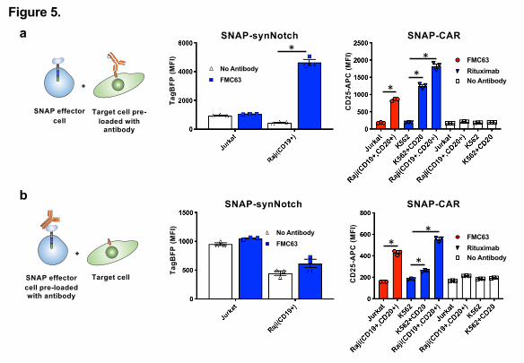
a
b
Target Cell
+
SNAP Effector Cell Pre-loaded with
Antibody
Target Cell Pre-loaded with Antibody
+
SNAP EffectorCell
SNAP-synNotch SNAP-CAR
SNAP-synNotch SNAP-CAR
FMC63
Rituxim
ab
K562+
CD20 Raji
No Antib
odyRaji
K562
K562+
CD200
200
400
600
800
CD
25-A
PC
(AU
)
CD25-APC
FMC63
RituximabNo Antibody
FMC63
Rituxim
ab
K562+
CD20 Raji
No Antib
odyRaji
K562
K562+
CD200
200
400
600
800
CD
25-A
PC
(AU
)
CD25-APC
FMC63
RituximabNo Antibody
Jurk
atRaji
0
2000
4000
6000
TagB
FP (a
u)SynNotch - Prestained Targets
No Antibody
FMC63
Jurk
atRaji
0
2000
4000
6000
TagB
FP (a
u)
SynNotch - Prestained Targets
No Antibody
FMC63
*
***
**
*
SNAP effector cell
Target cell pre-loaded with
antibody
SNAP effector cell pre-loaded with antibody
Target cell CD
25-A
PC (M
FI)
CD
25-A
PC (M
FI)
TagB
FP(M
FI)
TagB
FP(M
FI)
Figure 5.

a b
c
Fra
ctio
n o
f Ma
xim
al S
ign
al
kf1 KD1
Cetuximab
FMC63
Herceptin
Rituximab
Antibody (μg/mL)
Ternary body
(normalized)
Figure 6.
Simulation (literature values)
Simulation (fitted)
Experimental data
Tumor antigen (TA)
CAR T cells SynNotch T cells
Universal CAR T cells
kf1
kr1 = 0
kf1
kr1 = 0
kr2 kf2
+
+ + +
kr2 kf2