Embed Size (px)
Citation preview

lable at ScienceDirect
Biochimie 93 (2011) 18e31
Contents lists avai
Biochimie
journal homepage: www.elsevier .com/locate/b iochi
Review
Post-translational myristoylation: Fat matters in cellular life and death
Dale D.O. Martin 1, Erwan Beauchamp 1, Luc G. Berthiaume*
Department of Cell Biology, School of Molecular and Systems Medicine, MSB-5-55, Faculty of Medicine and Dentistry, University of Alberta, Edmonton, Alberta T6G 2H7, Canada
a r t i c l e i n f o
Article history:Received 1 October 2010Accepted 23 October 2010Available online 5 November 2010
Keywords:MyristoylationN-myristoyltransferaseApoptosisOmega alkynyl-myristateOmega azido-myristate
Abbreviations: ARF, ADP ribosylation factor; BimCAD, caspase activated DNAse; Caspase, cysteinyl-cytoplasmic dynein intermediate chain 2A; DED, ddihydroceramide delta4 desaturase; DISC, death induglutamate cysteine ligase; GFP, green fluorescence ptransferase; GPI, glycosylphosphatidylinositol; HIVvirus; HSC70, heat shock protein 71; LPS, lipopolysalated alanine-rich C kinase substrate; MBOAT, membraNIP71, NMT inhibitor protein 71; NMT, N-myristoyltrakinase 2; PAT, protein fatty acyl transferase; PKC3, prooxygen species; TRAM, TRIF-related adaptor molecuanion channel.* Corresponding author. Tel.: þ1 780 492 5146; fax
E-mail address: [email protected] (L.G.1 These authors contributed equally.
0300-9084/$ e see front matter � 2010 Elsevier Masdoi:10.1016/j.biochi.2010.10.018
a b s t r a c t
Myristoylation corresponds to the irreversible covalent linkage of the 14-carbon saturated fatty acid,myristic acid, to the N-terminal glycine of many eukaryotic and viral proteins. It is catalyzed byN-myristoyltransferase. Typically, the myristate moiety participates in protein subcellular localization byfacilitating protein-membrane interactions as well as proteineprotein interactions. Myristoylatedproteins are crucial components of a wide variety of functions, which include many signalling pathways,oncogenesis or viral replication. Initially, myristoylation was described as a co-translational reaction thatoccurs after the removal of the initiator methionine residue. However, it is now well established thatmyristoylation can also occur post-translationally in apoptotic cells. Indeed, during apoptosis hundredsof proteins are cleaved by caspases and in many cases this cleavage exposes an N-terminal glycine withina cryptic myristoylation consensus sequence, which can be myristoylated. The principal objective of thisreview is to provide an overview on the implication of myristoylation in health and disease with a specialemphasis on post-translational myristoylation. In addition, new advancements in the detection andidentification of myristoylated proteins are also briefly reviewed.
� 2010 Elsevier Masson SAS. All rights reserved.
1. Protein lipidation: an overview
Lipid modification of proteins plays many roles insideand outside of the cell including directing proteins to variouscellular membranes, sub-membrane domains and promoting pro-teineprotein interactions. Thus, protein lipidation adds anotherlevel of functionality to many proteins. Proteins can be co- or post-translationallymodifiedwith awide variety of lipids. These covalentlipid modifications of proteins are characterized by the identity ofthe lipid moiety attached, the nature of the covalent bond,the attachment site of the lipid on the protein and the enzymes
, Bcl-2 interacting mediator;aspartyl protease; CD-IC2A,eath effector domain; DES,cing signalling complex; GCL,rotein; GOAT, ghrelin-O-acyl-, human immunodeficiencyccharide; MARCKS, myristoy-ne bound O-acyl transferase;nsferase; PAK2, p21-activatedtein kinase C 3; ROS, reactivele; VDAC, voltage-dependent
: þ1 780 492 0450.Berthiaume).
son SAS. All rights reserved.
catalyzing the reactions involved. Four major types of lipid modifi-cation are known; cholesteroylation, prenylation, glypiation andfatty acylation [1]. In cholesteroylation, a cholesterol molecule isesterified to a C-terminal glycine residue in an autocatalytic process[2]. Prenylation involves the formation of a thioether bond betweeneither of the farnesyl or geranylgeranyl isoprenoids and one or twoC-terminally located cysteine residue(s) by a variety of prenyltransferases [3]. In glypiation, a glycosylphosphatidylinositol (GPI)anchor is added to the C-terminus of a protein by a transaminidasein the lumen of the endoplasmic reticulum [4]. Finally, fatty acyla-tion mainly consists of the covalent addition of palmitic or myristicfatty acids to proteins. In S-acylation, saturated and unsaturatedlong chain fatty acids of various carbon chain lengths can beattached to cysteine residues of a protein by a variety of protein fattyacyl transferases (PATs) belonging either to the zDHHC-PATs familyor the membrane bound O-acyl transferases (MBOATs) family [5,6].Because palmitic acid is the most abundant fatty acid, S-acylationtypically corresponds to the reversible attachment of the 16-carbonsaturated fatty acid palmitate to a cysteine residue via a thioesterbond and is thus typically referred to as palmitoylation [7]. In somecases, the palmitoyl moiety is linked to proteins via an amide bondon N-terminal cysteine or glycine residues [8,9]. Palmitoylation alsooccurs non-enzymatically (spontaneously) on several mitochon-drial proteins [10,11]. In myristoylation, various saturated andunsaturated 14-carbon fatty acids are also found on the N-terminalglycine of proteins [12], but, typically, myristoylation consists of the

D.D.O. Martin et al. / Biochimie 93 (2011) 18e31 19
covalent addition of the 14-carbon saturated fatty acid myristate tothe N-terminal glycine residue through a stable amide bond [13,14].The reaction is catalyzed by myristoyl-CoA:protein N-myristoyl-transferase (NMT) [15]. In some rare cases of myristoylation,including the insulin receptor [16] and interleukin 1 a and b [17],myristic acid is also attached to an internal lysine via an amide bond.Alternative forms of fatty acylation have also been reported. Theappetite-stimulating peptide hormone ghrelin is known to beacylated on serine 3 with octanoate [18], a reaction catalyzed byGhrelin-O-Acyltransferase (GOAT), a member of the MBOAT family[19]. GOAT appears to be regulated by dietary lipids [20]. Sinceoctanoylation is essential for ghrelin’s physiological activity [21],inhibitors of GOAT could represent attractive new appetitesuppressants helpful in the treatment of the metabolic syndrome.Furthermore, the signalling protein Wnt is not only known to befatty acylated with palmitic acid on Cys77 [22], but also modifiedwith unsaturated palmitoleic acid on ahighly conserved Ser209. Theacylation at both sites is catalyzed by the MBOAT Porcupine [23].Recently, Aquaporin 0 was demonstrated to be acylated with oleicacid on a lysine residue via an amide bond, whichmay contribute totarget the protein to lipid rafts [24].
The objective of this review is to provide insights on the roles ofmyristoylation in health and disease states with a special emphasison post-translational myristoylation in cell death.
2. Myristoylation: a matter of fat
Historically, N-myristoylation was initially described as anunusual blocking group, which prevented the direct use of Edmandegradation at the N-terminus of the catalytic subunit of cyclicAMP-dependent protein kinase [25] and calcineurin B [26]. It waslater determined by mass spectrometry that the blocking groupwas myristic acid. N-myristoylation was previously described asa co-translational modification [27,28] that occurs on the nascentpolypeptide following the removal of the initiator methionineresidue (Fig. 1A). Now it is well established that myristoylation canalso occur post-translationally on an internal glycine withina cryptic myristoylation consensus sequence exposed by the actionof caspases in apoptotic cells [29e34] (discussed in detail in thefollowing sections below) (Fig. 1B). Interestingly, removal of the
Fig. 1. Co- and post-translational attachment of myristate to proteins. Myristoylationinvolves the covalent addition of the saturated 14-carbon fatty acid myristate to theN-terminal glycine residues of (A) a nascent polypeptide following removal of theinitiator Met (co-translational myristoylation) or (B) a cryptic myristoylation sitefollowing exposure by caspase cleavage (post-translational myristoylation).
initiator methionine is a common modification of proteins, occur-ring in up to 80% of total proteins. The reaction is catalyzed bymethionine aminopeptidase [35] and is even more efficient whenthe amino acid residue following the initiator methionine isa glycine residue [36], yet only selected proteins are myristoylateddue to the selectivity of NMTs. Similarly, analysis of the caspase-cleaved proteome revealed that there is a high propensity to havea glycine residue after the various caspase-cleavage sites [37,38],suggesting that there might be numerous cases of post-trans-lational myristoylation yet to be uncovered.
2.1. How myristoylation affects proteins
Many myristoylated proteins play key roles in cellular signallingpathways [e.g. non-receptor protein tyrosine kinases, heterotrimericGa proteins, MARCKS and calcium binding proteins (e.g. Recoverin,Neurocalcin) reviewed in [6]]. For many of these proteins, the myr-istoyl moiety has been shown to mediate subcellular targeting,proteineprotein and protein-membrane interactions required forthe activities of these proteins [39]. Although myristoylation isrequired for membrane binding, it is not sufficient and must beaugmented by downstream interactions in order to provide stablemembrane anchoring [40]. This is usually achieved by an adjacent ordistant polybasic domain, by one or two nearby palmitoylatedcysteine residues or by a proteinelipid binding domain [41]. Myr-istoylation can alsoparticipate indifferential targeting tomembranesand sub-membrane domains known as lipid rafts [42,43].
Although myristate is irreversibly bound to proteins it allowsthe acylated protein to reversibly sample membranes because of itsweak hydrophobic nature. Indeed, the half-life of a membranebound myristoylated peptide is in the order of minutes incomparison to hours for palmitoylated or dually acylated(myristoylated and palmitoylated) peptides [44]. Again, althoughmyristoylation is an irreversible modification, the anchorage ofmyristoylated proteins to membranes can also be dynamicallyregulated via various “myristoyl switches”. One of these switchesrelies on a “ligand”-dependent conformational change of theprotein leading to exposure of the myristoyl moiety previouslysequestered in a hydrophobic pocket to the cytosol [45]. Thisconcept is exemplified by the binding of GTP to the ADP ribosyla-tion factor (ARF) proteins or calcium to recoverin that allow thesubsequent exposure of the myristoyl moiety and targeting of theseproteins to membranes [46,47]. Another variation of this mode ofregulation is referred to as the myristoyl electrostatic switch. Forinstance, protein kinase C dependant phosphorylation of serineresidues of the polybasic domain of MARCKS protein releases theprotein from the membrane by reducing the positive charge of thepolybasic domain that contributes to the membrane binding [48].A variation on the above, linking acylation and phosphorylation, isexemplified by c-Abl, which is known to be regulated by a “myr-istoyl/phosphotyrosine” switch [49]. In that case, the myristoylmoiety binds to a hydrophobic pocket and induces a conforma-tional modification of the protein allowing the docking of the SH2and SH3 domain onto the kinase domain and auto-inhibition of theenzyme [50]. Interestingly, a similar pocket has recently beenpredicted to exist in the c-Src kinase, but in that case, myr-istoylation acted in a different manner and enhanced c-Src activity[51]. This suggests that myristoylation of many other non-receptortyrosine kinases (e.g. Blk, Lyn, Fyn, Fgr) might not only be requiredfor the regulation of proper localization but also of their activity.
2.2. N-myristoyltransferases
In vertebrates, N-myristoylation is catalyzed by twoNMTs,NMT1and NMT2, which are members of the GCN5 acetyltransferase

D.D.O. Martin et al. / Biochimie 93 (2011) 18e3120
superfamily [52]. NMTs have been purified and cloned from manyorganisms [53e57]. To date, an NCBI database search for NMTannotated entries revealed the presence of the gene encoding forNMT in 53 organisms from taxa spanning animals, plants, apicom-plexan parasites (e.g. Plasmodium falciparum), fungi and yeast. Ofthese 53 organisms, 16 contained both NMT1 and NMT2. The NMTorthologues from Saccharomyces cerevisiae is the most extensivelycharacterized and has provided the majority of informationregarding the enzymology of the reaction [58,59]. NMTs performtheir catalysis via an ordered Bi Bi reaction mechanism with myr-istoyl-CoA binding first, peptide binding second followed by a directnucleophilic additioneelimination reaction and the sequentialrelease of CoA and the myristoyl-peptide [60]. The two genesencoding for the two isoforms of NMT were identified in humansand share about 76% amino acid sequence identity. The two NMTshave unique as well as overlapping substrate specificities [56,61]. Inorganisms where there is only one isoform of NMT, it has beenshown to be essential for survival (e.g. S. cerevisiae [53] andDrosophila [62]). NMT1�/� mice also die during embryogenesissuggesting that NMT2 is not able to rescue N-myristoylation ofproteins for the proper development of the mice embryos.This clearly indicates that both NMTs must have their own subsetsof unique substrates and that NMT1, or some proteins perhapsstrictly myristoylated by NMT1, play some crucial roles in devel-opment [61].
NMTs are highly selective for the substrate myristoyl-CoA inin vitro enzymatic assays [63,64]. This selectivity was in partexplained by looking at the 3D structure of NMT, which revealeda bent active site cavity only large enough to optimally accommo-date a myristoyl moiety [65]. However, different in vitro studieshave shown that NMT can catalyze the linkage of a syntheticpeptide that mimics the N-terminal region of a protein to differentfatty acids, such as lauric, tridecanoic, pentadecanoic and palmiticacid [66,67]. In the retina, several proteins are heterogeneouslymyristoylated with the unsaturated fatty acids C14:1 n-9 and C14:2n-6 (reviewed in [12]), but no retinal isoform specific of NMTable touse various fatty acyl-CoA substrates has been identified so far [68].
Cellular concentrations of myristoyl-CoA available for N-myr-istoylation are very low; approximately 5 nM in animal cells [69]. Inrat hepatocytes, only 0.05% of radiolabeled myristic acid added tothe cells is dedicated to the N-myristoylation of proteins [70].Because NMT can use palmitic acid as a substrate in vitro, albeit ata reduced efficiency compared to myristate [67,71,72], and the factthat cellular palmitic acid concentrations are significantly higherthan myristic acid suggest that the access of cytosolic NMTs toa specific pool of myristoyl-CoA is of critical importance [39]. Forinstance, it has recently been suggested that myristic acid capturedvia the CD36 receptor may be specifically dedicated to the myr-istoylation of tyrosine kinase pp59fyn [73].
The general consensus peptide sequence recognized by NMTs isGly-X3-X4-X5-(Ser/Thr/Cys)6 where X represents most amino acids,except for proline, aromatic or charged residues in position X3. Theamino-terminal Gly residue is absolutely required for myr-istoylation to occur and substitution of this residue to any otherabrogates myristoylation. While Ser, Thr, Cys are preferred atposition X6, other amino acids can be tolerated at this position (e.g.Ala found in Annexin XIII and Gly found in c-Abl tyrosine kinase)[74,75]. Moreover, the combination of amino acids at position X3, X6
and X7 play important roles for the N-myristoylation of thecandidate protein [76]. For instance, a Lys residue at position 7reduces the stringency for certain amino acids at position 3 and isalso important in conferring membrane binding properties [77].These studies and others have formed the basis for the develop-ment of myristoylation predictionmodels [78e81]. Using computeranalysis, the 17 N-terminal residues following the initial
methionine were demonstrated to be required for the prediction ofthe myristoylation status of the protein [82]. Using these computeralgorithms, it is estimated that 0.5e3% of the mammalian and plantproteomes consist of myristoylated proteins [78e81].
2.3. Regulation of myristoylation in health and disease
Little is known about the regulation of NMT activity and it ispresumed that levels of NMTactivity are necessary and sufficient toefficiently acylate endogenous proteins, but it was also suggestedthat NMT activity might actually be limited by the scarce levels ofmyristoyl-CoA itself [83,84]. In that context, the fact that NMTenzyme levels are elevated in several types of tumours is puzzling[85].
It has also been suggested that NMT has endogenous inhibitorsin the cell. An NMT inhibitor named NMT inhibitor protein 71(NIP71) was isolated from bovine brain using an in vitro NMT assay[86]. NIP71 shares 43% identity with the heat shock cognate protein70 (HSC70). In addition, the same group also demonstrated that theglycolytic enzyme enolase is able to inhibit N-myristoylation invitro [87]. Whether these inhibitors play a significant role in cellswas not investigated.
2.3.1. Crosstalk between myristoylation and phosphorylationPhosphorylation of hNMT1 was also shown to regulate the
activity of the enzyme. A Y100F mutation in NMT1 resulted ina 98% decrease of NMT activity [88]. However, the extent and thesite(s) of phosphorylation were not fully characterized since thismutant was still partially phosphorylated (17% as compared towild type). Interestingly, NMT is phosphorylated by Src familytyrosine kinase members of Lyn and Fyn [88] that are, in turn,myristoylated [85]. These results underline a possible complexregulation of the phosphorylated NMT by myristoylated tyrosinekinases. It is well established that those myristoylated tyrosinekinases (pp60src, pp60yes, pp56lck, pp59fyn/syn and c-Abl) [89]display elevated activity in many human cancers [90]. In addi-tion, increased protein and mRNA levels of pp60src were shown inhuman colon carcinoma, and many human cancer cell lines suchas HT29, COLO 201 and COLO 205 [91,92]. These data suggestedthat the regulation of pp60src happens at the level of transcriptionwith two distinct promoters (SRC1A and SRC1a) [93]. Inhibition ofpp60src myristoylation decreased colony formation, cell prolifera-tion and targeting to the membrane demonstrating the impor-tance of pp60src myristoylation for tumorigenicity [94]. Moreover,myristoylation seems to be critical for the dephosphorylation ofpp60src and its subsequent kinase activity [95]. Myristoylationincreases pp60src kinase activity but also regulates the stability ofthe protein. Recently, it was shown that myristoylation-dependantmembrane targeting of pp60src regulates the degradation andubiquitination of the protein [51] and the myristoyl moiety wasrecently proposed to possibly reside in a hydrophobic pocket likeit does in c-Abl [49], but would stimulate rather than inhibitkinase activity [51].
Also linking phosphorylation and myristoylation is the fact thatthe N-terminal domain of hNMT1 interacts with the Lyn tyrosinekinase in a phosphorylation dependant manner [85]. Because,NMT1 and NMT2 have their major amino acid sequence differencesat their N-termini [56], this suggests that differential phosphory-lation might allow for the fine tuning of the two NMT activitiesinside the cell. Interestingly, although the N-terminal region is notrequired for the activity of NMTs and because it exhibits significantsequence differences between the two enzymes, it has beenpostulated that the N-terminus allows the differential targeting ofNMTs to various cellular compartments, as well as provide access to

D.D.O. Martin et al. / Biochimie 93 (2011) 18e31 21
cellular myristoyl-CoA pools [96] and the differential binding toribosomes [14].
2.3.2. Myristoylation and cancerMagnuson et al. [97] were the first to demonstrate that NMT
activity was increased in rat colonic tumours, human colorectaltumours, adenocarcinomas [98] and stage B1 tumours, suggestingthat NMT is important in the early stages of colonic carcinogenesis[97]. This increase is correlated with higher expression levels ofNMT [98], and particularly the second isoform NMT2 [99].Increased expression of NMT was also found in gallbladder cancer[100], breast cancer [101] and brain tumours [102]. It is easy tospeculate that the overexpression of the myristoylated oncogenickinases may require increased NMT activity for their proper acyl-ation and, ultimately, function.
2.3.3. Spurious myristoylation in diseaseRecently, spurious myristoylation of the SHOC2 protein con-
taining a S2G mutation was documented and corresponds toa disease phenotype. Cordeddu et al. [103] identified an aberrantmyristoylation of the SHOC2 protein in the development of theNoonan-like syndrome with loose anagen hair in twenty-fivepatients. This syndrome is a rare developmental disorder resultingin reduced growth, facial dysmorphism, cognitive deficits andmalignancies [104,105]. The authors found that the SHOC2S2G
mutant incorporated [3H]-myristic acid and was targeted to the cellmembrane instead of the nucleus leading to increased activation ofRas and the MAPK pathway. This is the first demonstration of thedirect involvement of myristoylation in the development ofa human disease and the first example of a spontaneous geneticmutation leading to aberrant myristoylation.
2.3.4. Myristoylation in infectious diseasesThe covalent attachment of myristate to the N-terminal glycine
residue occurs on many mammalian [78] in addition to viral andeven secreted bacterial [106] proteins, and, is essential to theseproteins’ ultimate function [13,14]. Therefore, NMTs representattractive therapeutical drug targets. The NMT protein substratespecificity has been well characterized in S. cerevisiae usingsynthetic peptides corresponding to the N-terminus of protein[15,107,108]. Using this system for mammalian NMT1, the first tenamino acids were shown to be necessary for effective myristoyl-transferase activity. Interestingly, human and yeast NMTs havedifferent yet overlapping substrate specificities [109]. The differ-ences were significant enough to allow for the development ofselective inhibitors of yNMT [110] paving the way to the design ofNMT inhibitors against pathogenic yeasts and fungi such as Candidaalbicans, Cryptococcus neoformans and Histoplasma capsulatum[54,111]. The fact that the NMTactivities appear to be critical for thevirulence of C. albicans and C. neoformans in an immunosuppressedmodel, again highlights the potential of NMTas a useful therapeutictarget. Of note, several parasitic protozoa such as Leishmania majorand Leishmania donovani (leishmaniasis desease), Trypanosomabrucei (African sleeping sickness disease) and P. falciparum(malaria) also possess their own NMT that appears to be essentialfor their survival [112e115]. Investigations are now focusing on thevalidation of inhibitors of protozoan NMTs. Different strategieshave been used towards the development of these inhibitors andwere based on the use of either peptidomimetics able to bind to theNMT active sites or small molecule inhibitors of NMTs. Recently,a pyrazole sulphonamide specific inhibitor of the T. brucei NMT hasbeen identified [116]. Its selectivity over human NMT is 100-foldand a tight correlation between NMT activity and T. brucei prolif-eration is observed suggesting that NMTactivity is the target of thiscompound. Pyrazole sulphonamide competes with the peptide
binding site of NMT. This compound rapidly kills T. brucei andeliminates the bloodstream form of T. brucei. Moreover, thiscompound not only works in vitro, but also cured all animals ina mouse model of human African trypanosomiasis. Therefore, itshows promising therapeutic properties to treat sleeping sickness.
Of note, myristoylation is required for assembly and replicationof viruses such as lentiviruses (e.g. HIV) or picornaviridae [106]. Inthe case of HIV replication, both the structural proteins Gag and Nefare myristoylated by the host cell NMT. The Gag protein consists ofa polyprotein precursor containing several domains, includinga matrix domain, which is myristoylated and directs the protein tothe plasma membrane in combination with its polybasic domain[117]. Subsequently, this anchoring is critical for the downstreamcleavage of the polyprotein and the release of the mature myr-istoylated matrix domain to the cytosol [118]. These crucial stepsare required for the formation of the viral capsid, as inhibition ofmyristoylation causes the accumulation of the Gag proteinprecursor in the cytosol and blocks formation of a competent viralprogeny [119]. Recent studies suggest that NMT1 and NMT2 displaydifferent affinity for Gag and Nef, allowing the possible develop-ment of selective inhibitors [120,121]. In addition, the competitionfor the myristoyl-CoA substrate pool appeared to be critical in thispathophysiological situation. It is estimated that the concentrationof myristoyl-CoA necessary for the maturation of viral particles isabout 1 mM [13], while the endogenous cellular concentration isabout 5 nM [69]. This suggests that the production of myristoyl-CoAmight be a rate-limiting step and that the size of the myristoyl-CoApool might be regulated during viral infection [122].
Certain types of bacteria are known to inject proteins (via typeIII secretion) containing a myristoylatable N-terminal glycineresidue directly into the cytoplasm of the host cell. The injectedprotein is thenmyristoylated by the host NMTand relocalizes to theplasma membrane. Myristoylation of the protein is essential formaximum virulence [106,123]. This type of myristoylation of typeIII secreted bacterial proteins was demonstrated in Pseudomonassyringae [123] and predicted for Shigella sonnei [124]. Drugs tar-geting eukaryotic acylation of bacterial proteins may represent aninteresting strategy to fight plant and animal infections.
Finally, the examples listed above highlight the importance ofunderstanding and characterizing myristoylation under normaland pathological conditions. For instance, because apoptosis ulti-mately results in caspase activation and is often deregulated incancer, the importance of characterizing post-translational myr-istoylation of proteins cleaved by caspases during apoptosis isundeniable, may be critical to understand the development andprogression of cancer and will be discussed in the next section.
3. Post-translational myristoylation: different meansto an end
3.1. Post-translational myristoylation during apoptosis
Until about 10 years ago, myristoylation was thought to beexclusively a co-translational process, wherein myristate is addedto the amino-terminal glycine via an amide bond following theremoval of the initiator methionine from the nascent polypeptideby a methionyl aminopeptidase (Fig. 1A) [13,14]. However, the pro-apoptotic protein Bid was shown to be post-translationally myr-istoylated on an internal glycine residue exposed after caspasecleavage during apoptosis [33]. Since then, we and others haveidentified several proteins post-translationally myristoylatedduring apoptosis and now it is well established that myristoylationcan also occur post-translationally upon caspase cleavage andexposure of an internal glycine within a cryptic myristoylationconsensus sequence (Fig. 1B) [30e32,34].
D.D.O. Martin et al. / Biochimie 93 (2011) 18e3122
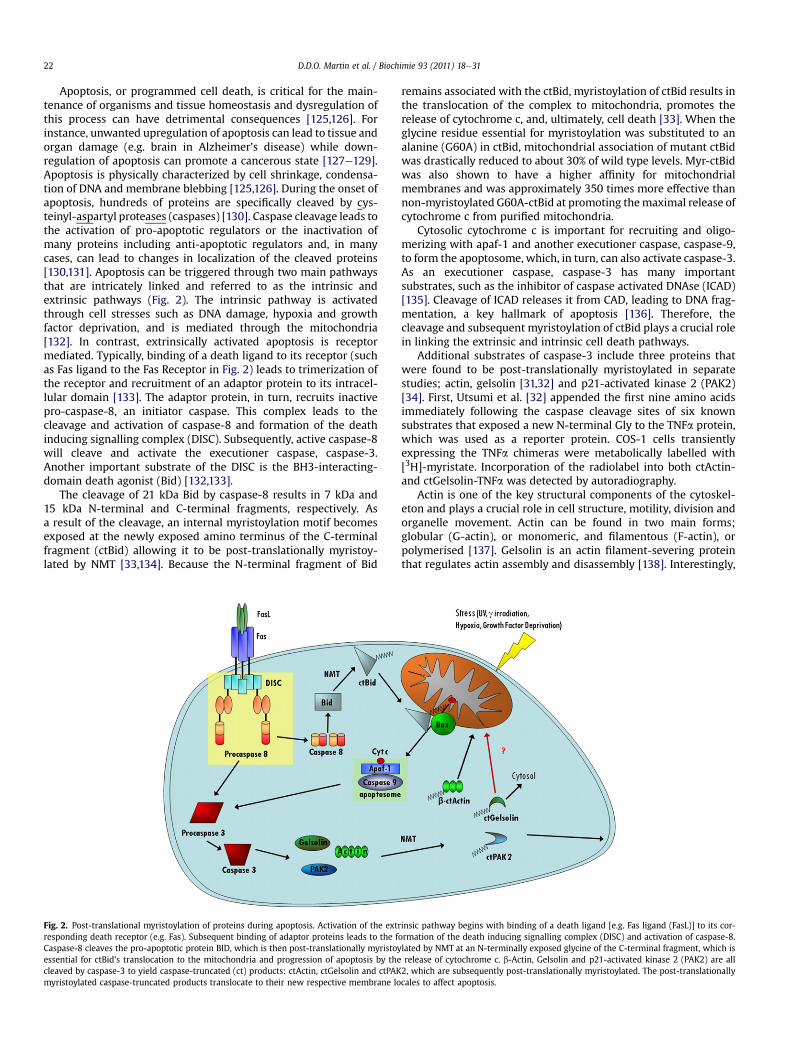
Apoptosis, or programmed cell death, is critical for the main-tenance of organisms and tissue homeostasis and dysregulation ofthis process can have detrimental consequences [125,126]. Forinstance, unwanted upregulation of apoptosis can lead to tissue andorgan damage (e.g. brain in Alzheimer’s disease) while down-regulation of apoptosis can promote a cancerous state [127e129].Apoptosis is physically characterized by cell shrinkage, condensa-tion of DNA and membrane blebbing [125,126]. During the onset ofapoptosis, hundreds of proteins are specifically cleaved by cys-teinyl-aspartyl proteases (caspases) [130]. Caspase cleavage leads tothe activation of pro-apoptotic regulators or the inactivation ofmany proteins including anti-apoptotic regulators and, in manycases, can lead to changes in localization of the cleaved proteins[130,131]. Apoptosis can be triggered through two main pathwaysthat are intricately linked and referred to as the intrinsic andextrinsic pathways (Fig. 2). The intrinsic pathway is activatedthrough cell stresses such as DNA damage, hypoxia and growthfactor deprivation, and is mediated through the mitochondria[132]. In contrast, extrinsically activated apoptosis is receptormediated. Typically, binding of a death ligand to its receptor (suchas Fas ligand to the Fas Receptor in Fig. 2) leads to trimerization ofthe receptor and recruitment of an adaptor protein to its intracel-lular domain [133]. The adaptor protein, in turn, recruits inactivepro-caspase-8, an initiator caspase. This complex leads to thecleavage and activation of caspase-8 and formation of the deathinducing signalling complex (DISC). Subsequently, active caspase-8will cleave and activate the executioner caspase, caspase-3.Another important substrate of the DISC is the BH3-interacting-domain death agonist (Bid) [132,133].
The cleavage of 21 kDa Bid by caspase-8 results in 7 kDa and15 kDa N-terminal and C-terminal fragments, respectively. Asa result of the cleavage, an internal myristoylation motif becomesexposed at the newly exposed amino terminus of the C-terminalfragment (ctBid) allowing it to be post-translationally myristoy-lated by NMT [33,134]. Because the N-terminal fragment of Bid
Fig. 2. Post-translational myristoylation of proteins during apoptosis. Activation of the extrresponding death receptor (e.g. Fas). Subsequent binding of adaptor proteins leads to the fCaspase-8 cleaves the pro-apoptotic protein BID, which is then post-translationally myristoyessential for ctBid’s translocation to the mitochondria and progression of apoptosis by thecleaved by caspase-3 to yield caspase-truncated (ct) products: ctActin, ctGelsolin and ctPAKmyristoylated caspase-truncated products translocate to their new respective membrane lo
remains associated with the ctBid, myristoylation of ctBid results inthe translocation of the complex to mitochondria, promotes therelease of cytochrome c, and, ultimately, cell death [33]. When theglycine residue essential for myristoylation was substituted to analanine (G60A) in ctBid, mitochondrial association of mutant ctBidwas drastically reduced to about 30% of wild type levels. Myr-ctBidwas also shown to have a higher affinity for mitochondrialmembranes and was approximately 350 times more effective thannon-myristoylated G60A-ctBid at promoting themaximal release ofcytochrome c from purified mitochondria.
Cytosolic cytochrome c is important for recruiting and oligo-merizing with apaf-1 and another executioner caspase, caspase-9,to form the apoptosome, which, in turn, can also activate caspase-3.As an executioner caspase, caspase-3 has many importantsubstrates, such as the inhibitor of caspase activated DNAse (ICAD)[135]. Cleavage of ICAD releases it from CAD, leading to DNA frag-mentation, a key hallmark of apoptosis [136]. Therefore, thecleavage and subsequent myristoylation of ctBid plays a crucial rolein linking the extrinsic and intrinsic cell death pathways.
Additional substrates of caspase-3 include three proteins thatwere found to be post-translationally myristoylated in separatestudies; actin, gelsolin [31,32] and p21-activated kinase 2 (PAK2)[34]. First, Utsumi et al. [32] appended the first nine amino acidsimmediately following the caspase cleavage sites of six knownsubstrates that exposed a new N-terminal Gly to the TNFa protein,which was used as a reporter protein. COS-1 cells transientlyexpressing the TNFa chimeras were metabolically labelled with[3H]-myristate. Incorporation of the radiolabel into both ctActin-and ctGelsolin-TNFa was detected by autoradiography.
Actin is one of the key structural components of the cytoskel-eton and plays a crucial role in cell structure, motility, division andorganelle movement. Actin can be found in two main forms;globular (G-actin), or monomeric, and filamentous (F-actin), orpolymerised [137]. Gelsolin is an actin filament-severing proteinthat regulates actin assembly and disassembly [138]. Interestingly,
insic pathway begins with binding of a death ligand [e.g. Fas ligand (FasL)] to its cor-ormation of the death inducing signalling complex (DISC) and activation of caspase-8.lated by NMT at an N-terminally exposed glycine of the C-terminal fragment, which isrelease of cytochrome c. b-Actin, Gelsolin and p21-activated kinase 2 (PAK2) are all2, which are subsequently post-translationally myristoylated. The post-translationallycales to affect apoptosis.

D.D.O. Martin et al. / Biochimie 93 (2011) 18e31 23
myr-ctActineFLAG co-localized with mitochondria, like myr-ctBID,but it was not found to have any effect on cellularmorphology, actinnetworks, nor was any effect on apoptosis established [31,32].
In a follow up study by the same authors, myr-ctGelsolineHAwas shown to be primarily cytosolic. Indeed, cell fractionationstudies showed ctGelsolineHA predominantly in the cytosolicfraction regardless if it was myristoylated or not. In addition, myr-ctGelsolin was also concluded to be cytosolic based on indirectimmunofluorescence data [31]. Regardless, cells expressing myr-ctGelsolin had an increased protection from apoptosis induced bythe topoisomerase inhibitor etoposide, when compared to non-myr-ctGelsolin or vector alone. Once again, this demonstrates theimportance of post-translational myristoylation in regulating thefunction of caspase-cleaved proteins during apoptosis. Interest-ingly, previous studies have shown that both the full-length formand ctGelsolin were anti-apoptotic through a mechanism that isthought to involve the voltage-dependent anion channel (VDAC) atthe mitochondrial membrane [139,140]. In that context, it would beinteresting to compare the potency of full-length versus caspase-cleaved myristoylated gelsolin in order to assess whether myr-istoylation augments the ability of gelsolin to suppress apoptosis.Interestingly, ctBid is also thought to promote apoptosis by asso-ciating with VDAC and promoting an open pore conformation [141].Although it should be noted that in that study, post-translationalmyristoylation was blocked by the presence of an N-terminalepitope tag on ctBid. It may be of interest to determine whethermyr-ctActin, which translocates to the mitochondria, also interactswith the VDAC. Of note, it has been suggested that destabilization ofthe interaction between cardiolipin and the mitochondrialpermeability transition pore, of which VDAC is a main component,promotes an open conformation of the pore during apoptosis toallow the release of cytochrome c [142]. In the absence of car-diolipin, Bid does not localize to the mitochondria [142]. Theseresults suggest that post-translationally myristoylated proteinsmay be directed to VDAC in a cardiolipin dependent manner toregulate the open/closed formation of the pore complex.
In addition to ctBid, ctActin and ctGelsolin, our laboratory hasidentified another post-translationally myristoylated protein: PAK2[34]. PAK2 is a Ser/Thr kinase whose activity is regulated by Rac andCdc42 [143]. In dividing cells, PAK2 activity stimulates cell growth,cell motility and cell survival [144]. In apoptotic cells, PAK2 is cleavedby caspase-3 resulting in the release of the highly pro-apoptoticconstitutively active C-terminal kinase domain from the N-terminalregulatory domain [145]. We have shown that the newly exposed N-terminal glycineof the caspase-cleavedPAK2kinasedomain (ctPAK2)is post-translationally myristoylated [34]. It was observed thatmyr-ctPAK2 was a more potent activator of apoptosis than its non-myristoylated counterpart. Indeed, after 12 hof transient transfectionof ctPAK2-Myc, more than 50% of myr-ctPAK2-Myc expressing cellswere apoptotic compared to only 23% in both the vector control andG213A-ctPAK2-Myc transfected cells. While WTctPAK-Myc induced50% of cell death within 12 h, the non-myristoylated G2A mutantreached a maximal cell death of 50% after 24 h. In addition,a concomitant 5-fold and 3.5-fold increase in phosphorylation ofthe stress-activated signalling kinase JNK was observed in cellsexpressing the myristoylated- and non-myristoylated ctPAK2respectively, over vector alone. Conversely, themyristoylated form ofctPAK2 was found to be hypophosphorylated compared to its non-myristoylated counterpart suggesting that loss ofmyristoylation leadto a dysregulation of PAK2 phosphorylation. In addition, myr-ctPAK2was found to localize to plasma membrane ruffles and early endo-somes while the non-myristoylated ctPAK2 remained primarilycytosolic, thus suggesting once more that myristoylation leads toproper localization and enhanced biological activity. Interestingly,and excitingly, cellular death induced by expression of myr-ctPAK2
bypassed several key hallmarks of apoptosis. Indeed, cells under-going myr-ctPAK2 mediated cell death did not release cytochrome cfrom the mitochondria nor lose mitochondrial potential. In addition,these cells did not expose phosphatidylserine at the cell surfaceduring the cell death process. These observations suggest that myr-ctPAK2mediates its actions downstream of themitochondrial “step”.
Because post-translational myristoylation potentiates the pro-apoptotic activity of ctBid and ctPAK2, post-translational myr-istoylation of caspase-cleaved proteins could represent an elegantmeans developed by the cell to expand the functionality of itsencoded proteins.
3.2. Potential roles for co-translational myristoylationand myristate in the regulation of apoptosis
Myristoylation may also play an important role in the metabo-lism of sphingolipids and ceramides, two types of molecules knownto be involved in the regulation of apoptosis. For instance, severalproteins involved in these pathways contain N-terminal glycinesthat may be myristoylated, such as ceramide kinase [146] andneutral sphingomyelinase [147]. In fact, dihydroceramide delta4desaturase (DES) has been shown to be myristoylated and itsactivity is even upregulated when “free” myristate is added to cells[148,149]. Myristoylation of DES targets the enzyme to mitochon-dria where sphingolipid metabolism may be altered [150].Furthermore, this acylation induces ceramide production, cyto-chrome c release from mitochondria and, consequently, apoptosisin COS-7 cells. It is well known that ceramides induce apoptosis bytargeting mitochondrial metabolism via the formation of channelsin the outer mitochondrial membrane and release of cytochrome c[151]. Ceramides also interact with key components of the electrontransport chain [152] and induce the production of ROS species[153] leading to the induction of apoptosis. We can also hypothe-size that changes in myristate concentration may alter the outermitochondrial membrane composition in phospholipids and car-diolipins by structurally altering their fatty acid constituents and,therefore, change the mitochondrial membrane permeability.
Overall, it thus appears both co- and post-translational myr-istoylated proteins play important roles in the regulation of mito-chondrial integrity and the generation of pro-death or pro-survivalsignals. Altogether these are key components of the cell’s ability tomake life and death decisions.
4. Detection of myristoylation: click chemistryand the solution to long film exposures
4.1. Detection using radioactive fatty acids
The key roles played by co- and post-translationally myristoy-lated proteins in the generation of pro-survival or pro-death signalswarrant the identification of the co- and post-translationalmyristoylated protein proteomes or “myristoylomes”. Indeed, inaddition to the previously identified post-translationally myristoy-lated proteins, we have observed the presence of nine other post-translationally myristoylated proteins using metabolic labeling ofapoptotic Jurkat cell lysates with [3H]-myristic acid [30]. However,the use of radioactive [3H]-myristic acid has a low sensitivity, is verytime consuming, expensive and represents a health hazard. Overall,progress in the identification and characterization of myristoylatedproteins has been impeded by the long exposure times required tomonitor incorporation of radioactive myristate into proteins (typi-cally 1e3 months). Still using [3H]-myristic acid as a label, theUtsumi laboratory recently developed cell free assays that utilizerabbit reticulocyte lysates or insect cell lysates for the synthesis andlabeling of potentially myristoylated proteins [154e156]. In their
D.D.O. Martin et al. / Biochimie 93 (2011) 18e3124
paper, using these cell free systems myristoylation detection wasreduced to days/weeks film exposure using fluorography. Thisallowed for the identification of 27myristoylated proteins, of which18 were new myristoylated proteins [155]. Despite identifyinga number of novel myristoylated proteins, this approach was rathertime consuming and still relied on metabolic labeling techniqueslinked to rather lengthy fluorographic film exposures and anexhaustive mass spectrometric/proteomic approach.
To alleviate the lengthy fluorographic exposures inherent to theuse of [3H]-myristic acid to label cells, an [125I]-iodomyristate hasalso been synthesized and used to reduce exposure time to days orless, but this requires the handling of large quantities (mCi) of thehazardous high energy 125I radioisotope [157,158]. Unfortunately,this compound is not commercially available and must be gener-ated in the laboratory [158].
4.2. Chemical biology and click chemistry
In order to circumvent this and to bypass the use of radioactivity,we and other laboratories have developed a number of non-radioactive techniques for the labeling of myristoylated proteinstaking advantage of bio-orthogonal analogs of fatty acids that canbe incorporated into proteins and selectively reacted with probesfacilitating their detection [29,30,159e164]. These new techniquescould ultimately be used for affinity purification and identificationof co- and post-translationally myristoylated proteins. Initially, thevery specific reaction between an alkyl-azide and a phosphineusing the Staudinger ligation (Fig. 3A) was exploited to detectvarious fatty acylated proteins [30,159]. In this method, the isostericand bio-orthogonal analog of myristate, azido-myristate (12 carbonwith an u-azido group; Az-C12), can be specifically incorporatedco- or post-translationally into proteins at the N-terminal glycineresidues of various proteins. Subsequently, the azido moiety waschemoselectively ligated to triarylphosphines bearing FLAG orbiotin and detected by Western blotting with short exposure timesto film [30,159]. For instance, detection of azido-myristoylatedctPAK2 in apoptotic Jurkat cell lysates was reduced from 2 monthsusing [3H]-myristic acid to 1 s after ligation and detection of thebiotin-phosphine probe [30]. This represents over a million-foldsignal amplification in comparison to using radioactive labelingmethods. This novel use of a myristate analogue that can bechemically ligated to probes was shown to be specific for N-terminal glycine residues of several proteins [30,159] and to rely onN-myristoyltransferases for their incorporation [30].
In order to discover new post-translationally myristoylatedproteins, we identified 48 protein candidates, from a review listing280 caspase substrates that exposed an N-terminal glycine aftertheir caspase cleavage sites [131]. The 48 amino acid sequencesdownstream of the cleavage sites were subjected to myristoylationprediction analysis using Myr Predictor and Myristoylator and 9proteins were predicted to be myristoylatable [165,166]. Followingan initiator methionine, the first ten amino acids of the C-terminalfragment of these 9 potential substrates were appended to theN-terminus of EGFP. Using metabolic labeling of COS-7 cells tran-siently expressing the chimeric GFPs with the Az-C12 analog, theincorporation of the Az-C12 into proteins was monitoredby chemical labeling with a biotinylated triarylphosphine, asdescribed in [30,159]. Doing so, we identified 5 new post-transla-tionally myristoylatable proteins [Protein Kinase C 3 (PKC3), cyto-plasmic dynein intermediate chain 2A (CD-IC2A), the pro-apoptoticprotein Bap31, mammalian Ste20-like kinase (MST3), and thecatalytic subunit of glutamate cysteine ligase (GCLC)] [30]. Becausemyristoylation is typically necessary and sufficient to exclude GFPfrom the nucleus [42], we used this exclusion criteria to assesswhether our chimeric EGFPs were bona fidemyristoylated proteins.
We demonstrated that chimeric EGFP bearing N-terminalsequences of caspase-cleaved fragments of PAK2, PKC3 and CD-IC2Awere extensively excluded from the nucleus while chimeras ofBap31, MST3 and GCLC led to little to no nuclear exclusion of EGFP.Therefore, PAK2, PKC3 and CD-IC2A chimeric EGFPs were assessedas strong candidates for myristoylation based on our criteria whilethe Bap31, MST3 and GCLC chimeric EGFPs may not be efficientlymyristoylated nor strong candidates for myristoylation. Critically, inour approach we originally used an expression system relying onco-translational myristoylation to investigate and assess the post-translational myristoylation potential of caspase-cleaved proteinsequences. Some of the observed discrepancies may thus beattributed, in part, to the substrate specificity of methionineaminopeptidase and its ability to remove the initiator methioninein these artificial substrates. Alternatively, post-translational myr-istoylation may rely selectively on one of the two NMT isoforms.Since NMT1 associates with ribosomes [14] and thus may playa more predominant role in co-translational myristoylation, NMT2might be the key NMT required for post-translational myr-istoylation. Furthermore, immunoprecipitation studies have shownthat NMT2 interacts with caspase-3 [85]. This finding may supportthe idea that NMT2 may play a greater role in post-translationalmyristoylation during apoptosis than NMT1. Therefore, the myr-istoylation of a protein normally post-translationally myristoylatedby NMT2 might be difficult to assess using a co-translationalexpression system possibly relying preferentially on NMT1.
Subsequently, we and others developed a chemical ligationtechnique that relies on the Cu (I) e catalyzed [3 þ 2] Huisgencycloaddition reaction (commonly referred to as Click chemistry)that uses an u-alkynyl-myristate analog (Alk-C14) and variousazido-probes (Fig. 3B) [29,162e164]. Like Az-C12, Alk-C14 is readilytaken up by cells and incorporated into proteins. Subsequently, thealkynyl moiety of the fatty acid analog can be covalently linked toan azido-probe using Click chemistry (Fig. 3B). The probe maycontain biotin for Western blot detection [29,162,164] and/oraffinity purification (as shown for palmitoylation) [164], a fluores-cent rhodamine [163,167] or a fluorogenic coumarin [29] for in-gelfluorescence detection. Use of the fluorescent azido-rhodamine andfluorogenic azido-coumarin allows flatbed in-gel detection of fattyacylated proteins separated by SDS-PAGE, thereby bypassingWestern blot analysis and facilitating detection and excision of fattyacylated proteins for identification by mass spectrometry.
The development of an assay that can assess post-translationalmyristoylation will be required to establish the post-translationalmyristoylation of cancer cells and evaluate the fatty acylation of thenewly discovered myristoylated proteins. Interestingly, we alsodemonstrated the existence of at least 15 post-translationallymyristoylated proteins in apoptotic Jurkat cell lysates using theStaudinger ligation approach [30], an observation we also corrob-orated using click chemistry [29]. Thus, the presence of numerouspost-translationally myristoylated proteins in apoptotic cellssuggests an underappreciated role for post-translational myr-istoylation in the regulation of cell death. Altogether these newtechniques at the chemical biology forefront will accelerate thedata acquisition process not only formyristoylated proteins but alsofor NMTs as well as other types of acylated and acylating proteins(please see [168] for a complete review).
5. Discussion
5.1. Potential roles of recently identified putativepost-translationally myristoylated proteins in apoptosis
The previous screen of Martin et al. [30] identified five proteinsthat were predicted to be cleaved by caspases to reveal an

Fig. 3. Schematic of the Copper (I)-catalyzed Azide-Alkyne cycloaddition and the Staudinger ligation used to detect myristoylation of proteins. (A) u-alkynyl-myristate is added tothe cells, which leads to its activation to the CoA ester and its NMT dependent addition to the N-terminal glycine residue of a protein. u-alkynyl-myristoylated proteins are reactedto a variety of tags using the Copper (I)-catalyzed Azide-Alkyne cycloaddition commonly referred to as “Click” reaction [29] (B). Similarly an azido-myristate analog (u-azido-dodecanoate, Az-C12) can be incorporated into cellular proteins and ligated to a variety of tagged-phosphine probes by the Staudinger ligation (C).
D.D.O. Martin et al. / Biochimie 93 (2011) 18e31 25

D.D.O. Martin et al. / Biochimie 93 (2011) 18e3126
N-terminal glycine on their C-terminal cleavage fragment. Asmentioned before, these included two kinases, PKC3 and MST3, theapoptotic regulator Bap31, the structural component CD-IC2A andfinally an enzyme involved in glutathione metabolism, GCLC.A novel assay aimed at strictly detecting post-translational myr-istoylation was recently developed in our laboratory (Martin et al.,manuscript in preparation). Using this assay, it appears that the first10 amino acids of ctPKC3 and ctCD-IC2A remain strong candidatesfor post-translational myristoylation, whereas Bap31, GCLC andMST3 are not (unpublished results). Using this assay, we alsorevealed several new candidates for post-translational myr-istoylation, including Mcl-1 (see below).
Like ctPAK2, caspase cleavage of PKC3 at D383 separates theN-terminal regulatory domain from the C-terminal kinase domain,which becomes constitutively active [169]. PKC3 contains a secondcaspase cleavage sites at D451 that leads to an inactive form of thekinase domain. The former cleavage site appears to be the primarysite of cleavage and is locatedwithin the hinge domain, between theregulatory and kinase domains, whereas cleavage at the second siteis delayed and is found within the catalytic domain and, presum-ably, inactivates the kinase. Full-length PKC3 binds diacylglycerideduring its activation. Therefore, loss of the diacylglyceride-bindingdomain in the N-terminal region may be substituted by post-translational myristoylation of the constitutively active ctPKC3 toprovide membrane binding during apoptosis.
The CD-IC proteins are required for the interaction betweencytoplasmic dynein and dynactin through binding of p150Glued, viatheir N-termini. Dynein and dynactin regulate the secretory andendocytic pathways, microtubule organization at interphase, andthe ER serves as cargo for cytoplasmic dynein in Xenopus eggextracts. During apoptosis, CD-IC2 is cleaved within the p150Glued
binding domain, but remains associated with the heavy and inter-mediate light chains of dynein, probably via its overlapping WD-40repeats [170]. Very interestingly, light chain 8 of dynein associateswith the pro-apoptotic protein Bcl-2 interacting mediator of celldeath (Bim) in normal cells and during apoptosis this complextranslocates to membranes of various organelles, including mito-chondria, where it binds and inhibits BCL-2 or functional homologs[171]. Alternatively, during apoptosis, molecular motors promotethe paranuclear clustering of mitochondria, which increases theapoptotic effect [172]. It is possible that myr-ctCD-IC2 may beinvolved in either of these processes to direct the progression ofapoptosis.
5.2. On the prevalence of post-translationallymyristoylated proteins
Since the identification of the new post-translationally myr-istoylatable substratesdescribedabove [30], thenumberof identifiedcaspase substrates with known caspase cleavage sites has increasedfrom 280 [131] to well over 400 [37,38,173]. Likewise, the number ofcleaved protein products with N-terminally exposed glycines hasincreased from 48 to 72 and this accounts forw17% of all the knowncaspase substrates, suggesting that many more potential substratesfor post-translational myristoylation may exist. In addition, a recentstudy looking at preferred caspase cleavage sites demonstrated thatglycine was the most favoured amino acid adjacent to the aspartateresidue (DXXDG) further strengthening the idea that more post-translationally myristoylated substrates exist [38]. Unfortunately, inthe same study [38] the technique used for the labeling, affinitypurification and identification of caspase-cleaved proteins by massspectrometry relied on subtiligase, a modified form of the bacterialprotease subtilisin BPN, which can perform ligation of peptides.Unfortunately, subtiligase requires a free N-terminus for its activityand, therefore, myristoylation would abrogate the addition of an
affinity purification tag by subtiligase and prevent the identificationof post-translationally myristoylated proteins by this method.Nonetheless, using the information obtained in that studyandothers[37,173], the newly identified caspase substrates were investigatedfor their potential myristoylation as we did before using a combina-tion of myristoylation prediction programs (e.g. Myr Predictor andMyristoylator) [165,166]. By doing so, we list over a dozen potentialsubstrates for post-translational myristoylation in Table 1. Ofparticular interest, one of the predicted putative post-translationallymyristoylatable proteins includes the anti-apoptotic protein Mcl-1.Once cleaved, the C-terminus of Mcl-1 becomes pro-apoptotic and,like ctBid, it promotes an open confirmation of the pore complex[141,174]. In fact, the new post-translational myristoylation assayused to confirm the post-translational myristoylation of the first tenamino acids of ctPKC3 and ctCD-IC2A suggests that ctMcl-1 isa substrate for post-translational myristoylation as well (unpub-lished results). If Mcl-1 is a bona fide post-translationally myristoy-lated protein, this would suggest, once again, that post-translationalmyristoylation may promote the translocation of various apoptoticregulators to mitochondria to promote pore complex assembly andpromote apoptotic progression. The eventual combination of a post-translationalmyristoylationdetection assayandclick chemistrywithazido-probes will lead to the identification and characterization ofmany more post-translationally myristoylated proteins.
5.3. New regulatory roles for myristoylation during cell deathand development
The fact that some post-translationally myristoylated proteinspromote apoptosis (myr-ctBid and myr-ctPAK2) [33,34] while othersprevent it (myr-ctGelsolin) [31] is paradoxical and suggests antago-nistic roles for post-translational myristoylation. This again suggestsa potential regulatory role forpost-translationalmyristoylation in celldeath/survival. The final outcomemay be decided by the identity andfunction of newly post-translationally myristoylated proteins. Inaddition, the fact that caspases are active and essential for cellproliferation and differentiation in various cell lines may suggesta potential involvement of post-translationally myristoylatedproteins in the developmental context as well [175e178]. Forexample, caspase-8 activity was shown to be required for the differ-entiation of monocytes intomacrophages and PAK2 has been proventobecleaved inacaspase-dependentmanner, atwhatappears tobeatvery low levels in those cells during this process [179]. Alkyne-myristate labelingofdifferentiatingU937 cells suggests that ctPAK2 isin fact post-translationally myristoylated under these conditions(unpublishedresults). Furthermore, gelsolin is alsocleavedduring thedifferentiation of megakaryocytes [180]. It seems likely that theseproteins would be post-translationally myristoylated in thesecontexts. Again, myr-ctGelsolin may aid in cell survival while myr-ctPAK2may promote cytoskeletal ormorphological changes requiredfor both apoptosis and differentiation, depending on the level ofcleavageoractivationofmyr-ctPAK2.Differentiationandproliferationmay provide yet another potential pool of post-translationally myr-istoylatedproteins to discover and another level of cellular regulationby NMTs and myristoylation.
NMT was also shown to play a role in the regulation of immunefunction and differentiation [181,182]. When embryonic stem cellsfrom homozygous NMT1 (�/�) were stimulated to undergodifferentiation with M-CSF a drastic decrease in macrophageproduction was observed [182]. These results led the authors toconclude that NMT1 is essential for proper monocytic differentia-tion. Furthermore, if NMT activity was reduced in inflamed bovinelungs, total NMT1 activity and expression were significantlyincreased in neutrophils and macrophages exposed to LPS in vitro[181]. Finally, NMT1 knockdown increases the cell death of normal
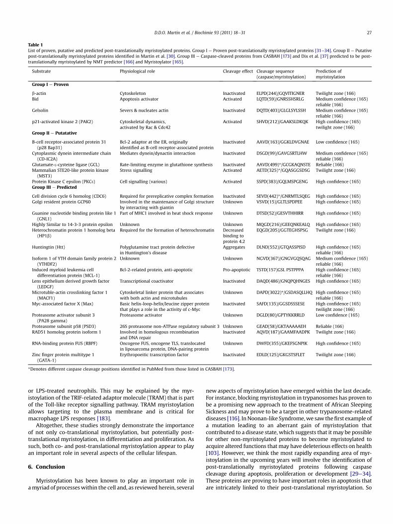
Table 1List of proven, putative and predicted post-translationally myristoylated proteins. Group I e Proven post-translationally myristoylated proteins [31e34]. Group II e Putativepost-translationally myristoylated proteins identified in Martin et al. [30]. Group III e Caspase-cleaved proteins from CASBAH [173] and Dix et al. [37] predicted to be post-translationally myristoylated by NMT predictor [166] and Myristoylator [165].
Substrate Physiological role Cleavage effect Cleavage sequence(caspase/myristoylation)
Prediction ofmyristoylation
Group I e Proven
b-actin Cytoskeleton Inactivated ELPD(244)/GQVITIGNER Twilight zone (166)Bid Apoptosis activator Activated LQTD(59)/GNRSSHSRLG Medium confidence (165)
reliable (166)Gelsolin Severs & nucleates actin Inactivated DQTD(403)/GLGLSYLSSH Medium confidence (165)
reliable (166)p21-activated kinase 2 (PAK2) Cytoskeletal dynamics,
activated by Rac & Cdc42Activated SHVD(212)/GAAKSLDKQK High confidence (165)
twilight zone (166)Group II e Putatative
B-cell receptor-associated protein 31(p28 Bap31)
Bcl-2 adaptor at the ER, originallyidentified as B-cell receptor-associated protein
Inactivated AAVD(163)/GGKLDVGNAE Low confidence (165)
Cytoplasmic dynein intermediate chain(CD-IC2A)
Mediates dynein/dynactin interaction Inactivated DSGD(99)/GAVGSRTLHW Medium confidence (165)reliable (166)
Glutamate-L-cysteine ligase (GCL) Rate-limiting enzyme in glutathione synthesis Inactivated AAVD(499)*/GCGKAQNSTE Reliable (166)Mammalian STE20-like protein kinase
(MST3)Stress signalling Activated AETD(325)*/GQASGGSDSG Twilight zone (166)
Protein Kinase C epsilon (PKC3) Cell signalling (various) Activated SSPD(383)/GQLMSPGENG High confidence (165)Group III e Predicted
Cell division cycle 6 homolog (CDC6) Required for prereplicative complex formation Inactivated SEVD(442)*/GNRMTLSQEG High confidence (165)Golgi resident protein GCP60 Involved in the maintenance of Golgi structure
by interacting with giantinUnknown VSVD(15)/GLTLSPDPEE High confidence (165)
Guanine nucleotide binding protein like 1(GNL1)
Part of MHC1 involved in heat shock response Unknown DTSD(52)/GESVTHHIRR High confidence (165)
Highly Similar to 14-3-3 protein epsilon Unknown Unknown MQGD(216)/GEEQNKEALQ High confidence (165)Heterochromatin protein 1 homolog beta
(HP1b)Required for the formation of heterochromatin Decreased
binding toprotein 4.2
EQGD(205)/GGTEGHSPSG Twilight zone (166)
Huntingtin (Htt) Polyglutamine tract protein defectivein Huntington’s disease
Aggregates DLND(552)/GTQASSPISD High confidence (165)reliable (166)
Isoform 1 of YTH domain family protein 2(YTHDF2)
Unknown Unknown NGVD(367)/GNGVGQSQAG Medium confidence (165)reliable (166)
Induced myeloid leukemia celldifferentiation protein (MCL-1)
Bcl-2-related protein, anti-apoptotic Pro-apoptotic TSTD(157)GSL PSTPPPA High confidence (165)reliable (166)
Lens epithelium derived growth factor(LEDGF)
Transcriptional coactivator Inactivated DAQD(486)/GNQPQHNGES High confidence (165)
Microtuble-actin crosslinking factor 1(MACF1)
Cytoskeletal linker protein that associateswith both actin and microtubules
Unknown DAPD(3022)*/GSDASQLLHQ High confidence (165)reliable (166)
Myc-associated factor X (Max) Basic helix-loop-helix/leucine zipper proteinthat plays a role in the activity of c-Myc
Inactivated SAFD(135)/GGSDSSSESE High confidence (165)twilight zone (166)
Proteasome activator subunit 3(PA28 gamma)
Proteasome activator Unknown DGLD(80)/GPTYKKRRLD Low confidence (165)
Proteasome subunit p58 (PSD3) 26S proteasome non-ATPase regulatory subunit 3 Unknown GEAD(58)/GKTAAAAAEH Reliable (166)RAD51 homolog protein isoform 1 Involved in homologous recombination
and DNA repairInactivated AQVD(187)/GAAMFAADPK Twilight zone (166)
RNA-binding protein FUS (RBPF) Oncogene FUS, oncogene TLS, translocatedin liposarcoma protein, DNA-pairing protein
Unknown DWFD(355)/GKEFSGNPIK High confidence (165)
Zinc finger protein multitype 1(GATA-1)
Erythropoeitic transcription factor Inactivated EDLD(125)/GKGSTSFLET Twilight zone (166)
*Denotes different caspase cleavage positions identified in PubMed from those listed in CASBAH [173].
D.D.O. Martin et al. / Biochimie 93 (2011) 18e31 27
or LPS-treated neutrophils. This may be explained by the myr-istoylation of the TRIF-related adaptor molecule (TRAM) that is partof the Toll-like receptor signalling pathway. TRAM myristoylationallows targeting to the plasma membrane and is critical formacrophage LPS responses [183].
Altogether, these studies strongly demonstrate the importanceof not only co-translational myristoylation, but potentially post-translational myristoylation, in differentiation and proliferation. Assuch, both co- and post-translational myristoylation appear to playan important role in several aspects of the cellular lifespan.
6. Conclusion
Myristoylation has been known to play an important role inamyriad of processeswithin the cell and, as reviewed herein, several
new aspects of myristoylation have emerged within the last decade.For instance, blockingmyristoylation in trypanosomes has proven tobe a promising new approach to the treatment of African SleepingSickness andmay prove to be a target in other trypanosome-relateddiseases [116]. InNoonan-like Syndrome,we saw thefirst example ofa mutation leading to an aberrant gain of myristoylation thatcontributed to a disease state, which suggests that it may be possiblefor other non-myristoylated proteins to become myristoylated toacquire altered functions thatmay have deleterious effects on health[103]. However, we think the most rapidly expanding area of myr-istoylation in the upcoming years will involve the identification ofpost-translationally myristoylated proteins following caspasecleavage during apoptosis, proliferation or development [29e34].These proteins are proving to have important roles in apoptosis thatare intricately linked to their post-translational myristoylation. So

D.D.O. Martin et al. / Biochimie 93 (2011) 18e3128
far, apoptosis induced by myr-ctBid and myr-ctPAK2 can be signifi-cantly reduced by abrogating myristoylation of these proteins[33,34]. Paradoxically, the anti-apoptotic effect of myr-ctGelsolin isalso linked to its myristoylation status [31]. This suggests that theremaybedual roles for post-translationallymyristoylatedproteins. It ispossible that some proteins promote cell survival until the cell hasreached an apoptotic threshold and has committed to cell death.Alternatively, some of the pro-survival factors may have a greaterrole in cell differentiation andproliferationwhere caspase cleavage isessential and at least PAK2 and gelsolin are targets for caspasecleavage and most likely post-translationally myristoylated[179,180]. With PAK2, it appears that the level of cleavage is not asgreat in differentiating cells as in apoptosis, which may suggest thatlower levels of constitutively active myr-ctPAK2 in differentiatingcells may not be toxic or is tightly regulated to prevent apoptosis[34,179]. As such, it may provide a structural role in differentiationand proliferation while myr-ctGelsolin would act as a pro-survivalfactor in this scenario.
Several new findings suggest that there remains many more, asof yet, unidentified co- and post-translationally myristoylatedproteins. For instance, in the cell free assay described above,Utsumi’s group identified 1.4% of the proteins as myristoylated ofthe 1929 proteins in the query proteome they investigated [155].Therefore, this may suggest that the myristoylome is larger thanpreviously predicted (0.5%) [78,82]. Interestingly, and of particularnote, they found that the two commonly used prediction analysisprograms used for myristoylation were strong predictors of myr-istoylation. However, while Myr Predictor [166] had more false-negative predictions, Myristoylator [165] had more false-positivepredictions, and both programs missed two proteins that wereshown to be myristoylated [155]. This demonstrates the impor-tance of testing for myristoylation in vitro and in vivo.
The possible existence of other post-translationally myristoy-lated proteins is supported by the fact that we have identified 16new proteins that are predicted to be post-translationally myr-istoylatable (Table 1). Furthermore, we also showed that there isa clear difference in the cellular contents of co- and post-transla-tionally myristoylated proteins in non-apoptotic and apoptoticJurkat cells and the identification of these myristoylated proteinsawaits [29,30]. Moreover, the fact that several studies havedemonstrated that the preferred amino acid following caspasecleavage sites is glycine [37,38], further suggests the existence ofmore potential sites for post-translational myristoylation. Further-more, cryptic myristoylation sites might also be found downstreamof other proteolytic sites (e.g. calpain, granzyme B, cathepsins, etc.).Using chemical biology as a means to speed up detection andisolation of post-translationally myristoylated proteins, the firstproteomic analyses of the post-translational “myristoylomes” ofnormal and metabolically compromised cells should emerge in thenear future. Because post-translational myristoylation occurs inapoptotic cells and apoptosis is often deregulated in cancer andneurodegenerative diseases, the importance of identifying andcharacterizing post-translationally myristoylated proteins isundeniable. A better understanding of novel molecular compo-nents involved in apoptosis will not only offer insights into path-ogenesis but could also allow for the development of diagnostic,prognostic and therapeutic tools.
References
[1] M.J. Nadolski, M.E. Linder, Protein lipidation, FEBS J. 274 (2007) 5202e5210.[2] R.K. Mann, P.A. Beachy, Cholesterol modification of proteins, Biochim. Bio-
phys. Acta 1529 (2000) 188e202.[3] D.N. Crowell, D.H. Huizinga, Protein isoprenylation: the fat of the matter,
Trends Plant Sci. 14 (2009) 163e170.
[4] M.G. Paulick, C.R. Bertozzi, The glycosylphosphatidylinositol anchor:a complex membrane-anchoring structure for proteins, Biochemistry 47(2008) 6991e7000.
[5] X. Liang, Y. Lu, M. Wilkes, T.A. Neubert, M.D. Resh, The N-terminal SH4 regionof the Src family kinase Fyn is modified by methylation and heterogeneousfatty acylation: role in membrane targeting, cell adhesion, and spreading,J. Biol. Chem. 279 (2004) 8133e8139.
[6] M.D. Resh, Trafficking and signaling by fatty-acylated and prenylatedproteins, Nat. Chem. Biol. 2 (2006) 584e590.
[7] Y. Fukata, M. Fukata, Protein palmitoylation in neuronal development andsynaptic plasticity, Nat. Rev. Neurosci. 11 (2010) 161e175.
[8] R.B. Pepinsky, C. Zeng, D. Wen, P. Rayhorn, D.P. Baker, K.P. Williams,S.A. Bixler, C.M. Ambrose, E.A. Garber, K. Miatkowski, F.R. Taylor, E.A. Wang,A. Galdes, Identification of a palmitic acid-modified form of human Sonichedgehog, J. Biol. Chem. 273 (1998) 14037e14045.
[9] C. Kleuss, E. Krause, Galpha(s) is palmitoylated at the N-terminal glycine,EMBO J. 22 (2003) 826e832.
[10] M.M. Corvi, C.L. Soltys, L.G. Berthiaume, Regulation of mitochondrial carba-moyl-phosphate synthetase 1 activity by active site fatty acylation, J. Biol.Chem. 276 (2001) 45704e45712.
[11] M.A. Kostiuk, M.M. Corvi, B.O. Keller, G. Plummer, J.A. Prescher,M.J. Hangauer, C.R. Bertozzi, G. Rajaiah, J.R. Falck, L.G. Berthiaume, Identifi-cation of palmitoylated mitochondrial proteins using a bio-orthogonal azido-palmitate analogue, FASEB J. 22 (2008) 721e732.
[12] J.C. DeMar Jr., D.R. Rundle, T.G. Wensel, R.E. Anderson, HeterogeneousN-terminal acylation of retinal proteins, Prog. Lipid Res. 38 (1999) 49e90.
[13] J.A. Boutin, Myristoylation, Cell. Signal. 9 (1997) 15e35.[14] T.A. Farazi, G. Waksman, J.I. Gordon, The biology and enzymology of protein
N-myristoylation, J. Biol. Chem. 276 (2001) 39501e39504.[15] D.A. Towler, S.R. Eubanks, D.S. Towery, S.P. Adams, L. Glaser, Amino-terminal
processing of proteins by N-myristoylation. Substrate specificity of N-myr-istoyl transferase, J. Biol. Chem. 262 (1987) 1030e1036.
[16] J.A. Hedo, E. Collier, A. Watkinson, Myristyl and palmityl acylation of theinsulin receptor, J. Biol. Chem. 262 (1987) 954e957.
[17] S.L. Bursten, R.M. Locksley, J.L. Ryan, D.H. Lovett, Acylation of monocyte andglomerular mesangial cell proteins. Myristyl acylation of the interleukin 1precursors, J. Clin. Invest. 82 (1988) 1479e1488.
[18] M. Kojima, H. Hosoda, Y. Date, M. Nakazato, H. Matsuo, K. Kangawa, Ghrelinis a growth-hormone-releasing acylated peptide from stomach, Nature 402(1999) 656e660.
[19] J. Yang, M.S. Brown, G. Liang, N.V. Grishin, J.L. Goldstein, Identification of theacyltransferase that octanoylates ghrelin, an appetite-stimulating peptidehormone, Cell 132 (2008) 387e396.
[20] H. Kirchner, J.A. Gutierrez, P.J. Solenberg, P.T. Pfluger, T.A. Czyzyk,J.A. Willency, A. Schurmann, H.G. Joost, R.J. Jandacek, J.E. Hale, M.L. Heiman,M.H. Tschop, GOAT links dietary lipids with the endocrine control of energybalance, Nat. Med. 15 (2009) 741e745.
[21] M. Kojima, K. Kangawa, Ghrelin: structure and function, Physiol. Rev. 85(2005) 495e522.
[22] K. Willert, J.D. Brown, E. Danenberg, A.W. Duncan, I.L. Weissman, T. Reya,J.R. Yates 3rd, R. Nusse, Wnt proteins are lipid-modified and can act as stemcell growth factors, Nature 423 (2003) 448e452.
[23] R. Takada, Y. Satomi, T. Kurata, N. Ueno, S. Norioka, H. Kondoh, T. Takao,S. Takada, Monounsaturated fatty acid modification of Wnt protein: its rolein Wnt secretion, Dev. Cell 11 (2006) 791e801.
[24] K.L. Schey, D.B. Gutierrez, Z. Wang, J. Wei, A.C. Grey, Novel fatty acidacylation of lens integral membrane protein aquaporin-0, Biochemistry(2010).
[25] S.A. Carr, K. Biemann, S. Shoji, D.C. Parmelee, K. Titani, n-Tetradecanoyl is theNH2-terminal blocking group of the catalytic subunit of cyclic AMP-depen-dent protein kinase from bovine cardiac muscle, Proc. Natl. Acad. Sci. U.S.A.79 (1982) 6128e6131.
[26] A. Aitken, P. Cohen, S. Santikarn, D.H. Williams, A.G. Calder, A. Smith,C.B. Klee, Identification of the NH2-terminal blocking group of calcineurin Bas myristic acid, FEBS Lett. 150 (1982) 314e318.
[27] C. Wilcox, J.S. Hu, E.N. Olson, Acylation of proteins with myristic acid occurscotranslationally, Science 238 (1987) 1275e1278.
[28] I. Deichaite, L.P. Casson, H.P. Ling, M.D. Resh, In vitro synthesis of pp60v-src:myristylation in a cell-free system, Mol. Cell. Biol. 8 (1988) 4295e4301.
[29] M.C. Yap, M.A. Kostiuk, D.D. Martin, M.A. Perinpanayagam, P.G. Hak,A. Siddam, J.R. Majjigapu, G. Rajaiah, B.O. Keller, J.A. Prescher, P. Wu,C.R. Bertozzi, J.R. Falck, L.G. Berthiaume, Rapid and selective detection of fattyacylated proteins using omega-alkynyl-fatty acids and click chemistry,J. Lipid Res. 51 (2010) 1566e1580.
[30] D.D. Martin, G.L. Vilas, J.A. Prescher, G. Rajaiah, J.R. Falck, C.R. Bertozzi,L.G. Berthiaume, Rapid detection, discovery, and identification of post-translationally myristoylated proteins during apoptosis using a bio-orthog-onal azidomyristate analog, FASEB J. 22 (2008) 797e806.
[31] N. Sakurai, T. Utsumi, Posttranslational N-myristoylation is required for theanti-apoptotic activity of human tGelsolin, the C-terminal caspase cleavageproduct of human gelsolin, J. Biol. Chem. 281 (2006) 14288e14295.
[32] T. Utsumi, N. Sakurai, K. Nakano, R. Ishisaka, C-terminal 15 kDa fragment ofcytoskeletal actin is posttranslationally N-myristoylated upon caspase-mediated cleavage and targeted to mitochondria, FEBS Lett. 539 (2003)37e44.

D.D.O. Martin et al. / Biochimie 93 (2011) 18e31 29
[33] J. Zha, S. Weiler, K.J. Oh, M.C. Wei, S.J. Korsmeyer, Posttranslational N-myr-istoylation of BID as a molecular switch for targeting mitochondria andapoptosis, Science 290 (2000) 1761e1765.
[34] G.L. Vilas, M.M. Corvi, G.J. Plummer, A.M. Seime, G.R. Lambkin,L.G. Berthiaume, Posttranslational myristoylation of caspase-activated p21-activated protein kinase 2 (PAK2) potentiates late apoptotic events, Proc.Natl. Acad. Sci. U.S.A. 103 (2006) 6542e6547.
[35] A.T. Matheson, M. Yaguchi, L.P. Visentin, The conservation of amino acids inthe n-terminal position of ribosomal and cytosol proteins from Escherichiacoli, Bacillus stearothermophilus, and Halobacterium cutirubrum, Can.J. Biochem. 53 (1975) 1323e1327.
[36] F. Frottin, A. Martinez, P. Peynot, S. Mitra, R.C. Holz, C. Giglione, T. Meinnel,The proteomics of N-terminal methionine cleavage, Mol. Cell. Proteomics 5(2006) 2336e2349.
[37] M.M. Dix, G.M. Simon, B.F. Cravatt, Global mapping of the topography andmagnitude of proteolytic events in apoptosis, Cell 134 (2008) 679e691.
[38] S. Mahrus, J.C. Trinidad, D.T. Barkan, A. Sali, A.L. Burlingame, J.A. Wells, Globalsequencing of proteolytic cleavage sites in apoptosis by specific labeling ofprotein N termini, Cell 134 (2008) 866e876.
[39] D.R. Johnson, R.S. Bhatnagar, L.J. Knoll, J.I. Gordon, Genetic and biochemicalstudies of protein N-myristoylation, Annu. Rev. Biochem. 63 (1994) 869e914.
[40] R.M. Peitzsch, S. McLaughlin, Binding of acylated peptides and fatty acids tophospholipid vesicles: pertinence to myristoylated proteins, Biochemistry 32(1993) 10436e10443.
[41] M.D. Resh, Membrane targeting of lipid modified signal transductionproteins, Subcell. Biochem. 37 (2004) 217e232.
[42] J.B. McCabe, L.G. Berthiaume, Functional roles for fatty acylated amino-terminal domains in subcellular localization, Mol. Biol. Cell 10 (1999)3771e3786.
[43] J.B. McCabe, L.G. Berthiaume, N-terminal protein acylation confers localiza-tion to cholesterol, sphingolipid-enriched membranes but not to lipid rafts/caveolae, Mol. Biol. Cell 12 (2001) 3601e3617.
[44] J.R. Silvius, PeptideeLipid Interactions, Current Topics in Membranes.Academic Press, San Diego, Calif.; London, 2002, xxi, 371e395 p. [315] p. ofplates.
[45] S. McLaughlin, A. Aderem, The myristoyl-electrostatic switch: a modulator ofreversible protein-membrane interactions, Trends Biochem. Sci. 20 (1995)272e276.
[46] J.C. Amor, D.H. Harrison, R.A. Kahn, D. Ringe, Structure of the human ADP-ribosylation factor 1 complexed with GDP, Nature 372 (1994) 704e708.
[47] J.B. Ames, T. Tanaka, L. Stryer, M. Ikura, Portrait of a myristoyl switch protein,Curr. Opin. Struct. Biol. 6 (1996) 432e438.
[48] J.T. Seykora, M.M. Myat, L.A. Allen, J.V. Ravetch, A. Aderem, Moleculardeterminants of the myristoyl-electrostatic switch of MARCKS, J. Biol. Chem.271 (1996) 18797e18802.
[49] O. Hantschel, B. Nagar, S. Guettler, J. Kretzschmar, K. Dorey, J. Kuriyan,G. Superti-Furga, A myristoyl/phosphotyrosine switch regulates c-Abl, Cell112 (2003) 845e857.
[50] B. Nagar, O. Hantschel, M.A. Young, K. Scheffzek, D. Veach, W. Bornmann,B. Clarkson, G. Superti-Furga, J. Kuriyan, Structural basis for the auto-inhibition of c-Abl tyrosine kinase, Cell 112 (2003) 859e871.
[51] P. Patwardhan, M.D. Resh, Myristoylation and membrane binding regulate c-Src stability and kinase activity, Mol. Cell. Biol. (2010).
[52] F. Dyda, D.C. Klein, A.B. Hickman, GCN5-related N-acetyltransferases:a structural overview, Annu. Rev. Biophys. Biomol. Struct. 29 (2000) 81e103.
[53] R.J. Duronio, D.A. Towler, R.O. Heuckeroth, J.I. Gordon, Disruption of the yeastN-myristoyl transferase gene causes recessive lethality, Science 243 (1989)796e800.
[54] J.K. Lodge, R.L. Johnson, R.A. Weinberg, J.I. Gordon, Comparison of myristoyl-CoA:protein N-myristoyltransferases from three pathogenic fungi: Crypto-coccus neoformans, Histoplasma capsulatum, and Candida albicans, J. Biol.Chem. 269 (1994) 2996e3009.
[55] M. Ntwasa, M. Egerton, N.J. Gay, Sequence and expression of Drosophilamyristoyl-CoA: protein N-myristoyl transferase: evidence for proteolyticprocessing and membrane localisation, J. Cell. Sci. 110 (Pt 2) (1997) 149e156.
[56] D.K. Giang, B.F. Cravatt, A second mammalian N-myristoyltransferase, J. Biol.Chem. 273 (1998) 6595e6598.
[57] V. Rioux, E. Beauchamp, F. Pedrono, S. Daval, D. Molle, D. Catheline,P. Legrand, Identification and characterization of recombinant and native ratmyristoyl-CoA: protein N-myristoyltransferases, Mol. Cell. Biochem. 286(2006) 161e170.
[58] D. Towler, L. Glaser, Protein fatty acid acylation: enzymatic synthesis of anN-myristoylglycyl peptide, Proc. Natl. Acad. Sci. U.S.A. 83 (1986) 2812e2816.
[59] D.A. Towler, S.P. Adams, S.R. Eubanks, D.S. Towery, E. Jackson-Machelski,L. Glaser, J.I. Gordon, Purification and characterization of yeast myristoylCoA:protein N-myristoyltransferase, Proc. Natl. Acad. Sci. U.S.A. 84 (1987)2708e2712.
[60] D.A. Rudnick, C.A. McWherter, W.J. Rocque, P.J. Lennon, D.P. Getman,J.I. Gordon, Kinetic and structural evidence for a sequential ordered Bi Bimechanism of catalysis by Saccharomyces cerevisiae myristoyl-CoA:proteinN-myristoyltransferase, J. Biol. Chem. 266 (1991) 9732e9739.
[61] S.H. Yang, A. Shrivastav, C. Kosinski, R.K. Sharma, M.H. Chen, L.G. Berthiaume,L.L. Peters, P.T. Chuang, S.G. Young, M.O. Bergo, N-myristoyltransferase 1is essential in early mouse development, J. Biol. Chem. 280 (2005)18990e18995.
[62] M. Ntwasa, S. Aapies, D.A. Schiffmann, N.J. Gay, Drosophila embryos lackingN-myristoyltransferase have multiple developmental defects, Exp. Cell Res.262 (2001) 134e144.
[63] N.S. Kishore, T.B. Lu, L.J. Knoll, A. Katoh, D.A. Rudnick, P.P. Mehta, B. Devadas,M. Huhn, J.L. Atwood, S.P. Adams, et al., The substrate specificity of Saccha-romyces cerevisiae myristoyl-CoA:protein N-myristoyltransferase. Analysisof myristic acid analogs containing oxygen, sulfur, double bonds, triplebonds, and/or an aromatic residue, J. Biol. Chem. 266 (1991) 8835e8855.
[64] R.S. Bhatnagar, E. Jackson-Machelski, C.A. McWherter, J.I. Gordon, Isothermaltitration calorimetric studies of Saccharomyces cerevisiae myristoyl-CoA:p-rotein N-myristoyltransferase. Determinants of binding energy and catalyticdiscrimination among acyl-CoA and peptide ligands, J. Biol. Chem. 269(1994) 11045e11053.
[65] R.S. Bhatnagar, K. Futterer, G. Waksman, J.I. Gordon, The structure of myr-istoyl-CoA:protein N-myristoyltransferase, Biochim. Biophys. Acta 1441(1999) 162e172.
[66] D.A. Rudnick, C.A. McWherter, S.P. Adams, I.J. Ropson, R.J. Duronio,J.I. Gordon, Structural and functional studies of Saccharomyces cerevisiaemyristoyl-CoA:protein N-myristoyltransferase produced in Escherichia coli.Evidence for an acyl-enzyme intermediate, J. Biol. Chem. 265 (1990)13370e13378.
[67] D.A. Rudnick, T. Lu, E. Jackson-Machelski, J.C. Hernandez, Q. Li, G.W. Gokel,J.I. Gordon, Analogs of palmitoyl-CoA that are substrates for myristoyl-CoA:protein N-myristoyltransferase, Proc. Natl. Acad. Sci. U.S.A. 89 (1992)10507e10511.
[68] D.R. Rundle, R.V. Rajala, R.A. Alvarez, R.E. Anderson, Myristoyl-CoA:proteinN-myristoyltransferases: isoform identification and gene expression inretina, Mol. Vis. 10 (2004) 177e185.
[69] N.J. Faergeman, J. Knudsen, Role of long-chain fatty acyl-CoA esters in theregulation of metabolism and in cell signalling, Biochem. J. 323 (Pt 1) (1997)1e12.
[70] V. Rioux, A. Galat, G. Jan, F. Vinci, S. D’Andrea, P. Legrand, Exogenous myristicacid acylates proteins in cultured rat hepatocytes, J. Nutr. Biochem. 13 (2002)66e74.
[71] N.S. Kishore, D.C. Wood, P.P. Mehta, A.C. Wade, T. Lu, G.W. Gokel, J.I. Gordon,Comparison of the acyl chain specificities of human myristoyl-CoA synthe-tase and human myristoyl-CoA:protein N-myristoyltransferase, J. Biol. Chem.268 (1993) 4889e4902.
[72] T. Suzuki, M. Ito, T. Ezure, M. Shikata, E. Ando, T. Utsumi, S. Tsunasawa,O. Nishimura, Preparation of N-acylated proteins modified with fatty acidshaving a specific chain length using an insect cell-free protein synthesissystem, Biosci. Biotechnol. Biochem. 71 (2007) 261e264.
[73] J.S. Isenberg, Y. Jia, J. Fukuyama, C.H. Switzer, D.A. Wink, D.D. Roberts,Thrombospondin-1 inhibits nitric oxide signaling via CD36 by inhibitingmyristic acid uptake, J. Biol. Chem. 282 (2007) 15404e15415.
[74] J. Turnay, E. Lecona, S. Fernandez-Lizarbe, A. Guzman-Aranguez,M.P. Fernandez, N. Olmo, M.A. Lizarbe, Structure-function relationship inannexin A13, the founder member of the vertebrate family of annexins,Biochem. J. 389 (2005) 899e911.
[75] P. Jackson, D. Baltimore, N-terminal mutations activate the leukemogenicpotential of the myristoylated form of c-abl, EMBO J. 8 (1989) 449e456.
[76] T. Utsumi, K. Nakano, T. Funakoshi, Y. Kayano, S. Nakao, N. Sakurai, H. Iwata,R. Ishisaka, Vertical-scanning mutagenesis of amino acids in a modelN-myristoylation motif reveals the major amino-terminal sequence require-ments for protein N-myristoylation, Eur. J. Biochem. 271 (2004) 863e874.
[77] L. Silverman, M.D. Resh, Lysine residues form an integral component ofa novel NH2-terminal membrane targeting motif for myristylated pp60v-src,J. Cell Biol. 119 (1992) 415e425.
[78] S. Maurer-Stroh, B. Eisenhaber, F. Eisenhaber, N-terminal N-myristoylationof proteins: prediction of substrate proteins from amino acid sequence,J. Mol. Biol. 317 (2002) 541e557.
[79] B. Boisson, C. Giglione, T. Meinnel, Unexpected protein families including celldefense components feature in the N-myristoylome of a higher eukaryote,J. Biol. Chem. 278 (2003) 43418e43429.
[80] S. Podell, M. Gribskov, Predicting N-terminal myristoylation sites in plantproteins, BMC Genomics 5 (2004) 37.
[81] M. Sugii, R. Okada, H. Matsuno, S. Miyano, Performance improvement inprotein N-myristoyl classification by BONSAI with insignificant indexingsymbol, Genome Inform. 18 (2007) 277e286.
[82] S. Maurer-Stroh, B. Eisenhaber, F. Eisenhaber, N-terminal N-myristoylationof proteins: refinement of the sequence motif and its taxon-specific differ-ences, J. Mol. Biol. 317 (2002) 523e540.
[83] G.J. van der Vusse, M. van Bilsen, J.F. Glatz, D.M. Hasselbaink, J.J. Luiken,Critical steps in cellular fatty acid uptake and utilization, Mol. Cell. Biochem.239 (2002) 9e15.
[84] S. Colombo, R. Longhi, S. Alcaro, F. Ortuso, T. Sprocati, A. Flora, N. Borgese,N-myristoylation determines dual targeting of mammalian NADH-cyto-chrome b5 reductase to ER and mitochondrial outer membranes bya mechanism of kinetic partitioning, J. Cell Biol. 168 (2005) 735e745.
[85] P. Selvakumar, A. Lakshmikuttyamma, A. Shrivastav, S.B. Das, J.R. Dimmock,R.K. Sharma, Potential role of N-myristoyltransferase in cancer, Prog. LipidRes. 46 (2007) 1e36.
[86] M.J. King, R.K. Sharma, Identification, purification and characterization ofa membrane-associated N-myristoyltransferase inhibitor protein frombovine brain, Biochem. J. 291 (Pt 2) (1993) 635e639.

D.D.O. Martin et al. / Biochimie 93 (2011) 18e3130
[87] A. Shrivastav, M.K. Pasha, P. Selvakumar, S. Gowda, D.J. Olson, A.R. Ross,J.R. Dimmock, R.K. Sharma, Potent inhibitor of N-myristoylation: a novelmolecular target for cancer, Cancer Res. 63 (2003) 7975e7978.
[88] R.V. Rajala, R.S. Datla, S.A. Carlsen, D.H. Anderson, Z. Qi, J.H. Wang,R.K. Sharma, Phosphorylation of human N-myristoyltransferase by N-myr-istoylated SRC family tyrosine kinase members, Biochem. Biophys. Res.Commun. 288 (2001) 233e239.
[89] F.R. Cross, E.A. Garber, D. Pellman, H. Hanafusa, A short sequence in thep60src N terminus is required for p60src myristylation and membraneassociation and for cell transformation, Mol. Cell. Biol. 4 (1984) 1834e1842.
[90] J.M. Summy, G.E. Gallick, Src family kinases in tumor progression andmetastasis, Cancer Metastasis Rev. 22 (2003) 337e358.
[91] S. Dehm, M.A. Senger, K. Bonham, SRC transcriptional activation in a subsetof human colon cancer cell lines, FEBS Lett. 487 (2001) 367e371.
[92] S.M. Dehm, K. Bonham, SRC gene expression in human cancer: the role oftranscriptional activation, Biochem. Cell Biol. 82 (2004) 263e274.
[93] K. Bonham, D.J. Fujita, Organization and analysis of the promoter region and50 non-coding exons of the human c-src proto-oncogene, Oncogene 8 (1993)1973e1981.
[94] S. Shoji, T. Kurosawa, H. Inoue, T. Funakoshi, Y. Kubota, Human cellular src geneproduct: identification of the myristoylated pp60c-src and blockage of itsmyristoyl acylationwith N-fatty acyl compounds resulted in the suppression ofcolony formation, Biochem. Biophys. Res. Commun. 173 (1990) 894e901.
[95] S. Bagrodia, S.J. Taylor, D. Shalloway, Myristylation is required for Tyr-527dephosphorylation and activation of pp60c-src in mitosis, Mol. Cell. Biol. 13(1993) 1464e1470.
[96] D.A. Rudnick, R.L. Johnson, J.I. Gordon, Studies of the catalytic activities andsubstrate specificities of Saccharomyces cerevisiae myristoyl-coenzyme A:protein N-myristoyltransferase deletion mutants and human/yeast Nmtchimeras in Escherichia coli and S. cerevisiae, J. Biol. Chem. 267 (1992)23852e23861.
[97] B.A. Magnuson, R.V. Raju, T.N. Moyana, R.K. Sharma, Increased N-myr-istoyltransferase activity observed in rat and human colonic tumors, J. Natl.Cancer Inst. 87 (1995) 1630e1635.
[98] R.V. Raju, T.N. Moyana, R.K. Sharma, N-Myristoyltransferase overexpressionin human colorectal adenocarcinomas, Exp. Cell Res. 235 (1997) 145e154.
[99] P. Selvakumar, E. Smith-Windsor, K. Bonham, R.K. Sharma, N-myristoyl-transferase 2 expression in human colon cancer: cross-talk between thecalpain and caspase system, FEBS Lett. 580 (2006) 2021e2026.
[100] R.V. Rajala, J.M. Radhi, R. Kakkar, R.S. Datla, R.K. Sharma, Increased expres-sion of N-myristoyltransferase in gallbladder carcinomas, Cancer 88 (2000)1992e1999.
[101] A. Shrivastav, S. Varma, A. Senger, R.L. Khandelwal, S. Carlsen, R.K. Sharma,Overexpression of Akt/PKB modulates N-myristoyltransferase activity incancer cells, J. Pathol. 218 (2009) 391e398.
[102] Y. Lu, P. Selvakumar, K. Ali, A. Shrivastav, G. Bajaj, L. Resch, R. Griebel,D. Fourney, K. Meguro, R.K. Sharma, Expression of N-myristoyltransferase inhuman brain tumors, Neurochem. Res. 30 (2005) 9e13.
[103] V. Cordeddu, E. Di Schiavi, L.A. Pennacchio, A. Ma’ayan, A. Sarkozy, V. Fodale,S. Cecchetti, A. Cardinale, J. Martin, W. Schackwitz, A. Lipzen, G. Zampino,L. Mazzanti, M.C. Digilio, S. Martinelli, E. Flex, F. Lepri, D. Bartholdi, K. Kutsche,G.B. Ferrero, C. Anichini, A. Selicorni, C. Rossi, R. Tenconi, M. Zenker, D. Merlo,B. Dallapiccola, R. Iyengar, P. Bazzicalupo, B.D. Gelb, M. Tartaglia, Mutation ofSHOC2 promotes aberrant protein N-myristoylation and causes Noonan-likesyndrome with loose anagen hair, Nat. Genet. 41 (2009) 1022e1026.
[104] L. Mazzanti, E. Cacciari, A. Cicognani, R. Bergamaschi, E. Scarano,A. Forabosco, Noonan-like syndrome with loose anagen hair: a newsyndrome? Am. J. Med. Genet. A 118A (2003) 279e286.
[105] S. Schubbert, K. Shannon, G. Bollag, Hyperactive Ras in developmentaldisorders and cancer, Nat. Rev. Cancer 7 (2007) 295e308.
[106] S. Maurer-Stroh, F. Eisenhaber, Myristoylation of viral and bacterial proteins,Trends Microbiol. 12 (2004) 178e185.
[107] D.A. Towler, S.P. Adams, S.R. Eubanks, D.S. Towery, E. Jackson-Machelski,L. Glaser, J.I. Gordon, Myristoyl CoA:protein N-myristoyltransferase activitiesfrom rat liver and yeast possess overlapping yet distinct peptide substratespecificities, J. Biol. Chem. 263 (1988) 1784e1790.
[108] D.A. Towler, J.I. Gordon, S.P. Adams, L. Glaser, The biology and enzymology ofeukaryotic protein acylation, Annu. Rev. Biochem. 57 (1988) 69e99.
[109] W.J. Rocque, C.A. McWherter, D.C. Wood, J.I. Gordon, A comparative analysisof the kinetic mechanism and peptide substrate specificity of human andSaccharomyces cerevisiae myristoyl-CoA:protein N-myristoyltransferase,J. Biol. Chem. 268 (1993) 9964e9971.
[110] B. Devadas, S.K. Freeman, C.A. McWherter, N.S. Kishore, J.K. Lodge,E. Jackson-Machelski, J.I. Gordon, J.A. Sikorski, Novel biologically activenonpeptidic inhibitors of myristoylCoA:protein N-myristoyltransferase,J. Med. Chem. 41 (1998) 996e1000.
[111] R.C. Wiegand, C. Carr, J.C. Minnerly, A.M. Pauley, C.P. Carron, C.A. Langner,R.J. Duronio, J.I. Gordon, The Candida albicans myristoyl-CoA:proteinN-myristoyltransferase gene. Isolation and expression in Saccharomycescerevisiae and Escherichia coli, J. Biol. Chem. 267 (1992) 8591e8598.
[112] J.A. Brannigan, B.A. Smith, Z. Yu, A.M. Brzozowski, M.R. Hodgkinson,A. Maroof, H.P. Price, F. Meier, R.J. Leatherbarrow, E.W. Tate, D.F. Smith,A.J. Wilkinson, N-myristoyltransferase from Leishmania donovani: structuraland functional characterisation of a potential drug target for visceral leish-maniasis, J. Mol. Biol. 396 (2010) 985e999.
[113] H.P. Price, M.R. Menon, C. Panethymitaki, D. Goulding, P.G. McKean,D.F. Smith, Myristoyl-CoA:protein N-myristoyltransferase, an essentialenzyme and potential drug target in kinetoplastid parasites, J. Biol. Chem.278 (2003) 7206e7214.
[114] C. Panethymitaki, P.W. Bowyer, H.P. Price, R.J. Leatherbarrow, K.A. Brown,D.F. Smith, Characterization and selective inhibition of myristoyl-CoA:proteinN-myristoyltransferase from Trypanosoma brucei and Leishmania major,Biochem. J. 396 (2006) 277e285.
[115] P.W. Bowyer, R.S. Gunaratne, M. Grainger, C. Withers-Martinez,S.R. Wickramsinghe, E.W. Tate, R.J. Leatherbarrow, K.A. Brown, A.A. Holder,D.F. Smith, Molecules incorporating a benzothiazole core scaffold inhibit theN-myristoyltransferase of Plasmodium falciparum, Biochem. J. 408 (2007)173e180.
[116] J.A. Frearson, S. Brand, S.P. McElroy, L.A. Cleghorn, O. Smid, L. Stojanovski,H.P. Price, M.L. Guther, L.S. Torrie, D.A. Robinson, I. Hallyburton,C.P. Mpamhanga, J.A. Brannigan, A.J. Wilkinson, M. Hodgkinson, R. Hui,W. Qiu, O.G. Raimi, D.M. van Aalten, R. Brenk, I.H. Gilbert, K.D. Read,A.H. Fairlamb, M.A. Ferguson, D.F. Smith, P.G. Wyatt, N-myristoyltransferaseinhibitors as new leads to treat sleeping sickness, Nature 464 (2010)728e732.
[117] A.M. Schultz, S. Oroszlan, In vivo modification of retroviral gag gene-encodedpolyproteins by myristic acid, J. Virol. 46 (1983) 355e361.
[118] P. Spearman, R. Horton, L. Ratner, I. Kuli-Zade, Membrane binding of humanimmunodeficiency virus type 1 matrix protein in vivo supports a confor-mational myristyl switch mechanism, J. Virol. 71 (1997) 6582e6592.
[119] K. Furuishi, H. Matsuoka, M. Takama, I. Takahashi, S. Misumi, S. Shoji,Blockage of N-myristoylation of HIV-1 gag induces the production ofimpotent progeny virus, Biochem. Biophys. Res. Commun. 237 (1997)504e511.
[120] K.E. Seaton, C.D. Smith, N-Myristoyltransferase isozymes exhibit differentialspecificity for human immunodeficiency virus type 1 Gag and Nef, J. Gen.Virol. 89 (2008) 288e296.
[121] N. Takamune, K. Gota, S. Misumi, K. Tanaka, S. Okinaka, S. Shoji, HIV-1production is specifically associated with human NMT1 long form in humanNMT isozymes, Microbes Infect. 10 (2008) 143e150.
[122] B.T. Hill, J. Skowronski, Human N-myristoyltransferases form stablecomplexes with lentiviral nef and other viral and cellular substrate proteins,J. Virol. 79 (2005) 1133e1141.
[123] Z. Nimchuk, E. Marois, S. Kjemtrup, R.T. Leister, F. Katagiri, J.L. Dangl,Eukaryotic fatty acylation drives plasma membrane targeting and enhancesfunction of several type III effector proteins from Pseudomonas syringae, Cell101 (2000) 353e363.
[124] R. Menard, P. Sansonetti, C. Parsot, The secretion of the Shigella flexneri Ipainvasins is activated by epithelial cells and controlled by IpaB and IpaD,EMBO J. 13 (1994) 5293e5302.
[125] A.H. Wyllie, Apoptosis: an overview, Br. Med. Bull. 53 (1997) 451e465.[126] A.H. Wyllie, C.O. Bellamy, V.J. Bubb, A.R. Clarke, S. Corbet, L. Curtis,
D.J. Harrison, M.L. Hooper, N. Toft, S. Webb, C.C. Bird, Apoptosis and carci-nogenesis, Br. J. Cancer 80 (Suppl. 1) (1999) 34e37.
[127] C.B. Thompson, Apoptosis in the pathogenesis and treatment of disease,Science 267 (1995) 1456e1462.
[128] M. Leist, M. Jaattela, Four deaths and a funeral: from caspases to alternativemechanisms, Nat. Rev. Mol. Cell Biol. 2 (2001) 589e598.
[129] G.I. Evan, K.H. Vousden, Proliferation, cell cycle and apoptosis in cancer,Nature 411 (2001) 342e348.
[130] D.W. Nicholson, Caspase structure, proteolytic substrates, and functionduring apoptotic cell death, Cell Death Differ. 6 (1999) 1028e1042.
[131] U. Fischer, R.U. Janicke, K. Schulze-Osthoff, Many cuts to ruin: a compre-hensive update of caspase substrates, Cell Death Differ. 10 (2003) 76e100.
[132] K.C. Zimmermann, C. Bonzon, D.R. Green, The machinery of programmed celldeath, Pharmacol. Ther. 92 (2001) 57e70.
[133] M. Ramaswamy, S.Y. Cleland, A.C. Cruz, R.M. Siegel, Many checkpoints on theroad to cell death: regulation of Fas-FasL interactions and Fas signaling inperipheral immune responses, Results Probl. Cell Differ. 49 (2009) 17e47.
[134] M. Mishkind, Morbid myristoylation, Trends Cell Biol. 11 (2001) 191.[135] S. Nagata, Apoptotic DNA fragmentation, Exp. Cell Res. 256 (2000) 12e18.[136] M. Enari, H. Sakahira, H. Yokoyama, K. Okawa, A. Iwamatsu, S. Nagata, A
caspase-activated DNase that degrades DNA during apoptosis, and itsinhibitor ICAD, Nature 391 (1998) 43e50.
[137] T.D. Pollard, L. Blanchoin, R.D. Mullins, Molecular mechanisms controllingactin filament dynamics in nonmuscle cells, Annu. Rev. Biophys. Biomol.Struct. 29 (2000) 545e576.
[138] L.D. Burtnick, D. Urosev, E. Irobi, K. Narayan, R.C. Robinson, Structure of theN-terminal half of gelsolin bound to actin: roles in severing, apoptosis andFAF, EMBO J. 23 (2004) 2713e2722.
[139] R.C. Koya, H. Fujita, S. Shimizu, M. Ohtsu, M. Takimoto, Y. Tsujimoto,N. Kuzumaki, Gelsolin inhibits apoptosis by blocking mitochondrialmembrane potential loss and cytochrome c release, J. Biol. Chem. 275 (2000)15343e15349.
[140] H. Kusano, S. Shimizu, R.C. Koya, H. Fujita, S. Kamada, N. Kuzumaki,Y. Tsujimoto, Human gelsolin prevents apoptosis by inhibiting apoptoticmitochondrial changes via closing VDAC, Oncogene 19 (2000) 4807e4814.
[141] T.K. Rostovtseva, B. Antonsson, M. Suzuki, R.J. Youle, M. Colombini,S.M. Bezrukov, Bid, but not Bax, regulates VDAC channels, J. Biol. Chem. 279(2004) 13575e13583.

D.D.O. Martin et al. / Biochimie 93 (2011) 18e31 31
[142] M.M. Wright, A.G. Howe, V. Zaremberg, Cell membranes and apoptosis: roleof cardiolipin, phosphatidylcholine, and anticancer lipid analogues, Biochem.Cell Biol. 82 (2004) 18e26.
[143] U.G. Knaus, S. Morris, H.J. Dong, J. Chernoff, G.M. Bokoch, Regulation ofhuman leukocyte p21-activated kinases through G proteinecoupled recep-tors, Science 269 (1995) 221e223.
[144] G.M. Bokoch, Biology of the p21-activated kinases, Annu. Rev. Biochem. 72(2003) 743e781.
[145] T. Rudel, G.M. Bokoch, Membrane and morphological changes in apoptoticcells regulated by caspase-mediated activation of PAK2, Science 276 (1997)1571e1574.
[146] M. Sugiura, K. Kono, H. Liu, T. Shimizugawa, H. Minekura, S. Spiegel,T. Kohama, Ceramide kinase, a novel lipid kinase. Molecular cloning andfunctional characterization, J. Biol. Chem. 277 (2002) 23294e23300.
[147] S. Chatterjee, H. Han, S. Rollins, T. Cleveland, Molecular cloning, character-ization, and expression of a novel human neutral sphingomyelinase, J. Biol.Chem. 274 (1999) 37407e37412.
[148] E. Beauchamp, D. Goenaga, J. Le Bloc’h, D. Catheline, P. Legrand, V. Rioux,Myristic acid increases the activity of dihydroceramide Delta4-desaturase 1through its N-terminal myristoylation, Biochimie 89 (2007) 1553e1561.
[149] V. Rioux, F. Pedrono, P. Legrand, Regulation of mammalian desaturases bymyristic acid: N-terminal myristoylation and other modulations, Biochim.Biophys. Acta (2010).
[150] E. Beauchamp, X. Tekpli, G. Marteil, D. Lagadic-Gossmann, P. Legrand,V. Rioux, N-Myristoylation targets dihydroceramide Delta4-desaturase 1 tomitochondria: partial involvement in the apoptotic effect of myristic acid,Biochimie 91 (2009) 1411e1419.
[151] J. Stiban, D. Fistere, M. Colombini, Dihydroceramide hinders ceramidechannel formation: Implications on apoptosis, Apoptosis 11 (2006) 773e780.
[152] T.I. Gudz, K.Y. Tserng, C.L. Hoppel, Direct inhibition of mitochondrial respi-ratory chain complex III by cell-permeable ceramide, J. Biol. Chem. 272(1997) 24154e24158.
[153] C. Garcia-Ruiz, A. Colell, M. Mari, A. Morales, J.C. Fernandez-Checa, Directeffect of ceramide on the mitochondrial electron transport chain leads togeneration of reactive oxygen species. Role of mitochondrial glutathione,J. Biol. Chem. 272 (1997) 11369e11377.
[154] T. Suzuki, M. Ito, T. Ezure, M. Shikata, E. Ando, T. Utsumi, S. Tsunasawa,O. Nishimura, N-Terminal protein modifications in an insect cell-free proteinsynthesis system and their identification by mass spectrometry, Proteomics6 (2006) 4486e4495.
[155] T. Suzuki, K. Moriya, K. Nagatoshi, Y. Ota, T. Ezure, E. Ando, S. Tsunasawa,T. Utsumi, Strategy for comprehensive identification of human N-myristoy-lated proteins using an insect cell-free protein synthesis system, Proteomics10 (2010) 1780e1793.
[156] N. Sakurai, K. Moriya, T. Suzuki, K. Sofuku, H. Mochiki, O. Nishimura,T. Utsumi, Detection of co- and posttranslational protein N-myristoylation bymetabolic labeling in an insect cell-free protein synthesis system, Anal.Biochem. 362 (2007) 236e244.
[157] I. Deichaite, L. Berthiaume, S.M. Peseckis, W.F. Patton, M.D. Resh, Novel use ofan iodo-myristyl-CoA analog identifies a semialdehyde dehydrogenase inbovine liver, J. Biol. Chem. 268 (1993) 13738e13747.
[158] M.A. Kostiuk, B.O. Keller, L.G. Berthiaume, Palmitoylation of ketogenicenzyme HMGCS2 enhances its interaction with PPARalpha and transcriptionat the Hmgcs2 PPRE, FASEB J. 24 (2010) 1914e1924.
[159] H.C. Hang, E.J. Geutjes, G. Grotenbreg, A.M. Pollington, M.J. Bijlmakers,H.L. Ploegh, Chemical probes for the rapid detection of Fatty-acylatedproteins in Mammalian cells, J. Am. Chem. Soc. 129 (2007) 2744e2745.
[160] W.P. Heal, S.R. Wickramasinghe, P.W. Bowyer, A.A. Holder, D.F. Smith,R.J. Leatherbarrow, E.W. Tate, Site-specific N-terminal labelling of proteins invitro and in vivo using N-myristoyl transferase and bioorthogonal ligationchemistry, Chem. Commun. (Camb.) (2008) 480e482.
[161] W.P. Heal, S.R. Wickramasinghe, R.J. Leatherbarrow, E.W. Tate, N-Myristoyltransferase-mediated protein labelling in vivo, Org. Biomol. Chem. 6 (2008)2308e2315.
[162] G. Charron, M.M. Zhang, J.S. Yount, J. Wilson, A.S. Raghavan, E. Shamir,H.C. Hang, Robust fluorescent detection of protein fatty-acylation withchemical reporters, J. Am. Chem. Soc. 131 (2009) 4967e4975.
[163] R.N. Hannoush, N. Arenas-Ramirez, Imaging the lipidome: omega-alkynylfatty acids for detection and cellular visualization of lipid-modified proteins,ACS Chem. Biol. 4 (2009) 581e587.
[164] B.R. Martin, B.F. Cravatt, Large-scale profiling of protein palmitoylation inmammalian cells, Nat. Methods 6 (2009) 135e138.
[165] G. Bologna, C. Yvon, S. Duvaud, A.L. Veuthey, N-Terminal myristoylationpredictions by ensembles of neural networks, Proteomics 4 (2004) 1626e1632.
[166] F. Eisenhaber, B. Eisenhaber, W. Kubina, S. Maurer-Stroh, G. Neuberger,G. Schneider, M. Wildpaner, Prediction of lipid posttranslational modifica-tions and localization signals from protein sequences: big-Pi, NMT and PTS1,Nucleic Acids Res. 31 (2003) 3631e3634.
[167] G. Charron, J. Wilson, H.C. Hang, Chemical tools for understanding proteinlipidation in eukaryotes, Curr. Opin. Chem. Biol. 13 (2009) 382e391.
[168] R.N. Hannoush, J. Sun, The chemical toolbox for monitoring protein fattyacylation and prenylation, Nat. Chem. Biol. 6 (2010) 498e506.
[169] A. Basu, D. Lu, B. Sun, A.N. Moor, G.R. Akkaraju, J. Huang, Proteolytic activationof protein kinase C-epsilon by caspase-mediated processing and transductionof antiapoptotic signals, J. Biol. Chem. 277 (2002) 41850e41856.
[170] J.D. Lane, M.A. Vergnolle, P.G. Woodman, V.J. Allan, Apoptotic cleavage ofcytoplasmic dynein intermediate chain and p150(Glued) stops dynein-dependent membrane motility, J. Cell Biol. 153 (2001) 1415e1426.
[171] H. Puthalakath, D.C. Huang, L.A. O’Reilly, S.M. King, A. Strasser, The proa-poptotic activity of the Bcl-2 family member Bim is regulated by interactionwith the dynein motor complex, Mol. Cell 3 (1999) 287e296.
[172] J.E. Aslan, G. Thomas, Death by committee: organellar trafficking andcommunication in apoptosis, Traffic 10 (2009) 1390e1404.
[173] A.U. Luthi, S.J. Martin, The CASBAH: a searchable database of caspasesubstrates, Cell Death Differ. 14 (2007) 641e650.
[174] C. Weng, Y. Li, D. Xu, Y. Shi, H. Tang, Specific cleavage of Mcl-1 by caspase-3in tumor necrosis factor-related apoptosis-inducing ligand (TRAIL)-inducedapoptosis in Jurkat leukemia T cells, J. Biol. Chem. 280 (2005) 10491e10500.
[175] C. Schwerk, K. Schulze-Osthoff, Non-apoptotic functions of caspases incellular proliferation and differentiation, Biochem. Pharmacol. 66 (2003)1453e1458.
[176] M. Lamkanfi, N. Festjens, W. Declercq, T. Vanden Berghe, P. Vandenabeele,Caspases in cell survival, proliferation and differentiation, Cell Death Differ.14 (2007) 44e55.
[177] N. Droin, S. Cathelin, A. Jacquel, L. Guery, C. Garrido, M. Fontenay,O. Hermine, E. Solary, A role for caspases in the differentiation of erythroidcells and macrophages, Biochimie 90 (2008) 416e422.
[178] J. Fujita, A.M. Crane, M.K. Souza, M. Dejosez, M. Kyba, R.A. Flavell,J.A. Thomson, T.P. Zwaka, Caspase activity mediates the differentiation ofembryonic stem cells, Cell Stem Cell 2 (2008) 595e601.
[179] S. Cathelin, C. Rebe, L. Haddaoui, N. Simioni, F. Verdier, M. Fontenay,S. Launay, P. Mayeux, E. Solary, Identification of proteins cleaved down-stream of caspase activation in monocytes undergoing macrophage differ-entiation, J. Biol. Chem. 281 (2006) 17779e17788.
[180] S. De Botton, S. Sabri, E. Daugas, Y. Zermati, J.E. Guidotti, O. Hermine,G. Kroemer, W. Vainchenker, N. Debili, Platelet formation is the consequenceof caspase activation within megakaryocytes, Blood 100 (2002) 1310e1317.
[181] A. Shrivastav, S.S. Suri, R. Mohr, K.S. Janardhan, R.K. Sharma, B. Singh,Expression and activity of N-myristoyltransferase in lung inflammation ofcattle and its role in neutrophil apoptosis, Vet. Res. 41 (2010) 9.
[182] A. Shrivastav, S. Varma, Z. Lawman, S.H. Yang, S.A. Ritchie, K. Bonham,S.M. Singh, A. Saxena, R.K. Sharma, Requirement of N-myristoyltransferase 1in the development of monocytic lineage, J. Immunol. 180 (2008)1019e1028.
[183] D.C. Rowe, A.F. McGettrick, E. Latz, B.G. Monks, N.J. Gay, M. Yamamoto,S. Akira, L.A. O’Neill, K.A. Fitzgerald, D.T. Golenbock, The myristoylation ofTRIF-related adaptor molecule is essential for Toll-like receptor 4 signaltransduction, Proc. Natl. Acad. Sci. U.S.A. 103 (2006) 6299e6304.


![MSc in Translational (Neuroscience) · PDF fileMSc in Translational Pathology [Neuroscience] Why Translational Pathology? The MSc Translational Pathology (Neuroscience) course combines](https://img.pdfslide.net/doc/110x75/5a7454947f8b9a0d558bb440/msc-in-translational-neuroscience-a-msc-in-translational-pathology-neuroscience.jpg)