Embed Size (px)
Citation preview
Brain and Language 74, 395–431 (2000)
doi:10.1006/brln.2000.2370, available online at http://www.idealibrary.com on
POSTER SESSION 2: APHASIA RECOVERY
Long-Term Recovery of Naming and Word Finding in NarrativeDiscourse in Aphasia
Patricia Marinaro Fitzpatrick,*,† Lisa Tabor Connor,*Loraine K. Obler,*,‡ and Avron Spiro III*,§
*Harold Goodglass Aphasia Research Center, Boston University School of Medicine;†Speech Pathology and Audiology Service, VA Boston Healthcare System; ‡City University
of New York Graduate Center; and §Normative Aging Study, VA Boston HealthcareSystem, and Boston University School of Public Health
Introduction
Models of lexical retrieval that differentiate between access for singleword naming and access for sentence-planning propose multiple lexical asso-ciation networks (Kohn & Cragnolino, 1998; Chertkow, Bub, & Caplan,1992). Clinical support for this dissociation has been documented in studiesby Williams & Canter (1982, 1987). However, no study has investigatedwhether this dissociation persists over time. Most studies of recovery inaphasia describe a decelerating curve—steepest in the first month and thenflattening out, reaching a plateau 6–12 months postonset with minimal recov-ery after 1 year (Basso, 1992).
In this retrospective study, we have examined the long-term recovery pat-terns on these two different word retrieval tasks—visual confrontation nam-ing (VCN) and word-finding in discourse (WFD) in participants with fluentand nonfluent aphasia. The following issues were addressed: (1) Does singleword VCN improve beyond one year? (2) Does WFD improve beyond oneyear? (3) Are there differences in the patterns of recovery of WFD and VCNwith respect to aphasia type?
Methods
Participants. Participants were 20 aphasic stroke patients (19 males and1 female) participating in a longtitudinal study of aphasia recovery. Elevenparticipants had nonfluent aphasia and 9 participants had fluent aphasia re-sulting from unilateral left hemisphere CVAs.
3950093-934X/00 $35.00
Copyright 2000 by Academic PressAll rights of reproduction in any form reserved.
396 ACADEMY OF APHASIA MEETING
Procedure. Boston Diagnostic Aphasia Examination (BDAE) VCN scoresand scores for the key elements named during a narrative discourse descrip-tion of the BDAE Cookie Theft picture were obtained as part of clinicalevaluations. Each subject had complete scores for each task at two or morepoints in time starting at 1 year postonset. The number of test data pointsranged from two to six (M 5 3.85). Only data beginning at 1 year postonsetwere analyzed. The latest final point of testing was 15.9 years postonset witha median time of tests at 7.9 years postonset. Regression analyses were usedto examine whether the fluent/nonfluent aphasia types changed differentlyover time.
Results
The data for each task were plotted by months postonset and multipleregression was used to generate a curve that described how the test scoreschanged over time. Analyses were conducted on the overall recovery of theentire group as well as a separate analysis of participants with fluent andnonfluent types of aphasia for each task. The regression model examiningthe effect on VCN scores of years postonset (YPO), aphasia type, and theirinteraction resulted in an R2 of 39.3. YPO was a significant predictor of VCNscore (p , .005) as was aphasia type (p 5 .01), indicating that VCN doesimprove beyond 1 year for both groups. The interaction term (aphasia type3 YPO) (p . .75) was not a significant predictor.
The regression model examining the effect on WFD scores of YPO, apha-sia type, and their interaction resulted in an R2 of 39.4. Aphasia type was asignificant factor (p 5 , .001) in predicting WFD score. YPO was not asignificant factor nor was the interaction (p , .25). Therefore, although flu-ent aphasic participants performed better than nonfluent aphasic participants,there was no discernible improvement over time in either group on WFD.
Conclusions
The results indicate that long-term improvement of VCN in chronic apha-sia is greater than the aphasia literature currently suggests. In contrast, WFDdoes not show long-term improvement in aphasic participants—a pattern thatwas identical in fluent and nonfluent participants. These results document adissociation between VCN and WFD that persists throughout periods oflong-term recovery, suggesting independence in the rate of recovery amongdifferent language functions.
Parallel distributed processing models (Chertkow et al., 1992; Kohn &Cragnolino, 1998) that propose more than one lexical association networkprovide a theoretical framework for the interpretation of these results—i.e.,when naming a picture (VCN), participants may be accessing one lexicalnetwork, and when retrieving words in discourse, a different network is in-volved.
ACADEMY OF APHASIA MEETING 397
Single word naming is a structured task whereas word finding in narrativediscourse is a more functionally communicative task. Future research willinclude a wide spectrum of WFD tasks to explore and quantify recovery offunctional communication.
REFERENCES
Basso, A. 1992. Prognostic factors in aphasia. Aphasiology, 6, 337–348.
Chertkow, H., Bub, D., & Caplan, D. 1992. Constraining theories of semantic memory pro-cessing: Evidence from dementia. Cognitive Neuropsychology, 9(4), 327–365.
Kohn, S. E., & Cragnolino, A. 1998. The role of lexical co-occurrence in aphasic sentenceproduction. Applied Psycholinguistics, 19, 631–646.
Williams, S. E., & Canter, G. J. 1982. The influence of situational context on naming perfor-mance in aphasic syndromes. Brain and Language, 17, 92–106.
Williams, S. E., & Canter, G. J. 1987. Action-naming performance in four syndromes ofaphasia. Brain and Language, 32, 124–136.
Clinical Significance of Recovery in Aphasia: Social Validation ofImprovement in a Selected Aspect of Communication Change
Leonard L. LaPointe,* Richard C. Katz,† and Cindy Braden‡
*Department of Communication Disorders, Florida State University, Tallahassee, Florida32306-2007; †VA Medical Center, Phoenix, Arizona; and ‡Department of Speech and
Hearing Science, Arizona State University, Tempe, Arizona 85287-0102
A theme that has attracted recent attention in aphasiology, though it haslain relatively fallow since the time of Broca, is the issue of recovery fromaphasia and the efficacy of treatment paradigms that facilitate recovery. Ina recent review of the efficacy of language intervention in aphasia, Holland,Fromm, DeRuyter, & Stein (1996) reported clinical and experimental evi-dence to support the notion that people with aphasia can obtain measurablebenefit from treatment. Although controversy still abounds regarding the na-ture and facilitators of recovery during the evolution of aphasia, increasingsupport for levels of effectiveness of aphasia treatment is apparent. Implantedin the issue of treatment efficacy is the concept of clinical meaningfulnessof changes that occur. Within the global storm of concern and angst about thedelivery and reimbursement for health care services, especially for chronicconditions such as aphasia, is the question of ‘‘When are changes in languageperformance meaningful?’’
Clinical significance of change in aphasia has assumed a more prominentposition in the era of managed health care and the necessity to documentfunctional outcomes of behavior during the course of rehabilitative services(LaPointe, 1999). Though changes in neurolinguistic behavior and specific
398 ACADEMY OF APHASIA MEETING
communication skills have been measured and documented, the interpreta-tion of these changes remains unclear.
What constitutes social relevance? Social relevance and thus clinical sig-nificance may vary depending upon who is conducting the interpretation ofcommunication change. The perspectives of health care professionals, com-munity members, family members, or individuals with aphasia may differ,blurring perspectives of clinical significance.
The process of evaluation of changes in performance based on perceptionsfrom people other than experimenters or examiners has become known as‘‘social validation’’ and assumes importance in determining clinical signifi-cance of rehabilitation. Social validation procedures sample the opinions ofappropriate consumers of clinical treatments and lend insight into whetheror not the changes experts measure in communication are perceived andappreciated by consumers.
Few studies have dealt with issues of social validation or clinical meaning-fulness of recovery in aphasia. Studies by Doyle, Goldstein, & Bourgeois(1987) and Massaro & Tompkins (1994) have been reported, but most docu-mentation of treatment effects in aphasia have neglected issues of socialvalidation of measured changes.
Purposes
We believe that the establishment of an agenda of clinical research inaphasiology that attempts to define clinical significance of change in aphasiais crucial. Third-party payers and other external sources of reimbursementfor aphasia treatment will insist increasingly on the establishment of criteriathat speak to social validation of communication change. The purposes ofthis paper are to advocate the need for research in clinical significance ofchange in aphasia and to present an example of how social validationof measured changes can be accomplished.
Methods
We investigated social validation of changes in writing performance ofsubjects with aphasia. For a series of randomized, paired writing samples,judgments were made by a wide range of members of the community as towhich sample was ‘‘better’’ (grammatically, syntactically, orthographicallyaccurate) or ‘‘no different.’’ Writing samples were selected from the re-sponses of three individuals with aphasic writing disturbances. Each samplewas elicited as part of the sentence writing response to Graphic Subtest Aof the Porch Index of Communicative Ability. Each pair of writing samplesrepresented:
(a) a baseline sample taken prior to treatment upon entry into a largerstudy; and
(b) a sample taken after approximately 6 months had passed.
ACADEMY OF APHASIA MEETING 399
One-hundred forty-one (141) subjects were selected from the communityas judges of the writing samples. Judges ranged in age from 16–94 years,with a mean of 40.3 years. Education of judges ranged from 8 to 22 yearswith a mean of 15.9 years. Judges were selected to represent a wide spectrumof occupations and included students, teachers, professors, clerical workers,engineers, firefighters, fitness instructors, retirees, business workers, lab tech-nicians, waitresses, laborers, homemakers, physicians, a minister, and amodel.
Twenty-four pairs of ‘‘Pre–Post’’ samples were included in each packetpresented to judges. In addition, ‘‘Reliability Sets’’ of writing samples wereconstructed. Six items were selected for repeat presentation (‘‘RepeatedPairs’’) and six pairs of items were included which samples A and B werephotocopied exact duplicates (‘‘Duplicate Pairs’’). Order of items was coun-terbalanced.
Results
For the 24 ‘‘Pre–Post’’ judgments made by the 141 subjects, agreementwith the actual measured change in Pre–Post PICA scores was a mean of18 (75%) (SD 5 4.24) of the paired samples. Judges were able to judge theposttreatment sample as ‘‘better’’ for 75% of actual pairs of items that re-flected measured change. For the ‘‘Repeated Pairs’’ judgments, subjectswere consistent with their previous judgments 73% of the time. On the sixsamples that were exact duplicates (‘‘Duplicate Pairs), subjects made ‘‘nodifference’’ judgments on 96% of the items. Judgments were statisticallysignificant at the .05 level.
Discussion
The results of this study provide evidence for social validation of measuredchanges in writing samples of individuals with aphasia. By providing evi-dence that changes in aphasia can be translated to outcomes that are per-ceived and validated by segments of society, we can begin weaving a con-vincing and trustworthy case that intervention services for people withaphasia are genuine, necessary, and reimbursable.
REFERENCES
Doyle, P. J., Goldstein, H., & Bourgeois, M. S. 1987. Experimental analysis of syntax trainingin Broca’s aphasia: A generalization and social validation study. Journal of Speech andHearing Disorders, 52, 143–155.
Holland, A. L., Fromm, D. S., DeRuyter, F., & Stein, M. 1996. Treatment efficacy: Aphasia.Journal of Speech and Hearing Research, 39, S27–S36.
Massaro, M., & Tompkins, C. 1994. Feature analysis for treatment of communication disordersin traumatically brain-injured patients: An efficacy study. In M. L. Lemme (Ed.), ClinicalAphasiology, 22, 245–256.
400 ACADEMY OF APHASIA MEETING
LaPointe, L. L. 1999. An enigma: Outcome measurement in speech-language therapy. Ad-vances in speech-language pathology: Journal of the Speech Pathology Association ofAustralia (Vol. 1, No. 1, p. 57). San Diego: Singular Publishing Group.
POSTER SESSION 2: FUNCTIONAL IMAGING STUDIESOF LANGUAGE AND APHASIA
Localization of Syntactic Processing in Sentence Comprehensionby Event-Related fMRI
David Caplan,* Sujith Vijayan,* Gina Kuperberg,* Caroline West,*Gloria Waters,† Doug Greve,‡ and Anders M. Dale‡
*Massachusetts General Hospital, Neuropsychology Lab, Boston, Massachusetts; †BostonUniversity, Department of Communication Disorders, Boston, Massachusetts; and‡Massachusetts General Hospital, Neuroimaging Center, Boston, Massachusetts
Functional neuroimaging studies using PET and fMRI are beginning toprovide data regarding the localization of syntactic processing in sentencecomprehension (Stromswold, Caplan et al., 1996; Caplan, Alpert, & Waters,1998, 1999; Caplan Alpert, Waters, & Olivieri, 2000; Just, Carpenter, Keller,Eddy, & Thulborn, 1996; Dapretty & Bookheimer, 1999). The recently de-veloped method of event-related fMRI offers significant advantages in exper-imental design (Dale & Buckner, 1997). These include allowing intermixingof stimulus types and the measurement of the time course of activity associ-ated with specified types of sentences and with specific parts of sentences.
Methods
We used event-related fMRI to study cerebral activity associated withprocessing-relative clauses. We examined two sentence types with relativeclauses that differ in the difficulty they pose for syntactic analysis. In thesimpler ‘‘subject subject’’ (SS) sentence type (The reporter who admiredthe photographer appreciated the award ), the head noun of the relativeclause (the reporter) and the verb of the relative clause (admired ) occurwithout an intervening noun. In the more complex ‘‘subject object’’ (SO)sentences (The reporter who the photographer admired appreciated theaward), subjects need to maintain the head noun of the relative clause (thereporter) in a memory buffer while they encounter another noun (the photog-rapher) before they encounter the verb of the clause. We tested the hypothe-sis that BOLD signal would increase in association with the relative clauseof SO compared to SS sentences. To test this hypothesis, we presented sub-jects 144 pairs of matched SO and SS sentences in a plausibility judgmenttask. Each pair of sentences began with the same words and ended witheither an SS or an SO clause. BOLD signal was predicted not to differ in
ACADEMY OF APHASIA MEETING 401
the initial, identical, portions of the two sentence types and to increase inparts of the left perisylvian association cortex in association with the relativeclause in the SO compared to the SS sentences.
We piloted these materials using the self-paced reading technique, inwhich subjects pushed a response key interfaced with the computer to seeeach successive word in the sentence. Ten subjects participated in this study.Reading times were longer on the main verb of the SO than the SS sentences(509 vs. 461 ms; F 5 4.6, p , .01), indicating an increase in processingload at the expected point in the SO sentences.
In the fMRI experiment, we presented these materials using word-by-wordrapid serial visual presentation. Word exposure durations were 500 ms, basedupon the reading time data obtained in the pilot study. Nine blocks of fourbaseline fixations and 16 sentences were presented in each of two sessions.Each sentence trial was 14 words long, beginning with 500 ms of fixationand 500 ms of a blank screen and followed by 4 s of a blank screen. Stimuliwere pseudorandomized so as to meet the requirements for analysis of event-related fMRI data.
Ten subjects (mean age 24.8 years, range 18–31 years; mean years ofeducation 17, range 12–21 year) took part in the experiment after givinginformed consent. They made plausibility judgments by pressing a responsekey with their right hand. They were tested in two sessions of 2 h each; a third0.5-h session was used to obtain structural images. fMRI data acquisition andanalysis utilized methods developed by Dale & Buckner (1997).
Results
Subjects’ error rates and response times to the judgments did not differfor the two sentence types. This was expected, because the increased pro-cessing load in SO sentences occurred well before the end of the sentence,and subjects were able to assign the structure and meaning of both sentencestypes before making their judgments regarding plausibility.
BOLD signal activity associated with processing the two sentence typeswas compared to that associated with simple visual fixation. At a point whenthe first hemodynamic responses to the presentation of the sentences wasexpected, BOLD signal was seen in both occipital regions. A short timelater, BOLD signal occurred in the left perisylvian association cortex, andsomewhat later it appeared in the left premotor and left motor regions. Thelocation of these BOLD signal changes is consistent with what is knownabout the localization of the visual, linguistic, and motor planning and execu-tion aspects of the task.
To investigate the location of the brain region involved in syntactic pro-cessing, we compared the time course of BOLD signal for the more complexSO and less complex SS sentences, looking for differences late in the timecourse of each presentation that reflected processing the more complex re-
402 ACADEMY OF APHASIA MEETING
gion of the SO sentences. We compared BOLD signal associated with plausi-ble sentences only. This analysis revealed a significantly greater BOLD sig-nal for the SO sentences in the late time periods in the left angular gyrusand a trend toward such an increase in the adjacent portion of the first tempo-ral gyrus (Wernicke’s area). There were no areas in which BOLD signal wasreduced when subjects processed the more complex SO sentences.
Conclusions
This study demonstrates the sensitivity of event-related fMRI to hemody-namic changes in regions of the brain involved in sentence processing. Forthe comparison of all sentences against fixation, the location of BOLD signalcorresponded to present knowledge regarding gross functional neuroanat-omy: bioccipital activation reflecting perception and early analysis of thevisually presented stimuli; left perisylvian activation reflecting language pro-cessing; and left motor and premotor activation reflecting the subjects’ man-ual responses with their right hands. In addition, we were able to identify adistinct pattern of BOLD signal specifically associated with syntactic pro-cessing. BOLD signal increased in the left angular gyrus when subjectsviewed the more syntactically complex plausible sentences, at a point in timethat corresponds to the processing of the syntactically complex portion ofthose sentences. This demonstration of a regional increase in BOLD signalassociated with processing the more syntactically complex portion of a sen-tence provides evidence that one aspect of syntactic processing can be local-ized to this region.
REFERENCES
Stromswold, K., Caplan, D., et al. 1996. Brain and Language, 52, 452–473.
Caplan, D., Alpert, N., & Waters, G. 1998. Journal of Cognitive Neuroscience, 10, 541–552.Caplan, D., Alpert, N., & Waters, G. 1999. NeuroImage, 9, 343–351.
Caplan, D., Alpert, N., Waters, G., & Olivieri, A. 2000. Human Brain Mapping, 9, 65–71.
Just, M. A., Carpenter, P. A., Keller, T. A., Eddy, W. F., & Thulborn, K. R. 1996. Science,274, 114–116.
Dapretto, M., & Bookheimer, Y. S. 1999. Neuron, 24, 427–432.
Dale, A. M., & Buckner, R. L. 1997. Human Brain Mapping, 5, 329–340.Rosen, B., Buckner, R., et al. 1998. Proceedings of the National Academy of Sciences of the
USA, 95, 773–780.
Corresponding author: David Caplan, Neuropsychology Laboratory, Vincent Burnham, 827Massachusetts General Hospital, Fruit Street, Boston, MA 02114. Fax: (617) 726-2353.E-mail: [email protected].
ACADEMY OF APHASIA MEETING 403
Functional Imaging Studies of Language in Patientswith Dominant-Hemisphere Brain Lesions
D. Klein, B. Milner, R. Visca, A. Olivier, and A. Bastos
Cognitive Neuroscience Unit/Montreal Neurological Institute, McGill University,Montreal, Quebec, Canada
We have conducted several functional imaging studies (PET and fMRI)to explore the neural substrates of the processing of language in normal vol-unteers (Klein, Milner, Zatorre, Meyer, & Evans,1995; Klein, Zatorre, Mil-ner, Johnsrude, Nikelski, Meyer, & Evans,1996; Klein, Milner, Zatorre,Zhao, & Nikelski, 1999) and in presurgical patients with brain lesions thatborder on cortical areas critical for language (Klein, Olivier, Milner, Zatorre,Johnsrude, Meyer, & Evansl, 1997).
We present our analysis of a large series of neurosurgical patients whohave been scanned using functional neuroimaging techniques for preopera-tive language localization. To date, many studies have argued for the utilityof functional neuroimaging techniques to determine hemispheric dominancefor language and whether these less invasive methods may ultimately replacethe sodium amobarbital test (e.g., Pardo & Fox, 1993; Desmond, Sum,Wagner, Demb, Shera, Glover et al., 1994). But even studies of normal vol-unteer subjects have shown that the answer may not be that simple; for exam-ple, the lateralization of frontal activations to the left is only relative, withright prefrontal involvement observed in a good number of normal right-handed subjects scanned (Warburton, Price, Swinburn, & Wise, 1999; Kleinet al., 1996). Moreover, little has been written about hemispheric specializa-tion as derived from PET or fMRI in individuals who differ from the typicalpattern of left-hemisphere dominance for primary language function.
We now have perhaps the largest collection of patients scanned with PETpreoperative language mapping (n 5 140), with data for several differenttasks tapping different linguistic processes (e.g., passive listening, word repe-tition, synonym generation, word reading, calculation; across-language trans-lation) and in some instances with repeat scans for a particular patient acrosstime. A number of patients in our series have also undergone preoperativefMRI scans, intracarotid sodium amobarbital speech testing, and intraopera-tive cortical stimulation mapping, thus enabling us to correlate our findingswith these different methods.
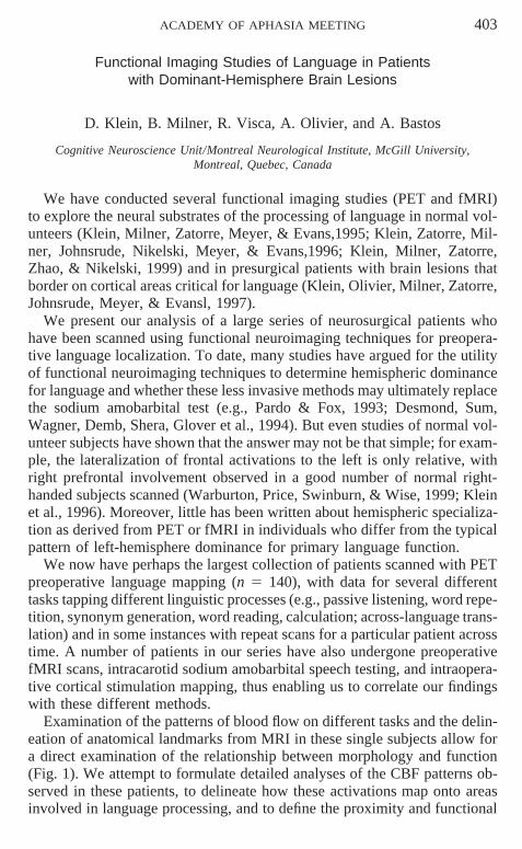
Examination of the patterns of blood flow on different tasks and the delin-eation of anatomical landmarks from MRI in these single subjects allow fora direct examination of the relationship between morphology and function(Fig. 1). We attempt to formulate detailed analyses of the CBF patterns ob-served in these patients, to delineate how these activations map onto areasinvolved in language processing, and to define the proximity and functional
404 ACADEMY OF APHASIA MEETING
FIG. 1. Example of the patterns of cerebral blood flow (CBF) observed with positronemission tomography (PET) for the subtraction of word repetition from synonym generationin two patients with lesions in the left inferior frontal region. (a) Early-onset static lesionsecondary to fetal anoxia; right-hemisphere speech representation as determined by intra-carotid sodium amobarbital testing and PET activation in the right inferior frontal region.(b) Late-onset space occupying lesion (glioma); left-hemisphere speech representation. Theactivity is in the left inferior frontal region, but is spatially shifted by the tumor.
significance of these regions in relation to specific brain lesions. To this end,we explore: (a) early- and late-onset left-hemisphere lesions; (b) whetherthere is activation of compensatory areas following lesions occurring duringmaturation or in adulthood; (c) changes in CBF patterns as a consequenceof brain tumor evolution; and (d) language organization in multilingual pa-tients. In addition to the major theoretical interest of these issues, they areextremely important for localizing the lesion and its immediate topographicalrelationships and for addressing the practical problems raised by the treat-ment of patients with focal brain lesions.
REFERENCES
Desmond, J. E., Sum, J. M., Wagner, A. D., Demb, J. B., Shera, P. K., Glover, G. H., et al.1995. Functional MRI measurement of language lateralization in Wada-tested patients.Brain, 118, 1411–1419.
Klein, D., Milner, B., Zatorre, R. J., Meyer, E., & Evans, A. C. 1995. The neural substratesunderlying word generation: A bilingual functional-imaging study. Proceedings of theNational Academy of Sciences of the USA, 92, 2899–2903.
Klein, D., Zatorre, R. J., Milner, B., Johnsrude, J., Nikelski, J., Meyer, E., & Evans, A. C.1996. CBF patterns during synonym generation: Group vs. individual study. NeuroImage,3(3), S444.
Klein, D., Olivier, A., Milner, B., Zatorre, R. J., Johnsrude, I., Meyer, E., & Evans, A. C. 1997.Obligatory role of the LIFG in synonym generation: Evidence from PET and corticalstimulation. NeuroReport, 8, 3275–3279.
ACADEMY OF APHASIA MEETING 405
Klein, D., Milner, B., Zatorre, R. J., Zhao, V., & Nikelski, J. 1999. Cerebral organization inbilinguals: A PET study of Chinese–English verb generation. NeuroReport, 10, 2841–2846.
Pardo, J. V., & Fox, P. T. 1993. Preoperative assessment of the cerebral hemispheric domi-nance language with CBF PET. Human Brain Mapping, 1, 57–68.
Warburton, E., Price, C. J., Swinburn, K., & Wise, R. J. 1999. Mechanisms of recovery fromaphasia: Evidence from positron emission tomography studies. Journal of Neurology,Neurosurgery & Psychiatry, 66(2), 155–161.
Stability of Functional Neuroanatomy of Auditory Sentence Processingin an Aphasic Patient
Martha W. Burton,* Charlotte C. Mitchum,* and Steven L. Small†
*Department of Neurology, University of Maryland; and †Department of Neurology,University of Chicago
Introduction
The present functional magnetic resonance imaging (fMRI) study investi-gates the functional neuroanatomy of auditory sentence processing in anaphasic patient. Previous studies of normal subjects performing auditory sen-tence listening using neuroimaging methods have shown distributed activa-tion along the superior and middle temporal gyri (e.g., Mazoyer et al., 1993;Schlosser, Aoyagi, Fulbright, Gore, & McCarthy, 1998; Small, Burton, Per-fetti, & Noll, 1999). Several of these studies report some degree of left later-alization with passive listening; however, close inspection reveals that theirdata appear strikingly bilateral and often symmetrical. The questions we ad-dress in the present study are (1) how the presence of a large left-hemispherelesion will affect the distributed processing networks in the left and righthemispheres involved in auditory sentence processing and (2) whether audi-tory sentence comprehension activation patterns are stable in a patient overa series of fMRI scans.
To date, most neuroimaging studies of language in patients with aphasiahave focused on determining whether recovery is mediated by increased righthemisphere activity or whether unaffected regions of the left hemisphereplay a greater role. Although there seems to be little question that the righthemisphere is active to some extent during language recovery, some evi-dence suggests activation or (reactivation) of left temporal areas may beassociated with patients’ showing recovery of language function (Small &Burton, in press).
We studied a patient with an impairment in auditory sentence comprehen-sion which resulted in difficulty understanding semantically reversible sen-tences. We report three functional scans, in which the patient performed a
406 ACADEMY OF APHASIA MEETING
sentence–picture verification task in an event-related fMRI study that tookplace over the course of 3 months.
Method
A 61-year-old right-handed man (JQ) participated in the study followinga left temporoparietaloccipital hemorrhagic stroke (2 years postonset) witha resulting deficit in sentence comprehension. On a computer-administeredsentence–picture verification task comparing active and passive sentences,performed outside of the scanner prior to the study, JQ’s performance wasat chance (overall proportion correct, .49; active 5 0.72, passive 5 0.26).
During the functional scans, he was presented with six semantically re-versible sentences, each in the active and passive voices, matched with apicture that corresponded to the sentence. In addition, a set of distractor sen-tence–picture pairs was created in which the six sentences were presentedwith pictures that showed a reversal of noun thematic roles. Thus, there werea total of 24 different sentence–picture combinations presented during ascan. Each session consisted of four functional scans of approximately 10min each.
The patient performed a delayed sentence–picture verification task inwhich he decided whether a picture presented approximately 4 s followingthe end of the sentence matched the sentence. His response was indicatedby a button press. The stimuli were presented to the patient using an event-related design to allow random presentation of sentences and to analyze sepa-rate components of the trials (e.g., auditory sentence comprehension). Eachtrial lasted a total of 24 s, consisting of a warning tone, an active or passivesentence (mean duration 5 approximately 2.5 s) presented over headphones1.5 s following trial onset, and a picture presented 8 s after the onset of thetrial. A fixation cross remained on the screen during the entire trial exceptduring picture presentation. Functional image acquisition and analysis meth-ods were performed according to previously described methods (Burton &Small, 1999).
The proportion of correct responses was calculated. FMRI data were ana-lyzed using a t test in which the MR signal intensity during the intervalbetween sentence and picture presentation (where the activity related to audi-tory sentence comprehension should be at its peak due to hemodynamic lag)was compared to the baseline at the end of the trial. A threshold t value of 5.0was used to determine significant differences in activation between sentencecomprehension and baseline.
Results
Behavioral data. Behavioral data indicated that performance on the de-layed sentence–picture verification task during the fMRI scans was similarto performance on the sentence–picture verification task without delay tested
ACADEMY OF APHASIA MEETING 407
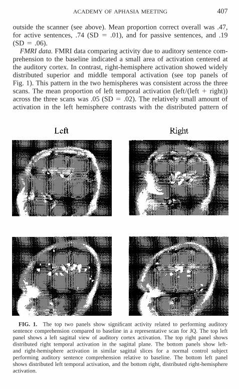
outside the scanner (see above). Mean proportion correct overall was .47,for active sentences, .74 (SD 5 .01), and for passive sentences, and .19(SD 5 .06).
FMRI data. FMRI data comparing activity due to auditory sentence com-prehension to the baseline indicated a small area of activation centered atthe auditory cortex. In contrast, right-hemisphere activation showed widelydistributed superior and middle temporal activation (see top panels ofFig. 1). This pattern in the two hemispheres was consistent across the threescans. The mean proportion of left temporal activation (left/(left 1 right))across the three scans was .05 (SD 5 .02). The relatively small amount ofactivation in the left hemisphere contrasts with the distributed pattern of
FIG. 1. The top two panels show significant activity related to performing auditorysentence comprehension compared to baseline in a representative scan for JQ. The top leftpanel shows a left sagittal view of auditory cortex activation. The top right panel showsdistributed right temporal activation in the sagittal plane. The bottom panels show left-and right-hemisphere activation in similar sagittal slices for a normal control subjectperforming auditory sentence comprehension relative to baseline. The bottom left panelshows distributed left temporal activation, and the bottom right, distributed right-hemisphereactivation.

408 ACADEMY OF APHASIA MEETING
bilateral activation seen in a normal control subject (female, age 21 years)performing the same task (see bottom panels of Fig. 1), and an age-matchedcontrol subject listening to grammatical sentences (male, age 52 years, whoparticipated in a passive listening experiment described in Small, Burton,Perfetti, & Noll, 1999). In contrast to JQ, the proportion of left temporalactivation for these control subjects was .57 and .61, respectively.
Discussion
The results of this study demonstrate a highly consistent pattern of activa-tion (focal left auditory cortex activity, distributed right temporal activity)in an aphasic patient performing an auditory sentence comprehension taskover a series of several scans. While auditory processing of the speech signalremains intact in both hemispheres, the presence of a large left-hemispherelesion strongly affects the distributed pattern of left activity found in normals,reducing it to a small focused area of auditory cortex, whereas right hemi-sphere activation appears remarkably similar to that of normals. The lack ofdistributed left-hemisphere activity corresponds to impaired performance ofthe sentence—picture verification task. Importantly, this pattern can be reli-ably shown over several separate scanning sessions in a patient with languagecomprehension impairment. Furthermore, consistent activation patterns andbehavioral performance in a single-trial design allow the possibility of morefine-grained analysis of trial components in which activation related to audi-tory sentence comprehension can be separated from activation due to othertrial components (e.g., picture verification).
REFERENCES
Burton, M. W., & Small, S. L. 1999. An introduction to functional magnetic resonance im-aging. The Neurologist, 5(3), 145–158.
Mazoyer, B. M., Tzourio, N., Frak, V., Syrota, A., Murayama, N., Levrier, O., Salamon, G.,Dehaene, S., Cohen, L., & Mehler, J. 1993. The cortical representation of speech. Journalof Cognitive Neuroscience, 5(4), 467–479.
Schlosser, M. J., Aoyagi, N., Fulbright, R. K., Gore, J. C., & McCarthy, G. 1998. FunctionalMRI studies of auditory comprehension. Human Brain Mapping, 6(1), 1–13.
Small, S. L., & Burton, M. W. (in press). Functional neuroimaging of language. In F. Boller &J. Grafman (Eds.), Handbook of neuropsychology. New York: Elsevier.
Small, S. L., Burton, M. W., Perfetti, C. A., & Noll, D. C. 1999. Sentence listening with andwithout responding. Brain and Language, 64(1), 325–328.
Corresponding author: Martha W. Burton, Department of Neurology, University ofMaryland School of Medicine, 12-011 Bressler Research Building, 655 West BaltimoreStreet, Baltimore, MD 21201-1559. Fax: (410) 706-0324. E-mail: [email protected].
ACADEMY OF APHASIA MEETING 409
Processing of Homonyms: A Functional MRI Study on theSeparation of Word Forms from Concepts
Walter Huber, Susanne Weis, Marion Grande, Stefan Pollrich,and Klaus Willmes
Department of Neurology, University of Technology (RWTH), Aachen, Germany
Introduction
It is a linguistic truism that words are lexical units combining semanticor conceptual information with sound structures. Classical brain center mod-els of language functions assumed separate anatomical representations forlexical and conceptual representations. Modern functional imaging studiesin normal subjects have readdressed the issue by trying to localize differentcortical networks that are specialized for sublexical, lexical, and conceptualprocessing of auditory and visual words (cf. recent reviews and studies byWarburton et al., 1996; Binder, Frost, Hammeke, Cox, Rao, & Prieto, 1997;Bookheimer et al., 1998; Price, 1998; Chee, O’Craven, Bergida, Rosen, &Savoy, 1999; Fujimaki et al., 1999). These studies have demonstrated seman-tic word processing to be distributed across left prefrontal, extrasylvian tem-poral, and/or posterior parietal areas. Tasks relying on sublexical phonologi-cal processing elicited classical perisylvian language areas (BA 40/22 andBA 6/44).
It is so far unresolved to what extend the processing of word form canbe separated from conceptual information. The purpose of the present studyis to explore the hypothesis that conceptual and lexical–semantic word pro-cessing activate different cortical networks. Following the classical model,we assume that the nonoverlapping part of these cortical networks lies withinthe posterior perisylvian cortex, possibly in the posterior part of the superiortemporal gyrus (i.e., in Wernicke’s area).
Methods
We made use of homonyms to distinguish between conceptual processingand semantic access to word form knowledge. Homonyms are words whichhave two or more unrelated meanings but he same phonological form, e.g.,the word bank with the meanings ‘‘bank of a river’’ and ‘‘financial institu-tion.’’ Lexical homonymy is specific to individual languages. In German,for example, the word Bank also expresses the furniture ‘‘bench.’’ Anotherexclusively German example is ‘‘Schloss,’’ which expresses both ‘‘lock’’and ‘‘castle.’’
Right-handed male subjects (all university students, aged between 20 and35 years) were scanned while performing three different tasks on visuallypresented pairs of word stimuli, which consisted of German nouns:
410 ACADEMY OF APHASIA MEETING
1. Passive reading of the two nouns and silent repetition;2. Mental search for a homonym with each of its meanings associated
with one of the two presented nouns (e.g., river/money ⇒ bank); and3. Mental search for a conceptual association related to both presented
nouns (e.g., river/lake ⇒ water)Each of the tasks was performed in a separate run in an event-related
design with 42 pairs of stimuli each. The interstimulus interval was 12.5 swith the word pairs being shown for 6 s and a blank screen for the remaining6 s. The subjects were not informed about the last task while performing thefirst two tasks in order to minimize conceptual associations already duringtasks 1 and 2.
Magnetic resonance imaging was performed on a 1.5-T Philips NT Gy-roscan using a standard bird-cage head coil and EPI T2*-weighted sequences(TR 2500 ms; FA 40°; matrix 64 3 64; FOV 224 mm; 19 continuous slicesparallel to the AC-PC line, comprising the neocerebrum; slice thickness5 mm, no interslice gap). Data were analyzed using Matlab 5.3 and SPM99software (Wellcome Department of Cognitive Neurology, London, UK).
Results
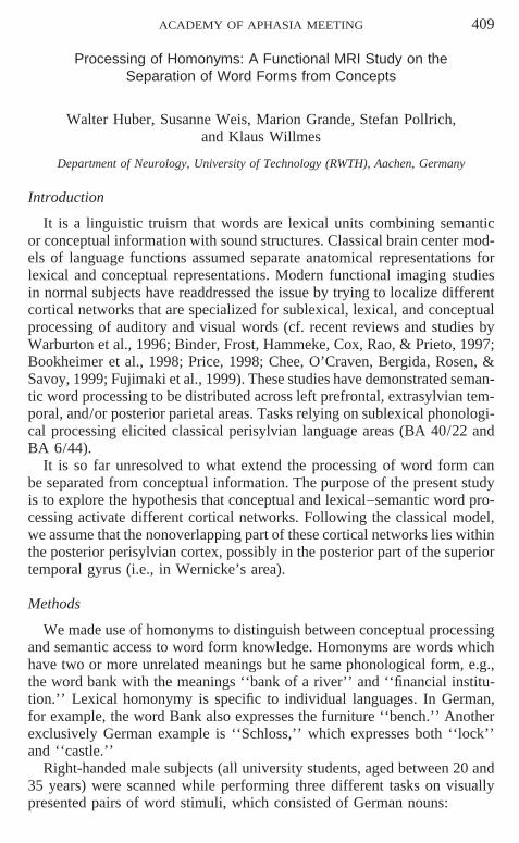
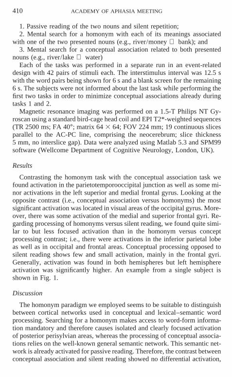
Contrasting the homonym task with the conceptual association task wefound activation in the parietotemporooccipital junction as well as some mi-nor activations in the left superior and medial frontal gyrus. Looking at theopposite contrast (i.e., conceptual association versus homonyms) the mostsignificant activation was located in visual areas of the occipital gyrus. More-over, there was some activation of the medial and superior frontal gyri. Re-garding processing of homonyms versus silent reading, we found quite simi-lar to but less focused activation than in the homonym versus conceptprocessing contrast; i.e., there were activations in the inferior parietal lobeas well as in occipital and frontal areas. Conceptual processing opposed tosilent reading shows few and small activation, mainly in the frontal gyri.Generally, activation was found in both hemispheres but left hemisphereactivation was significantly higher. An example from a single subject isshown in Fig. 1.
Discussion
The homonym paradigm we employed seems to be suitable to distinguishbetween cortical networks used in conceptual and lexical–semantic wordprocessing. Searching for a homonym makes access to word-form informa-tion mandatory and therefore causes isolated and clearly focused activationof posterior perisylvian areas, whereas the processing of conceptual associa-tions relies on the well-known general semantic network. This semantic net-work is already activated for passive reading. Therefore, the contrast betweenconceptual association and silent reading showed no differential activation,
ACADEMY OF APHASIA MEETING 411
FIG. 1. Left hemisphere activation at the junction of BA 39/37 for homonym findingminus conceptual association in one subject (right-handed male, 23 years).
which demonstrates that even when only reading words conceptual associa-tions cannot be avoided. This makes it so difficult to detect word-form pro-cessing in modern activation studies.
REFERENCES
Binder J. R., Frost, J. A., Hammeke, T. A., Cox, R. W., Rao, S. M., & Prieto, T. 1997. Humanbrain language areas identified by functional magnetic resonance imaging. Journal ofNeuroscience, 17, 353–362.
Bookheimer, S. Y., Zeffiro, T. A., Blaxton, T. A., Gaillard, W. D., Malow, B., & Theodore,W. H. 1998. Regional cerebral blood flow during auditory responsive naming: evidencefor cross-modality neural activation. NeuroReport, 9, 2409–2413.
Chee, M. W. L., O’Craven, K. M., Bergida, R., Rosen, B. R., & Savoy, R. L. 1999. Auditoryand visual word processing studied with fMRI. Human Brain Mapping, 7, 15–28.
Fujimaki, N., Miyauchi, S., Putz, B., Sasaki, Y., Takino, R., Sakai, K., & Tamada, T. 1999.Functional magnetic resonance imaging of neural activity related to orthographic, phono-logical and lexico-semantic judgments of visually presented characters and words. HumanBrain Mapping, 8, 44–59.
Price, C. J. 1998. The functional anatomy of word comprehension and production. Trends inCognitive Sciences, 2, 281–288.
Warburton, E., Wise, R. J., Price, C. J., Weiller, C., Hadar, U., Ramsay, S., et al. 1996. Nounand verb retrieval by normal subjects: Studies with PET. Brain, 119, 159–179.
412 ACADEMY OF APHASIA MEETING
Task Demands Influence the Activation Patternof Auditory Semantic Priming in fMRI
Sonja A. Kotz,* Angela D. Friederici,* Stefano F. Cappa,†and D.Yves von Cramon*
*Max Planck Institute of Cognitive Neuroscience, Leipzig, Germany; and †Facolta diPsicologia, Universita Vita Salute San Raffaele, Milano, Italy
Both normal and lesion studies indicate that priming effects can vary asa function of semantic information. For example, data resulting from reactiontimes and event-related potentials showed that associative priming (mouse–cheese) seems more left lateralized while categorical priming (chicken–cat)seems more right lateralized (Chiarello & Richards, 1992; Hagoort,Brown, & Swaab, 1996; Kotz, 1998; Swaab, Baynes, & Knight, 1998). Sev-eral fMRI investigations of word and picture processing have indicated theexistence of a ‘‘modality-independent’’ semantic network, which includesthe temporal lobe (BA 21), the temporoparietal junction, and the frontal lobe(i.e., Cappa, Perani, Schnur, Tettamanti, & Fazio, 1999). We recently re-ported single trial fMRI data (Kotz, Cappa, von Cramon, & Friederici, 1999)exploring auditory priming via means of a lexical decision task. The resultsindicate that priming of semantic information can be qualified by increasedbilateral activation of the Heschl’s gyri, the planum temporale, and planumpolare for unrelated targets. However, there was additional left hemisphericactivation of the posterior insula, the thalamus, and right hemispheric activa-tion of the superior temporal gyrus, mainly along the superior temporal sul-cus for the unrelated targets. No differentiation of associative and categoricaltargets was found. We also did not find activation in the left inferior frontalcortex which is assumed to be part of the semantic network. Two recentfMRI studies have explored the function of the left frontal activation.Thompson-Schill, D’Esposito, Aguirre, & Farah (1997) have suggested thatthe left frontal activation is due to selectional demands or the choice amongmultiple alternatives in semantic tasks. Demb et al. (1995) have shown thatactivation associated with semantic encoding is not related to task difficulty.
The goal of the current study was to specify the neuroanatomical correlatesof semantic information processing via means of an auditory semanticcategorization/priming task. We were interested to see whether the inferiorfrontal cortex (left/right) is activated as a function of task type and increasedtask demand comparing the activation of conceptual vs. semantic priming(see Kotz et al., 1999). We expected to replicate our previous fMRI primingdata and to find activation of the inferior frontal cortex as well as increasedactivation in the right hemisphere for categorical items as a function of tasktype and demand.
ACADEMY OF APHASIA MEETING 413
Methods. A pseudo-randomized list of 320 word/pseudo-word pairs form-ing four conditions (associatively, categorically related, unrelated, and pseu-dowords) were presented auditorily via head phones in a single-trial para-digm. Thirteen subjects (8 female) were required to perform a ‘‘go’’semantic categorization task (respond to food/drink items). The ISI betweenprime and target items was 100 ms, and the ITI 6.8 s. Four echo planarimages (TE 5 40 ms, TR 5 2 s) per trial were acquired in eight axial slices(thickness 5 4 mm, interslice distance 5 2 mm) using a Brucker 3T/100scanner. The calculation of activation maps for each subject utilized voxel-wise Pearson correlation coefficients. After normalizing images to Talairachspace group averages were calculated. Activation correlated with each criti-cal condition was evaluated by means of z score (p , .05, z . 5 2).
Results. Comparing the activation of associatively and categorically re-lated targets to unrelated targets the following picture emerged: There wasbilateral activation of the Heschl’s gyri (HG), the posterior superior temporalgyri (STG), and the anterior STG with a linear decrease of activation in allareas for unrelated vs. categorically related vs. associatively related targets.Furthermore, there was bilateral activation (though, more accentuated in theleft hemisphere) in the inferior frontal gyri (IFG) with unrelated targets morestrongly activated than categorically related targets. Activation for associa-tively related targets was only found in the left IFG. Activation for ‘‘food’’and ‘‘drink’’ targets displayed a similar pattern with bilateral activation ofthe posterior STG and the IFG (right accentuated), but also bilateral activa-tion in the posterior cingulate (BA 23).
Discussion. The current results support the notion that activation of theinferior frontal cortex can vary as a function of task type and demand. Fur-thermore, the linear decrease of activity comparing unrelated and relatedtargets suggests that activation reflecting lexical–semantic processing variesas a function of degree of relatedness. The increase of right hemisphere acti-vation for categorically related and unrelated targets confirms previous datafrom visual hemifield (Chiarello & Richards, 1992) and lesion studies (Ha-goort, Brown, & Swaab, 1996). However, this increase varied as a functionof task and demand (see Kotz et al., 1999).
REFERENCES
Cappa, S. F., Perani, D., Schnur, T., Tettamanti, M., & Fazio, F. 1998. The effects of semanticcategory and knowledge-type on lexical-semantic access: A PET study, NeuroImage, 8,350–359.
Chiarello, C., & Richards, L. 1992. Another look at categorical priming in cerebral hemi-spheres. Neuropsychologia, 30, 81–392.
Demb, J. B., Desmond, J. E., Wagner, A. D., Vaidya, C. J., Glover, G. H., & Gabrieli,J. D. E. 1995. Semantic encoding and retrieval in the left inferior prefrontal cortex: A
414 ACADEMY OF APHASIA MEETING
functional MRI study of task difficulty and process specificity. Journal of Neuroscience,15, 5870–5878.
Hagoort, P., Brown, C., & Swaab, T. Y. 1996. Lexical–semantic event-related potential effectsin patients with left hemisphere lesions and aphasia, and patients with right hemispherelesions without aphasia. Brain, 119, 627–649.
Kotz, S. A. 1998. Comparing the auditory and visual sequential priming paradigm: An event-related potential study. 1998. Supplement of the Journal of Cognitive Neuroscience, S54.
Kotz, S. A., Cappa, S. F., von Cramon, D. Y., & Friederici, A. D. 1999. Priming as a functionof semantic information types: An fMRI investigation. NeuroImage, 9(6), S1000.
Swaab, T. Y., Baynes, K., & Knight, R. T. 1998. Coarse semantic coding in the right hemi-sphere: An ERP study. Supplement of the Journal of Cognitive. Neuroscience, S34.
Thompson-Schill, S., D’Esposito, M., Aguirre, G. K., & Farah, M. J.. 1997. Role of left inferiorprefrontal cortex in retrieval of semantic knowledge: A reevaluation. Proceedings of theNational Academy of Sciences, USA, 94, 14792–14797.
POSTER SESSION 2: LEXICAL SEMANTICS
The Status of Zero in the Semantic System:A Neuropsychological Study
Manuela Cacciatori, Alessia Grana, Luisa Girelli, and Carlo Semenza
Department of Psychology, University of Trieste, Italy
Introduction
Zero as a number appeared very late in the history of mathematics (Butter-worth, 1999). The delay of its appreciation by infants (Wynn, 1998) andpreschoolers and the difficulties tied with its use may be grounded in itsunique mathematical function. In fact, while adding a null quantity or sub-tracting it from a given quantity can be easily represented (e.g., by visualimaging), it is less obvious which representation may be invoked in multipli-cation or division by 0. In particular, zero as an operand makes any givenquantity disappear despite the fact that, by multiplying, we generally obtainthe opposite result, that is, a significant increment of the quantity.
Double dissociations between problem types and specific patterns of errorshave led to the assumption that nonzero problems are individually stored inlong-term memory (e.g., 3 3 6) while 0-problems are solved by referenceto general rules (e.g., N 1 0 5 N, for any N). Moreover, it has been shownthat the same 0-rule may be correctly retrieved as part of a multistep calcula-tion procedure while being not available in answering simple rule-basedproblems (Sokol, McCloskey, Cohen, & Aliminosa, 1991).
This paper describes two neuropsychological patients whose specific pat-terns of preserved/impaired performance with single and multidigit multipli-cations shed light on the mechanisms mediating the manipulation of 0.
ACADEMY OF APHASIA MEETING 415
Case Reports
AF is a 71-year-old Broca’s aphasic with a short-term memory withinnormal limits. FV is a 55-year-old geometer who developed a Wernicke’saphasia following a left CVA. Some signs of frontal dysfunction and poorshort-term memory were detected. Both patients showed specific difficultiesin simple and complex calculation, despite efficient production and compre-hension of up to three-digit Arabic numerals.
Experimental Investigation
Material. Overall, 196 single-digit operations (50 additions, 50 subtrac-tions, and 96 multiplications) including both fact-based and rule-based prob-lems and 66 multidigit problems (14 additions, 16 subtractions, and 36 multi-plications) were visually presented to be answered in written modality.Problems varied in complexity in terms of carrying or borrowing require-ments. Moreover, factors may or may not include ‘‘0,’’ and the relative posi-tion of zero within the numerals was balanced across problems (e.g.,NNNxN0N; NN0xNNN). Patients were asked to work out the solution ofeach problem and were encouraged to comment on the operational stepsused.
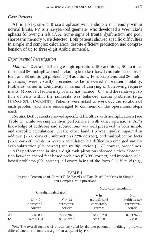
Results. Both patients showed specific difficulties with multiplications (seeTable 1) while varying in their performance with other operations. AF’sknowledge of additions and subtractions was well preserved in both simpleand complex calculations. On the other hand, FS was equally impaired inaddition (76% correct), subtraction (72% correct), and multiplication facts(76% correct), while in written calculation his difficulties emerged mainlywith subtraction (0% correct) and multiplication (5.6% correct) procedures.
AF’s performance in single-digit multiplications showed a clear dissocia-tion between spared fact-based problems (95.8% correct) and impaired rule-based problems (0% correct), all errors being of the form 0 3 N 5 N (e.g.,
TABLE 1Patient’s Percentage of Correct Rule-Based and Fact-Based Problems in Simple
and Complex Multiplications
Multi-digit calculationOne-digit calculation
0 in 0 inN 3 0 N 3 M multiplicand multiplicator
correct/n% correct/n% correct/n% correct/n%correct correct correct correct
AF 0/16 0.0 77/80 96.3 16/50 32.0 31/33 94.2FV 16/16 100 62/80 77.5 0/14 0.0 0/9 0.0
Note. The overall number of 0-facts answered by the two patients in multidigit problemsdiffered due to the incorrect algorithm adopated by FV.
416 ACADEMY OF APHASIA MEETING
3 3 0 5 3, 0 3 5 5 5). Interestingly, in multidigit problems AF appliedalways the correct algorithm but his approach to the 0-facts varied with thestructure of the operands: when 0 was in the multiplicand he failed to retrievethe rule (32% correct), while when 0 was in the multiplicator he bypassedthe repeated application of the 0-rule, writing directly as many 0s as thenumber of digits in the multiplicand. By applying this special-case procedurehe succeeded in 94.2% of the problems.
FV’s performance was in the opposite direction. In simple multiplications,his difficulties were limited to fact-based problems (75% correct), being rule-based problems error free. Instead, the solution of multidigit multiplicationswas affected by both procedural and arithmetic facts difficulties: besides in-correctly multiplying each number only by the number in the same column,FV was totally unable to apply adequately the 0 3 N 5 0-rule (0% correct)so that he could easily retrieve in simple calculation.
Discussion
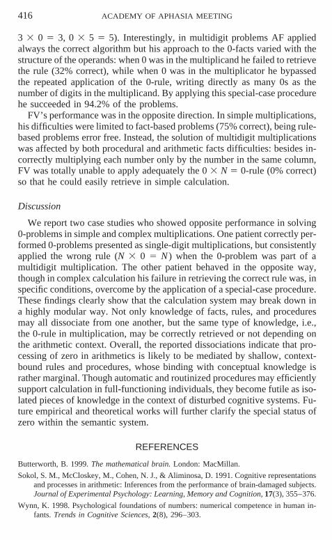
We report two case studies who showed opposite performance in solving0-problems in simple and complex multiplications. One patient correctly per-formed 0-problems presented as single-digit multiplications, but consistentlyapplied the wrong rule (N 3 0 5 N ) when the 0-problem was part of amultidigit multiplication. The other patient behaved in the opposite way,though in complex calculation his failure in retrieving the correct rule was, inspecific conditions, overcome by the application of a special-case procedure.These findings clearly show that the calculation system may break down ina highly modular way. Not only knowledge of facts, rules, and proceduresmay all dissociate from one another, but the same type of knowledge, i.e.,the 0-rule in multiplication, may be correctly retrieved or not depending onthe arithmetic context. Overall, the reported dissociations indicate that pro-cessing of zero in arithmetics is likely to be mediated by shallow, context-bound rules and procedures, whose binding with conceptual knowledge israther marginal. Though automatic and routinized procedures may efficientlysupport calculation in full-functioning individuals, they become futile as iso-lated pieces of knowledge in the context of disturbed cognitive systems. Fu-ture empirical and theoretical works will further clarify the special status ofzero within the semantic system.
REFERENCES
Butterworth, B. 1999. The mathematical brain. London: MacMillan.
Sokol, S. M., McCloskey, M., Cohen, N. J., & Aliminosa, D. 1991. Cognitive representationsand processes in arithmetic: Inferences from the performance of brain-damaged subjects.Journal of Experimental Psychology: Learning, Memory and Cognition, 17(3), 355–376.
Wynn, K. 1998. Psychological foundations of numbers: numerical competence in human in-fants. Trends in Cognitive Sciences, 2(8), 296–303.
ACADEMY OF APHASIA MEETING 417
Address for correspondence: Dr. Luisa Girelli, Department of Psychology, University ofTrieste, Via S. Anastasio 12 34124, Trieste, Italy. E-mail: [email protected].
Processing Idiomatic Expressions in the Living–Nonliving Dissociation
Francesca Borgo,* Teresa Maria Sgaramella,† Silvia Pontin,‡and Carlo Semenza‡
*Cognitive Neuroscience Sector, SISSA-ISAS, Trieste; †Neurology Clinic OspedaleS. Bortolo, Vicenza; and ‡Department of Psychology and BRAIN Neuroscience Center,
University of Trieste, Italy
Introduction
Knowledge related to the living things class is disproportionately affectedin some patients with respect to a relatively unimpaired performance withnonliving things (Warrington & Shallice, 1984). This effect has been ob-served, in a variety of tasks, in relation to concrete referents. However, alarge number of words indicating both living and nonliving entities has, inmany languages, entered the common use with an additional idiomatic, crys-tallized meaning. The performance is described here of a patient with a livingthings deficit when asked to process idiomatic meaning. The results arethought to be informative about idioms in the semantic system and to shedlight on the nature of the living–nonliving dissociation.
Case Report
MU was a 32-year-old male, with 13 years of education. He suffered fromherpes simplex encephalitis in 1994; an MRI scan showed an extensive dam-age affecting bilaterally the temporal lobes, portions of the frontal lobes, andportions of the right occipital lobe.
MU was administered a variety of tasks classically aimed at exploring theliving–nonliving dissociation: naming on visual confrontation, naming afterverbal description, word-to-picture matching, and knowledge of semanticfeatures (i.e., ‘‘Does the leopard run fast?’’). Over all these tasks (adminis-tered twice, like most of those in the experimental study, with an intervalof 1 year between administrations, see Table 1) a dramatic and persistentover time loss of information concerning living things emerged, while non-living things knowledge was relatively spared.
Experimental Study
The Living–Nonliving Dissociation
In order to obtain a direct comparison, a picture naming task was adminis-tered on the same lexical items that were used for the ‘‘idiomatic’’ tasks
418 ACADEMY OF APHASIA MEETING
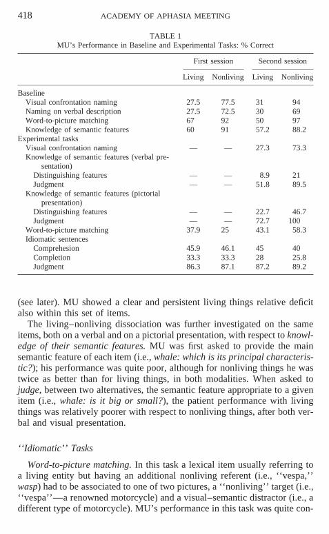
TABLE 1MU’s Performance in Baseline and Experimental Tasks: % Correct
First session Second session
Living Nonliving Living Nonliving
BaselineVisual confrontation naming 27.5 77.5 31 94Naming on verbal description 27.5 72.5 30 69Word-to-picture matching 67 92 50 97Knowledge of semantic features 60 91 57.2 88.2
Experimental tasksVisual confrontation naming — — 27.3 73.3Knowledge of semantic features (verbal pre-
sentation)Distinguishing features — — 8.9 21Judgment — — 51.8 89.5
Knowledge of semantic features (pictorialpresentation)
Distinguishing features — — 22.7 46.7Judgment — — 72.7 100
Word-to-picture matching 37.9 25 43.1 58.3Idiomatic sentences
Comprehesion 45.9 46.1 45 40Completion 33.3 33.3 28 25.8Judgment 86.3 87.1 87.2 89.2
(see later). MU showed a clear and persistent living things relative deficitalso within this set of items.
The living–nonliving dissociation was further investigated on the sameitems, both on a verbal and on a pictorial presentation, with respect to knowl-edge of their semantic features. MU was first asked to provide the mainsemantic feature of each item (i.e., whale: which is its principal characteris-tic?); his performance was quite poor, although for nonliving things he wastwice as better than for living things, in both modalities. When asked tojudge, between two alternatives, the semantic feature appropriate to a givenitem (i.e., whale: is it big or small?), the patient performance with livingthings was relatively poorer with respect to nonliving things, after both ver-bal and visual presentation.
‘‘Idiomatic’’ Tasks
Word-to-picture matching. In this task a lexical item usually referring toa living entity but having an additional nonliving referent (i.e., ‘‘vespa,’’wasp) had to be associated to one of two pictures, a ‘‘nonliving’’ target (i.e.,‘‘vespa’’—a renowned motorcycle) and a visual–semantic distractor (i.e., adifferent type of motorcycle). MU’s performance in this task was quite con-
ACADEMY OF APHASIA MEETING 419
sistent over time and equally deficient in both living and nonliving catego-ries.
Sentence comprehension. The patient had to explain the meaning of idio-matic sentences (i.e., ‘‘mangiare come un bue’’ eat like an ox; ‘‘essere unavolpe’’ to be like a fox); two independent judges assessed the patient’s re-sponses with no disagreement on the scores. No difference emerged betweenliving and nonliving items.
Sentence completion. The patient had to complete a series of idiomaticexpressions with the correct idiomatic lexical item (i.e., ‘‘lento comeuna . . . tartaruga’’ slow as a . . . turtle); again no living/nonliving differencewas found.
Judgment. Judgment between two alternatives of the appropriate meaning(i.e., ‘‘essere un’acciuga significa essere magro o grasso?’’ being like ananchovy means being thin or fat?). In the judgment task MU’s performancewas relatively well preserved: again no major differences were observed be-tween living and nonliving things.
Age and education-matched controls scored at ceiling in all these tasks.
Conclusion
MU, a patient with a classic dissociation between knowledge of the com-mon meaning of living things (defective) and of artifacts (relatively spared),did not show the same difference in tasks involving the processing of theidiomatic meaning of the same items.
This finding suggests that the living/nonliving dissociation may be limitedto common meaning, thus reflecting a different brain organization of com-mon and idiomatic semantics.
REFERENCE
Warrington, E. K., & Shallice, T. 1984. Category specific semantic impairments. Brain, 107,829–854.
Repeated Sampling of Word Retrieval in Aphasia: Stability of Errors
Julie L. Wambaugh, Michele N. Alegre, Aida L. Martinez,and Craig W. Linebaugh*
VA Salt Lake City Healthcare System and University of Utah, Salt Lake City, Utah;and *The George Washington University, Washington, DC
Numerous investigators have stressed the importance of determining thelocus of lexical retrieval deficit to better understand the nature of aphasicnaming errors, as well as to plan effective treatment (Drew & Thompson,
420 ACADEMY OF APHASIA MEETING
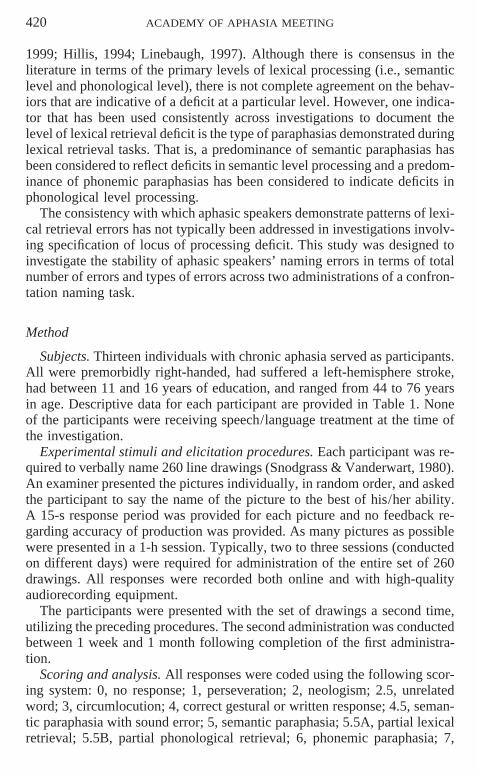
1999; Hillis, 1994; Linebaugh, 1997). Although there is consensus in theliterature in terms of the primary levels of lexical processing (i.e., semanticlevel and phonological level), there is not complete agreement on the behav-iors that are indicative of a deficit at a particular level. However, one indica-tor that has been used consistently across investigations to document thelevel of lexical retrieval deficit is the type of paraphasias demonstrated duringlexical retrieval tasks. That is, a predominance of semantic paraphasias hasbeen considered to reflect deficits in semantic level processing and a predom-inance of phonemic paraphasias has been considered to indicate deficits inphonological level processing.
The consistency with which aphasic speakers demonstrate patterns of lexi-cal retrieval errors has not typically been addressed in investigations involv-ing specification of locus of processing deficit. This study was designed toinvestigate the stability of aphasic speakers’ naming errors in terms of totalnumber of errors and types of errors across two administrations of a confron-tation naming task.
Method
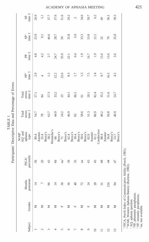
Subjects. Thirteen individuals with chronic aphasia served as participants.All were premorbidly right-handed, had suffered a left-hemisphere stroke,had between 11 and 16 years of education, and ranged from 44 to 76 yearsin age. Descriptive data for each participant are provided in Table 1. Noneof the participants were receiving speech/language treatment at the time ofthe investigation.
Experimental stimuli and elicitation procedures. Each participant was re-quired to verbally name 260 line drawings (Snodgrass & Vanderwart, 1980).An examiner presented the pictures individually, in random order, and askedthe participant to say the name of the picture to the best of his/her ability.A 15-s response period was provided for each picture and no feedback re-garding accuracy of production was provided. As many pictures as possiblewere presented in a 1-h session. Typically, two to three sessions (conductedon different days) were required for administration of the entire set of 260drawings. All responses were recorded both online and with high-qualityaudiorecording equipment.
The participants were presented with the set of drawings a second time,utilizing the preceding procedures. The second administration was conductedbetween 1 week and 1 month following completion of the first administra-tion.
Scoring and analysis. All responses were coded using the following scor-ing system: 0, no response; 1, perseveration; 2, neologism; 2.5, unrelatedword; 3, circumlocution; 4, correct gestural or written response; 4.5, seman-tic paraphasia with sound error; 5, semantic paraphasia; 5.5A, partial lexicalretrieval; 5.5B, partial phonological retrieval; 6, phonemic paraphasia; 7,
ACADEMY OF APHASIA MEETING 421T
AB
LE
1Pa
rtic
ipan
ts’
Des
crip
tive
Dat
aan
dPe
rcen
tage
ofE
rror
s
WA
Bb
AQ
can
dT
otal
Tot
alM
onth
sPI
CA
aap
hasi
aer
rors
erro
rsPP
dPP
SPe
SPSu
bjec
tG
ende
rpo
ston
set
perc
entil
ety
petim
e1
time
2tim
e1
time
2tim
e1
time
2
1M
1456
80.4
54.7
57.1
2.9
4.8
25.9
26.9
Ano
mic
2M
720
35.4
7884
44.
29.
15.
1B
roca
’s3
M96
4358
.263
.757
.31.
22.
746
.637
.7W
erni
cke’
s4
M14
6666
44.9
33.2
12.2
24.7
28.7
37.6
Bro
ca’s
5F
36na
f71
24.2
2522
.635
.950
51.6
Bro
ca’s
6M
3639
62.4
46.9
44.1
8.3
22.1
35.8
29.2
Bro
ca’s
7F
4835
39.4
9396
.51.
70.
45.
92
Bro
ca’s
8M
72na
58.6
58.6
611.
31.
933
.334
.6B
roca
’s9
F24
6382
.931
.328
.17.
516
.753
.859
.7A
nom
ic10
M28
4361
.280
.982
.41.
41.
912
.16.
2C
ondu
ctio
n11
M31
6888
.440
.235
.29.
715
.632
40A
nom
ic12
M21
644
56.8
50.8
51.6
16.2
13.6
7056
.1B
roca
’s13
M94
4753
.746
.954
.74.
23.
635
.839
.3B
roca
’s
aPI
CA
,Po
rch
Inde
xof
Com
mun
icat
ive
Abi
lity
(Por
ch,
1981
).b
WA
B,
Wes
tern
Aph
asia
Bat
tery
(Ker
tesz
,19
82).
cA
Q,
apha
sia
quot
ient
.d
PP,
phon
emic
para
phas
ias.
eSP
,se
man
ticpa
raph
asia
s.fna
,no
tav
aila
ble.
422 ACADEMY OF APHASIA MEETING
self-correction; 8, correct response with a delay of .5 s; 8.5, regular plural/singular error; 8.9, minor articulatory distortion; and 9, immediate, accurateresponse.
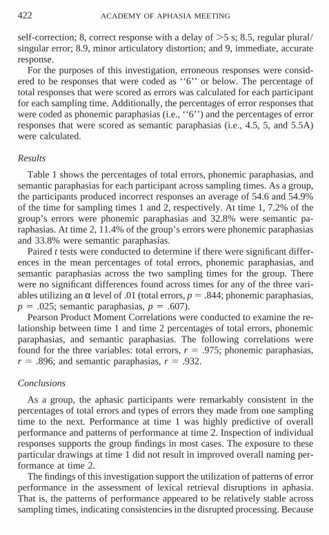
For the purposes of this investigation, erroneous responses were consid-ered to be responses that were coded as ‘‘6’’ or below. The percentage oftotal responses that were scored as errors was calculated for each participantfor each sampling time. Additionally, the percentages of error responses thatwere coded as phonemic paraphasias (i.e., ‘‘6’’) and the percentages of errorresponses that were scored as semantic paraphasias (i.e., 4.5, 5, and 5.5A)were calculated.
Results
Table 1 shows the percentages of total errors, phonemic paraphasias, andsemantic paraphasias for each participant across sampling times. As a group,the participants produced incorrect responses an average of 54.6 and 54.9%of the time for sampling times 1 and 2, respectively. At time 1, 7.2% of thegroup’s errors were phonemic paraphasias and 32.8% were semantic pa-raphasias. At time 2, 11.4% of the group’s errors were phonemic paraphasiasand 33.8% were semantic paraphasias.
Paired t tests were conducted to determine if there were significant differ-ences in the mean percentages of total errors, phonemic paraphasias, andsemantic paraphasias across the two sampling times for the group. Therewere no significant differences found across times for any of the three vari-ables utilizing an α level of .01 (total errors, p 5 .844; phonemic paraphasias,p 5 .025; semantic paraphasias, p 5 .607).
Pearson Product Moment Correlations were conducted to examine the re-lationship between time 1 and time 2 percentages of total errors, phonemicparaphasias, and semantic paraphasias. The following correlations werefound for the three variables: total errors, r 5 .975; phonemic paraphasias,r 5 .896; and semantic paraphasias, r 5 .932.
Conclusions
As a group, the aphasic participants were remarkably consistent in thepercentages of total errors and types of errors they made from one samplingtime to the next. Performance at time 1 was highly predictive of overallperformance and patterns of performance at time 2. Inspection of individualresponses supports the group findings in most cases. The exposure to theseparticular drawings at time 1 did not result in improved overall naming per-formance at time 2.
The findings of this investigation support the utilization of patterns of errorperformance in the assessment of lexical retrieval disruptions in aphasia.That is, the patterns of performance appeared to be relatively stable acrosssampling times, indicating consistencies in the disrupted processing. Because
ACADEMY OF APHASIA MEETING 423
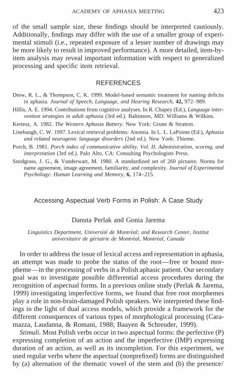
of the small sample size, these findings should be interpreted cautiously.Additionally, findings may differ with the use of a smaller group of experi-mental stimuli (i.e., repeated exposure of a lesser number of drawings maybe more likely to result in improved performance). A more detailed, item-by-item analysis may reveal important information with respect to generalizedprocessing and specific item retrieval.
REFERENCES
Drew, R. L., & Thompson, C. K. 1999. Model-based semantic treatment for naming deficitsin aphasia. Journal of Speech, Language, and Hearing Research, 42, 972–989.
Hillis, A. E. 1994. Contributions from cognitive analyses. In R. Chapey (Ed.), Language inter-vention strategies in adult aphasia (3rd ed.). Baltimore, MD: Williams & Wilkins.
Kertesz, A. 1982. The Western Aphasia Battery. New York: Grune & Stratton.
Linebaugh, C. W. 1997. Lexical retrieval problems: Anomia. In L. L. LaPointe (Ed.), Aphasiaand related neurogenic language disorders (2nd ed.). New York: Thieme.
Porch, B. 1981. Porch index of communicative ability. Vol. II. Administration, scoring, andinterpretation (3rd ed.). Palo Alto, CA: Consulting Psychologists Press.
Snodgrass, J. G., & Vanderwart, M. 1980. A standardized set of 260 pictures: Norms forname agreement, image agreement, familiarity, and complexity. Journal of ExperimentalPsychology: Human Learning and Memory, 6, 174–215.
Accessing Aspectual Verb Forms in Polish: A Case Study
Danuta Perlak and Gonia Jarema
Linguistics Department, Universite de Montreal; and Research Center, Institutuniversitaire de geriatrie de Montreal, Montreal, Canada
In order to address the issue of lexical access and representation in aphasia,an attempt was made to probe the status of the root—free or bound mor-pheme—in the processing of verbs in a Polish aphasic patient. Our secondarygoal was to investigate possible differential access procedures during therecognition of aspectual forms. In a previous online study (Perlak & Jarema,1999) investigating imperfective forms, we found that free root morphemesplay a role in non-brain-damaged Polish speakers. We interpreted these find-ings in the light of dual access models, which provide a framework for thedifferent consequences of various types of morphological processing (Cara-mazza, Laudanna, & Romani, 1988; Baayen & Schreuder, 1999).
Stimuli. Most Polish verbs occur in two aspectual forms: the perfective (P)expressing completion of an action and the imperfective (IMP) expressingduration of an action, as well as its incompletion. For this experiment, weused regular verbs where the aspectual (nonprefixed) forms are distinguishedby (a) alternation of the thematic vowel of the stem and (b) the presence/
424 ACADEMY OF APHASIA MEETING
absence of a suffix and thematic vowel alternation. We constructed twogroups of verbs (24 stimuli in each group). In the first group (G1), all verbsfeature root morphemes that are words in the language: they are homopho-nous with the second person singular imperative of perfective forms. In thesecond group (G2), roots are bound morphemes. In each group we testedperfective infinitives, imperfective infinitives, and roots. The verbs were se-mantically and phonologically transparent and were controlled for frequencyand syllable length. Half of the stimuli tested were nonwords. We usednouns, adjectives, and inflected verbs as fillers.
Subjects. The subject of this study, KL, was a 61-year-old female patient,born and educated in Poland. She presented with a nonfluent aphasia and amild deficit in speech production. At time of testing, she was 6 years post-onset. We also tested three female controls with the same level of educationas KL, 59, 62, and 62 years old, respectively.
Procedure. All subjects were tested on an online lexical decision task inthe visual modality. The patient and control subjects were asked to identifythe item that appeared on the screen of a computer as a word or nonwordin Polish by pressing either a designated ‘‘yes’’ or ‘‘no’’ key. Presentationof the stimuli and recording of response latencies was controlled by an AppleMacintosh laptop computer. The 300 experimental trials were divided intofive tests and were run in five sessions for the patient and in two sessionsfor the control subjects. A two-factor ANOVA (lexical status of roots byaspectual forms) was performed on all subjects response latencies.
Results and discussion. Errors and reactions times (RTs) over 1500 msfor controls and 2000 ms for KL were removed. Analysis of the mean RTsrevealed a significant main effect of root status for both the controls and KL(F(1,28) 5 6.48, p 5 .02; and F(1,22) 5 3.46, p 5 .04, respectively). InG1, results did not reveal any significant differences between the two as-pectual forms for both the controls (F 5 0.32, p 5 57) and KL (F 5 2.08,p 5 16). In G2, the controls showed a significant form effect in a plannedcomparison (F 5 4.12, p 5 .05), but no significant difference was obtainedfor KL (F 5 0.8, p 5 .78).
The overall differential recognition pattern obtained for G1 and G2 acrosscontrols and the aphasic subject suggests that free and bound root morphemeshave a distinct status in the mental lexicon and that the status of the root playsa role in the processing of words. The absence of any differences betweenperfective and imperfective forms in G1 for both the controls and KL sug-gests that all forms with free root morpheme are accessed in the same man-ner: decomposition by virtue of the word status of the root and whole-wordaccess (see Baayen & Schreuder, 1999) as shown by the faster RTs for con-trols on G1 when compared to G2. G2 forms are thus assumed to be accessedvia only one route: whole-word access. It has been proposed by Grzegor-czykowa, Laskowski, & Wrobel (1998) that in Polish perfectives are thebase forms of verbs. This is reflected in the faster RTs for perfectives when
ACADEMY OF APHASIA MEETING 425
compared to imperfectives in G2 in the control group. The fact that G2 im-perfectives are slower for controls indicates that these forms involve a cost,that of activating their corresponding perfective base. KL does not showthis pattern, which seems to point to a weakening of the links between thederivationally related forms, imperfective forms being accessed without theconcomitant activation of their perfective counterparts.
Conclusion. Our results support dual access models of the mental lexiconand suggest that the Polish-speaking controls and the aphasic subject investi-gated in the present study process aspectual forms with root morphemes thatare words in the language via two routes: decomposition and whole-wordaccess. In contrast, forms with bound root morpheme are accessed via thewhole-word route only.
REFERENCES
Baayen, R. H., & Schreuder, R. 1999. Morphemes and full forms in a noninteractive activationparallel dual route model. Brain and Language, 68, 27–32.
Caramazza, A., Laudanna, A., & Romani, C. 1989. Lexical access and inflectional morphol-ogy. Cognition, 28, 297–332.
Grzegorczykowa, R., Laskowski, R., & Wrobel, H. 1998. Gramatyka wspolczesnego jezykapolskiego: Morfologia. Warszawa: PWN.
Perlak, D., & Jarema, G. 1999. Recognition and storage of aspectual verbal forms in the mentallexicon: Evidence from Polish. Communication in the 3rd European Conference on For-mal Description of Slavic Languages, FDSL 3, Leipzig, Allemagne, 1–3 decembre 1999.
Naming Objects and Actions: A Case Study
Simona Collina,* Paola Marangolo,† and Patrizia Tabossi*
*University of Trieste, Italy; and †IRCCS Clinica S. Lucia, Rome, Italy
Agrammatic patients often experience greater difficulties in producingverbs than nouns and this impairment is typically interpreted as a selectivegrammatical class deficit (Miceli, Silveri, Villa, & Caramazza, 1984; Zin-geser & Berndt, 1990).
In this study, we tested the hypothesis that many agrammatics’ difficultieswith verbs may be due, at least in part, to the different structural complexitytypically associated with nouns and verbs: while most nouns denote objects,all verbs express some relations among objects, and the specification of theserelations render their representation more complex than that of nouns. Infact, unlike most nouns, it is generally agreed that the semantic representationof a verb (e.g., to destroy) should include not only its core meaning (e.g.,the action of breaking to pieces), but also the thematic roles specified by theverb (e.g., the agent, the destroyer; and the theme, the entity being de-
426 ACADEMY OF APHASIA MEETING
stroyed). Moreover, at the syntactic level, the representation of a verb con-tains, along with the specification of the syntactic category (e.g.,V), informa-tion relative to its subcategorization frame(s) (e.g., (NP)) and its argumentstructure (e.g., (x,y)).
To test our hypothesis, we relied on the fact that although most nouns arenot relational, some are (e.g., destruction) and like verbs have an argumentstructure (Grimshaw, 1990). Accordingly, we selected four sets of Italianwords: nonargumental nouns (e.g., medaglia–medal), argumental nouns(e.g., pianto–crying), one-place argument verbs (e.g., dormire–to sleep), andtwo-place argument verbs (e.g., distruggere–to destroy). We then tested theability of MN, an Italian agrammatic patient with clear noun/verb dissocia-tion in the assessment test, to produce the different types of words in a picturenaming and a sentence completion task. If MN’s selective deficit is gram-matic class specific, argument structure complexity should leave the dissocia-tion unchanged. However, if structural complexity has an effect, it shouldmodify the patient’s pattern of errors within and across syntactic categories.
Methods
Patient description. When tested, MN was an agrammatic patient withseverly reduced spontaneous speech. On the BADA battery (Miceli, Lau-danna, & Burani, 1994), he had no difficulties in the comprehension tasks.In production, he showed a selective verb deficit in both the oral and thewritten modality, and serious difficulties in generating grammatical correctsentences.
Materials. Four lists of Italian words were created. List N0 included 20nonargumental nouns, and list N1 included 30 argumental nouns; list V1included 22 one-place argument verbs, and V2 included 22 two-place argu-ment verbs. Across lists, words were matched for syllable length, frequency,familiarity, concreteness, and imageability.
For each word, a black and white drawing and a sentential frame werecreated to use in the picture naming and in the sentence completion task,respectively.
Procedure. MN performed the two tasks in different days. For picturenaming, he was instructed to produce nouns with N0 and N1 and verbs withV1 and V2. In the sentence completion task, the experimenter read a senten-tial frame and the patient was instructed to complete it with one word. Prac-tice trials were given.
Results
In the picture naming task, the percentage of errors made by MN in thefour type of words was: N0 5%, N1 73%, V1 27%, and V2 55%. The follow-ing differences were reliable (p , .5): N0 vs. V112 (χ2(1) 5 8.49), N0 vs.N1(χ2(1) 5 22.56), N1 vs. V112 (χ2(1) 5 7.55). The difference betweenV1 and V2 also approached significance (χ2(1) 5 3.39, p 5 .07).
ACADEMY OF APHASIA MEETING 427
In the sentence completion task, MN produced the following pattern oferrors: N0 20%, N1 60%, V1 35%, and V2 67%. N0 differed reliably fromV112 (χ2(1) 5 4.26) and from N1 (χ2 (1) 5 6.43). Also, V1 vs. V2 ap-proached significativity (χ2(1) 5 3.44, p 5 .06).
The pattern of results suggest that MN’s performance is affected by argu-ment complexity, and the initially observed noun/verb dissociation disap-peared once argument complexity was considered. However, argument com-plexity cannot be invoked to explain MN’s exceptionally poor performancewith N1. One obvious candidate is morphological complexity. In Italian ar-gumental nouns are typically derived from verbs and more than half of thewords in list N1 contained more than two morphemes. Therefore, eventhough M.N. showed no morphological deficits during assessment, morpho-logical complexity may have introduced an extra source of difficulty, possi-bly flowing the comparisons involving argumental nouns. To overcome thedifficulty, we reconsidered the data, restricting the analyses to two-morphe-mic words. In the picture naming task, the percentage of errors produced byM.N was 0% for N0, 57% for N1, and 44% for V112. N0 reliably differedfrom N1 (χ2(1) 5 11.2), but N1 and V112 did not. In the sentence comple-tion task, MN produced 27% of errors with N0, 50% with N1, and 47% withV112. Although clear, the former difference was not statistically reliable,whereas N1 and V112 were almost identical.
Discussion
We tested the ability of an agrammatic patient with a noun/verb dissocia-tion to produce nonargumental and argumental nouns and one-place and two-place argument verbs in two tasks. In both tasks, the results showed thatargument complexity affected the patient’s performace such that nonargu-mental nouns were easier to produce than argumental nouns and verbs, andone-place argument verbs were easier to produce than two-place argumentverbs, whereas no noun/verb dissociation appeared when MN’s ability wastested with words of comparable argument complexity.
Though preliminary, the study indicates that argument complexity mayeasily be confounded with grammatical class. However, the two dimensionsmay be separated. When this is done, argument complexity can produce itseffects, and verb-specific deficits may disappear.
REFERENCES
Grimshaw, J. 1990. Argument structure. Cambridge: MIT Press.
Miceli, G., Laudanna, A., & Burani, C. 1994. BADA. Milano: CEPSAG.
Miceli, G., Silveri, C., Villa, G., & Caramazza, A. 1984. The basis for the agrammatic’sdifficulty in producing main verbs. Cortex, 20, 207–220.
Zingeser, L. B., & Berndt, R. S. 1990. retrieval of nouns and verbs in agrammatism andanomia. Brain and Language, 39, 14–32.
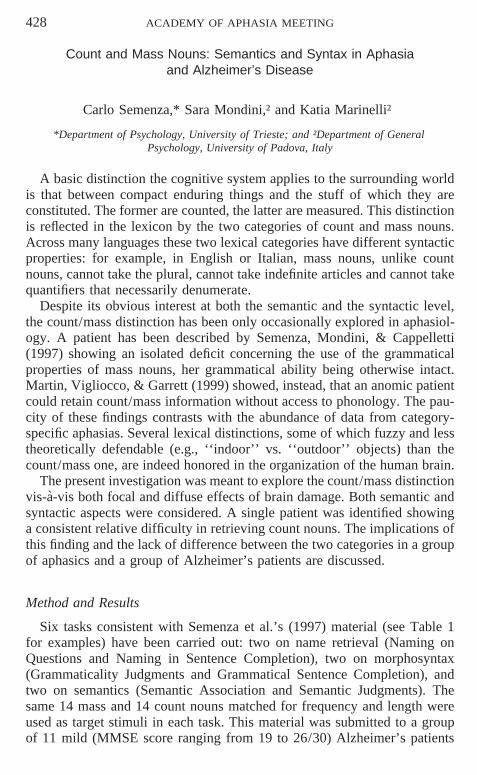
428 ACADEMY OF APHASIA MEETING
Count and Mass Nouns: Semantics and Syntax in Aphasiaand Alzheimer’s Disease
Carlo Semenza,* Sara Mondini,† and Katia Marinelli†
*Department of Psychology, University of Trieste; and †Department of GeneralPsychology, University of Padova, Italy
A basic distinction the cognitive system applies to the surrounding worldis that between compact enduring things and the stuff of which they areconstituted. The former are counted, the latter are measured. This distinctionis reflected in the lexicon by the two categories of count and mass nouns.Across many languages these two lexical categories have different syntacticproperties: for example, in English or Italian, mass nouns, unlike countnouns, cannot take the plural, cannot take indefinite articles and cannot takequantifiers that necessarily denumerate.
Despite its obvious interest at both the semantic and the syntactic level,the count/mass distinction has been only occasionally explored in aphasiol-ogy. A patient has been described by Semenza, Mondini, & Cappelletti(1997) showing an isolated deficit concerning the use of the grammaticalproperties of mass nouns, her grammatical ability being otherwise intact.Martin, Vigliocco, & Garrett (1999) showed, instead, that an anomic patientcould retain count/mass information without access to phonology. The pau-city of these findings contrasts with the abundance of data from category-specific aphasias. Several lexical distinctions, some of which fuzzy and lesstheoretically defendable (e.g., ‘‘indoor’’ vs. ‘‘outdoor’’ objects) than thecount/mass one, are indeed honored in the organization of the human brain.
The present investigation was meant to explore the count/mass distinctionvis-a-vis both focal and diffuse effects of brain damage. Both semantic andsyntactic aspects were considered. A single patient was identified showinga consistent relative difficulty in retrieving count nouns. The implications ofthis finding and the lack of difference between the two categories in a groupof aphasics and a group of Alzheimer’s patients are discussed.
Method and Results
Six tasks consistent with Semenza et al.’s (1997) material (see Table 1for examples) have been carried out: two on name retrieval (Naming onQuestions and Naming in Sentence Completion), two on morphosyntax(Grammaticality Judgments and Grammatical Sentence Completion), andtwo on semantics (Semantic Association and Semantic Judgments). Thesame 14 mass and 14 count nouns matched for frequency and length wereused as target stimuli in each task. This material was submitted to a groupof 11 mild (MMSE score ranging from 19 to 26/30) Alzheimer’s patients
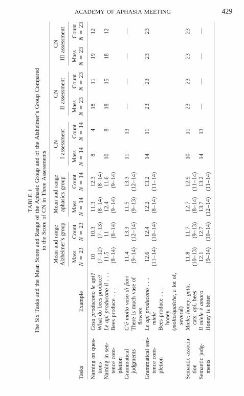
ACADEMY OF APHASIA MEETING 429
TA
BL
E1
The
Six
Tas
ksan
dth
eM
ean
Scor
ean
dR
ange
ofth
eA
phas
icG
roup
and
ofth
eA
lzhe
imer
’sG
roup
Com
pare
dto
the
Scor
eof
CN
inT
hree
Ass
essm
ents
Mea
nan
dra
nge
Mea
nan
dra
nge
CN
CN
CN
Alz
heim
er’s
grou
pap
hasi
csgr
oup
Ias
sess
men
tII
asse
ssm
ent
III
asse
ssm
ent
Mas
sC
ount
Mas
sC
ount
Mas
sC
ount
Mas
sC
ount
Mas
sC
ount
Tas
ksE
xam
ple
N5
23N
523
N5
14N
514
N5
14N
514
N5
23N
523
N5
23N
523
Nam
ing
onqu
es-
Cos
apr
oduc
ono
leap
i?10
10.3
11.3
12.3
84
1811
1912
tions
Wha
tdo
bees
prod
uce?
(7–1
2)(7
–13)
(8–1
4)(8
–14)
Nam
ing
inse
n-L
eap
ipr
oduc
ono
il.
..
11.5
1112
.411
.610
818
1518
12te
nce
com
-B
ees
prod
uce
..
.(8
–14)
(8–1
4)(9
–14)
(9–1
4)pl
etio
nG
ram
mat
ical
C’e
mol
tova
sodi
fiori
11.4
13.3
11.5
13.3
1113
——
——
judg
men
tsT
here
ism
uch
vase
of(9
–14)
(12–
14)
(9–1
3)(1
2–14
)flo
wer
sG
ram
mat
ical
sen-
Le
api
prod
ucon
o.
..
12.6
12.4
12.2
13.2
1411
2323
2323
tenc
eco
m-
mie
le(1
1–14
)(1
0–14
)(8
–14)
(11–
14)
plet
ion
Bee
spr
oduc
e.
..
hone
y(m
olto
/qua
lche
,a
lot
of,
seve
ral)
Sem
antic
asso
cia-
Mie
le,
hone
y;ga
tti,
11.8
11.7
12.7
12.9
1011
2323
2323
tion
cats
;ap
i,be
es(1
0–13
)(9
–13)
(8–1
4)(1
1–14
)Se
man
ticju
dg-
Ilm
iele
eam
aro
12.1
12.7
13.7
13.2
1413
——
——
men
tsH
oney
isbi
tter
(9–1
4)(1
0–14
)(1
2–14
)(1
1–14
)
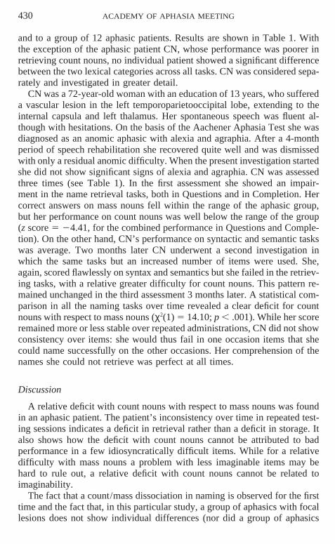
430 ACADEMY OF APHASIA MEETING
and to a group of 12 aphasic patients. Results are shown in Table 1. Withthe exception of the aphasic patient CN, whose performance was poorer inretrieving count nouns, no individual patient showed a significant differencebetween the two lexical categories across all tasks. CN was considered sepa-rately and investigated in greater detail.
CN was a 72-year-old woman with an education of 13 years, who suffereda vascular lesion in the left temporoparietooccipital lobe, extending to theinternal capsula and left thalamus. Her spontaneous speech was fluent al-though with hesitations. On the basis of the Aachener Aphasia Test she wasdiagnosed as an anomic aphasic with alexia and agraphia. After a 4-monthperiod of speech rehabilitation she recovered quite well and was dismissedwith only a residual anomic difficulty. When the present investigation startedshe did not show significant signs of alexia and agraphia. CN was assessedthree times (see Table 1). In the first assessment she showed an impair-ment in the name retrieval tasks, both in Questions and in Completion. Hercorrect answers on mass nouns fell within the range of the aphasic group,but her performance on count nouns was well below the range of the group(z score 5 24.41, for the combined performance in Questions and Comple-tion). On the other hand, CN’s performance on syntactic and semantic taskswas average. Two months later CN underwent a second investigation inwhich the same tasks but an increased number of items were used. She,again, scored flawlessly on syntax and semantics but she failed in the retriev-ing tasks, with a relative greater difficulty for count nouns. This pattern re-mained unchanged in the third assessment 3 months later. A statistical com-parison in all the naming tasks over time revealed a clear deficit for countnouns with respect to mass nouns (χ2(1) 5 14.10; p , .001). While her scoreremained more or less stable over repeated administrations, CN did not showconsistency over items: she would thus fail in one occasion items that shecould name successfully on the other occasions. Her comprehension of thenames she could not retrieve was perfect at all times.
Discussion
A relative deficit with count nouns with respect to mass nouns was foundin an aphasic patient. The patient’s inconsistency over time in repeated test-ing sessions indicates a deficit in retrieval rather than a deficit in storage. Italso shows how the deficit with count nouns cannot be attributed to badperformance in a few idiosyncratically difficult items. While for a relativedifficulty with mass nouns a problem with less imaginable items may behard to rule out, a relative deficit with count nouns cannot be related toimaginability.
The fact that a count/mass dissociation in naming is observed for the firsttime and the fact that, in this particular study, a group of aphasics with focallesions does not show individual differences (nor did a group of aphasics
ACADEMY OF APHASIA MEETING 431
tested in Semenza et al., 1997) may be interpreted as indicating that theanatomical systems responsible for retrieval of count and mass nouns largelyoverlap. The fact that a group of Alzheimer’s patients does not show individ-ual differences seems to suggest that the single dissociation happening inCN is not due to a gradient of difficulty in the experimental material.
REFERENCES
Martin, R. C., Vigliocco, G., & Garrett, M. 1999. Access to count/mass information withoutaccess to phonology in an anomic patient. Brain and Language, 69(3), 426–428.
Semenza, C., Mondini, S., & Cappelletti, M. 1997. The grammatical properties of mass nouns:An aphasia case study. Neuropsychologia, 35(5), 669–675.
Acknowledgment: This study was supported by MRCI Grant 412-95-0006, MURST ’99.