Embed Size (px)
Citation preview
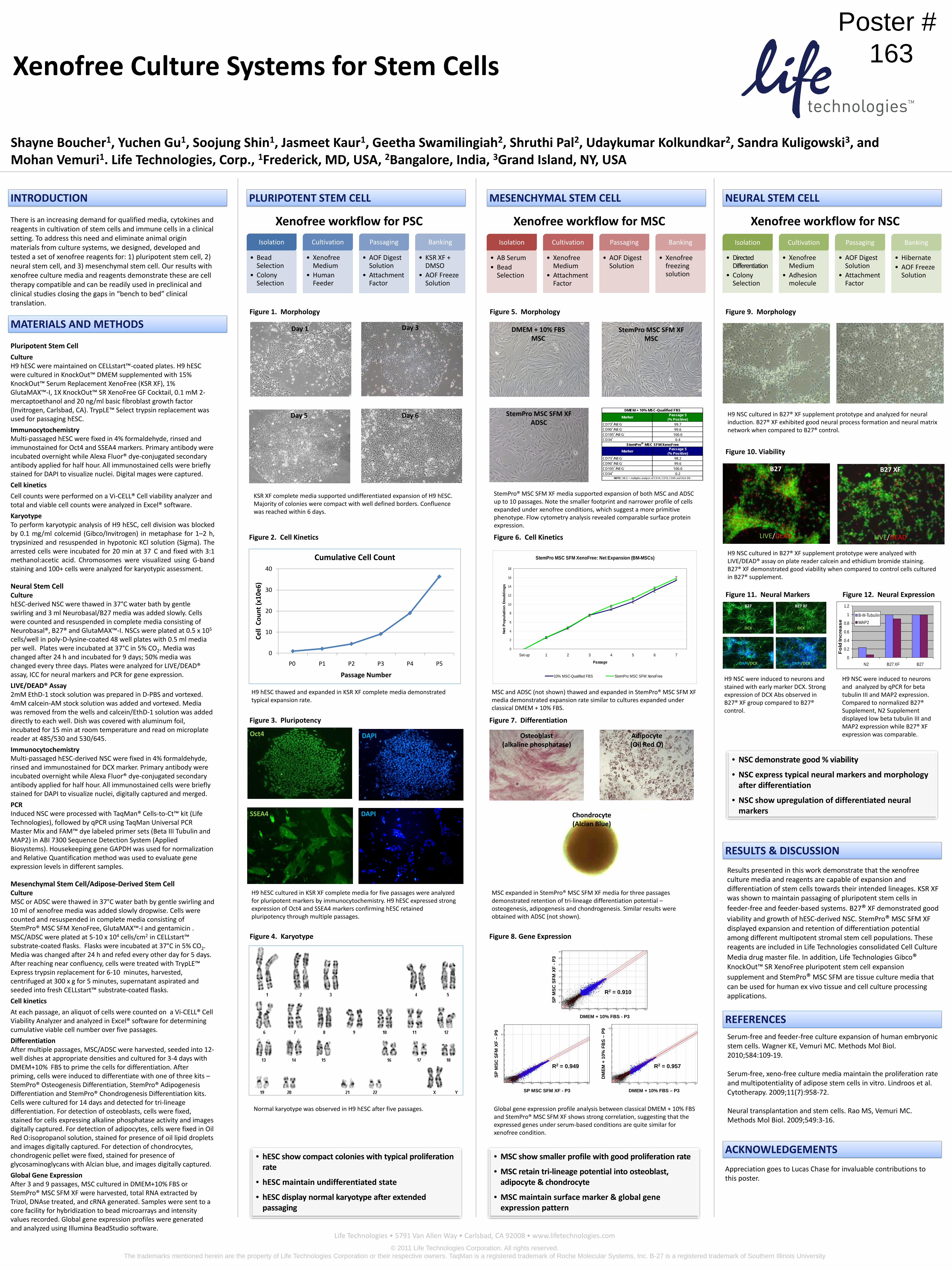
Shayne Boucher1, Yuchen Gu1, Soojung Shin1, Jasmeet Kaur1, Geetha Swamilingiah2, Shruthi Pal2, Udaykumar Kolkundkar2, Sandra Kuligowski3, and Mohan Vemuri1. Life Technologies, Corp., 1Frederick, MD, USA, 2Bangalore, India, 3Grand Island, NY, USA
Figure 1. Morphology
KSR XF complete media supported undifferentiated expansion of H9 hESC. Majority of colonies were compact with well defined borders. Confluence was reached within 6 days.
Figure 2. Cell Kinetics
H9 hESC thawed and expanded in KSR XF complete media demonstrated typical expansion rate.
INTRODUCTION
Results presented in this work demonstrate that the xenofree culture media and reagents are capable of expansion and differentiation of stem cells towards their intended lineages. KSR XF was shown to maintain passaging of pluripotent stem cells in feeder-free and feeder-based systems. B27® XF demonstrated good viability and growth of hESC-derived NSC. StemPro® MSC SFM XF displayed expansion and retention of differentiation potential among different multipotent stromal stem cell populations. These reagents are included in Life Technologies consolidated Cell Culture Media drug master file. In addition, Life Technologies Gibco®KnockOut™ SR XenoFree pluripotent stem cell expansion supplement and StemPro® MSC SFM are tissue culture media that can be used for human ex vivo tissue and cell culture processing applications.
Xenofree Culture Systems for Stem Cells
Life Technologies • 5791 Van Allen Way • Carlsbad, CA 92008 • www.lifetechnologies.com
There is an increasing demand for qualified media, cytokines and reagents in cultivation of stem cells and immune cells in a clinical setting. To address this need and eliminate animal origin materials from culture systems, we designed, developed and tested a set of xenofree reagents for: 1) pluripotent stem cell, 2) neural stem cell, and 3) mesenchymal stem cell. Our results with xenofree culture media and reagents demonstrate these are cell therapy compatible and can be readily used in preclinical and clinical studies closing the gaps in “bench to bed” clinical translation.
MATERIALS AND METHODS
PLURIPOTENT STEM CELL NEURAL STEM CELL
RESULTS & DISCUSSION
REFERENCESSerum-free and feeder-free culture expansion of human embryonic stem cells. Wagner KE, Vemuri MC. Methods Mol Biol. 2010;584:109-19.
Serum-free, xeno-free culture media maintain the proliferation rate and multipotentiality of adipose stem cells in vitro. Lindroos et al. Cytotherapy. 2009;11(7):958-72.
Neural transplantation and stem cells. Rao MS, Vemuri MC. Methods Mol Biol. 2009;549:3-16.
Appreciation goes to Lucas Chase for invaluable contributions to this poster.
ACKNOWLEDGEMENTS
MESENCHYMAL STEM CELL
Pluripotent Stem Cell
CultureH9 hESC were maintained on CELLstart™-coated plates. H9 hESC were cultured in KnockOut™ DMEM supplemented with 15% KnockOut™ Serum Replacement XenoFree (KSR XF), 1% GlutaMAX™-I, 1X KnockOut™ SR XenoFree GF Cocktail, 0.1 mM 2-mercaptoethanol and 20 ng/ml basic fibroblast growth factor (Invitrogen, Carlsbad, CA). TrypLE™ Select trypsin replacement was used for passaging hESC.
ImmunocytochemistryMulti-passaged hESC were fixed in 4% formaldehyde, rinsed and immunostained for Oct4 and SSEA4 markers. Primary antibody were incubated overnight while Alexa Fluor® dye-conjugated secondary antibody applied for half hour. All immunostained cells were briefly stained for DAPI to visualize nuclei. Digital mages were captured.
Cell kinetics
Cell counts were performed on a Vi-CELL® Cell viability analyzer and total and viable cell counts were analyzed in Excel® software.
KaryotypeTo perform karyotypic analysis of H9 hESC, cell division was blockedby 0.1 mg/ml colcemid (Gibco/Invitrogen) in metaphase for 1–2 h,trypsinized and resuspended in hypotonic KCl solution (Sigma). Thearrested cells were incubated for 20 min at 37 C and fixed with 3:1methanol:acetic acid. Chromosomes were visualized using G-bandstaining and 100+ cells were analyzed for karyotypic assessment.
Neural Stem CellCulturehESC-derived NSC were thawed in 37°C water bath by gentle swirling and 3 ml Neurobasal/B27 media was added slowly. Cells were counted and resuspended in complete media consisting of Neurobasal®, B27® and GlutaMAX™-I. NSCs were plated at 0.5 x 10⁵
cells/well in poly-D-lysine-coated 48 well plates with 0.5 ml media per well. Plates were incubated at 37°C in 5% CO2. Media was changed after 24 h and incubated for 9 days; 50% media was changed every three days. Plates were analyzed for LIVE/DEAD® assay, ICC for neural markers and PCR for gene expression.
LIVE/DEAD® Assay2mM EthD-1 stock solution was prepared in D-PBS and vortexed. 4mM calcein-AM stock solution was added and vortexed. Media was removed from the wells and calcein/EthD-1 solution was added directly to each well. Dish was covered with aluminum foil, incubated for 15 min at room temperature and read on microplate reader at 485/530 and 530/645.
ImmunocytochemistryMulti-passaged hESC-derived NSC were fixed in 4% formaldehyde, rinsed and immunostained for DCX marker. Primary antibody were incubated overnight while Alexa Fluor® dye-conjugated secondary antibody applied for half hour. All immunostained cells were briefly stained for DAPI to visualize nuclei, digitally captured and merged.
PCRInduced NSC were processed with TaqMan® Cells-to-Ct™ kit (Life Technologies), followed by qPCR using TaqMan Universal PCR Master Mix and FAM™ dye labeled primer sets (Beta III Tubulin and MAP2) in ABI 7300 Sequence Detection System (Applied Biosystems). Housekeeping gene GAPDH was used for normalization and Relative Quantification method was used to evaluate gene expression levels in different samples.
Normal karyotype was observed in H9 hESC after five passages.
Figure 3. Pluripotency
Figure 4. Karyotype
Xenofree workflow for PSC
Figure 9. Morphology
H9 NSC were induced to neurons and stained with early marker DCX. Strong expression of DCX Abs observed in B27® XF group compared to B27® control.
Figure 11. Neural Markers Figure 12. Neural Expression
Xenofree workflow for NSC
Figure 5. Morphology
StemPro® MSC SFM XF media supported expansion of both MSC and ADSC up to 10 passages. Note the smaller footprint and narrower profile of cells expanded under xenofree conditions, which suggest a more primitive phenotype. Flow cytometry analysis revealed comparable surface protein expression.
Global gene expression profile analysis between classical DMEM + 10% FBS and StemPro® MSC SFM XF shows strong correlation, suggesting that the expressed genes under serum-based conditions are quite similar for xenofree condition.
Isolation
• AB Serum• Bead
Selection
Cultivation
• Xenofree Medium
• Attachment Factor
Passaging
• AOF Digest Solution
Banking
• Xenofree freezing solution
Xenofree workflow for MSC
Day 1
Day 6Day 5
Day 3
B27 B27 XF
DCX
DAPI/DCX
0
10
20
30
40
P0 P1 P2 P3 P4 P5
Cell
Cou
nt (x
10e6
)
Passage Number
Cumulative Cell Count
Isolation
• Bead Selection
• Colony Selection
Cultivation
• Xenofree Medium
• Human Feeder
Passaging
• AOF Digest Solution
• Attachment Factor
Banking
• KSR XF + DMSO
• AOF Freeze Solution
H9 hESC cultured in KSR XF complete media for five passages were analyzed for pluripotent markers by immunocytochemistry. H9 hESC expressed strong expression of Oct4 and SSEA4 markers confirming hESC retained pluripotency through multiple passages.
Oct4 DAPI
SSEA4 DAPI
• hESC show compact colonies with typical proliferation rate
• hESC maintain undifferentiated state
• hESC display normal karyotype after extended passaging
H9 NSC cultured in B27® XF supplement prototype and analyzed for neural induction. B27® XF exhibited good neural process formation and neural matrix network when compared to B27® control.
H9 NSC cultured in B27® XF supplement prototype were analyzed with LIVE/DEAD® assay on plate reader calcein and ethidium bromide staining. B27® XF demonstrated good viability when compared to control cells cultured in B27® supplement.
H9 NSC were induced to neurons and analyzed by qPCR for beta tubulin III and MAP2 expression. Compared to normalized B27® Supplement, N2 Supplement displayed low beta tubulin III and MAP2 expression while B27® XF expression was comparable.
B27 B27 XF
LIVE/DEADLIVE/DEAD
• NSC demonstrate good % viability
• NSC express typical neural markers and morphology after differentiation
• NSC show upregulation of differentiated neural markers
DCX
DAPI/DCX
Isolation
• Directed Differentiation
• Colony Selection
Cultivation
• Xenofree Medium
• Adhesion molecule
Passaging
• AOF Digest Solution
• Attachment Factor
Banking
• Hibernate• AOF Freeze
Solution
Figure 10. Viability
• MSC show smaller profile with good proliferation rate
• MSC retain tri-lineage potential into osteoblast, adipocyte & chondrocyte
• MSC maintain surface marker & global gene expression pattern
Poster # 163
DMEM + 10% FBSMSC
StemPro MSC SFM XFMSC
StemPro MSC SFM XFADSC
Figure 6. Cell Kinetics
StemPro MSC SFM XenoFree: Net Expansion (BM-MSCs)
0
2
4
6
8
10
12
14
16
18
Set-up 1 2 3 4 5 6 7
Passage
Net
Po
pu
lati
on
Do
ub
lin
gs
10% MSC-Qualified FBS StemPro MSC SFM XenoFree
MSC and ADSC (not shown) thawed and expanded in StemPro® MSC SFM XF media demonstrated expansion rate similar to cultures expanded under classical DMEM + 10% FBS.
Figure 7. Differentiation
MSC expanded in StemPro® MSC SFM XF media for three passages demonstrated retention of tri-lineage differentiation potential –osteogenesis, adipogenesis and chondrogenesis. Similar results were obtained with ADSC (not shown).
Osteoblast(alkaline phosphatase)
Adipocyte(Oil Red O)
Chondrocyte(Alcian Blue)
Figure 8. Gene Expression
Marker Passage 5(% Positive)
CD73+/NEG- 99.7CD90+/NEG- 99.6CD105+/NEG- 100.0CD34+ 0.4
Marker Passage 5(% Positive)
CD73+/NEG- 98.2CD90+/NEG- 99.6CD105+/NEG- 100.0CD34+ 0.2
DMEM + 10% MSC-Qualified FBS
NOTE: NEG = multiplex analysis of CD14, CD19, CD45 and HLA-DR.
StemPro MSC SFM XenoFree
R2 = 0.910
DMEM + 10% FBS - P3
SP M
SC S
FM X
F -P
3
R2 = 0.957
DMEM + 10% FBS – P3
DM
EM +
10%
FB
S –
P9
R2 = 0.949
SP MSC SFM XF - P3
SP M
SC S
FM X
F –
P9
Mesenchymal Stem Cell/Adipose-Derived Stem CellCultureMSC or ADSC were thawed in 37°C water bath by gentle swirling and 10 ml of xenofree media was added slowly dropwise. Cells were counted and resuspended in complete media consisting of StemPro® MSC SFM XenoFree, GlutaMAX™-I and gentamicin . MSC/ADSC were plated at 5-10 x 104 cells/cm2 in CELLstart™ substrate-coated flasks. Flasks were incubated at 37°C in 5% CO2. Media was changed after 24 h and refed every other day for 5 days. After reaching near confluency, cells were treated with TrypLE™ Express trypsin replacement for 6-10 minutes, harvested, centrifuged at 300 x g for 5 minutes, supernatant aspirated and seeded into fresh CELLstart™ substrate-coated flasks.
Cell kinetics
At each passage, an aliquot of cells were counted on a Vi-CELL® Cell Viability Analyzer and analyzed in Excel® software for determining cumulative viable cell number over five passages.
DifferentiationAfter multiple passages, MSC/ADSC were harvested, seeded into 12-well dishes at appropriate densities and cultured for 3-4 days with DMEM+10% FBS to prime the cells for differentiation. After priming, cells were induced to differentiate with one of three kits –StemPro® Osteogenesis Differentiation, StemPro® Adipogenesis Differentiation and StemPro® Chondrogenesis Differentiation kits. Cells were cultured for 14 days and detected for tri-lineage differentiation. For detection of osteoblasts, cells were fixed, stained for cells expressing alkaline phosphatase activity and images digitally captured. For detection of adipocytes, cells were fixed in Oil Red O:isopropanol solution, stained for presence of oil lipid droplets and images digitally captured. For detection of chondrocytes, chondrogenic pellet were fixed, stained for presence of glycosaminoglycans with Alcian blue, and images digitally captured.
Global Gene ExpressionAfter 3 and 9 passages, MSC cultured in DMEM+10% FBS or StemPro® MSC SFM XF were harvested, total RNA extracted by Trizol, DNAse treated, and cRNA generated. Samples were sent to a core facility for hybridization to bead microarrays and intensity values recorded. Global gene expression profiles were generated and analyzed using Illumina BeadStudio software.
© 2011 Life Technologies Corporation. All rights reserved. The trademarks mentioned herein are the property of Life Technologies Corporation or their respective owners. TaqMan is a registered trademark of Roche Molecular Systems, Inc. B-27 is a registered trademark of Southern Illinois University
0
0.2
0.4
0.6
0.8
1
1.2
N2 B27 XF B27
Fo
ld In
crea
se
B-III-TubulinMAP2