Embed Size (px)
Citation preview

HAL Id: hal-01136778https://hal.archives-ouvertes.fr/hal-01136778
Submitted on 28 Mar 2015
HAL is a multi-disciplinary open accessarchive for the deposit and dissemination of sci-entific research documents, whether they are pub-lished or not. The documents may come fromteaching and research institutions in France orabroad, or from public or private research centers.
L’archive ouverte pluridisciplinaire HAL, estdestinée au dépôt et à la diffusion de documentsscientifiques de niveau recherche, publiés ou non,émanant des établissements d’enseignement et derecherche français ou étrangers, des laboratoirespublics ou privés.
Postural- and respiratory-related activities of abdominalmuscles during post-exercise hyperventilation
Pascal David, Jérémy Terrien, Michel Petitjean
To cite this version:Pascal David, Jérémy Terrien, Michel Petitjean. Postural- and respiratory-related activities of ab-dominal muscles during post-exercise hyperventilation. Gait and Posture, Elsevier, 2015, 41 (4),pp.899-904. 10.1016/j.gaitpost.2015.03.012. hal-01136778

Accepted Manuscript
Title: Postural- and respiratory-related activities of abdominalmuscles during post-exercise hyperventilation
Author: Pascal David Jeremy Terrien Michel Petitjean
PII: S0966-6362(15)00081-8DOI: http://dx.doi.org/doi:10.1016/j.gaitpost.2015.03.012Reference: GAIPOS 4449
To appear in: Gait & Posture
Received date: 12-11-2014Revised date: 23-2-2015Accepted date: 19-3-2015
Please cite this article as: David P, Terrien J, Petitjean M, Postural- and respiratory-related activities of abdominal muscles during post-exercise hyperventilation, Gait andPosture (2015), http://dx.doi.org/10.1016/j.gaitpost.2015.03.012
This is a PDF file of an unedited manuscript that has been accepted for publication.As a service to our customers we are providing this early version of the manuscript.The manuscript will undergo copyediting, typesetting, and review of the resulting proofbefore it is published in its final form. Please note that during the production processerrors may be discovered which could affect the content, and all legal disclaimers thatapply to the journal pertain.

Page 1 of 25
Accep
ted
Man
uscr
ipt
Postural- and respiratory-related activities
of abdominal muscles during post-exercise hyperventilation
Pascal Davida*
, Jérémy Terrienb and Michel Petitjean
a,c,d
a Université de Versailles Saint-Quentin en Yvelines, Montigny-le-Bretonneux, France
b Service d’électronique, Université de Technologie de Compiègne, Compiègne, France
c Unité 1179 INSERM, Montigny-le-Bretonneux, France
d Service de Physiologie-Explorations Fonctionnelles, Hôpital Ambroise Paré, Groupe
Hospitalier Paris Ile-de-France Ouest, Boulogne-Billancourt, France
* Corresponding author: Pascal David, PhD, Research Associate, Université de Versailles
Saint-Quentin en Yvelines, Montigny le Bretonneux
E-mail address: [email protected] (P. David)
Conflict of interest statement
The authors declare that there is no conflict of interest.
Acknowledgements
We are grateful to the anonymous reviewer from ServiceScape for the English revision of the
manuscript.
Submission type: Original Article (full paper)
Abstract word count: 196
Manuscript word count: 2,993 (excluding figures, tables, reference, and abstract)
Number of tables: 1
Number of figures: 4
*4. Title Page (with authors and addresses)

Page 2 of 25
Accep
ted
Man
uscr
ipt
1 2 3 4 5 6 7 8 9 10 11 12 13 14 15 16 17 18 19 20 21 22 23 24 25 26 27 28 29 30 31 32 33 34 35 36 37 38 39 40 41 42 43 44 45 46 47 48 49 50 51 52 53 54 55 56 57 58 59 60 61 62 63 64 65
1
Postural- and respiratory-related activities
of abdominal muscles during post-exercise hyperventilation
Pascal Davida*
, Jérémy Terrienb and Michel Petitjean
a,c,d
a Université de Versailles Saint-Quentin en Yvelines, Montigny-le-Bretonneux, France
b Service d’électronique, Université de Technologie de Compiègne, Compiègne, France
c Unité 1179 INSERM, Montigny-le-Bretonneux, France
d Service de Physiologie-Explorations Fonctionnelles, Hôpital Ambroise Paré, Groupe
Hospitalier Paris Ile-de-France Ouest, Boulogne-Billancourt, France
* Corresponding author: Pascal David, PhD, Research Associate, Université de Versailles
Saint-Quentin en Yvelines, Montigny le Bretonneux
E-mail address: [email protected] (P. David)
Conflict of interest statement
The authors declare that there is no conflict of interest.
Acknowledgements
We are grateful to the anonymous reviewer from ServiceScape for the English revision of the
manuscript.
Submission type: Original Article (full paper)
Abstract word count: 196
Manuscript word count: 2,993 (excluding figures, tables, reference, and abstract)
Number of tables: 1
Number of figures: 4
Page 3 of 25
Accep
ted
Man
uscr
ipt
1 2 3 4 5 6 7 8 9 10 11 12 13 14 15 16 17 18 19 20 21 22 23 24 25 26 27 28 29 30 31 32 33 34 35 36 37 38 39 40 41 42 43 44 45 46 47 48 49 50 51 52 53 54 55 56 57 58 59 60 61 62 63 64 65
2
Abstract
The present study focuses on the role of superficial abdominal muscles revealed by
electromyographic recordings during the maintenance of a bipedal stance perturbed by post-
exercise hyperventilation. Twelve healthy subjects performed six 30-second postural tests:
one pre-exercise test while breathing quietly, then one test every minute for the five minutes
immediately following a maximum-intensity, incremental cycling exercise test. Displacement
of the centre of pressure in the sagittal plane was monitored over time. Myoelectric activities
of the obliquus externus (OE), obliquus internus (OI) and rectus abdominis (RA) muscles
were recorded by surface electromyography (EMG). Metabolic parameters were measured
with a portable telemetric device. The change in ventilatory drive induced by exercise was
accompanied by a significant increase in both postural sway parameters and EMG activities.
For OE and OI, the increased EMG activities were prominent during expiration, whereas OI
was silent during inspiration. OE and RA were activated during both expiration and
inspiration. It is concluded that the compensation of respiratory disturbances of the erect
posture appears to be less effective when minute ventilation increases. The patterns of muscle
activity suggest that abdominal muscles are controlled differentially and that their functional
coordination is dependent on the respiratory demand.
Keywords: Electromyography; biomechanics; postural control; respiration

Page 4 of 25
Accep
ted
Man
uscr
ipt
1 2 3 4 5 6 7 8 9 10 11 12 13 14 15 16 17 18 19 20 21 22 23 24 25 26 27 28 29 30 31 32 33 34 35 36 37 38 39 40 41 42 43 44 45 46 47 48 49 50 51 52 53 54 55 56 57 58 59 60 61 62 63 64 65
3
1. Introduction
The ability to maintain balance plays a fundamental role in motor programming. In
many physical and sporting activities, this ability is considered as a key factor for supporting
voluntary movement [1]. In addition to gravity and other external forces, internal forces
related to voluntary movement and cardiorespiratory activities may disturb postural stability.
In an upright stance at rest, it is well known that quiet breathing perturbs body balance in the
sagittal and frontal planes, and that the effects of compensatory motions of the trunk and
lower limbs are more pronounced in the sagittal plane [2]. When the respiratory demand
increases, the deeper and faster rib cage movements lead to greater postural sways, which
seem to be more compensated by the postural control system when respiration is under
automatic control, suggesting the existence of functional links between respiration and
postural control centres [2,3].
Abdominal muscles that constitute the anterolateral wall of the abdomen contribute
significantly to postural control [4]. In an upright stance, De Troyer [5] reported tonic
abdominal muscle activity, reflecting the antigravitational function of these muscles. Due to
their anatomical arrangement, the abdominal muscles have distinguishable actions on trunk
movement, and thus, have an important role in the control and movement of the lumbar spine
and pelvis [6]. In addition, abdominal muscles participate in a wide range of postural
adjustments, for instance, they are activated to preserve the whole body balance in response to
external biomechanical perturbations [7]. They can also be activated prior to the onset of
voluntary movements to compensate predictable movement-related postural disturbances [8].
In addition to their contribution to postural activities, abdominal muscles also
contribute to respiration [9]. The myoelectric activities of these muscles are reduced during
quiet breathing because expiration depends primarily on the elastic recoil of the respiratory
system. However, abdominal muscles become increasingly active and show phasic,

Page 5 of 25
Accep
ted
Man
uscr
ipt
1 2 3 4 5 6 7 8 9 10 11 12 13 14 15 16 17 18 19 20 21 22 23 24 25 26 27 28 29 30 31 32 33 34 35 36 37 38 39 40 41 42 43 44 45 46 47 48 49 50 51 52 53 54 55 56 57 58 59 60 61 62 63 64 65
4
expiratory-related activity in parallel with increasing peripheral oxygen demand due to
exercise [10,11]. By their contraction, they contribute to the regulation of the end-expiratory
lung volume and expiratory airflow, and assist inspiration by regulating the length of the
diaphragm [5,12]. Thus, abdominal muscles are generally regarded as the principal muscles of
active expiration.
Several studies have investigated the strategies used by the central nervous system
(CNS) to adequately coordinate the postural and respiratory functions of abdominal muscles
during daily activities [13,14]. From these studies, it seems that: (i) both postural and
respiratory functions can be performed simultaneously; and (ii) the CNS seems to be able to
prioritize one function over the other if necessary. In this context, high-intensity physical
activity represents a physiological challenge for the CNS; it requires rapid adjustments of
respiratory function essential for the maintenance of blood gas homeostasis, as well as the
involvement of the postural control system to compensate for respiratory-related postural
disturbances [2,3]. Accordingly, the aim of this study was to investigate the influence of post-
exercise hyperventilation on the recruitment of the abdominal muscles during standing in
healthy young adults. The increased metabolic and postural sway parameters expected
following a cycling exercise test [15,16] should reveal the abdominal activation strategies
selected by the CNS to appropriately manage postural disturbances and respiratory demand.
2.1 Materials and Methods
2.1 Participants
Twelve healthy university students participated in this study (10 males and 2 females;
mean age: 20.6 ± 1.8 yrs; mean weight: 69.2 ± 9 kg; mean height: 1.7 ± 0.1 m). All subjects
had been fully informed of the experiment’s aims and procedure (in accordance with the
Declaration of Helsinki) and gave their written consent. None of them had any known

Page 6 of 25
Accep
ted
Man
uscr
ipt
1 2 3 4 5 6 7 8 9 10 11 12 13 14 15 16 17 18 19 20 21 22 23 24 25 26 27 28 29 30 31 32 33 34 35 36 37 38 39 40 41 42 43 44 45 46 47 48 49 50 51 52 53 54 55 56 57 58 59 60 61 62 63 64 65
5
diseases or injuries that might have impaired balance or respiration. In order to evaluate the
subjects' pulmonary capacity, routine spirometric tests using a Fleisch pneumotachograph
(Masterlab, Jaeger, France) were performed by the same experimenter. The normality of
parameters such as tidal volume (VT) or vital capacity (VC) was judged according to the
reference values proposed by the European Respiratory Society [17].
2.2 Data collection
A piezoelectric force plate (type 9281, Kistler AG, Winterthur, Switzerland) was used
to continuously record the displacement of the centre of pressure (CoP). Since respiratory
movements mainly concern the sagittal plane, we only considered the anteroposterior
displacement of the CoP (CoPA-P). The force plate signal was sampled at 1,000 Hz using a 12-
bit analogue-to-digital converter (Daq Card AI-16E-4, National Instruments, Austin, TX) and
then stored on a computer for off-line processing.
Electromyographic (EMG) signals were recorded unilaterally from the right obliquus
externus (OE) abdominis, right obliquus internus (OI) abdominis and right rectus abdominis
(RA) muscles in a bipolar mode using two 8-mm-diameter silver chloride surface electrodes
(Beckman, Fullerton, CA). Pairs of electrodes (centre-to-centre spacing: 20 mm) were placed
about 2 cm lateral to the midline and 1 cm above the umbilicus for the RA, 15 cm lateral to
the umbilicus for the OE and 1 cm medial to the anterior superior iliac spine (ASIS) and
beneath a line joining both ASISs for the OI, following SENIAM recommendations [18].
Impedance of the skin-electrode interface was maintained below 5 kΩ. A ground electrode
was placed over the sternum. Surface EMG electrodes were connected to a bipolar multi-
channel amplifier (Gould 6600, Gould Electronics, Cleveland, Ohio), with a bandwidth
frequency ranging from 10 to 500 Hz and the gain adjusted to between 1,000 and 5,000.
Page 7 of 25
Accep
ted
Man
uscr
ipt
1 2 3 4 5 6 7 8 9 10 11 12 13 14 15 16 17 18 19 20 21 22 23 24 25 26 27 28 29 30 31 32 33 34 35 36 37 38 39 40 41 42 43 44 45 46 47 48 49 50 51 52 53 54 55 56 57 58 59 60 61 62 63 64 65
6
Electromyographic signals were sampled at 1,000 Hz using the 12-bit analogue-to-digital
converter described above and then stored on the computer for off-line processing.
Oxygen uptake ( 2OV ) was measured telemetrically using a portable gas analyser
(Cosmed K2, Vacumetrics Inc., Ventura, CA). Minute ventilation ( EV ) was also measured,
along with the tidal volume (VT) and breathing frequency (fR) components. Heart rate (HR)
was obtained from a chest strap (Xtrainer Plus, Polar Electro Oy, Kempele, Finland). The
respiratory signal was obtained by measuring variations in the timing of breaths via a custom-
made connection realized inside the Cosmed K2 portable unit. When the subject expires
through the face mask, the revolutions of the turbine produce a voltage signal. Conversely,
when the subject inspires, the turbine stops and leads to the cancellation of the voltage signal.
The square-wave output signal was transmitted simultaneously with the CoPA-P and EMG
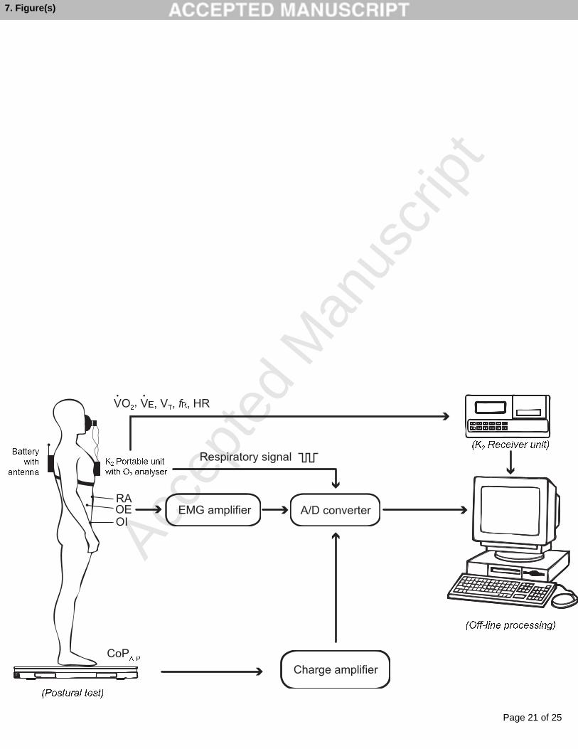
signals to the 12-bit analogue-to-digital converter acquisition system. The experimental set-up
is illustrated in Fig. 1.
2.3 Experimental procedure
Subjects performed a total of 6 postural tests, each corresponding to the maintenance
of a bipedal stance for 30 s with their eyes open. Subjects were asked to stand barefoot on the
force plate, relax with their arms hanging alongside the trunk and focus on a target placed 2 m
in front of the subject at eye level. The foot position was standardized by placing foot prints
on the force plate. The foot prints were drawn so that the respective longitudinal axes formed
a 30° angle and intersected the force plate's anteroposterior axis, with the heels 2 cm apart. A
reference test (pre-effort postural test) was carried out in quiet conditions prior to a maximal
incremental cycling exercise (ICE) test. The exercise testing procedure has been described
previously in detail [16]. Briefly, the exercise test was performed on a friction-loaded cycle
ergometer (Monark 824E, Stockholm, Sweden). The increase in workload was individualized

Page 8 of 25
Accep
ted
Man
uscr
ipt
1 2 3 4 5 6 7 8 9 10 11 12 13 14 15 16 17 18 19 20 21 22 23 24 25 26 27 28 29 30 31 32 33 34 35 36 37 38 39 40 41 42 43 44 45 46 47 48 49 50 51 52 53 54 55 56 57 58 59 60 61 62 63 64 65
7
based on each subject’s predicted maximum oxygen uptake and converted into maximum
watts (Wmax) to make it easier to increase the load during the test. The test involved two
consecutive periods: a 3-min warm-up (with a workload corresponding to 20% of Wmax) and
a 10-min period of incremental exercise from 20 to 100% of Wmax, with an increased
workload stage every minute. Five post-effort postural tests (PEPTs) were then carried out:
immediately after the ICE (PEPT0) and during the first 30 seconds of each of the next 4
minutes (PEPT1 to PEPT4).
2.4 Data analysis
Off-line signal processing was performed in Matlab 6.5 (The Mathworks Inc., Natick
MA). The respiratory and CoPA-P signals were passed through a 4th-order low-pass
Butterworth filter with a cut-off frequency of 15 Hz. Kinematics of the CoPA-P were analyzed
by computing the sway path (SP) and the mean amplitude (∆x). The cardiorespiratory values
were averaged every 5 seconds and stored for further analysis. For each subject, the VT to VC
ratio was used as an index of ventilatory change. The post-processing of raw EMG data
included offset correction and full-wave rectification using an absolute function. Due to the
proximity of the electrode sites to the heart, the three EMG signals were passed through a 4th-
order high-pass Butterworth filter with a cut-off frequency of 30 Hz [19]. During the postural
tests, myoelectrical activities were quantified by computing the root mean square (RMS)
value. The RMS values were then averaged according to the phase of the respiratory cycle
and expressed as percentages of each subject's baseline values acquired during the pre-effort
postural test.

Page 9 of 25
Accep
ted
Man
uscr
ipt
1 2 3 4 5 6 7 8 9 10 11 12 13 14 15 16 17 18 19 20 21 22 23 24 25 26 27 28 29 30 31 32 33 34 35 36 37 38 39 40 41 42 43 44 45 46 47 48 49 50 51 52 53 54 55 56 57 58 59 60 61 62 63 64 65
8
2.5 Statistical analysis
Data distributions were checked for normality using a Kolmogorov-Smirnov test and
for equality of variance using a Fisher-Snedecor test. Non-normally distributed data
underwent log-transformation. A simple analysis of variance with repeated-measures was
performed on the postural test to assess the effects caused by ICE on metabolic ( 2OV , EV ,
VT, VT/VC, fR, HR), electromyographic (OE RMS, OI RMS, RA RMS), and postural sway
(SP, ∆x) parameters. Post hoc testing using the Tukey honestly significant difference test was
used to identify local differences. Differences were considered significant when p < 0.05.
Data are presented as mean ± one standard deviation. All data analyses were performed with
Statview software (SAS institute Inc, Cary, NC).
3. Results
3.1 Effects of ICE on metabolic and postural parameters
The mean maximal power output produced by the subjects at the end of ICE was
249.6 ± 49.9 W. As shown in Table 1, measurements taken at PEPT0, immediately following
ICE, prompted a significant increase in all cardiorespiratory parameters (p < 0.01). Significant
increases in postural sway parameters were also observed (p < 0.05). The analysis of the
CoPA-P signal immediately after ICE showed that CoPA-P displacement was cyclical (Fig. 2).
The simultaneous recording of the mechanical and respiration signals revealed displacements
of CoPA-P backwards during inspiration and correspondingly forwards during expiration. All
cardiorespiratory and postural parameters remained significantly higher (p < 0.05) 4 minutes
after ICE, with the exception of ∆x, which returned to pre-effort values two minutes after
ICE.
3.2 Abdominal muscle activities

Page 10 of 25
Accep
ted
Man
uscr
ipt
1 2 3 4 5 6 7 8 9 10 11 12 13 14 15 16 17 18 19 20 21 22 23 24 25 26 27 28 29 30 31 32 33 34 35 36 37 38 39 40 41 42 43 44 45 46 47 48 49 50 51 52 53 54 55 56 57 58 59 60 61 62 63 64 65
9
In comparison to reference values, ICE led to a significant (p < 0.01) increase in OE
(+249%), OI (+116%) and RA (+183%) RMS values. All myoelectric activities remained
significantly higher (p < 0.01) 4 minutes after ICE, with the exception of OI, which returned
to pre-effort values one minute after ICE (Fig. 3A).
Although no phasic abdominal EMG activity was observed during the pre-effort
postural test (Fig. 4A), the activity of these muscles recorded immediately after ICE clearly
displayed phasic bursts, especially with OE and OI. A typical time course of respiration and
abdominal EMG recordings is illustrated in Fig. 4B. From the mean patterns of EMG activity
(calculated across all analogous respiratory cycles), the results showed that phasic bursts of
OA and OI EMG activities occurred simultaneously with expiration (Fig. 2). From the whole
group, quantification of EMG activities within the respiratory cycle revealed a significant (p <
0.05) increase in RMS values for all three abdominal muscles during expiration (Fig. 3B). For
OE and RA, the results also showed a significant (p < 0.05) increase in RMS values during
inspiration, yet these increases were less pronounced, especially for OE (p < 0.05).
4. Discussion
The results show that post-exercise hyperventilation in standing posture involves: (i) a
periodic movement of the CoP that is time-locked to respiration; and (ii) different abdominal
muscle activation strategies.
Abdominal muscles are thought to contribute to respiration, particularly if the oxygen
demand is increased by exercise. Indeed, previous studies have reported respiratory-related
abdominal muscle activity thresholds for minute ventilation equal to or greater than 40 L/min
[10,11]. Persistent EMG activity in abdominal muscles throughout the post-exercise recovery
for minute ventilation values above 40 L/min suggests that these muscles played an important
role in respiration. Although pulmonary ventilation remained significantly higher one minute

Page 11 of 25
Accep
ted
Man
uscr
ipt
1 2 3 4 5 6 7 8 9 10 11 12 13 14 15 16 17 18 19 20 21 22 23 24 25 26 27 28 29 30 31 32 33 34 35 36 37 38 39 40 41 42 43 44 45 46 47 48 49 50 51 52 53 54 55 56 57 58 59 60 61 62 63 64 65
10
after ICE (60-90 litres/min), the EMG activity of OI returned to pre-effort values. Thus, our
results revealed differential activity and presumably different physiological roles of
abdominal muscles during standing, as well as during respiratory muscle mass derecruitment
following the onset of the post-exercise recovery. Synchronization of abdominal muscle
activities with expiration leaves no doubt about the expiratory contribution of these muscles,
especially OE and OI. These results are in good agreement with those obtained in previous
work [12], where increases in intra-abdominal and intra-thoracic pressures at the end of
expiration during heavy exercises were reported, suggesting the stiffening of the abdomen by
abdominal muscle contraction.
The comparison of muscle activation patterns between abdominal muscles showed
that only OE and RA were activated during inspiration. Several possible explanations could
account for the selective activation of these muscles during inspiration. During high-intensity
physical activity, the OE and RA muscles could be stretched with the greater excursion of the
rib cage motion as attested by the backward CoP displacement and value of VT/VC measured
in this study, resulting in a muscle stretch reflex. Therefore, the activity of these muscles
would increase during inspiration, presumably to maintain upright posture as suggested
previously by Beith and Harrison [20]. Another possible explanation involves the gradual,
controlled relaxation of the abdominal muscles during inspiration, known to prevent rib cage
distortion and to assist diaphragmatic function by providing agonist-antagonist muscles
cooperation [21]. Lastly, the increase in RA myoelectric activity observed during both
inspiration and expiration could express a specific postural strategy, intended to enhance the
stiffness and stability of the trunk by increasing intra-abdominal pressure [22]. The levels of
activity in the OE and RA muscles, which remained significantly higher 4 minutes after ICE
despite minute ventilation values below 40 L/min, suggest that their contribution was
dedicated to postural regulation rather than respiratory supply.

Page 12 of 25
Accep
ted
Man
uscr
ipt
1 2 3 4 5 6 7 8 9 10 11 12 13 14 15 16 17 18 19 20 21 22 23 24 25 26 27 28 29 30 31 32 33 34 35 36 37 38 39 40 41 42 43 44 45 46 47 48 49 50 51 52 53 54 55 56 57 58 59 60 61 62 63 64 65
11
In healthy upright subjects at rest, the respiratory-related postural disturbances are
compensated by motion of the trunk and lower limbs that are phase-locked to respiration [2].
However, the degree to which the respiratory disturbances are compensated has been shown
to depend on various factors, including mobility of the postural chain [23] and low back pain
[24]. Our results show that the degree of compensation also depends on the respiratory
demand. Indeed, standing subjects displayed CoPA-P displacements of greater amplitude,
backwards during inspiration and correspondingly forwards during expiration, suggesting a
reduced postural compensation for respiration when breathing at high levels of minute
ventilation. These results are consistent with previous studies that report a higher coherence
between respiratory movement and CoP displacement with increased respiration [2,16]. It this
context, it was suggested that the lumbar spine and pelvis (for which the abdominal muscles
play an important role in their control and movement [6]) are considered as a key factor for
compensating for respiratory disturbances [2,23]. Thus, a decreased postural compensation to
respiration could be due to changes affecting postural and respiratory activation of the
abdominal muscles. Previous studies have shown dual modulation of trunk muscle activity
with regard to respiration and posture [14,25]. Based on these studies, a lower contribution to
the maintenance of posture by the OE and OI muscles immediately after the cessation of
cycling exercise could account for a reduced postural compensation for respiration [2], and
thus the greater postural oscillations measured in the present study.
The posturographic and EMG examinations associated with breathing pattern analysis
reflect the complex and multivariate strategies used by the CNS to adequately coordinate the
postural and respiratory functions of the abdominal muscles. As evidenced by the patterns of
muscle activity, the present data suggest that: (i) both postural and respiratory functions can
be performed simultaneously; and (ii) the CNS seems able to prioritize one function over the
other when respiration increases, suggesting a selective coordination between the rhythmical

Page 13 of 25
Accep
ted
Man
uscr
ipt
1 2 3 4 5 6 7 8 9 10 11 12 13 14 15 16 17 18 19 20 21 22 23 24 25 26 27 28 29 30 31 32 33 34 35 36 37 38 39 40 41 42 43 44 45 46 47 48 49 50 51 52 53 54 55 56 57 58 59 60 61 62 63 64 65
12
respiratory drive and the postural drive to the abdominal muscles. This supports the
observations reported by Hodges et al. [25], Saunders et al. [14] and more recently by David
et al. [3], which showed that different respiratory drives impact postural control differently, at
least at the ankle level. Otherwise, if our results reinforce the existence of functional links
between respiration and postural control centres, the strategies used by the CNS could also be
influenced by the state of functional systems involved in the movement [26]. Reduced
posturo-kinetic capacity, through physiological limitations of the coordination of the postural
and respiratory functions of the trunk muscles [27], would explain the impaired mechanism of
compensation for increased respiration in low back pain subjects [24]. In our study, all
subjects succeeded in maintaining an upright posture despite minute ventilation values of up
to 100 L/min. Their experience in various sports activities, reflecting not only greater
neuromuscular control of trunk muscles [28], but also a greater ability to maintain and restore
balance in challenging conditions [29], had probably improved the ability of their CNS to
adequately manage respiratory drive and postural control. Insofar as effects of respiration on
postural sway increase with age and disease [30], the present results have clinical
implications, suggesting that interventions such as abdominal wall muscle training may
increase compensatory abilities in posturally unstable patients. Moreover, rehabilitation
programs aimed at improving the motor control of trunk stability must be coordinated with
hyperventilation challenges.
In conclusion, the compensation of periodic respiratory disturbance to posture is less
effective when the respiratory demand increases. The patterns of muscle activity suggest that
abdominal muscles are controlled differentially and that their functional coordination is
dependent on the level of ventilation, reinforcing the existence of functional links between
respiratory centres and the postural control system. The present study was focused on young
healthy adults. Changes in respiratory and postural conditions tested on various populations

Page 14 of 25
Accep
ted
Man
uscr
ipt
1 2 3 4 5 6 7 8 9 10 11 12 13 14 15 16 17 18 19 20 21 22 23 24 25 26 27 28 29 30 31 32 33 34 35 36 37 38 39 40 41 42 43 44 45 46 47 48 49 50 51 52 53 54 55 56 57 58 59 60 61 62 63 64 65
13
for which: (i) the neuromuscular system’s functional status is impaired; or (ii) respiratory
resistance or postural instability are increased, would allow a better understanding of the
coordination between the respiratory and postural components of the abdominal muscles.
References
[1] Bouisset S, Do MC. Posture, dynamic stability, and voluntary movement. Clin
Neurophysiol 2008;38:345-62.
[2] Hodges PW, Gurfinkel VS, Brumagne S, Smith TC, Cordo PC. Coexistence of stability
and mobility in postural control: evidence from postural compensation for respiration. Exp
Brain Res 2002;144:293-302.
[3] David P, Laval D, Terrien J, Petitjean M. Postural control and ventilatory drive during
voluntary hyperventilation and carbon dioxide rebreathing. Eur J Appl Physiol 2012;112:145-
54.
[4] Urquhart DM, Hodges PW, Story IH. Postural activity of the abdominal muscles varies
between regions of these muscles and between body positions. Gait Posture 2005;22:295-301.
[5] De Troyer A. Mechanical role of the abdominal muscles in relation to posture. Respir
Physiol 1983;53:341-53.
[6] Hodges P. Abdominal mechanism and support of the lumbar spine and pelvis. In:
Therapeutic Exercise for Lumbopelvic Stabilization, Edinburgh: Churchill Livingstone; 2004,
p. 31-57.
[7] Horak FB, Nashner LM. Central programming of postural movements: adaptation to
altered support-surface configurations. J Neurophysiol 1986;55:1369-81.
[8] Hodges PW, Richardson CA. Contraction of the abdominal muscles associated with
movement of the lower limb. Phys Ther 1997;77:132-42.

Page 15 of 25
Accep
ted
Man
uscr
ipt
1 2 3 4 5 6 7 8 9 10 11 12 13 14 15 16 17 18 19 20 21 22 23 24 25 26 27 28 29 30 31 32 33 34 35 36 37 38 39 40 41 42 43 44 45 46 47 48 49 50 51 52 53 54 55 56 57 58 59 60 61 62 63 64 65
14
[9] De Troyer A, Loring SH. Action of the respiratory muscles. In: Macklem PT, Mead J,
editors. Handbook of Physiology: The Respiratory System, Bethesda: American Physiological
Society; 1986, p. 443-61.
[10] Campbell EJ, Green JH. The behaviour of the abdominal muscles and the intra-
abdominal pressure during quiet breathing and increased pulmonary ventilation; a study in
man. J Physiol 1955;127:423-6.
[11] Abraham KA, Feingold H, Fuller DD, Jenkins M, Mateika JH, Fregosi RF. Respiratory-
related activation of human abdominal muscles during exercise. J Physiol 2002;541:653-63.
[12] Henke KG, Sharratt M, Pegelow D, Dempsey JA. Regulation of end-expiratory lung
volume during exercise. J Appl Physiol (1985) 1988;64:135-46.
[13] Hodges P, Saunders S. Coordination of the respiratory and locomotor activities of the
abdominal muscles during walking in humans. In: Proceedings of International Union of
Physiological Sciences; 2001.
[14] Saunders SW, Rath D, Hodges PW. Postural and respiratory activation of the trunk
muscles changes with mode and speed of locomotion. Gait Posture 2004;20:280-90.
[15] Vuillerme N, Hintzy F. Effects of a 200 W-15 min cycling exercise on postural control
during quiet standing in healthy young adults. Eur J Appl Physiol 2007;100:169-75.
[16] David P, Mora I, Terrien J, Lelard T, Petitjean M. Leg muscles activities during
hyperventilation following a cycling exercise. Electromyogr Clin Neurophysiol 2010;50:39-
45.
[17] European Respiratory Society. Volumes pulmonaires et débits ventilatoires forcés. Rev
Mal Respir 2001;18:S13-52.
[18] Hermens HJ, Freriks B, Disselhorst-Klug C, Rau G. Development of recommendations
for SEMG sensors and sensor placement procedures. J Electromyogr Kinesiol 2000;10:361-
74.

Page 16 of 25
Accep
ted
Man
uscr
ipt
1 2 3 4 5 6 7 8 9 10 11 12 13 14 15 16 17 18 19 20 21 22 23 24 25 26 27 28 29 30 31 32 33 34 35 36 37 38 39 40 41 42 43 44 45 46 47 48 49 50 51 52 53 54 55 56 57 58 59 60 61 62 63 64 65
15
[19] Drake JD, Callaghan JP. Elimination of electrocardiogram contamination from
electromyogram signals: An evaluation of currently used removal techniques. J Electromyogr
Kinesiol 2006;16:175-87.
[20] Beith ID, Harrison PJ. Stretch reflexes in human abdominal muscles. Exp Brain Res
2004;159:206-13.
[21] Kenyon CM, Cala SJ, Yan S, Aliverti A, Scano G, Duranti R, et al. Rib cage mechanics
during quiet breathing and exercise in humans. J Appl Physiol (1985) 1997;83:1242-55.
[22] Hodges PW, Cresswell AG, Thorstensson A. Intra-abdominal pressure response to
multidirectional support-surface translation. Gait Posture 2004;20:163-70.
[23] Kantor E, Poupard L, Le Bozec S, Bouisset S. Does body stability depend on postural
chain mobility or stability area? Neurosci Lett 2001;308:128-32.
[24] Hamaoui A, Do Mc, Poupard L, Bouisset S. Does respiration perturb body balance more
in chronic low back pain subjects than in healthy subjects? Clin Biomech (Bristol, Avon)
2002;17:548-50.
[25] Hodges PW, Heijnen I, Gandevia SC. Postural activity of the diaphragm is reduced in
humans when respiratory demand increases. J Physiol 2001;537:999-1008.
[26] Bouisset S, Le Bozec S. Posturo-kinetic capacity and postural function in voluntary
movements. In: Latash ML, editor. Progress in motor control, vol 2: Structure-function
relations in voluntary movements, Pennstate: Human kinetics; 2002, p.25-52.
[27] Smith MD, Russell A, Hodges PW. Disorders of breathing and continence have a
stronger association with back pain than obesity and physical activity. Aust J Physiother
2006;52:11-6.
[28] David P, Mora I, Pérot C. Neuromuscular efficiency of the rectus abdominis differs with
gender and sport practice. J Strength Cond Res 2008;22:1855-61.
Page 17 of 25
Accep
ted
Man
uscr
ipt
1 2 3 4 5 6 7 8 9 10 11 12 13 14 15 16 17 18 19 20 21 22 23 24 25 26 27 28 29 30 31 32 33 34 35 36 37 38 39 40 41 42 43 44 45 46 47 48 49 50 51 52 53 54 55 56 57 58 59 60 61 62 63 64 65
16
[29] Mouchnino L, Aurenty R, Massion J, Pedotti A. Coordination between equilibrium and
head-trunk orientation during leg movement: a new strategy build up by training. J
Neurophysiol 1992;67:1587-98.
[30] Manor BD, Hu K, Peng CK, Lipsitz LA, Novak V. Posturo-respiratory synchronization:
effects of aging and stroke. Gait Posture 2012;36:254-9.
Page 18 of 25
Accep
ted
Man
uscr
ipt
1 2 3 4 5 6 7 8 9 10 11 12 13 14 15 16 17 18 19 20 21 22 23 24 25 26 27 28 29 30 31 32 33 34 35 36 37 38 39 40 41 42 43 44 45 46 47 48 49 50 51 52 53 54 55 56 57 58 59 60 61 62 63 64 65
17
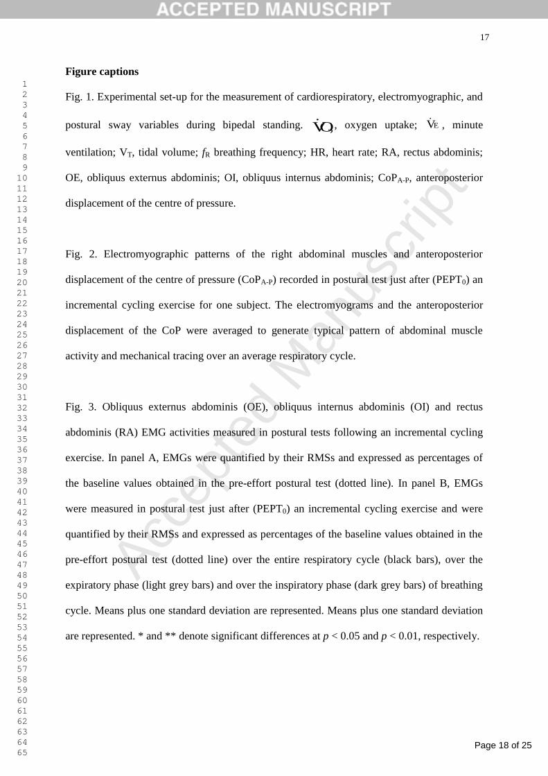
Figure captions
Fig. 1. Experimental set-up for the measurement of cardiorespiratory, electromyographic, and
postural sway variables during bipedal standing. 2OV , oxygen uptake; EV , minute
ventilation; VT, tidal volume; fR breathing frequency; HR, heart rate; RA, rectus abdominis;
OE, obliquus externus abdominis; OI, obliquus internus abdominis; CoPA-P, anteroposterior
displacement of the centre of pressure.
Fig. 2. Electromyographic patterns of the right abdominal muscles and anteroposterior
displacement of the centre of pressure (CoPA-P) recorded in postural test just after (PEPT0) an
incremental cycling exercise for one subject. The electromyograms and the anteroposterior
displacement of the CoP were averaged to generate typical pattern of abdominal muscle
activity and mechanical tracing over an average respiratory cycle.
Fig. 3. Obliquus externus abdominis (OE), obliquus internus abdominis (OI) and rectus
abdominis (RA) EMG activities measured in postural tests following an incremental cycling
exercise. In panel A, EMGs were quantified by their RMSs and expressed as percentages of
the baseline values obtained in the pre-effort postural test (dotted line). In panel B, EMGs
were measured in postural test just after (PEPT0) an incremental cycling exercise and were
quantified by their RMSs and expressed as percentages of the baseline values obtained in the
pre-effort postural test (dotted line) over the entire respiratory cycle (black bars), over the
expiratory phase (light grey bars) and over the inspiratory phase (dark grey bars) of breathing
cycle. Means plus one standard deviation are represented. Means plus one standard deviation
are represented. * and ** denote significant differences at p < 0.05 and p < 0.01, respectively.
Page 19 of 25
Accep
ted
Man
uscr
ipt
1 2 3 4 5 6 7 8 9 10 11 12 13 14 15 16 17 18 19 20 21 22 23 24 25 26 27 28 29 30 31 32 33 34 35 36 37 38 39 40 41 42 43 44 45 46 47 48 49 50 51 52 53 54 55 56 57 58 59 60 61 62 63 64 65
18
Fig. 4. Simultaneous recordings of respiration (inspiratory phase: low-magnitude pulse signal)
and raw electromyograms of the right abdominal muscles in postural test before (panel A) and
just after (panel B) an incremental cycling exercise for one subject.

Page 20 of 25
Accep
ted
Man
uscr
ipt
Tables
Table 1. Cardiorespiratory ( 2OV , EV , VT, VT/VC, fR, HR) and postural sway (SP, ∆x)
parameters measured in postural tests before (pre-effort test), just after (PEPT0) and over the
next 4 minutes following (PEPT1 to PEPT4) an incremental cycling exercise (mean ± standard
deviation). * and ** denotes significant differences between the pre-effort postural test and
the post-effort postural test at p < 0.05 and p < 0.01, respectively.
Pre-effort test PEPT0 PEPT1 PEPT2 PEPT3 PEPT4
2OV (LSTPD.min-1
) 0.36 ± 0.05 3.43 ± 0.70** 2.35 ± 0.41** 1.19 ± 0.2 ** 0.77 ± 0.14** 0.66 ± 0.16**
EV (LBTPS.min-1
) 12.8 ± 1.4 102.8 ± 19.1** 71.0 ± 11.2** 47.4 ± 8.5** 33.9 ± 4.7** 28.4 ± 5.5**
VT (LBTPS) 0.75 ± 0.10 2.70 ± 0.65** 2.40 ± 0.61** 1.95 ± 0.43** 1.47 ± 0.28** 1.28 ± 0.30**
VT/VC (%) 14.5 ± 1.9 51.5 ± 9.0** 45.6 ± 8.4** 37 ± 5** 27.9 ± 3.4** 24.3 ± 4.4**
fR (Hz) 0.29 ± 0.05 0.66 ± 0.19** 0.52 ± 0.15** 0.43 ± 0.15** 0.4 ± 0.07** 0.38 ± 0.08**
HR (b.min-1
) 83.9 ± 10.5 186.2 ± 9.8** 165.6 ± 13.5** 140.7 ± 14.8** 128.5 ± 15.8** 123.6 ± 15.2**
SP (mm) 164.1 ± 30.7 365.8 ± 104.9** 256.5 ± 76.2** 222.1 ± 63.4** 202.5 ± 46.4* 205. 6 ± 53.8*
∆x (mm) 3.4 ± 0.8 5.9 ± 1.6** 4.5 ± 0.9** 4.0 ± 0.8 3.8 ± 1.0 4.0 ± 1.3
6. Table(s)
Page 21 of 25
Accep
ted
Man
uscr
ipt
AP (Offline processing)(Postural test)
(K2 Receiver unit)K2 Portable unitwith O2 analyser2 E T RBatterywithantenna
. .
7. Figure(s)

Page 22 of 25
Accep
ted
Man
uscr
ipt
7. Figure(s)

Page 23 of 25
Accep
ted
Man
uscr
ipt
0 1 2 3 4
**
**
** ** ** **
**
**
***
*
**
**
**
*
*
7. Figure(s)

Page 24 of 25
Accep
ted
Man
uscr
ipt
μ
μ
μ
7. Figure(s)
Page 25 of 25
Accep
ted
Man
uscr
ipt
Highlights
Post-exercise hyperventilation involves periodic movement of the CoP in phase with
respiration.
EMG activities of OE and OI were prominent during expiration.
OI was silent during inspiration.
OE and RA were activated during both expiration and inspiration.
Functional coordination of abdominal muscles is dependent on the level of ventilation.
*Research Highligts