Embed Size (px)
Citation preview
RESEARCH ARTICLE Open Access
Potential of bacteriophages as disinfectantsto control of Staphylococcus aureus biofilmsJun Song†, Hongri Ruan†, Li Chen, Yuqi Jin, Jiasan Zheng, Rui Wu* and Dongbo Sun*
Abstract
Background: Staphylococcus aureus is the causative agent of chronic mastitis, and can form a biofilm that is difficultto completely remove once formed. Disinfectants are effective against S. aureus, but their activity is easily affected byenvironmental factors and they are corrosive to equipment and chemically toxic to livestock and humans. Therefore,we investigated the potential utility of a bacteriophage as a narrow-spectrum disinfectant against biofilms formed by S.aureus. In this study, we isolated and characterized bacteriophage vB_SauM_SDQ (abbreviated to SDQ) to determine itsefficacy in removing S. aureus biofilms.
Results: SDQ belongs to the family Myoviridae and consists of a hexagonal head, long neck, and short tail. This phage cansterilize a 109 CFU/mL culture of S. aureus in 12 h and multiply itself 1000-fold in that time. Biofilms formed on polystyrene,milk, and mammary-gland tissue were significantly reduced after SDQ treatment. Fluorescence microscopy and scanningelectron microscopy showed that SDQ destroyed the biofilm structure. Moreover, the titer of SDQ remained relatively highafter the lysis of the bacteria and the removal of the biofilm, exerting a continuous bacteriostatic effect. SDQ also retained itsfull activity under conditions that mimic common environments, i.e., in the presence of nonionic detergents, tap water, ororganic materials. A nonionic detergent (Triton X-100) enhanced the removal of biofilm by SDQ.
Conclusions: Our results suggest that SDQ, a specific lytic S. aureus phage, can be used to control biofilm infections. SDQmaintains its full activity in the presence of nonionic detergents, tap water, metal chelators, and organic materials, and canbe used in combination with detergents. We propose this phage as a narrow-spectrum disinfectant against S. aureus, toaugment or supplement the use of broad-spectrum disinfectants in the prevention and control of the mastitis and dairyindustry contamination caused by S. aureus.
Keywords: Bacteriophage, Staphylococcus aureus, Biofilm, Disinfectant
BackgroundMastitis, a persistent inflammatory reaction in the uddertissue of dairy cattle, is one of the most important dis-eases throughout the world [1]. Mastitis not only affectsthe health of milk-producing animals, with severe finan-cial losses for dairy farmers, but also affects animal wel-fare [2]. Bovine intramammary infections can be causedby a wide variety of bacteria, fungi, and mycoplasmas [3,
4]. Staphylococcus aureus is one of the major pathogenscausing bovine mastitis, and seriously affects the healthand milk quality of dairy cows [5]. Biofilms play an im-portant role in the spread of S. aureus and the persist-ence of infections [6, 7]. A biofilm is a reticulatedpolymeric matrix produced by bacteria that can wrapthe bacterial cells, and greatly enhances the bacterium’sresistance to the external environment. Several studieshave shown that biofilm bacteria are up to 1000-timesmore resistant to antibiotics than planktonic bacteria [8].Therefore, it is necessary to prevent and control both S.aureus colonization and biofilm formation.
© The Author(s). 2021 Open Access This article is licensed under a Creative Commons Attribution 4.0 International License,which permits use, sharing, adaptation, distribution and reproduction in any medium or format, as long as you giveappropriate credit to the original author(s) and the source, provide a link to the Creative Commons licence, and indicate ifchanges were made. The images or other third party material in this article are included in the article's Creative Commonslicence, unless indicated otherwise in a credit line to the material. If material is not included in the article's Creative Commonslicence and your intended use is not permitted by statutory regulation or exceeds the permitted use, you will need to obtainpermission directly from the copyright holder. To view a copy of this licence, visit http://creativecommons.org/licenses/by/4.0/.The Creative Commons Public Domain Dedication waiver (http://creativecommons.org/publicdomain/zero/1.0/) applies to thedata made available in this article, unless otherwise stated in a credit line to the data.
* Correspondence: [email protected]; [email protected]†Jun Song and Hongri Ruan contributed equally to this work.Heilongjiang Provincial Key Laboratory of Prevention and Control of BovineDiseases, College of Animal Science and Veterinary Medicine, HeilongjiangBayi Agricultural University, No. 5 Xinfeng Road, Daqing 163319, P. R. China
Song et al. BMC Microbiology (2021) 21:57 https://doi.org/10.1186/s12866-021-02117-1
Bovine mastitis prevention strategies, including goodinfection control and hygiene practices, vaccination, theculling of infected cows, and the cleaning of equipment,reduce the risk of bacterial infection and transmission[9]. It is extremely important to disinfect and sanitizedairy farms. In general, the commonly used broad-spectrum disinfectants belong to chemical categoriesthat have been shown to be flammable, light sensitive,corrosive to metals, irritating, carcinogenic, and/or toxicto livestock and humans [10, 11]. Importantly, severalstudies have shown that chemical disinfectants can selectfor mutant bacteria, increasing the risk of the emergenceof resistant strains [12, 13]. Tap water, organic materials,or detergents can also reduce the efficacy of chemicaldisinfectants. Therefore, a new disinfectant is requiredthat can be used in combination with detergents with nodiminution of its effectiveness.Since their discovery in 1915, bacteriophages have
been used extensively in human and veterinary medicine,the food industry, and various agricultural settings. Theyare mainly used to control common bacterial contami-nants, such as Staphylococcus aureus, Pseudomonas aer-uginosa, and Listeria monocytogenes [14].. As biocontrolagents, phages have the following advantages over con-ventional antibiotics: high specificity, self-replication,self-limitation, continuous adaption to altered host sys-tems, low inherent toxicity, and easy isolation. Phageshave also been shown to effectively remove bacterial bio-films, including those of S. aureus, Streptococcus agalac-tiae, Escherichia coli, and other major pathogens thatcause dairy cow mastitis [15–17]. In this study, a biofilmmodel was established in mammary-gland tissue to
evaluate the removal of biofilms by SDQ [18]. We alsoinvestigated the stability and bactericidal activity of SDQin detergents and other agents. The objective of thisstudy were to investigate the potential utility of SDQ asa disinfectant to help control the mastitis and food con-tamination caused by S. aureus, to lay the foundation forthe development of phage-based narrow-spectrum bio-logical disinfectants.
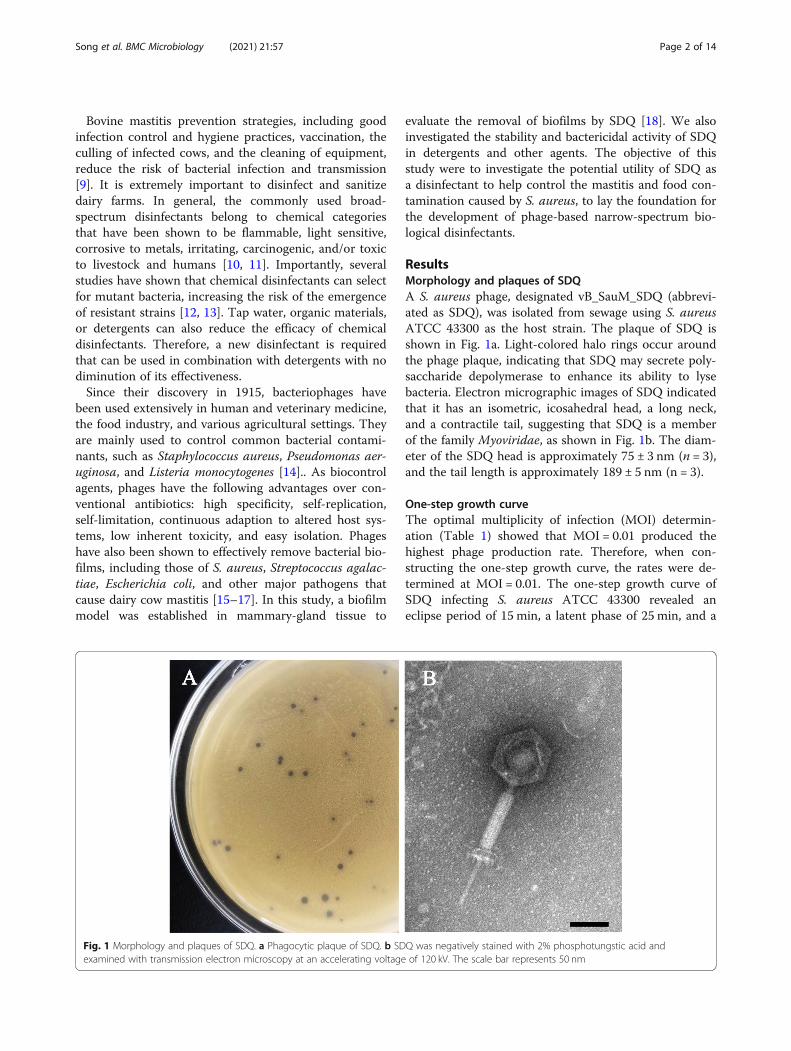
ResultsMorphology and plaques of SDQA S. aureus phage, designated vB_SauM_SDQ (abbrevi-ated as SDQ), was isolated from sewage using S. aureusATCC 43300 as the host strain. The plaque of SDQ isshown in Fig. 1a. Light-colored halo rings occur aroundthe phage plaque, indicating that SDQ may secrete poly-saccharide depolymerase to enhance its ability to lysebacteria. Electron micrographic images of SDQ indicatedthat it has an isometric, icosahedral head, a long neck,and a contractile tail, suggesting that SDQ is a memberof the family Myoviridae, as shown in Fig. 1b. The diam-eter of the SDQ head is approximately 75 ± 3 nm (n = 3),and the tail length is approximately 189 ± 5 nm (n = 3).
One-step growth curveThe optimal multiplicity of infection (MOI) determin-ation (Table 1) showed that MOI = 0.01 produced thehighest phage production rate. Therefore, when con-structing the one-step growth curve, the rates were de-termined at MOI = 0.01. The one-step growth curve ofSDQ infecting S. aureus ATCC 43300 revealed aneclipse period of 15 min, a latent phase of 25 min, and a
Fig. 1 Morphology and plaques of SDQ. a Phagocytic plaque of SDQ. b SDQ was negatively stained with 2% phosphotungstic acid andexamined with transmission electron microscopy at an accelerating voltage of 120 kV. The scale bar represents 50 nm
Song et al. BMC Microbiology (2021) 21:57 Page 2 of 14
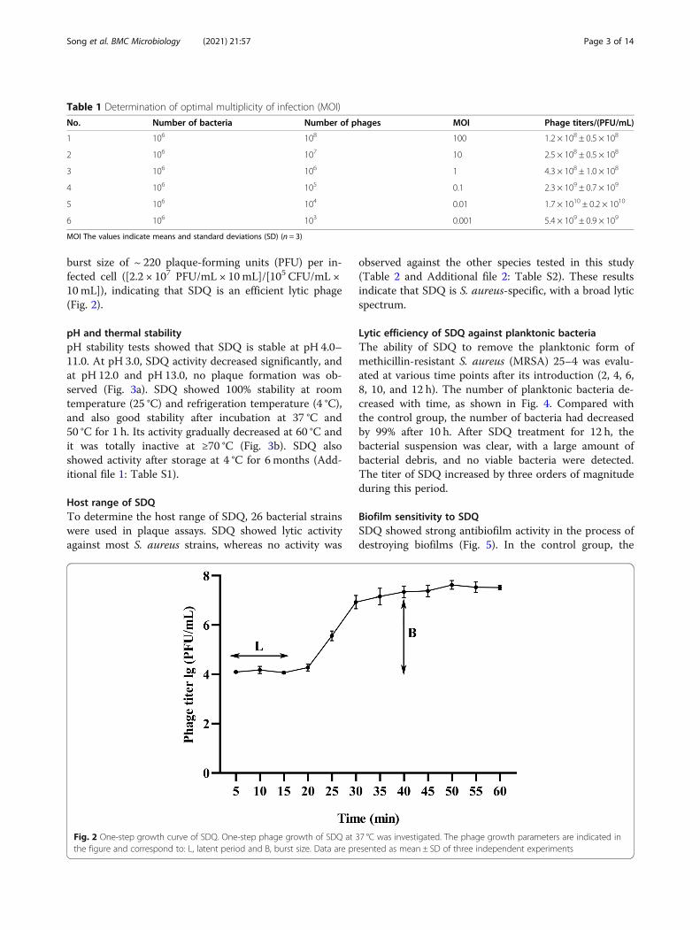
burst size of ~ 220 plaque-forming units (PFU) per in-fected cell ([2.2 × 10⁷ PFU/mL × 10mL]/[105 CFU/mL ×10mL]), indicating that SDQ is an efficient lytic phage(Fig. 2).
pH and thermal stabilitypH stability tests showed that SDQ is stable at pH 4.0–11.0. At pH 3.0, SDQ activity decreased significantly, andat pH 12.0 and pH 13.0, no plaque formation was ob-served (Fig. 3a). SDQ showed 100% stability at roomtemperature (25 °C) and refrigeration temperature (4 °C),and also good stability after incubation at 37 °C and50 °C for 1 h. Its activity gradually decreased at 60 °C andit was totally inactive at ≥70 °C (Fig. 3b). SDQ alsoshowed activity after storage at 4 °C for 6 months (Add-itional file 1: Table S1).
Host range of SDQTo determine the host range of SDQ, 26 bacterial strainswere used in plaque assays. SDQ showed lytic activityagainst most S. aureus strains, whereas no activity was
observed against the other species tested in this study(Table 2 and Additional file 2: Table S2). These resultsindicate that SDQ is S. aureus-specific, with a broad lyticspectrum.
Lytic efficiency of SDQ against planktonic bacteriaThe ability of SDQ to remove the planktonic form ofmethicillin-resistant S. aureus (MRSA) 25–4 was evalu-ated at various time points after its introduction (2, 4, 6,8, 10, and 12 h). The number of planktonic bacteria de-creased with time, as shown in Fig. 4. Compared withthe control group, the number of bacteria had decreasedby 99% after 10 h. After SDQ treatment for 12 h, thebacterial suspension was clear, with a large amount ofbacterial debris, and no viable bacteria were detected.The titer of SDQ increased by three orders of magnitudeduring this period.
Biofilm sensitivity to SDQSDQ showed strong antibiofilm activity in the process ofdestroying biofilms (Fig. 5). In the control group, the
Table 1 Determination of optimal multiplicity of infection (MOI)
No. Number of bacteria Number of phages MOI Phage titers/(PFU/mL)
1 106 108 100 1.2 × 10⁸ ± 0.5 × 10⁸
2 106 107 10 2.5 × 10⁸ ± 0.5 × 10⁸
3 106 106 1 4.3 × 10⁸ ± 1.0 × 10⁸
4 106 105 0.1 2.3 × 10⁹ ± 0.7 × 10⁹
5 106 104 0.01 1.7 × 10¹⁰ ± 0.2 × 10¹⁰
6 106 103 0.001 5.4 × 10⁹ ± 0.9 × 10⁹
MOI The values indicate means and standard deviations (SD) (n = 3)
Fig. 2 One-step growth curve of SDQ. One-step phage growth of SDQ at 37 °C was investigated. The phage growth parameters are indicated inthe figure and correspond to: L, latent period and B, burst size. Data are presented as mean ± SD of three independent experiments
Song et al. BMC Microbiology (2021) 21:57 Page 3 of 14
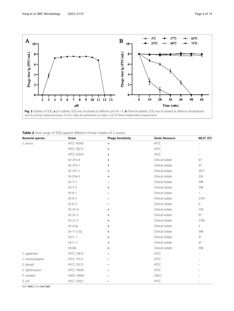
Fig. 3 Stability of SDQ. a pH stability: SDQ was incubated at different pHs for 1 h. b Thermal stability: SDQ was incubated at different temperaturesand its activity measured every 10min. Data are presented as mean ± SD of three independent experiments
Table 2 Host range of SDQ against different clinical isolates of S. aureus
Bacterial species Strain Phage Sensitivity Strain Resource MLST (ST)
S. aureus ATCC 43300 + ATCC –
ATCC 29213 + ATCC –
ATCC 25923 + ATCC –
SA 2Y3–4 + Clinical isolate 97
SA 2Y9–1 + Clinical isolate 97
SA 2Y7–1 + Clinical isolate 5817
SA 2Y4–3 + Clinical isolate 239
SA 7–1 – Clinical isolate 398
SA 7–3 + Clinical isolate 398
SA 8–1 – Clinical isolate 1
SA 8–2 – Clinical isolate 2154
SA 8–3 – Clinical isolate 9
SA 25–4 + Clinical isolate 239
SA 25–5 + Clinical isolate 97
SA 27–2 + Clinical isolate 5796
SA 4-2p + Clinical isolate 5
SA-11-2-2p + Clinical isolate 398
SA11–1 + Clinical isolate 97
SA11–2 + Clinical isolate 97
SA16b + Clinical isolate 398
S. agalactiae ATCC 13813 – ATCC –
L. monocytogenes ATCC 19115 – ATCC –
E. faecalis ATCC 29212 – ATCC –
S. Typhimurium ATCC 14028 – ATCC –
P. mirabilis CMCC 49005 – CMCC –
E. coli ATCC 25922 – ATCC –
(+) = lytic; (−) = non lytic
Song et al. BMC Microbiology (2021) 21:57 Page 4 of 14
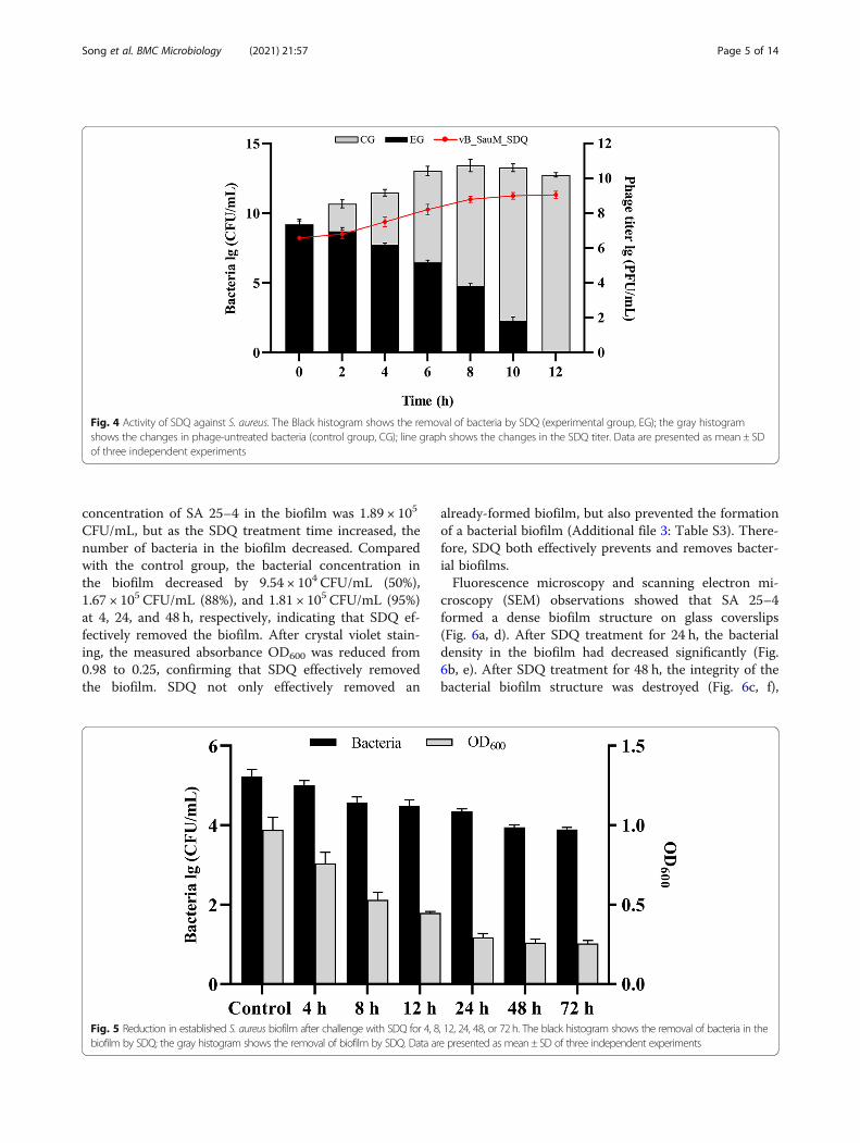
concentration of SA 25–4 in the biofilm was 1.89 × 10⁵
CFU/mL, but as the SDQ treatment time increased, thenumber of bacteria in the biofilm decreased. Comparedwith the control group, the bacterial concentration inthe biofilm decreased by 9.54 × 10⁴ CFU/mL (50%),1.67 × 105 CFU/mL (88%), and 1.81 × 10⁵ CFU/mL (95%)at 4, 24, and 48 h, respectively, indicating that SDQ ef-fectively removed the biofilm. After crystal violet stain-ing, the measured absorbance OD600 was reduced from0.98 to 0.25, confirming that SDQ effectively removedthe biofilm. SDQ not only effectively removed an
already-formed biofilm, but also prevented the formationof a bacterial biofilm (Additional file 3: Table S3). There-fore, SDQ both effectively prevents and removes bacter-ial biofilms.Fluorescence microscopy and scanning electron mi-
croscopy (SEM) observations showed that SA 25–4formed a dense biofilm structure on glass coverslips(Fig. 6a, d). After SDQ treatment for 24 h, the bacterialdensity in the biofilm had decreased significantly (Fig.6b, e). After SDQ treatment for 48 h, the integrity of thebacterial biofilm structure was destroyed (Fig. 6c, f),
Fig. 4 Activity of SDQ against S. aureus. The Black histogram shows the removal of bacteria by SDQ (experimental group, EG); the gray histogramshows the changes in phage-untreated bacteria (control group, CG); line graph shows the changes in the SDQ titer. Data are presented as mean ± SDof three independent experiments
Fig. 5 Reduction in established S. aureus biofilm after challenge with SDQ for 4, 8, 12, 24, 48, or 72 h. The black histogram shows the removal of bacteria in thebiofilm by SDQ; the gray histogram shows the removal of biofilm by SDQ. Data are presented as mean± SD of three independent experiments
Song et al. BMC Microbiology (2021) 21:57 Page 5 of 14
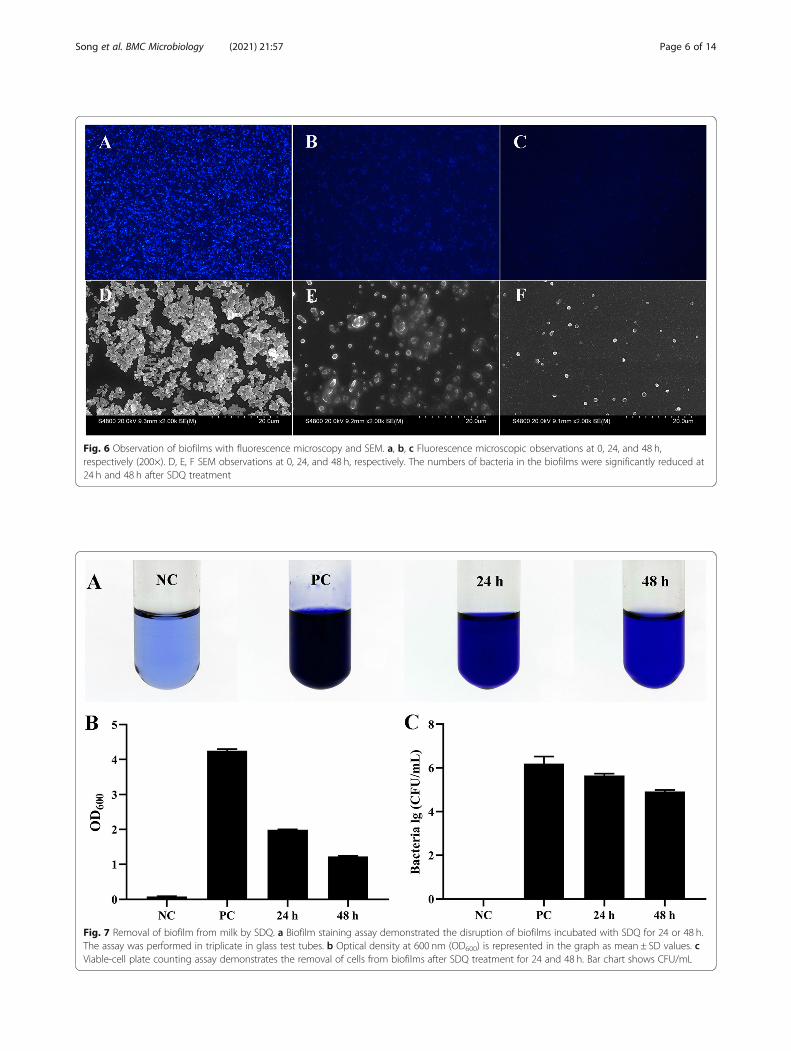
Fig. 6 Observation of biofilms with fluorescence microscopy and SEM. a, b, c Fluorescence microscopic observations at 0, 24, and 48 h,respectively (200×). D, E, F SEM observations at 0, 24, and 48 h, respectively. The numbers of bacteria in the biofilms were significantly reduced at24 h and 48 h after SDQ treatment
Fig. 7 Removal of biofilm from milk by SDQ. a Biofilm staining assay demonstrated the disruption of biofilms incubated with SDQ for 24 or 48 h.The assay was performed in triplicate in glass test tubes. b Optical density at 600 nm (OD600) is represented in the graph as mean ± SD values. cViable-cell plate counting assay demonstrates the removal of cells from biofilms after SDQ treatment for 24 and 48 h. Bar chart shows CFU/mL
Song et al. BMC Microbiology (2021) 21:57 Page 6 of 14
indicating that SDQ has a strong capacity to kill bacteriaand destroy the biofilm structure.
Removal of biofilm from milk by SDQSDQ showed strong antibiofilm activity when destroyingbiofilms in milk (Fig. 7). Crystal violet staining showedthat after treatment with SDQ for 24 h, the OD600 de-creased from 4.21 to 1.95 (by 54% compared with thepositive control group), and at 72 h, the OD600 had de-creased by 3.02 units (71%) (Fig. 7a, b), indicating thatSDQ effectively removed the biofilm from milk. In thepositive control group, the concentration of SA 25–4 inthe biofilm was 1.57 × 10⁶ CFU/mL, but as the SDQtreatment time increased, the number of bacteria in thebiofilm decreased. Compared with the positive controlgroup, the bacterial concentration in the biofilm at 24 hhad decreased by 1.09 × 10⁶ CFU/mL (69%), and at 48 h,the bacterial concentration had decreased by 1.48 × 10⁶
CFU/mL (95%) (Fig. 7c), confirming that SDQ effectivelyremoved the biofilm from milk.
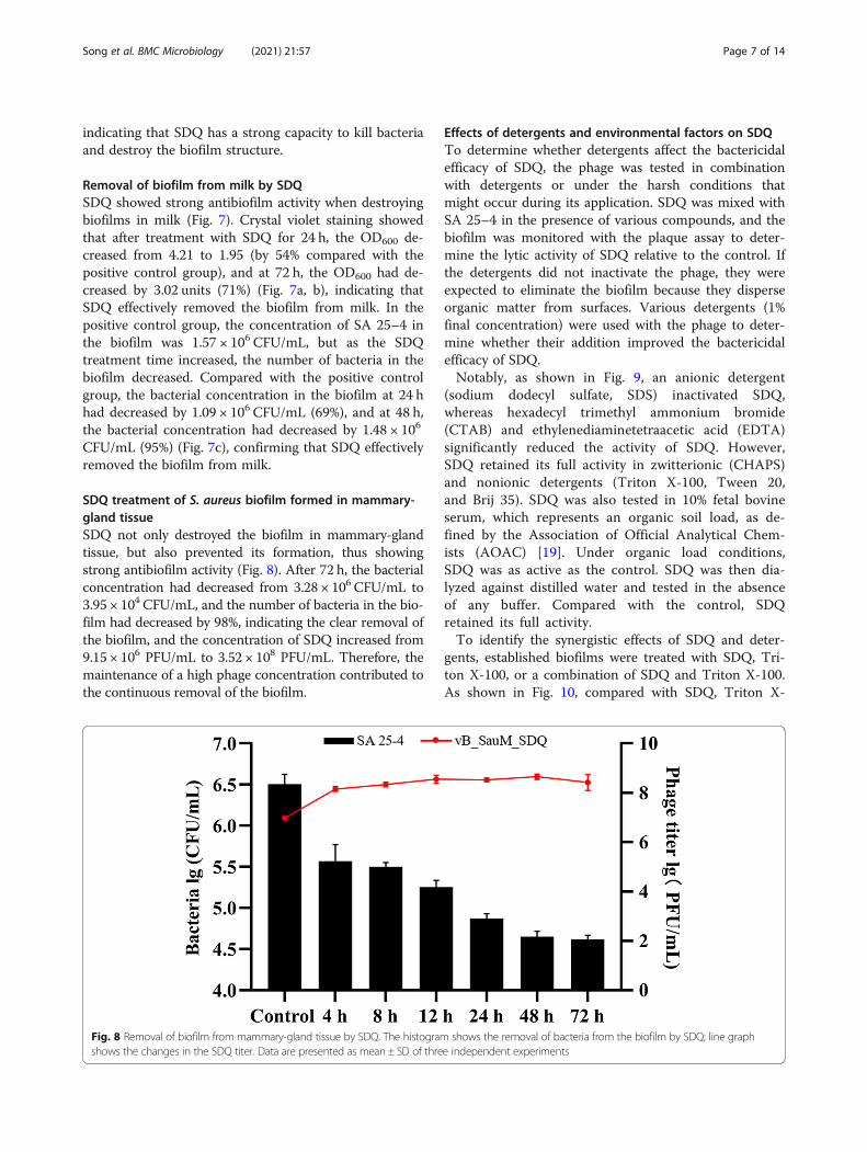
SDQ treatment of S. aureus biofilm formed in mammary-gland tissueSDQ not only destroyed the biofilm in mammary-glandtissue, but also prevented its formation, thus showingstrong antibiofilm activity (Fig. 8). After 72 h, the bacterialconcentration had decreased from 3.28 × 10⁶ CFU/mL to3.95 × 104 CFU/mL, and the number of bacteria in the bio-film had decreased by 98%, indicating the clear removal ofthe biofilm, and the concentration of SDQ increased from9.15 × 106 PFU/mL to 3.52 × 10⁸ PFU/mL. Therefore, themaintenance of a high phage concentration contributed tothe continuous removal of the biofilm.
Effects of detergents and environmental factors on SDQTo determine whether detergents affect the bactericidalefficacy of SDQ, the phage was tested in combinationwith detergents or under the harsh conditions thatmight occur during its application. SDQ was mixed withSA 25–4 in the presence of various compounds, and thebiofilm was monitored with the plaque assay to deter-mine the lytic activity of SDQ relative to the control. Ifthe detergents did not inactivate the phage, they wereexpected to eliminate the biofilm because they disperseorganic matter from surfaces. Various detergents (1%final concentration) were used with the phage to deter-mine whether their addition improved the bactericidalefficacy of SDQ.Notably, as shown in Fig. 9, an anionic detergent
(sodium dodecyl sulfate, SDS) inactivated SDQ,whereas hexadecyl trimethyl ammonium bromide(CTAB) and ethylenediaminetetraacetic acid (EDTA)significantly reduced the activity of SDQ. However,SDQ retained its full activity in zwitterionic (CHAPS)and nonionic detergents (Triton X-100, Tween 20,and Brij 35). SDQ was also tested in 10% fetal bovineserum, which represents an organic soil load, as de-fined by the Association of Official Analytical Chem-ists (AOAC) [19]. Under organic load conditions,SDQ was as active as the control. SDQ was then dia-lyzed against distilled water and tested in the absenceof any buffer. Compared with the control, SDQretained its full activity.To identify the synergistic effects of SDQ and deter-
gents, established biofilms were treated with SDQ, Tri-ton X-100, or a combination of SDQ and Triton X-100.As shown in Fig. 10, compared with SDQ, Triton X-
Fig. 8 Removal of biofilm from mammary-gland tissue by SDQ. The histogram shows the removal of bacteria from the biofilm by SDQ; line graphshows the changes in the SDQ titer. Data are presented as mean ± SD of three independent experiments
Song et al. BMC Microbiology (2021) 21:57 Page 7 of 14
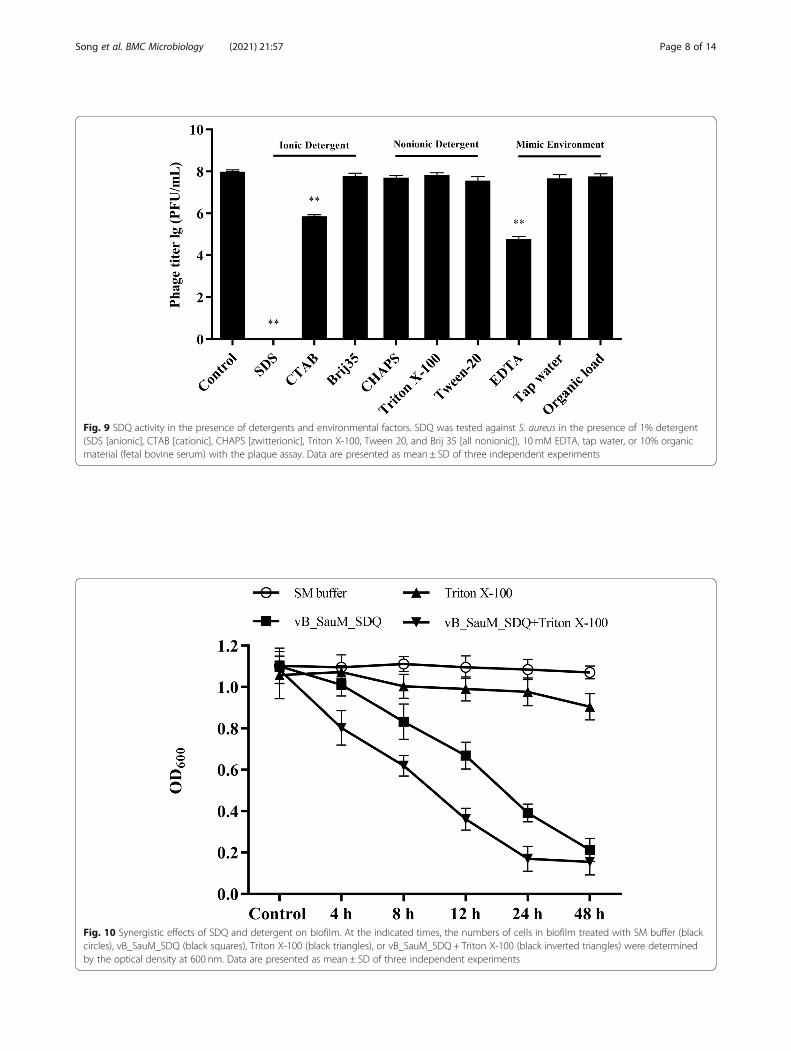
Fig. 9 SDQ activity in the presence of detergents and environmental factors. SDQ was tested against S. aureus in the presence of 1% detergent(SDS [anionic], CTAB [cationic], CHAPS [zwitterionic], Triton X-100, Tween 20, and Brij 35 [all nonionic]), 10 mM EDTA, tap water, or 10% organicmaterial (fetal bovine serum) with the plaque assay. Data are presented as mean ± SD of three independent experiments
Fig. 10 Synergistic effects of SDQ and detergent on biofilm. At the indicated times, the numbers of cells in biofilm treated with SM buffer (blackcircles), vB_SauM_SDQ (black squares), Triton X-100 (black triangles), or vB_SauM_SDQ + Triton X-100 (black inverted triangles) were determinedby the optical density at 600 nm. Data are presented as mean ± SD of three independent experiments
Song et al. BMC Microbiology (2021) 21:57 Page 8 of 14
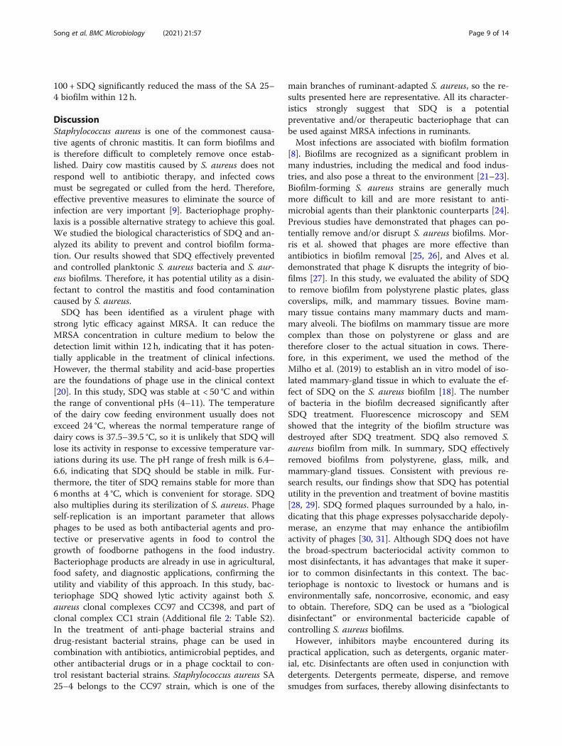
100 + SDQ significantly reduced the mass of the SA 25–4 biofilm within 12 h.
DiscussionStaphylococcus aureus is one of the commonest causa-tive agents of chronic mastitis. It can form biofilms andis therefore difficult to completely remove once estab-lished. Dairy cow mastitis caused by S. aureus does notrespond well to antibiotic therapy, and infected cowsmust be segregated or culled from the herd. Therefore,effective preventive measures to eliminate the source ofinfection are very important [9]. Bacteriophage prophy-laxis is a possible alternative strategy to achieve this goal.We studied the biological characteristics of SDQ and an-alyzed its ability to prevent and control biofilm forma-tion. Our results showed that SDQ effectively preventedand controlled planktonic S. aureus bacteria and S. aur-eus biofilms. Therefore, it has potential utility as a disin-fectant to control the mastitis and food contaminationcaused by S. aureus.SDQ has been identified as a virulent phage with
strong lytic efficacy against MRSA. It can reduce theMRSA concentration in culture medium to below thedetection limit within 12 h, indicating that it has poten-tially applicable in the treatment of clinical infections.However, the thermal stability and acid-base propertiesare the foundations of phage use in the clinical context[20]. In this study, SDQ was stable at < 50 °C and withinthe range of conventional pHs (4–11). The temperatureof the dairy cow feeding environment usually does notexceed 24 °C, whereas the normal temperature range ofdairy cows is 37.5–39.5 °C, so it is unlikely that SDQ willlose its activity in response to excessive temperature var-iations during its use. The pH range of fresh milk is 6.4–6.6, indicating that SDQ should be stable in milk. Fur-thermore, the titer of SDQ remains stable for more than6months at 4 °C, which is convenient for storage. SDQalso multiplies during its sterilization of S. aureus. Phageself-replication is an important parameter that allowsphages to be used as both antibacterial agents and pro-tective or preservative agents in food to control thegrowth of foodborne pathogens in the food industry.Bacteriophage products are already in use in agricultural,food safety, and diagnostic applications, confirming theutility and viability of this approach. In this study, bac-teriophage SDQ showed lytic activity against both S.aureus clonal complexes CC97 and CC398, and part ofclonal complex CC1 strain (Additional file 2: Table S2).In the treatment of anti-phage bacterial strains anddrug-resistant bacterial strains, phage can be used incombination with antibiotics, antimicrobial peptides, andother antibacterial drugs or in a phage cocktail to con-trol resistant bacterial strains. Staphylococcus aureus SA25–4 belongs to the CC97 strain, which is one of the
main branches of ruminant-adapted S. aureus, so the re-sults presented here are representative. All its character-istics strongly suggest that SDQ is a potentialpreventative and/or therapeutic bacteriophage that canbe used against MRSA infections in ruminants.Most infections are associated with biofilm formation
[8]. Biofilms are recognized as a significant problem inmany industries, including the medical and food indus-tries, and also pose a threat to the environment [21–23].Biofilm-forming S. aureus strains are generally muchmore difficult to kill and are more resistant to anti-microbial agents than their planktonic counterparts [24].Previous studies have demonstrated that phages can po-tentially remove and/or disrupt S. aureus biofilms. Mor-ris et al. showed that phages are more effective thanantibiotics in biofilm removal [25, 26], and Alves et al.demonstrated that phage K disrupts the integrity of bio-films [27]. In this study, we evaluated the ability of SDQto remove biofilm from polystyrene plastic plates, glasscoverslips, milk, and mammary tissues. Bovine mam-mary tissue contains many mammary ducts and mam-mary alveoli. The biofilms on mammary tissue are morecomplex than those on polystyrene or glass and aretherefore closer to the actual situation in cows. There-fore, in this experiment, we used the method of theMilho et al. (2019) to establish an in vitro model of iso-lated mammary-gland tissue in which to evaluate the ef-fect of SDQ on the S. aureus biofilm [18]. The numberof bacteria in the biofilm decreased significantly afterSDQ treatment. Fluorescence microscopy and SEMshowed that the integrity of the biofilm structure wasdestroyed after SDQ treatment. SDQ also removed S.aureus biofilm from milk. In summary, SDQ effectivelyremoved biofilms from polystyrene, glass, milk, andmammary-gland tissues. Consistent with previous re-search results, our findings show that SDQ has potentialutility in the prevention and treatment of bovine mastitis[28, 29]. SDQ formed plaques surrounded by a halo, in-dicating that this phage expresses polysaccharide depoly-merase, an enzyme that may enhance the antibiofilmactivity of phages [30, 31]. Although SDQ does not havethe broad-spectrum bacteriocidal activity common tomost disinfectants, it has advantages that make it super-ior to common disinfectants in this context. The bac-teriophage is nontoxic to livestock or humans and isenvironmentally safe, noncorrosive, economic, and easyto obtain. Therefore, SDQ can be used as a “biologicaldisinfectant” or environmental bactericide capable ofcontrolling S. aureus biofilms.However, inhibitors maybe encountered during its
practical application, such as detergents, organic mater-ial, etc. Disinfectants are often used in conjunction withdetergents. Detergents permeate, disperse, and removesmudges from surfaces, thereby allowing disinfectants to
Song et al. BMC Microbiology (2021) 21:57 Page 9 of 14
work. However, the activity of disinfectants is often re-duced or they are inactivated by detergents [32]. In thisstudy, we examined the effect of detergents on the bac-tericidal efficacy of SDQ. Ionic detergents reduced theactivity or inactivated SDQ, but these are seldom used ascleaning agents, whereas SDQ maintained full bacterio-lytic activity in the presence of nonionic detergents (Tri-ton X-100, Tween 20, and Brij 35). In practice, nonionicdetergents have good emulsification, penetration, anddispersion properties, rendering them effective deter-gents [33–35]. Therefore, these detergents can assistSDQ to disrupt the S. aureus biofilms.According to the AOAC, 10% fetal bovine serum rep-
resents an organic soil load [19]. To test the practical ef-fectiveness of SDQ, it was diluted with tap water andtested in the absence of any buffer. Compared with thecontrol, the full activity of SDQ was retained in both tapwater and fetal bovine serum. In some region, problemssuch as hard water might be encountered, which canbind to either disinfectants or detergents, interferingwith their effectiveness [36]. Metal chelators, such asEDTA, are commonly used to treat hard water. How-ever, the addition of 10 mM EDTA did not reduce thelytic activity of SDQ for S. aureus cells. Therefore, theuse of this phage combined with a nonionic detergentcan better eliminate a biofilm than the biofilm alone andis unaffected by organic matter or tap water.
ConclusionsIn this study, SDQ not only prevented and removed S.aureus biofilms, but also multiplied during the processof infection, making it a good candidate for further pre-ventative and therapeutic development. SDQ alsoretained its full activity in the presence of nonionic de-tergents, tap water, a metal chelator, and organic mater-ial, and can be used in combination with detergents.Although bacteriophage will never replace traditionaldisinfectants, they have great potential utility as narrow-spectrum biological disinfectants for controlling S. aur-eus infections and the devastating effects of MRSA andrelated biofilms, such as occur on medical equipmentand in the food industry and livestock farming.
MethodsBacterial strains and culture conditionsA selection of 26 different bacterial strains, including S.aureus, Streptococcus agalactiae, Enterobacter faecalis,Salmonella typhimurium, Proteus mirabilis, Listeriamonocytogenes, and E. coli, were purchased from theAmerican Type Culture Collection (ATCC, Manassas,VA, USA) and the National Center for Medical CultureCollections (CMCC, Beijing, China), and 17 clinical S.aureus isolates were provided by Heilongjiang ProvincialKey Laboratory for the Prevention and Control of Bovine
Diseases (Daqing, Heilongjiang, China). These clinicalstrains were tested with PCR amplification of the mecAgene to confirm that the isolates were MRSA. We identi-fied the multilocus sequence typing (MLST) types of thesestrains, and the 17 clinical strains of S. aureus includednine ST types (Additional file 2: Table S2). All the strainswere streaked on tryptic soy agar (Hopebio, Qingdao,China) before experimentation, and single colonies wererecovered by culture in tryptic soy broth (Hopebio) over-night at 37 °C to ensure the purity of the bacterial stocks.All bacterial strains were preserved in 20% (v/v) glyceroland the stocks were maintained at − 80 °C.
Biological characteristics of the phageIsolation of the S. aureus phagePhage SDQ was isolated from a dairy farm sewage sys-tem using S. aureus subsp. aureus Rosenbach (ATCC43300) as the host strain. Sewage samples were collectedfrom a cattle farm sewer system (Daqing, Heilongjiang,China), centrifuged (9000×g, 4 °C, 10 min), and filteredthrough 0.22 μm membrane filters (SteriFlip, Millipore).The filtered raw sewage (100 mL) was placed in sterile100 mL flasks with double-strength TSB. S. aureusATCC 43300 and the prepared sewage samples werethen cocultured overnight at 37 °C. The cocultures werecentrifuged at 10,000×g for 10 min at 4 °C, and the su-pernatants were collected and filtered (0.22 μm). Adouble-layer agar plate assay was used to detect and pur-ify the phage from the supernatant [37]. Briefly, wepicked a single spot from a double-layer plate and100 μL of the host strain and mixed them with 5mL ofmolten soft agar (0.75%) medium. This was then over-lain on the surface of solidified basal Luria–Bertani (LB)agar. The sample was then incubated for 6–8 h at 37 °C.This was repeated three times to obtain the purifiedphage lysate. The purified phage was then amplified andstored in dimethyl sulfoxide (DMSO, 3:1 [v/v]) at either4 °C or − 80 °C.Following large-scale culture, SDQ was precipitated
with 10% (w/v) polyethylene glycol (PEG) 8000 and 1MNaCl. The phage sample was then placed on the top of adiscontinuous CsCl gradient (1.32, 1.45, 1.50, and 1.70 g/mL) and centrifuged at 35,000×g for 3 h at 4 °C. Thephage band was collected and dialyzed against a suspen-sion medium (SM) buffer (0.01% gelatin, 100mM NaCl,50 mM Tris-HCl, 10 mM MgSO4) at 4 °C.
Morphology of SDQThe morphology of SDQ was examined with transmis-sion electron microscopy (TEM; JEM-2100, JEOL,Tokyo, Japan). SDQ was purified and transferred tocarbon-coated copper film for 1 min and negativelystained with 2% phosphotungstic acid (pH 7.0). The ex-cess liquid was removed, and the sample air-dried on
Song et al. BMC Microbiology (2021) 21:57 Page 10 of 14

the carbon-coated copper grid. The stained SDQ wasobserved under TEM (120 kV).
One-step growth‘MOI’ refers to the ratio of the phage to the host bacteriaduring the processes of infection [38]. S. aureus ATCC43300 was grown to logarithmic phase and transferredinto fresh LB broth at a final concentration of 107 CFU/mL. SDQ was then added at different MOIs (phage/bac-teria = 0.001, 0.01, 0.1, 1, 10, or 100) and the mixtureswere incubated for 8 h. Immediately after serial dilution,the phage titer in each sample was determined with thedouble-layer agar plate method.To construct the one-step growth curve, SDQ was
added to an exponential-phase culture of S. aureus (1 ×10⁵ CFU/mL) at an MOI of 0.01 and allowed to adsorbfor 10 min at 37 °C. The mixture was then centrifuged at12,000×g for 5 min at 4 °C, and the pellet was resus-pended in 10mL of LB broth. The suspension was thenincubated at 37 °C with shaking at 180 rpm. The samplewas collected at 5 min intervals until 60 min. The phagetiter of the lysates was quantified with the double-layeragar plate method and the growth curve for SDQ wasconstructed from these data [39]. The experiments wereperformed in triplicate.
pH and thermal stabilityTo measure the stability characteristics of SDQ, its sur-vival rate was determined after treatment with diverseranges of pHs and temperatures. Briefly, for the pH sta-bility test, SDQ was incubated at pH 2.0, 3.0, 4.0,5.0, 6.0,7.0, 8.0, 9.0, 10.0, 11.0, 12.0, and 13.0 for 1 h at 37 °C.For the thermal stability test, SDQ was incubated at 4,25, 37, 50, 60, and 70 °C, and the phage titers were mea-sured every 10 min. After treatment, all the samples werediluted and tested immediately with the double-layeragar plate method. To test SDQ stability after long-termstorage, aliquots of phage suspensions were stored at4 °C for 6 months. All the experiments were performedin triplicate.
Antimicrobial activity of SDQDetermination of SDQ the host rangeThe host range of SDQ was determined with spottests against a panel of 21 strains [40]. In the spottest experiment, 100 μL droplets of phage stock (109
PFU/mL) were spotted onto freshly seeded lawns ofthe indicated bacterial strains. The production of lyticspots was assessed after incubation for 12 h at 37 °C.The details of the bacterial strains used in the studyare listed in Table 2.
Lytic efficiency of SDQ against planktonic bacteriaThe experimental strain to be tested (SA 25–4) wasgrown overnight in LB broth. The overnight bacterialculture (100 μL) was diluted in LB broth to a final titerof approximately 109 CFU/mL. SDQ was added at anMOI of 0.01 and incubated at 37 °C with shaking at 180rpm in an incubator. The colonies in the culture brothwere counted at different time points (0, 2, 4, 6, 8, 10,and 12 h), and a phage-free treatment was used as thecontrol. The bacterial count was calculated from thenumber of colonies on the plate. The change in phagetiter was determined with the double-layer agar platemethod [41]. All the experiments were performed intriplicate.
Antibiofilm activity of SDQPhage treatment of S. aureus biofilm in 96-well cell cultureplatesA 96-well cell culture plate was used to detect the in-hibitory effect of SDQ on biofilm formation at MOI =0.01, and to investigate the removal of the biofilm bySDQ. In each well of a 96-well cell culture plate, TSBwas inoculated with SA 25–4 at a final concentration of106 CFU/mL. For the biofilm formation inhibition ex-periment, SDQ was added to the bacterial culture to afinal titer of 104 PFU/mL, and incubated statically at37 °C for 48 h. For the biofilm removal experiment, SA25–4 was initially incubated under the conditions de-scribed above for 48 h to allow biofilm formation, andthen treated with SDQ at a final titer of 107 PFU/mL for4, 8, 12, 24, 48, or 72 h. A phage-free treatment was usedas the control. Viable-cell plate counting was used to de-termine the numbers of bacteria in the biofilms at differ-ent time points, and biofilm removal was measured withcrystal violet staining. Each well was rinsed five timeswith sterile phosphate-buffered saline (PBS) and allowedto air-dry. The SDQ-treated biofilm in each well wasstained with 5% crystal violet solution (Becton Dickin-son, Sparks, MD) at 25 °C for 60 min, eluted with 33%acetic acid, and the OD600 measured with a spectropho-tometer (Beijing Purkinje General Instrument Co,Beijing, China) [42]. All the experiments were performedin triplicate.
Microscopic imaging of biofilmsThe effects of SDQ on the biofilms were assessed withHoechst 33342 stain (Beyotime Biotechnology, Shanghai,China) and visualized with fluorescence microscopy(Ti2, Nikon Corporation, Japan). Bacterial biofilms weregrown on glass coverslips as described above. The non-adherent cells were removed and the wells were washedthree times with sterile PBS. The biofilms were treatedwith SDQ at a final titer of 107 PFU/mL for 0, 24, or 48h. The glass coverslip was then washed once with sterile
Song et al. BMC Microbiology (2021) 21:57 Page 11 of 14
PBS. The biofilm was stained with Hoechst 33342 ac-cording to the manufacturer’s protocol and visualizedwith fluorescence microscopy.For DSEM, the biofilms were cultured as for fluores-
cence microscopy. The glass coverslips were washedonce with sterile PBS. Each biofilm was immobilizedwith 5% glutaraldehyde, dehydrated a graded series ofethanol concentrations (20, 50, 70, 90, and 100%), andthen freeze-dried before SEM analysis (Hitachi S-4800;Hitachi High-Technologies Europe GmbH, Krefeld,Germany).
Removal of biofilm from milk by SDQTo study the removal of biofilm from milk by SDQ, weused pasteurized milk as the culture medium for the S.aureus biofilm. In this experiment, a constant temperatureoscillator was used to keep the milk in a flowing statewhile the biofilm was cultured in a glass test tube. Alogarithmic-growth-phase culture of SA 25–4 at a finalconcentration of 106 CFU/mL was added to 1mL of milkand cultured at 37 ° and 120 rpm for 48 h. The nonadher-ent cells were removed by washing the biofilm three timeswith sterile PBS. SDQ at a final titer of 107 PFU/mL wasadded for 24 or 48 h. A phage-free treatment was used asthe positive control, and milk was used as the negativecontrol to compare the removal of the biofilm by thephage. The number of bacteria in the biofilm at differenttime points was determined with viable-cell plate count-ing, and biofilm removal was measured with crystal violetstaining. Each sample was rinsed five times with sterilePBS and allowed to air-dry. The SDQ-treated biofilms ineach sample were stained with 0.5% crystal violet solution(Becton Dickinson) for 60min at 25 °C and eluted with33% acetic acid. The OD600 of the eluate was measuredwith a spectrophotometer (Beijing Purkinje General In-strument Co.). All the experiments were performed intriplicate.
Phage treatment of S. aureus biofilm formed in mammary-gland tissueThe animal trial in this study was approved by the Insti-tutional Animal Care and Use Committee (IACUC) ofHeilongjiang Bayi Agricultural University, and conven-tional animal welfare regulations and standards werefollowed. A mammary gland from a healthy dairy cowwas obtained from a local farm. The mammary glandwas immediately sterilized with a previously describedmethod [43]. Briefly, the mammary gland was washedwith saline for 10 min, and then placed in alcohol (75%)for 2–3 min. This was repeated twice. The gland wasplaced in a desiccator for 30 min and washed three timeswith sterile PBS. Cuboids (10 × 10 × 5mm3) of tissuewere cut with a scalpel and frozen at − 20 °C until use.The treated mammary-gland tissue was transferred to a
24-well cell culture plate, and 1.8 mL of TSB mediumwas added together with 200 μL of 1 × 10⁶ CFU/mL SA25–4. The samples were incubated at 37 °C for 48 h. Toremove the nonadherent cells, the wells were washedfive times with sterile PBS. SDQ, at a final titer of 107
PFU/mL, was added for 24, 48, or 72 h, and a phage-freetreatment was used as the control group. PBS (10 mL)was then added and the samples shaken vigorously. Thechange in bacterial titer was determined with the viable-cell plate counting method and the change in phage titerwas measured with the double-layer agar plate method.All the experiments were performed in triplicate.
Effects of added detergents and environmental factors onSDQ eradication of S. aureusTo determine the infectivity of SDQ in the presence of de-tergents, the plaque-forming ability of the phage wasassessed in various types of detergent. Briefly, SDQ wasstandardized to a titer of 108 PFU/mL. Molten soft agar(5mL, 0.75%) was mixed with 100 μL of SDQ and 100 μLof bacterial culture, and overlain on the surface of solidi-fied basal LB agar. An SDQ suspension (108 PFU/mL) wastested against S. aureus in the presence of detergents(SDS, CTAB, Tween 20, Triton X-100, CHAPS, or Brij35) at concentrations of 1%, or with tap water, 10mMEDTA, or 10% fetal bovine serum. The plaques werecounted after incubation for 6–8 h at 37 °C. Each test wasrepeated three times. Established biofilms were alsotreated with SDQ, detergent, or a combination of phage(MOI = 10) + detergent. Biofilm formation was assessed at0, 4, 8, 12, and 24 h. All the experiments were performedin triplicate.
Statistical analysisAll data are presented as the means ± standard deviation(SD) of three or more independent experiments (n ≥ 3).One-way ANOVA followed by a t test (GraphPad Soft-ware Inc., San Diego, CA, USA) was used to evaluate dif-ferences between bacterial titers and between phagetiters. Differences with P < 0.05 or P < 0.01 were consid-ered significant (*) or highly significant (**), respectively.
Supplementary InformationThe online version contains supplementary material available at https://doi.org/10.1186/s12866-021-02117-1.
Additional file 1: Table S1. Storage stability of SDQ under refrigeratedtemperature.
Additional file 2: Table S2. List of S. aureus. Strains used in this study.
Additional file 3: Table S3. SDQ inhibited biofilm formation.
Additional file 4. Authors’ original data for Fig. 2
Additional file 5. Authors’ original data for Fig. 3.
Additional file 6. Authors’ original data for Fig. 4.
Additional file 7. Authors’ original data for Fig. 5.
Song et al. BMC Microbiology (2021) 21:57 Page 12 of 14
Additional file 8. Authors’ original data for Fig. 7.
Additional file 9. Authors’ original data for Fig. 8.
Additional file 10. Authors’ original data for Fig. 9.
Additional file 11. Authors’ original data for Fig. 10.
AbbreviationsATCC: American Type Culture Collection; CMCC: National Center for MedicalCulture Collections; MRSA: methicillin-resistant Staphylococcus aureus;LB: Luria Bertani; TEM: Transmission electron microscopy; MOI: Multiplicity ofinfection; PFU: Plaque forming units; CFU: Colony-forming units;PBS: Phosphate buffer saline; SEM: Scanning electron microscopy
AcknowledgementsNot applicable.
Authors’ contributionsWR, SDB and ZJS designed the study, RHR, CH and JYQ performed theexperimental work. SJ and RHR performed data analysis, SJ and RHR draftedthe manuscript. All authors were involved in revising the manuscript. Theauthor(s) read and approved the final manuscript.
FundingThis study was supported by the National Natural Science Foundation of China(project no. 31802226), Natural Science Foundation of Heilongjiang Province ofChina (QC2017021), Heilongjiang Postdoctoral Science Foundation Grant (LBH-Z17185), Key research projects on Heilongjiang Farms & Land ReclamationAdministration (HKKY190307), Heilongjiang Bayi Agricultural University SupportProgram for San Heng San Zong (TDJH201903).
Availability of data and materialsThe datasets used and/or analysed during the current study are availablefrom the corresponding author on reasonable request.
Ethics approval and consent to participateThe animal experiment was approved by the Institutional Animal Care andUse Committee (IACUC) of Heilongjiang Bayi Agricultural University, Daqing,China. The animal experiment conventional animal welfare regulations andstandards were taken into account.
Consent for publicationNot applicable.
Competing interestsThe authors declare that they have no competing interests.
Received: 17 September 2020 Accepted: 4 February 2021
References1. Bradley A. Bovine mastitis: an evolving disease. Vet J. 2002;164(2):116–28.2. Krömker V, Leimbach S. Mastitis treatment-reduction in antibiotic usage in
dairy cows. Reprod Domest Anim. 2017;52(Suppl 3):21–9.3. Fox LK. Prevalence, incidence and risk factors of heifer mastitis. Vet
Microbiol. 2009;134(1–2):82–8.4. Jasper DE. Bovine mycoplasmal mastitis. Adv Vet Sci Comp Med. 1981;25:
121–57.5. Shahzad W, Altaf M, Ahmad M, Munir R, Amin MT, Khan MS, Sagar MS, Khan
MA, Avais M, Akbar GJBB. Prevalence and molecular diagnosis ofstaphylococcus aureus subclinical mastitis in lactating Nili-Ravi buffaloes(Bubalus bubalis) at livestock Experiment Station, Bahadurnagar, Okara,Pakistan. Buffalo Bulletin. 2013;32(6):1041–5.
6. Gomes F, Saavedra MJ, Henriques M. Bovine mastitis disease/pathogenicity:evidence of the potential role of microbial biofilms. Pathogens Dis. 2016;74(3):ftw006.
7. Costerton JW, Stewart PS, Greenberg EP. Bacterial biofilms: a common causeof persistent infections. Science. 1999;284(5418):1318–22.
8. Del Pozo JL. Biofilm-related disease. Expert Rev Anti-Infect Ther. 2018;16(1):51–65.
9. van Soest FJS, Santman-Berends I, Lam T, Hogeveen H. Failure andpreventive costs of mastitis on Dutch dairy farms. J Dairy Sci. 2016;99(10):8365–74.
10. Dwyer RM. Environmental disinfection to control equine infectious diseases.Vet Clin North Am Equine Pract. 2004;20(3):531–42.
11. Laport MS, Marinho PR, Santos OC, de Almeida P, Romanos MT, Muricy G,Brito MA, Giambiagi-deMarval M. Antimicrobial activity of marine spongesagainst coagulase-negative staphylococci isolated from bovine mastitis. VetMicrobiol. 2012;155(2–4):362–8.
12. Randall L, Cooles S, Coldham N, Penuela E, Mott A, Woodward MJ, PiddockL, Webber M. Commonly used farm disinfectants can select for mutantSalmonella enterica serovar Typhimurium with decreased susceptibility tobiocides and antibiotics without compromising virulence. J AntimicrobChemother. 2007;60(6):1273–80.
13. Kim M, Weigand MR, Oh S, Hatt JK, Krishnan R, Tezel U, Pavlostathis SG,Konstantinidis KT. Widely used Benzalkonium chloride disinfectants canpromote antibiotic resistance. Appl Environ Microbiol. 2018;84(17):e01201–18.
14. Sillankorva SM, Oliveira H, Azeredo J. Bacteriophages and their role in foodsafety. Int J Microbiol. 2012;2012:863945.
15. Kaistha SD, Umrao PD. Bacteriophage for mitigation of multiple drug resistantbiofilm forming pathogens. Recent Pat Biotechnol. 2016;10(2):184–94.
16. Schuch R, Khan BK, Raz A, Rotolo JA, Wittekind M. Bacteriophage Lysin CF-301, a potent Antistaphylococcal biofilm agent. Antimicrob AgentsChemother. 2017;61(7):e02666–16.
17. Wroe JA, Johnson CT, García AJ. Bacteriophage delivering hydrogels reducebiofilm formation in vitro and infection in vivo. J Biomed Mater Res A. 2020;108(1):39–49.
18. Milho C, Andrade M, Vilas Boas D, Alves D, Sillankorva S. Antimicrobialassessment of phage therapy using a porcine model of biofilm infection. IntJ Pharm. 2019;557:112–23.
19. Wilson J, Margolin AB. Efficacy of glutaraldehyde disinfectant againstCryptosporidium parvum in the presence of various organic soils. J AOAC Int.2003;86(1):96–100.
20. Jończyk-Matysiak E, Łodej N, Kula D, Owczarek B, Orwat F, Międzybrodzki R,Neuberg J, Bagińska N, Weber-Dąbrowska B, Górski A. Factors determiningphage stability/activity: challenges in practical phage application. Expert RevAnti-Infect Ther. 2019;17(8):583–606.
21. Arciola CR, Campoccia D, Montanaro L. Implant infections: adhesion, biofilmformation and immune evasion. Nat Rev Microbiol. 2018;16(7):397–409.
22. Vergara A, Normanno G, Di Ciccio P, Pedonese F, Nuvoloni R, Parisi A,Santagada G, Colagiorgi A, Zanardi E, Ghidini S, et al. Biofilm formation andits relationship with the molecular characteristics of food-related methicillin-resistant Staphylococcus aureus (MRSA). J Food Sci. 2017;82(10):2364–70.
23. Di Pippo F, Di Gregorio L, Congestri R, Tandoi V, Rossetti S. Biofilm growthand control in cooling water industrial systems. FEMS Microbiol Ecol. 2018;94(5):044.
24. Lister JL, Horswill AR. Staphylococcus aureus biofilms: recent developmentsin biofilm dispersal. Front Cell Infect Microbiol. 2014;4:178.
25. Morris J, Kelly N, Elliott L, Grant A, Wilkinson M, Hazratwala K, McEwen P.Evaluation of bacteriophage anti-biofilm activity for potential control oforthopedic implant-related infections caused by Staphylococcus aureus. SurgInfect. 2019;20(1):16–24.
26. Lehman SM, Mearns G, Rankin D, Cole RA, Smrekar F, Branston SD, MoralesS. Design and preclinical development of a phage product for thetreatment of antibiotic-resistant Staphylococcus aureus infections. Viruses.2019;11(1):88.
27. Alves DR, Gaudion A, Bean JE, Perez Esteban P, Arnot TC, Harper DR, Kot W,Hansen LH, Enright MC, Jenkins AT. Combined use of bacteriophage K anda novel bacteriophage to reduce Staphylococcus aureus biofilm formation.Appl Environ Microbiol. 2014;80(21):6694–703.
28. Breyne K, Honaker RW, Hobbs Z, Richter M, Żaczek M, Spangler T,Steenbrugge J, Lu R, Kinkhabwala A, Marchon B, et al. Efficacy and safety ofa bovine-associated Staphylococcus aureus phage cocktail in a murinemodel of mastitis. Front Microbiol. 2017;8:2348.
29. Geng H, Zou W, Zhang M, Xu L, Liu F, Li X, Wang L, Xu Y. Evaluation ofphage therapy in the treatment of Staphylococcus aureus-induced mastitisin mice. Front Microbiol. 2020;65(2):339–51.
30. Mi L, Liu Y, Wang C, He T, Gao S, Xing S, Huang Y, Fan H, Zhang X, Yu W,et al. Identification of a lytic Pseudomonas aeruginosa phage depolymeraseand its anti-biofilm effect and bactericidal contribution to serum. VirusGenes. 2019;55(3):394–405.
Song et al. BMC Microbiology (2021) 21:57 Page 13 of 14
31. Cai R, Wang Z, Wang G, Zhang H, Cheng M, Guo Z, Ji Y, Xi H, Wang X, XueY, et al. Biological properties and genomics analysis of vB_KpnS_GH-K3, aKlebsiella phage with a putative depolymerase-like protein. Virus Genes.2019;55(5):696–706.
32. Gosling RJ, Mawhinney I, Vaughan K, Davies RH, Smith RP. Efficacy ofdisinfectants and detergents intended for a pig farm environment whereSalmonella is present. Vet Microbiol. 2017;204:46–53.
33. Canioni P, Julien R, Rathelot J, Sarda L. Interaction of porcine pancreaticcolipase with a nonionic detergent, triton X-100: spectrophotometricstudies. Lipids. 1980;15(1):6–9.
34. Møller JV, le Maire M. Detergent binding as a measure of hydrophobicsurface area of integral membrane proteins. J Biol Chem. 1993;268(25):18659–72.
35. Champeil P, de Foresta B, Picard M, Gauron C, Georgin D, le Maire M, MøllerJV, Lenoir G, Montigny C. Interaction of detergents with biologicalmembranes: comparison of fluorescence assays with filtration protocols andimplications for the rates of detergent association, dissociation and flip-flop.PLoS One. 2019;14(10):e0222932.
36. Gotoh K, Horibe K, Mei Y, Tsujisaka T. Effects of water hardness on textiledetergency performance in aqueous cleaning systems. J Oleo Sci. 2016;65(2):123–33.
37. Jamalludeen N, Johnson RP, Friendship R, Kropinski AM, Lingohr EJ, GylesCL. Isolation and characterization of nine bacteriophages that lyse O149enterotoxigenic Escherichia coli. Vet Microbiol. 2007;124(1–2):47–57.
38. Abedon ST. Phage therapy dosing: the problem(s) with multiplicity ofinfection (MOI). Bacteriophage. 2016;6(3):e1220348.
39. Gu J, Xu W, Lei L, Huang J, Feng X, Sun C, Du C, Zuo J, Li Y, Du T, et al.LysGH15, a novel bacteriophage lysin, protects a murine bacteremia modelefficiently against lethal methicillin-resistant Staphylococcus aureus infection.J Clin Microbiol. 2011;49(1):111–7.
40. Peng C, Hanawa T, Azam AH, LeBlanc C, Ung P, Matsuda T, Onishi H,Miyanaga K, Tanji Y. Silviavirus phage ɸMR003 displays a broad host rangeagainst methicillin-resistant Staphylococcus aureus of human origin. ApplMicrobiol Biotechnol. 2019;103(18):7751–65.
41. Mazzocco A, Waddell TE, Lingohr E, Johnson RP. Enumeration ofbacteriophages using the small drop plaque assay system. Methods MolBiol. 2009;501:81–5.
42. Xu Z, Liang Y, Lin S, Chen D, Li B, Li L, Deng Y. Crystal violet and XTT assayson Staphylococcus aureus biofilm quantification. Curr Microbiol. 2016;73(4):474–82.
43. Yang Q, Phillips PL, Sampson EM, Progulske-Fox A, Jin S, Antonelli P, SchultzGS. Development of a novel ex vivo porcine skin explant model for theassessment of mature bacterial biofilms. Wound Repair Regen. 2013;21(5):704–14.
Publisher’s NoteSpringer Nature remains neutral with regard to jurisdictional claims inpublished maps and institutional affiliations.
Song et al. BMC Microbiology (2021) 21:57 Page 14 of 14