Embed Size (px)
Citation preview

Vol. 9(1), pp. 1-13, January 2017
DOI: 10.5897/JMA2015.0354
Article Number: BD2460762280
ISSN 2141-2308
Copyright © 2017
Author(s) retain the copyright of this article
http://www.academicjournals.org/JMA
Journal of Microbiology and Antimicrobials
Review
Potential pharmacological applications of enzymes associated with bacterial metabolism of aromatic
compounds
Ranjith N. Kumavath1*, Debmalya Barh2, Vasco Azevedo3 and Alan Prem Kumar 4,5,6,7**
1Department of Genomic Sciences, School of Biological Sciences, Central University of Kerala, P.O. Central University,
Kasaragod- 671314, India. 2Centre for Genomics and Applied Gene Technology, Institute of Integrative Omics and Applied
Biotechnology, Nonakuri, PurbaMedinipur, West Bengal 721172, India. 3 Instituto de Ciências Biológicas, Universidade Federal de Minas Gerais. MG, Brazil 4 Cancer Science Institute of Singapore, National University of Singapore, Singapore
5Department of Pharmacology, Yong Loo Lin School of Medicine, National University of Singapore, Singapore.
6Curtin Medical School, Faculty of Health Sciences, Curtin University, Perth, Western Australia.
7Department of Biological Sciences, University of North Texas, Denton, TX, USA.
Received 30 September, 2015; Accepted 3 January, 2016
Many purple anoxygenic bacteria contribute significantly to the catabolic and anabolic processes in the oxic/anoxic zones of several ecosystems. However, these bacteria are incapable of degrading the benzenoid ring during the biotransformation of aromatic hydrocarbons. The key enzymes in the aromatic amin acids metabolism of purple bacteria include 3,4-dihydroxyphenylalanine minotransferase (EC 2.6.1.49), 3,4-dihydroxyphenylalanine reductive deaminase (EC 4.3.1.22), 3,4- dihydroxyphenylalanine oxidative deaminase (EC 1.13.12.15), L-tryptophan aminotransferase (EC 2.6.1.27), 3,4-dihydroxyphenylalanine aminotransferase (EC 2.6.1.49), phenylalanine ammonia lyase (EC 4.3.1.24), tyrosine ammonia lyase (EC 4.3.1.23), phenylalanine/tyrosine ammonia lyase (EC 4.3.1.25), phenylacetate-CoA ligase (EC 6.2.1.30); histidine ammonia lyase (EC 4.3.1.3), tryptophanase (EC 4.1.99.1), tryptophan 2,3-dioxygenase (EC 1.13.11.11) and kynurenineformidase (EC 3.5.1.49). These enzymes have biological significance since these are known to have highly antioxidant, anti-cancer, anti HIV, antifungal/microbial, cyclooxygenase inhibitory phytohormonal activities and also display an impressive array of pharmacological applications viz. pigmenta, toxins, enzyme inhibitors, pesticides herbicides, antiparasitics, mycotoxins, antitumor agents, cytotoxic activities and growth promoter of animal and plants. Here, we reviewed anoxygenic bacterial novel enzymes and their biotechnological applications. Key words:Alkylester, biotransformation, bioprospect, indigo, indolmycin, purple bacteria, violacein.
INTRODUCTION Anoxygenic phototrophic bacteria preferably grow by a photoheterotrophic metabolism with organic substances
as electron donors during their photosynthetic activity. However, phototrophic purple non-sulfur bacteria (PNSB)
2 J. Microbiol. Antimicrob.

Figure 1.General microbial catabolic pathway of phenylalanine and tyrosine. [(1)L-phenylalanine ammonia lyase (PAL; EC 4.3.1.24);2) L-tyrosine ammonia lyase (TAL; EC 4.3.1.23); 3) phenylalanine hydroxylase; 4) tyrosine dehydrogenase; 5) 3,4-dihydroxyphenylalanine decarboxylase (DDC; EC 4.1.1.27); 6) tyrosine transaminase (TAT; EC2.6.1.5); 7) 4-hydroxyphenylpyruvate dioxygenase; 8) phenylalanine aminotransferase (PATs; EC 2.6.1.5); 9) phenylpyruvate decarboxylase (PDC); 10) phenyl acetaldehyde dehydrogenase; 11) phenyl acetate Co-A ligase (EC 6.2.1.30); 12) phenyl acetate Co-A α-oxidizing enzyme; 13) phenylglyoxylateoxidoreductase; 14) cytochrome P450; DOPA=3,4-dihydroxyphenylalanine; ? = Indicates not known.]
belong to a physiological group of photosynthetic prokaryotes, and are distributed in four different phyla that are able to grow under anaerobic conditions and carry out photosynthesis without oxygen liberation. These bacteria are widely distributed in anoxic habitats of various ecosystems (Bertoldi et al., 1999). They lack photosystem-II and carry out anoxygenic photosynthesis. They have metabolic versatility and depend on electron donors viz. reduced sulfur compounds, hydrogen and organic compounds, which get more reduced than water. The purple non- sulfur bacteria are capable of metabolizing a wide range of aliphatic organic compounds and are versatile in inducing metabolic routes in response to nutritional changes in the environment. The catabolism of L-phenylalanine/ L-tyrosine takes place through several aerobic or anaerobic routes (Rother et
al., 2002). Catabolism of L-phenylalanine/L-tyrosine by these bacteria is carried out by a peripheral pathway resulting in the formation of homogensitate as a central intermediate (Figure 1).
Homogensitatedioxygenase is involved in the opening of the aromatic ring of homogensitate producing maleylacetoacetate and fumarylacetoacetate. Finally these substrates are hydrolyzed by a specific hydrolase forming fumarate and acetoacetate. In photosynthetic bacteria, catabolism of L- tyrosine is also crucial since homogentisate is a precursor in the biosynthesis of photosynthetic pigments.
The primary step in the catabolism of L-phenylalanine/ L-tyrosine is either transamination or ammonia lyase process. In Rhodobactercapsulatus and Pseudomonas putida, L- phenylalanine and L-tyrosine are converted into
*Corresponding author. E-mail: [email protected], [email protected]. Tel: ±91-8547648620.
**E-mail: [email protected] Tel: +6565165456.
Author(s) agree that this article remain permanently open access under the terms of the Creative Commons Attribution
License 4.0 International License

phenylpyruvate and 4-hydroxypyruvate by a non- specific aromatic aminotransferase, respectively. In most cases, L-phenylalanine is converted into L-tyrosine in the presence of phenylalanine hydroxylase (Copeland et al., 2005). Besides, trans-cinnamic acid and p-hydroxycinnamic acid are the products of L-phenylalanine catabolism when the reaction is catalyzed by L-phenylalanine/L-tyrosine ammonia lyase (PAL/TAL; EC 4.3.1.25) (Kevin et al., 2006). In R. capsulatus and Rhodobactersphaeroides, p-hydroxycinnamic acid biosynthesis from L-tyrosine has been reported to be catalyzed by TAL (Duran et al., 1983). In Erwiniaherbicola, 3,4-dihydroxyphenylalanine (DOPA) biosynthesis has been reported from L-tyrosine. On the other hand, some methanogens convert L-tyrosine into p-cresol and phenol. The downstream processing of L-phenylpyruvate occurs through phenyl acetaldehyde leading to the formation of phenyl acetate, phenyl glyoxylate culminating in the biosynthesis of benzyl CoA (Figure 1) (Lee et al., 2004; Herrera and Ramos, 2007). Ammonia-lyases belong to the family of enzymes that catalyze deamination of amino acids. The well-studied enzymes are L- phenylalanine/ L-tyrosine ammonia lyase (PAL/TAL) and L-histidine ammonia lyase (HAL). Deamination of tyrosine to p-hydroxycinnamic acid (pHCA) is catalyzed by the PAL/TAL enzyme. Phenylalanine ammonia lyase (PAL; EC 4.3.1.24) has been found in both higher plants and various microorganisms. It is a bifunctional enzyme. It uses tyrosine also as a substrate, and therefore is also called TAL. Among various microbial PAL/TAL enzymes (Samaha et al., 1997), the enzyme from the fungus, Rhodotorulaglutinis (RgTAL) has been reported to have the highest TAL activity. The members of ammonia lyase family contain a conversed “Ala, Ser, Gly” amino acids motif (Table 1) that undergoes autocatalytic cyclization to generate a 3,5-dihydro-5-methylidene-4H-imidazol-4-one (MIO) group and acts as the catalytic electrophile for elimination of ammonia and a non-acidic β-proton from the amino acid substrate (Samaha et al., 1997). This mechanism has been supported by the X-ray crystal structure of the Pseudomonas-HAL and Rhodotorulaglutinis-PAL (Juana et al., 1997). It is worthy to note that most of the PAL and TAL enzymes are strongly inhibited by their products.
In plants, this enzyme catalyzes the first reaction of the phenylpropanoids pathway and converts phenylalanine to trans-cinnamic acids (CA). Further hydroxylation of trans-cinnamic acid produces para-hydroxycinnamic acid (pHCA), which plays a pivotal role in the production of a diverse array of plant secondary met abolites. The PAL from some bacteria and plants deaminates phenylalanine to trans-cinnamic acid (CA), which is ultimately converted into secondary metabolites such as lignins, flavonoids and coumarins in plant and several antibiotic compounds in bacteria. Human’s recombinant phenylalanine ammonia lyase has been explored for the treatment of
Kumavath et al. 3 phenylketonuria (PKU) by metabolizing excess dietary phenylalanine (Li et al., 2003). This enzyme is highly selective for L-tyrosine and synthesizes para-hydroxycinnamic acid (4-coumaric acid, pHCA) as a protein co-factor or antibiotic precursor in microorganisms. The TAL enzyme from the photosynthetic bacterium R. sphaeroides has been identified, cloned and functionally expressed in Escherichia coli. The 4-coumaric acid serves as antibiotic precursor in microorganisms and as a cofactor for the synthesis of a small 14 kDa water soluble protein designated as photoactive yellow protein (PYP) in Ectothiorhospirahalophilia, Rhodospirillumsalexigens and Chromatiumsalexigenes. Many research groups have reported that, TAL metabolic engineering of flavonoid and resveratrol biosynthesis pathways require 4-coumaric acid as a precursor (Regina et al., 2004). Since TAL forms 4-coumaric acid directly from L-tyrosine and uses it in heterologous expression systems, it circumvents the need to express both PAL and 4-coumaric acid hydroxylase, a membrane bound cytochrome P450 enzyme for conversion of L-phenylalanine to 4-coumaric acid. Structural and functional studies of TAL identified a histidine residue in the active site, which is essential for controlling substrate preference for L-tyrosine over L-phenylalanine (Bartling et al., 1994). Phenylalanine ammonia lyase and tyrosine ammonia lyase have been reported in a few microorganisms with possible involvement in the biosynthesis of secondary metabolites similar to their plant counterparts (Bartling et al., 1994). These compounds are of interest due to their potential use as starting material for chemical and enzymatic conversion to a wide array of commercially valuable biomecules including, flavors, fragrances, pharmaceuticals, biocosmetics and other secondary metabolites (Hoshino et al., 1990). Two potential microbial routes for the biosynthesis of pHCA from aromatic amino acids have been envisioned (Figure 1). In such reactions, the enzyme has been designated as tyrosine ammonia lyase and reaction product is 4-coumaric acid. There are reports on the identification, characterization, cloning and functional expression of a TAL from anoxygenic phototrophic bacteria in E. coli. The PAL/TAL has been identified in R. capsulatus (integrated Genomics accession number RRC01844) R. sphaeroides (integrated genomics accession numbers YP-355075) and in Saccharothrixespanaensis (Gene bank accession numbers ABC88669) (Table 1). The sequence is homologous to plant PAL from Petroselinumcrispum (CAA57056, 30% identical and 48% similar) (Hoshino et al., 1990). However, it has more homology to bacterial histidine ammonia lyase (HAL; EC 4.3.1.3) from Pseudomonas putida (A35251, 36% identical and 54% similar).The amino acid ammonia lyases were thought to use dehydroalanine as an electrophile in the reaction mechanism but the three dimensional structure of PAL and HAL indicated that these enzymes have a MIO group

4 J. Microbiol. Antimicrob.
Table 1. Few key enzymes involved in aromatic amino acids catabolism.
Enzyme(EC.No.) Organism Co-factor Substrate Co-substrate Products GeneBankNo.
PAL(EC4.3.1.24) Rhodotorulagluti- nis/R.rubra/
Rhodobactercapsulatus MIO Phenylalanine NA Transcinnamic acid ABC88669
PAL/TAL (EC4.3.1.25 Rhodotorul rubra
/Streptomycesmaritimus MIO Phenylalanine/ Tyrosine NA CA/pHCA AF254925/AAF81735
TAL (EC.4.3.1.23) Rhodobacter sphae-
roides/Rhodobactercapsulatus NA Tyrosine NA PhCA YP355075
HAL(EC4.3.1.3) Rhodopseudomonas
putida NA Histidine NA Urocanicacid RRC01844
WAT (EC2.6.1.27)
Brevibacteriaum
linens/Clostridium sporogenes/Enterrobactercloacae
PLP Tryptophan α-KGA Indolepyruvic
acid AK102509
Phenylalanine/Tyrosineami- notransferase(EC.2.6.1.5)
Klebsiellapneumoniae PLP Tyrosine α-KGA 4-Dihydroxyphe
nylpyruvicacid NC.000913.1
DOPA Transaminase (EC2.6.1.49)
Enterobacter cloacae/
Alcaligens faecalis/ Erviniagerbicola PLP
L-DOPA
α-KGA
3,4-Dihydroxyp
henylpyruvic acid
D55724
HistidineTransamiaseEC2.6.1.38)
Pseudomonas acidovo-
rans PLP Histidine α-KGA Imidazol-5 yl AE001380
TDC (EC4.1.1.28) Streptococcusfaecalis PLP Tryptophan NA Tryptamine M25151
DDC (EC 4.1.1.28) Streptococcusfaecalis
PLP L-DOPA NA Dopamine UO8597
PAL = phenylalanine ammonia lyase; TAL = tyrosine ammonia lyase; HAL = histidine ammonia lyase; WAT=tryptophan aminotrans- ferase; DOPAATS= 3,4-dihydroxyphenylalanine aminotransferase; TDC= tryptophan decarboxylase; DDC= 3,4-dihydroxyphenylalanine decarboxylase;PLP= Pyridoxal-5-phosphateDOPA= 3,4- dihydroxyphenylalanine; pHCA= p-hydroxycinnamic acid; CA/pHCA= Cinnamic a c i d /p-hydroxycinnamicacid;α- KGA=2-oxoglutarate;MIO=3,5-dihydro-5-methylidene-4H-imidazol-4-one; NA= not applicable).
for substrate activation. The compounds such as phenyl pyruvate, indole pyruvate and keto acids with more than six carbon atoms in a straight chain served as substrate for the decarboxylase, and a subsequent step is the formation of phenyl acetaldehyde from phenyl acetate, a reaction catalysed by a dehydrogenase where NAD± is a cofactor. Phenyl acetate is a known intermediate in the microbial metabolism of various aromatic substrates including phenylalanine (Wolfram and Oliver, 2002). In Thauera aromatic and
Rhodobacter sp. strain RHA1, phenyl acetate is oxidized under anoxic conditions to a common intermediate benzyl CoA via phenyl acetyl CoA (Wolfram and Oliver, 2002). Phenyl acetyl CoA is formed from phenyl acetate by specific phenyl acetate CoA ligase in both the aerobic and anaerobic pathways (Chen et al., 2005). Phenylglyoxylate is oxidized to benzyl CoA by the phenylglyoxylate: acceptor oxidoreductase complex enzyme, which ultimately transfers electrons to NAD± Deaminases are a group of
enzymes that catalyze the elimination of ammonia from organic substitutes and play an important role in the nitrogen cycle. Microbial L-amino acid deaminases identified so far include: L-tyrosine, L-phenylalanine, and L-arginine. In a route, 4-coumaric acid is formed from L-phenylalanine in a two-step process where PAL removes the (pro3S) hydrogen and NH3 from phenylalanine to yield trans-cinnamic acid (CA). In the next step, a cytochrome P450 enzymesystem hydroxylates CA to produce 4-coumaric acid. The most natural
Kumavath et al. 5
Figure 2. Metabolism of L-tryptophan in bacteria.[Tryptophan 2,3 dioxygenase (EC 1.13.11.11); 2) kynurenineformidase (EC 3.5.1.49); 3) kynureninase (EC 3.7.1.3); 4) tryptophanase (EC4.1.99.1); 5) Tryptophan aminotransferase (EC 2.6.1.27); 6) indole lactate dehydrogenase (EC 1.1.1.110);7) Tryptophan decarboxylase (EC 4.1.1.28); 8) tryptamine oxidase (EC 1.4.3.4); 9) Tryptophan side chain oxidase (EC 4.1.1.43); 10) indole acetaldehyde dehydrogenase (EC 1.2.1.3); 11) indole acetic acid oxidase; 12) indole acetaldehyde dehydrogenase; 13) Tryptophan 2-monooxygenase (EC 1.13.12.3); 14) indoleacetamide hydrolase (EC 3.5.1.0); 15) Nitrilase; 16) Nitrile hydratase;17) indole-3-pyruvic acid decarboxylase. The dotted lines (---) indicate a spontaneous reaction].
PAL/TAL enzymes from either plants or microbial sources prefer to use L -phenylalanine rather than L-tyrosine as their substrate. In addition to their ability to convert L-phenylalanine to trans-cinnamic acid, also accept L-tyrosine, tamate, serine and cytosine as substrate. Based on the mechanism, these are further classified as: oxidative, reductive and hydrolytic deamination (Nieminen et al., 2002). The 3,4- dihydroxyphenylalanine decarboxylase purified from a mutant strain of E. coli is a homodimeric enzyme belonging to the family of pyridoxal-5-phosphate enzymes. During catalysis, there is also generation of aromatic amine. DOPA decarboxylase catalyzes not only the decarboxylation of L-aromatic amino acids but also side reactions including half-transamination of aromatic amino acid and oxidative deaminase of aromatic amines.
Decarboxylation of L-aromatic amino acids is the main reaction where the enzyme has been identified to catalyze the side reactions that are the oxidative deamination of aromatic amines and the half transamination of aromatic amino acids accompanied by a pictet-spengler reaction. The reaction specificity of
DOPA decarboxylase does not change in the presence or absence of oxygen. The enzyme catalyzes reaction between 3, 4-dihydroxyphenylalanine and 2-oxoglutarate to form 3, 4-dihydroxyphenyl pyruvic acid and L-glutamate. This enzyme has been reported from animals and bacteria that is Alcaligenesfaecalis IAM 1015 and Enterobacter cloacae. The phenylpyruvate decarboxylase (EC. 4.1.1.43) and phenyl acetaldehyde dehydrogenase enzymes catalyze the reaction of phenyl pyruvate to phenyl acetate through phenyl acetaldehyde and also catalyze the non-oxidative decarboxylation of phenyl pyruvate where diphosphothiamin (DPT) and Mg
2± act as
cofactors. While a variety of chemical transformations related to the aerobic degradation of L-tryptophan and most of the genes and corresponding enzymes involved therein have been predominantly characterized in eukaryotes, relatively little is known about these pathways in bacteria (Figure 2). In E. coli and many other bacteria, the non-oxidative degradation of L-tryptophan to indole, pyruvate and ammonia occurs by pyridoxal-5-phospahte dependent tryptophanase (Tryptophan indole- lyase EC 4.1.99.1). The oxidative degradation of
6 J. Microbiol. Antimicrob. exogenous L- tryptophan via the anthranilate pathway has been implicated as a sole source of carbon and nitrogen. The gene encoding kynureninase (KYN, EC 3.7.1.3) has been cloned from Pseudomonas fluorescens and corresponding enzyme homologous to eukaryotic KYN has been characterized. The tryptophan 2, 3-dioxygenase (EC 1.13.11.42) activity has been described in some bacteria and the corresponding genes have been identified. Phenylalanine hydroxylase enzyme converts phenylalanine to tyrosine. It is an iron-containing protein that requires a co-factor, (6R)-L-erythro-5, 6, 7, 8-tetrahydrobiopterin (pterine) (Schneider et al., 1997). Tyrosine/phenylalanine aminotransferase (EC 2.6.1.5) from Bacillus caldolyticuscatalyzes the conversion of tyrosine into 4-hydroxyphenylpyruvic acid and also phenylalanine to phenylpyruvate. The transformation of 4-hydroxyphenylpyruvate to homogentisate is catalyzed by 4-hydroxyphenylpyruvate dioxygenase. DOPA is synthesized f r o m L-tyrosine of Erwiniaherbicola in a one-step oxidation reaction catalyzed by tyrosine phenol-lyase (EC 4.1.99.2). In E. coli, histidine ammonia lyase enzyme is apparently ubiquitous (Kodach et al., 2006). It is a red colored enzyme, which converts histidine into urocanic acid and ammonia. The enzyme catalyzes the non-oxidative elimination of the α-amino group of histidine and is closely related to the important plant enzyme, phenylalanine ammonia lyase and is further metabolized to glutamate (Srinivas et al., 2002). It is widely distributed in higher plants and bacteria viz. Pseudomonas fluorescens, Pseudomonas aerusinosa, Pseudomonas putida, Pseudomonas testosteron and Aerobacteraerogenes, filamentous fungi and yeast. The whole genome sequence of photosynthetic bacterium Rhodobactersphaeroides enzyme indicated the presence of probable histidine ammonia lyase.
ENZYMES MODE OF ACTION
Tryptophan indole-lyase is a pyridoxal-5-phosphate dependent enzyme found in many bacteria. It catalyzes the reversible hydrolytic decomposition and α, β- elimination of L-tryptophan leading to formation of indole, pyruvate and ammonia. Tryptophanase is a homotetrametic protein in which each monomer binds to one molecule of PLP, and forms an aldimine bound with a lysine residue (Srinivas et al., 2002). It has been shown that the tnaA gene encoding tryptophanse is necessary for biofilm formation in E. coli. It has been cloned from a virulent strain of Haemophilesinfluenzae, and the production of indole has been correlated with the ability of the strain to cause some infectious diseases viz. septicemia, epiglottitis and meningitis. Therefore, tryptophanase is a convenient target for elaboration of efficient inhibition, and can be used for treatment of meningitis. Tryptophan aminotransferase (EC 2.6.1.27) enzyme belongs to the family of transferase, and it catalyzes the conversion of L- tryptophan into indole-3-
pyruvic acid in the presence of 2-oxoglutarate (Figure3) and pyridoxal-5- phasphate in Enterobacter cloacae (Table 1). It has also been reported in plants and animals. In the anthranilic acid pathway, tryptophan is first converted to formylkynurenine by the enzyme, tryptophan 2,3-dioxygenase and the latter is involved in the formation of kynurenine and anthranilic acid by kynurenineformidase (Figure 2). Indole-3-pyruvic acid decarboxylase catalyzes the conversion of indole-3-pyruvic acid to indole-3-acetaldehyde and it has been isolated from Azospirillumbrasilense and Paenibacillspolymyxa E681 (Figure 2).
Indole-3-pyruvic acid can also be converted into indole acetaldehyde non-enzymatically (Panke et al., 2004). L-tryptophan is also converted into indole-3-acetaldehyde and IAA via the intermediate tryptapmine (Figure 2). The initial metabolism of tryptophan to tryptamine is catalyzed by tryptophan decarboxylase followed by conversion of tryptamine to indole-3-acetaldehyde by tryptamine oxidase. Indole lactate dehydrogenase (EC 1.1.1.110) enzyme has been isolated from Clostridium saprogenic. It catalyzes the conversion of indole-3-pyruvic acid in to indole-3-lactic acid, a reversible reaction and NAD± is used as a cofactor. Indole-3-acetonitrile is converted into IAA in the presence of nitrilase (Figure3). This enzyme has been identified in higher plants belonging to families Cruciferae, GramineaeMusaceae and also in microbes (Klebsiellaozonae, Alcaligenesfaecalis and Rhodococcusrhodochrous). Nitriles could be hydrolyzed directly to their corresponding acids in the presence of specific nitrilases or via a two-steps process involving an initial conversion to an amide by nitrile hydratase followed by hydrolysis of the amide to an acid by amidase or by acetamide hydrolase (Figure 2) (Koga, 1995). Indole-3-acetic acid (IAA) biosynthesis pathways in bacteria have been studied. The identification of intermediates led to five different pathways using L-tryptophan as a precursor for IAA biosynthesis (Figure 2).
The discovery of Indole-3-acetic acid as a plant growth regulator coincided with the first indication of the molecular mechanisms involved in tumorigenesis induced by Agrobacterium. It was later found that not only plants but also microorganisms including bacteria and fungi are able to synthesize indole-3-acetic acid (Phi et al., 2008). The production of IAA by Enterobacter and Pseudomonas strains harboring the gene for indole-3-pyruvic acid decarboxylase were higher than those produced by the same strains without the gene (Elsa et al., 2004). A number of studies have shown that IAA can be a signalling molecule in microorganisms for both IAA producing and IAA non-producing species. Catabolism of L- tryptophan to indole-3-acetic acid occurs via five distinct routes. Indoleacetamide pathway involves tryptophan 2-monooxygenase (EC 1.13.12.3) and indoleacetamide hydrolase (EC 3.1.1.51). Tryptophan side-chain oxidase pathway and tryptophan side chain oxidase (EC.4.1.1.43) activity have been found in
Kumavath et al. 7
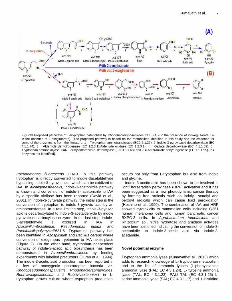
Figure3.Proposed pathways of L-tryptophan catabolism by Rhodobactersphaeroides OU5. (A = in the presence of 2-oxoglutarate; B= in the absence of 2-oxoglutarate). [The proposed pathway is based on the metabolites identified in this study and the evidence for some of the enzymes is from the literature. 1 = Tryptophan aminotransferase (EC2.6.1.27); 2=Indole-3-pyruvicacid decarboxylase (EC 4.1.1.74), 3 = Aldehyde dehydrogenase (EC 1.2.3.1)/Aldehyde oxidase (EC 1.2.3.1); 4 = Gallate decarboxylase (EC=4.1.1.59); 5= Tryptophan ammonialyase; 6=N-Formylanthranilate. deformylase (EC 3.5.1.68) and 7 = Anthranilate dehydrogenase (EC 1.1.1.95). ? = Enzymes not identified].
Pseudomonas fluorescens CHA0. In this pathway tryptophan is directly converted to indole-3acetaldehyde bypassing indole-3-pyruvic acid, which can be oxidized to IAA. In Alcaligenesfaecalis, indole-3-acetonitrile pathway is known and conversion of indole-3- acetonitrile to IAA by a specific nitrilase has been reported (David et al., 2001). In indole-3-pyruvate pathway, the initial step is the conversion of tryptophan to indole-3-pyruvic acid by an aminotransferase. In a rate limiting step, indole-3-pyruvic acid is decarboxylated to indole-3-acetaldehyde by indole pyruvate decarboxylase enzyme. In the last step, indole-3-acetaldehyde is oxidized in IAA in Azospirillumbrasilense, Pseudomonas putida and PaenibacillspolymyxaE681.5. Tryptamine pathway has been identified in Azospirillum and Bacillus cereus where conversion of exogenous tryptamine to IAA takes place (Figure 2). On the other hand, tryptophan-independent pathway of indole-3-acetic acid biosynthesis has been demonstrated in Azoperillumbrasilense by feeding experiments with labelled precursors (Duran et al., 1994). The indole-3-acetic acid production has been reported in a few of anoxygenic phototrophic bacteria viz. Rhodopseudomonaspalustris, Rhodobactersphaeroides, Rubrivivaxgelatinosus and Rubrivivaxtennius) in L-tryptophan grown culture where tryptophan production
occurs not only from L-tryptophan but also from indole and glycine.
Indole-3-acetic acid has been shown to be involved in light/ horseradish peroxidase (HRP) activation and it has been suggested as a new photodynamic cancer therapy by forming free radicals such as indolyl, statolyl and perxoyl radicals which can cause lipid peroxidation (Hoshino et al., 1990). The combination of IAA and HRP showed cytotoxicity to mammalian cells including G361 human melanoma cells and human pancreatic cancer BXPC-3 cells. In Agrobacterium tumefaciens and Rhizobium sp., nitrile hydratase and amidase activities have been identified indicating the conversion of indole-3-acetonitrile to indole-3-acetic acid via indole-3- acetamide. Novel potential enzyme Tryptophan ammonia lyase (Kumavathet al., 2015) which adds to research knowledge of L- tryptophan metabolism and to the list of ammonia lyases (L-phenylalanine ammonia lyase (PAL; EC 4.3.1.24), L- tyrosine ammonia lyase (TAL; EC 4.3.1.23), PAL/ TAL (EC 4.3.1.25) L-serine ammonia lyase (SAL; EC 4.3.1.17) and L-histidine
8 J. Microbiol. Antimicrob. ammonia lyase (HAL; EC 4.3.1.3) reported so far, hence went for purification and characterization of tryptophan ammonia lyase enzyme. Tryptophan ammonia lyase was isolated, purified and characterized as a ~225 kDaheterotetramer. The novelty of WAL was confirmed through MALDI-TOF (MS/MS) finger printing analysis. Among the four subunits, the 55 kDa subunit had ~54% score with a hypothetical protein in Rhodobactersphaeroides 2.4.1 while the remaining three had less significant match in database (NCBS, MSDB and SwissPort),the partial N-terminal sequences of WAL matched with the probable histidine ammonia lyase of Rhodobactersphaeroides 2.4.1. The molecular evidence of this enzyme is in progress.
Enzymes and their products
The production of secondary metabolites has been shown from aromatic amino acids. The metabolism of aromatic amino acids in microorganisms has been widely studied. Lots of microorganisms have evolved secondary metabolic pathways with the capacity to produce compounds displaying an impressive array of pharmacological applications which include pigments, toxins, enzyme inhibitors, pesticides, herbicides, antiparasitcs, mycotoxins, antitumor agents, antibiotics cytotoxicity activities and growth promotors of animal and plants (Bourinbaiar and Hung, 1994).
Indoles and its derivatives
Indoles are known for antimicrobial activities. In addition, molecules like indole-3-acetic acid (IAA) and indole-3-propionic acid (IPA), are natural auxins. Esters of indole acetic acid, indolemyoinositol esters; sphestrin and rhodestrin were reported to have phytoharmonal activity. The isolated rhodethrin also showed the phenol esters are an important group of bio- genic molecules reported from plants, bee propolis (Kumavathet al., 2011), yeasts like Candida and from a marine bacterium; microbulbifer. Alkyl esters of phenols are of biotechnological significance since they have antioxidant (Ranjith et al., 2008), anticancer, anti HIV and antimicrobial activities. The cytotoxicity of rhodophestrol against U937 (human leukemic monocyte lymphoma cell line) was determined (apoptosis bodies formation) on this cancer cell line even at low concentrations (50 nM). It has COX-I and COX-II inhibitory activity. Rhodophestrol is a potential anticancer and thus worth exploiting from Rubrivivaxbenzoatilyticus (Fonnum and Larsen, 1965). The literature suggested the production of other phenols during the bioprocessing of an aromatic amino acid like L-phenylalanine by Rubrivivaxbenzoatilyticus. Though most of theseare representatives of free phenols, which many researchers have already identified as microbial products, the microbially produced phenol terpenoids and related conjugates are novel bio-molecules, which are worth
exploiting. Indoles are extensively produced by the chemical industries for variety of applications including, pharmaceuticals, pesticides and dyes. They are widely used as analgesics, anti-inflammatory agents antihypertensive, anti HIV compounds and phytohormones (Ahn et al., 2004). Many indole esters are found as COX-2 selective enzyme inhibitors. They were also found to have anti lipid peroxidation activity and anti-superoxide formation. Indole esters are also found to have more phytohormonal activity than their corresponding acids in the auxin bioassay.
Rhodethrin
Rhodethrinhas A molecular mass of m/z 279 [M±H] ± and was named as rhodethrin (3-hydroxy-6-(1H-indole-33-yloxy)-4-methyl-hexanoic acid). This compound differs from sphestrin or rhodestrin in having an ether linkage rather than ester linkage and in the length of the terpenoid side chain. Esters of the myoinositol-indole acetate were only reported from plants. This metabolite was not a product of L-phenylalanine. The LC-MS analysis revealed a more wide range of indoles including those purified (m/z 279 m/z and 129m/z) and the other metabolites identified include indole-3-acrylaldehyde, idole-3-carboxaldehyde, indole-3-acetic acid and rhodethrin. It presumes the molecular mass of 171 (m/z) as indole-3-acrylaldehyde; this compound is not available in the literature, but a related compound indole-3-acrylic acid in known. Excretion of indole-3-acrylic acid in the urine and feces of L-tryptophan fed animals was attributed to the intervention of intestinal microorganisms. Thus, indole-3-acrylaldehyde may be an intermediate of L-tryptophan metabolism in Rhodobactersphaeroides. Production of indole-3-carboxaldehyde was reported earlier from an Acetenobacter sp. and by an unidentified fungus. Microbial production of indole-3-acetic acid is well known and was reported even from a few other purple bacteria (Rhodopseudomonaspalustris, Rhodobactersphaeroides, and Rubrivivaxtenuis) when grown on L- tryptophan or from indole ± glycine by Rhodobactersphaeroides. Low levels of some of the metabolites in the L-tryptophan induced culture supernatant were also observed in the absence of L-tryptophan, the notable one was indole-3-acetic acid. While in the absence of 2-oxoglutarate, none of the above metabolites were observed in LC-MS profiling One normally expects the production of indole by the enzymatic action of tryptophanase (EC 4.1.99.1) encoded by tnaA gene(Kar et al., 1999), which was not observed as confirmed through the LC-MS and enzyme assay.
Indole 3- acrylic acid
To the best of knowledge, the proposed production of indole 3-acrylic acid pathway is a novel biological

pathway. Indole 3-acrylic acid was not listed as a phyto- auxine(http://www.biologie.uni-hamburg.de/b online/e31/31.htm) though its microbial origin was suspected. Production of indole-3-acrylic acid and ammonia indicates that the enzyme as an ammonia lyase, since this family of enzymes cleaves at C-N bond of various substrates and results in the formation of ammonia and corresponding products.
Indigo
The blue dye indigo has been known since prehistoric times and is still one of the most economical important textile dyes. The first report of microbial indigo production was in 1928. It is biosynthesized in bacteria through the oxidation of indole by a naphthalene dioxygenase and subsequent oxidation and dimerization. The desire to achieve a competitive, alternative to the chemical production of indigo rejuvenated interest in microbial indigo production since many microorganisms expressing both monooxygenase and dioxygenase during growth on aromatic hydrocarbons have been shown to transform indole i n t o indigo. The work has been focused on the naphthalene dioxygenase from Pseudomonas putida PpG7 expressed in Escherichia coli. Some of the genes of indigo biosynthetic pathway have been cloned and used to construct “engineering bacteria”. More efficient fermentation systems for indigo production have been exploited with this kind of bacteria.
Violacein
Chromobacteriumviolaceum was first reported as an isolate from wet rice paste. One of the characteristics of this microorganism is the ability to produce a purple pigment known as violacein under aerobic conditions. The biological role of violacein in Chromobacteriumviolaceum, as well as its biosynthesis pathway and the role of tryptophan and other indole derivatives have been reported. Tryptophan appears to be the only precursor molecule in violacein biosynthesis. Its production is apparently essential for pigment production in Chromobacteriumviolaceum. The IUPAC name and molecular mass of violacein are (3-[1,2-dihydro-5-(5-hydroxy-1H-indol-3-yl)-2- oxo-3H-pyrrol-3-ylidene]-1,3-dihydro-2H-indol-2-one) and 343.34, respectively. Violacein has attracted interest owing to its important multiple biological activities and pharmacological potential such as antibiotic, bactericide, antitumoralgenotoxic properties (Forrest et al., 1993). The antioxidant efficiency has been reported against oxygen and nitrogen reactive species as a scavenger of hydroxyl, superoxide and nitric oxide radicals. In addition, it is capable of inducing apoptosis in cancer cell cultures and is effective against a panel of neoplastic cell lines including leukemia lineage cancer diseases.
Indolmycin Indolmycin is a secondary metabolite produced by
Kumavath et al. 9
Streptomyces griseus ATCC 1248 (formally Streptomyces albus BA 3972A), which was isolated from a sample of African soil. Indolmycin completely inhibits bacterial TrpRS (tryptophanyltRNAsynthetase) enzyme and it exhibits antimicrobial activity against gram positive and gram negative bacteria. Recently researchers have shown that indolmycin is active against mycobacteria and Helicobacter pylori. Indolmycin is also known as a major causative agent of chronic active gastritis.
Phenols and its derivatives
Alkyl esters of gallic acid
The chief source for obtaining gallic acid is through the hydrolysis of plant based products like tannins (Ahn et al., 2004). Microbial production of gallic acid has been reported using tannic acid as substrate. The esters iso-amyl- (iAG), n-amyl- (nAG), iso-butyl (iBG), n-butyl-(nBG) and isopropyl gallate (iPG) have been chemically synthesized from gallic acid. Gallic acid (3,4,5-trihydroxybenzoic acid) is an industrially important phenol and finds its applications in various fields. Alkyl esters of gallate form an important group of biogenic molecules which have been reported from plants, bee propolis and yeasts namely Candida (Forrest et al., 1993). These molecules are of biotechnological significance since these are known to have anti-oxidant, anticancer, anti HIV and antifungal/microbial activities (Forrest et al., 1993). Alkyl esters of gallic acid have antiviral, antibacterial, antifungal properties specifically against gram-positive bacteria (Kuniyoshi et al., 2003). The culture supernatants did not yield ammonia indicating that L-phenylalanine assimilation as nitrogen source is not through deamination process and hence possibility for transaminase activities is strong. Aromatic aminotransferase activity has been measured with L-phenylalanine, L-tyrosine and DOPA as substrates in the presence of 2-oxoglutarate and the substrate omission has been analyzed using HPLC. The enzyme activity has been reported to be 6, 10 and 21 units.mg protein
-1.min
-1. The increase in transaminase
activity with the substrates L-phenylalanine < L-tyrosine < DOPA suggested that transamination occurred at the level of DOPA. DOPA consumption stagnated in the absence of supplemented keto acceptor, which restored only in the presence of 2-oxoglutarate (Figure 3). The transaminated product of DOPA has been extracted into ethyl acetate, concentrated and analyzed using LC-MS. A mass of 196 (m/z) indicated the product as 3,4-dihydroxyphenylpyruvic acid.
3, 4-dihydroxyphenylalanine reductive deaminase
Ammonia, the product of deaminase, has been observed only in the absence of added 2-oxoglutarate. The deamination product of DOPA was extracted (after acidifying; pH 4) into ethyl acetate, concentrated and analyzed using LC-MS. A mass of 182 (m/z) indicated the product as 3,4-dihydroxyphenylpropionic acid (DPPA) and the enzyme as reductive deaminase. The enzyme L-

10 J. Microbiol. Antimicrob. phenylalanine ammonia lyase (PAL; EC. 4.3.1.24), which converts L-phenylalanine to trans-cinnamate is most commonly observed in plants and also in prokaryotes. Biological applications Anoxygenic phototrophic purple-non sulfur bacteria (PNSB) have the capability to degrade a wide range of low molecular weight aromatic hydrocarbons for growth (Poppe and Rétey, 2005) and thus help in the maintenance of biogeochemical cycles. The most extensively studied species among PNSB is Rhodopseudomonaspalustris, whose total genome analysis indicated the existence of at least five aromatic ring cleavage pathways, representing more aerobic (oxidative) mechanisms, than anaerobic. The other species of PNSB capable of utilizing benzoate for growth include: Phaeospirillumfulvum, Rubrivivaxpurpurens, Rhodomicrobiumvannielli and Rhubrivivaxbenzoatiliticus. On the other hand, the aromatic utilization for growth was not well reported among other purple bacteria, some incubation results indicated light dependent transformation of aromatic hydrocarbons by Rubrivivaxgelatinosus, RhodobactercapsulatusRhodobacterblasticus (Patten and Glick, 2002) and Rhodobactersphaeroides Correlation between L-phenylalanine consumption and simultaneous production of phenols during growth as sole nitrogen source by Rhodobactersphaeroides OU5 is similar to that observed with Rhodobactercapsulatus and differ from anaerobic chemotrophic bacterial metabolism, which produced benzoate. Anoxygenic production of phenyl gallates Gallate was the major product of L-phenylalanine meta- bolism in Rubrivivaxbenzoatilyticus. Gallate is a biotechnologically important compound (Ranjith et al., 2007), mainly produced by the hydrolysis of tannins and its microbial production by a few chemotrophic bacteria and fungi was observed earlier (Kumavathet al., 2007). Other phenols identified from the culture supernatant of Rhodobactersphaeroides include gallate, protocatechuate, catechol and caffeate, while homogensitate was reported from Rhodobactercapsulatus. Both gallate and caffeate had the same retention time under the experimental HPLC conditions used and their identity could be resolved and distinguished only through LC-MS analysis by their corresponding molecular masses. Rubrivivaxineand rhodophestrol Detailed characterization of these two of the purified
metabolites based on FT-IR; 1H NMR, 13C NMR and mass analysis confirmed the structures as 3,4-dihydroxybenzoicacid -5-carboxy-4-hydroxy-3-methyl-pentylester and 2,3,4 tryhydroxybenzoylterpenoid ester. The hydrolysis of the ester bond and of rhodophestrol and 2,3,4-tryhydroxybenzoyl terpenoid ester by the enzyme esterase (EC 1.1.1.49) and identifying the corresponding phenol acid and alcohol through HPLC/LC-MS analysis helped in the confirmation of the novel metabolites, whose yield was about 100 μmole/(65μg)/ml-1. Rubrivivaxine and 0.2 μ mol 2,3,4-tryhydroxybenzoyl terpenoid ester.
Phenol esters were identified through hydrolysis of the ester bonds of the metabolites in different fractions using esterase enzyme and the corresponding acid and alcohol were analyzed using LC-MS (56). These are broadly categorized as gallate, caffeate, genisitate and protocatechuate esters. While the esterase hydrolyzed products represented the corresponding acids, the alcohols were hydroxyalkanoates, terpenols and phenols.
The marine bacterial isolate identified as belonging to the genus Microbulbifer could produce 4-hydroxybenoate alkyl esters (butyl, heptyl and nonyl 4- hydroxybenzoate esters; commonly called as “parabens”), which were antimicrobial, which differ from the isolated conjugated metabolites from phenylalanine of Rubrivivaxbenzoatilyticus. 3,4- Dihydroxyphenylalanine was identified as the major product of L-phenylalanine (L-tyrosine) metabolism of Rhodobactersphaeroides OU5 (Sunayana et al., 2005).
The literature evidences suggest that DOPA is the downstream product of L-phenylalanine or L-tyrosine in Rhodobactersphaeroides OU5, thus differ from the homogentisate pathway observed in other bac- teria. Based on the earlier evidence of production of DOPA by Erwiniaherbicola from L-tyrosine together with isolation of the enzyme DOPAATS, in this study and from other bacteria, we propose the bacterial DOPAATS pathway of L-phenylalanine catabolism (Figure 4). The further downstream products of this metabolism were identified through the LC-MS analysis (52).
The role of alternative enzymes involved in the deamination of DOPA is also shown in the pathway (Figure 4). The comparison of the three enzymes i n v o l v e d in ammonia metabolism of DOPA in Rubrivivaxbenzoatilyticus are shown in Table 2. Para-dimethylaminobenzaldehyde reagent (Ehrlich reagent) is the most commonly and widely used reagents in the identification of indoles.
This reagent gives pink to red colour when reacted with indole and its derivatives, while yellow to brown with anthranilate. The culture supernatant of L-tryptophan induced Rubrivivaxbenzoatelitus and E. coli were tested with Ehrlich reagent. The orange and pink coloured reactions were observed with Rhodobactersphaeroides and E. coli, and its suggesting variations in the products of L- tryptophan metabolism.

Kumavath et al. 11
Figure 4.Proposed intracellular catabolism of L-phenylalanine by Rubrivivaxbenzoatilyticus. The pathway is based on experimental evidence observed in the present study. [1=L-phenylalaninehydrogenase;2=L-tyrosine dehydrogenase;3= 3,4-dihydroxyphenylalanine 2-oxoglutarate aminotransferase (DOPAATS;EC2.6.1.49);4=3,4-dihydroxyphenylalanine oxidative deaminase 5= 3,4- dihydroxyphenyalanine reductive Deaminase EC 4.3.1.22) ?= not knownone].
CONCLUSIONS 3,4-Dihydroxyphenylalanine as an intermediate of L-phenylalanine metabolism through intermediate of L-tyrosine in Rhodobactersphaeroides. Few enzymes reported in the ammonia metabolism of 3,4-dihydrophenylalanine 2-oxoglutarate aminotransferase; 3,4–dihydroxy- phenylalanine reductive deaminase and 3,4-Dihydroxy phenylalanine oxidative deaminase (Sunayana et al., 2005) were purified to homogeneity and characterized. An enzyme was identified in the downstream of L- tryptophan in Rubrivivaxbenzoatilyticus. Rhodethrin (Fonnum and Larsen, 1965) is one such novel molecule, which has Cox-2 inhibitory activity, cytotoxicity against cancer cell lines, phytoharmonal activity and antimicrobial activity. Rubrivivaxin is novel phenol terpenoid molecule, which has Cox-1 inhibitory activity, cytotoxicity against cancer cell lines (U937; Human leukemic monocyte lymphoma cell line) and antimicrobial activity.
This article suggested that production of a wide range of indoleterpenoids, and phenols during the bioprocessing of an aromatic amino acid like L- Tryptophan, L-phenylalanine and L-Tyrosine by Rubrivivaxbenzoatilyticus. Though most of these are representatives of free phenols, and indoles which most of the researchers had already identified as microbial enzyme products which are microbially produced phenol terpenoids and indoleterpenoids related conjugates are novel biomolecules, which are worth exploiting for microbial based drugs.
CONFLICT OF INTERESTS
The authors confirmed that this review article content have no conflict of interests.
ACKNOWLEDGMENT
The author’s thanks Prof. Anil Kumar (Devi Ahilya University, Indore, India), Prof. RamanaCh (University of Hyderabad, India), APK was supported by grants from National Medical Research Council of Singapore and by the National Research Foundation Singapore and the Singapore Ministry of Education under its Research Centers of Excellence initiative to Cancer Science Institute of Singapore, National University of Singapore. Dr. Vasco Azevedo (University of Federal de Minas Gerais, Brazil). Dr. Ranjith N Kumavath principal investigator thanks the Research grants of the DST- SERB and UGC. REFERENCES Ahn MR, Kumazawa S, Hamasaka T, Bang KS, Nakayama T (2004).
Antioxidant activity and constituents of propolis collected in various areas of Korea. J. Agric. Food. Chem. 52:7286-792.
Bartling D, Seedorf M, Schmidt RC, Weiler EW (1994). Molecular characterization of two cloned nitrilases from Arabidopsis thaliana: key enzymes in biosynthesis of the plant hormone indole-3-acetic acid. Proc. Natl. Acad. Aci. USA. 91:6021-6025.
Bertoldi M, Frigeri P, Paci M, Borri-Votattorni C (1999). Reaction specificity of native and nicked 3,4-dihydroxyphenylalanine

12 J. Microbiol. Antimicrob.
Table 2.Differentiating characters of the three enzymes of phenylalanine of Rubrivivaxbenzoatilyticus.involved in ammonia liberation /transformation of DOPA, an intermediate in the catabolism of L- phenylalanine of Rubrivivaxbenzoatilyticus.
Enzymes Products Co-substrates Co-factors Km(μM) Kcat (s-1) M.Wt. (kDa) Subunits(determinedby SDS-PAGE) (kDa)
L-DOPA-oxidative deaminase DOPP O2 Nil 11.84±1.80 0.680±0.023 ~190 Pentamer (54,43,34,25,22)
L-DOPA-reductive deaminase DPPA Nil NADH 21.23±0.09 0.0636±3.0 ~274 Hetero-tetra- mer (117,85,49,35)
L-DOPA-amino transfe- rase DOPP 2-KGA PLP 4.1 ND ND Homodimer
0.35±0.045 0.29±3.0 ~123 Heterodimer (60,63) [Results are means ± SD of three different determinations done in duplicates. *Isolated from Pig brain. α-KGA = 2-oxoglutarate; PLP = Pyridoxal-5-phosphate; NADH = Nicotinamide adenine dinucleotide; Nil = No requirement; kDa = Kilo Dalton; DOPP =3,4-dihydroxyphenyl pyruvic acid; DPPA = 3.4-dihydroxyphenyl pro- pionic acid; DOPA= 3,4-dihydroxyphenylalanine. ND = Not deter- mined].
decarboxylase. J. Biol. Chem. 274:5514-5521. Bourinbaiar AS, Hung SL (1994).The non-steroidal anti-
inflammatory drug indomethin as an inhibitor of HIV replication.FEBS Lett. 360:85-88.
Chen H, Li YL, Tu SS, Lei N, Ling Y, Xiao YH, Jing SH, Li PS, Yu L, Lu SS (2005).Apoptosis of pancreatic cancer BXPC-3 cells induced by indole-3-acetic acid in combination with horseradish peroxidase.World. J. Gastroenterol. 11(29):4519-4523.
Copeland A, Lucas S, Lapidus A, Barry K, Detter JC, Glavina T, Hammon N, Israni S, Pitluck S, Richardson P, Mackenzie C, Choudadhry M, Larimer F, Hauser LJ, Land M, Donohue TJ, Kaplan S (2005). Complete sequence of chromosome-1 of Rhodobacter sphaeroides2.4.1 submitted (SEP-2005) to the EMBL/GenBank/ DDBJ databases.
David PP, Aaron MG, Paul PT (2001). Purification and characterization of an L-amino acid deaminase used to prepare unnatural amino acids. J. Mol. Catal. B Enzym. 11:795-803.
Duran N, Antonio RV, Haun M, Pilli RA (1994). Biosynthesis of a trypanocide by Chromobacteriumviolaceum.World J. Microbiol.Biotechnol. 10:686-690.
Duran N, Erazo S, Campos V (1983). Bacterial chemistry-II: antimicrobial photoproduct from pigment of Chromobacteriumviolaceum. An Acad. Bras. Cienc. 55:231-234.
Elsa AB, Elias RO, Jose ML, Cristina F, Beatriz G, Jose LG, Eduardo D, Baltasar M (2004). The Homogentisate pathway: a central catabolic pathway involved in the degradation of L-phenylalanine, L-tyrosine, and 3-hydroxyphenylacetate in Pseudomonas putida. J. Bacteriol. 186 (15):5062-5077.
Fonnum F, Larsen K (1965). Purification and properties of dihydroxyphenylalanine transaminase from guinea pig brain.
J. Neurochem. 12:589-598. Forrest F, Nancy M, Keith AB (1993). Production of L-
Dihydroxyphenylalanine in Escherichia coli with the Tyrosine Phenol-Lyase Gene Cloned from Erwiniaherbicola. Appl. Environ. Microbiol. 59 (9):3070-3075.
Herrera CM, Ramos JL (2007). Catabolism of phenylalanine by Pseudomonas putida: the NtrC-family PhhR regulator binds to two sites upstream from the PhhA gene and stimulates transcription with σ70. J. Mol. Biol. 366:1374-1386.
Hoshino T, Ogasawara N (1990). Biosynthesis of violacein: evidence for the intermediary of 5- hydroxy-L-tryptophan and the structure of a new pigment, oxyviolacein, produced by the metabolism of 5-hydroxytryptophan. Agric. Biol. Chem. 64:2339-2345.
Juana M, Marianna, Patrauchan A, Stewart G, Davies J, Lindsay Eltis D, William M (1999). Phenylacetae catabolism in Rhodococcus spp. RHA1: a central Pathway for Degradation of Aromatic Compounds. J.Bacteriol. 187:4497-4504.
Kar B, Banerjee R, Bhattacharya BC (1999). Microbial production of gallic acid by modified solid- state fermentation. J. Ind. Micro. Biotechnol. 23:173-177.
Kevin TW, Benjamin NM, Pyung CL, Andew JM, Claudia SD (2006). Discovery of a substrate selectivity switch in tyrosineammonialyase, a member of the aromatic amino acid lyase family. Chem. Biol. 13:1317-1326.
Kodach LL, Bos CL, Duran N, Peppelenbosch MP, Ferreira CV,Hardwich JC. (2006). Violacein synergistically increases 5-fluorouracil Cytotoxicity, induces apoptosis and inhibits Akt-mediated signal transduction in human colorectal cancer cells. Carcinogenesis 27:508-516.
Koga J (1995). Structure and function of indole pyruvate decarboxylase, a key enzyme in indole-3- acetic acid
biosynthesis.Biochem.Biophys.Acta 1249:1-13. Kumavath RN, SasikalaCh, RamanaCh (2007b). Rhodethrin: a
novelindoleterpenoid ether produced by Rhodobactersphaeroides has cytotoxic and phytohormal activities. Biotechnol.Lett. 29:1399-1402.
Kumavath RN, SasikalaCh, RamanaChV (2011). Rubrivivaxin, a New Cytotoxic and Cyclooxygenase- I Inhibitory Metabolite from Rubrivivaxbenzoatilyticus JA2.World J. Microbiol.Biotechnol. 27:11-16.
Kumavath RN, SasikalaCh, RamanaChV, DedmalyaBarh, Alan premkumar, Vasco Azivedo (2015). Isolation and Characterization ofTryptophan ammonia lyase by Rubrivivaxbenzoatilyticus JA2.Curr.Protein Pept.
Sci.16(8):775-781.
Kuniyoshi S, Xingglian G, Mika H, Hiroto S, Syoko F, Deji Y, Ryuichiro K, Mototake T, Ikuo S (2003). In- dole-3-carbaldehyde: a tyrosine inhibitor from fungus YL 185. J. Wood Sci. 49:349-354.
Lee S, Flores-Encarnacion M, Contreras-Zentella M, Garcia-Flores L, Escamilla JE, Kennedy C (2004). Indole 3-acetic acid biosynthesis is deficient in Gluconacetobacterdiazotrophicus strains with mutations in cytochrome c biogenesis genes. J. Bacteriol. 186:5384-5391.
Li SC, Li H, Zhang F, Li ZJ, Cui JR (2003). Anticancer activities of substituted cinnamic acid phenethyl esters on human cancer cell lines. J. Chin. Pharm. Sci. 12:184-187.
Nieminen SM, Maki Paakkanen J, Hirvonen MR, Roponen M, Von wright A (2002).Genotoxicity of gliotoxin, a secondary metabolite of Aspergillusfumigatus in a battery of short term test system.Mutat. Res. 520:161-170.
Panke S, Held M, Wubbolts M (2004). Trends and innovations in industrial biocatalysis for the production of fine chemicals.Curr.Opin.Biotechnol. 15:272-279.

Patten CL, Glick BR (2002). Role of Pseudomonas putida indo- leacetic
acid in development of the host plant root system. Appl. Environ. Microbiol. 68:3795-3801.
Phi QT, Park YM, Ryu CM, Park SH, Ghim SY (2008). Functional identification and expression of indole-3-pyruvate de- carboxylase from PaenibacillspolymyxaE681. J. Microbiol. Biotechnol. 18(7):1235-1244.
Poppe L, Rétey JF (2005). Friedel-Crafts-type mechanism for the enzymatic elimination of ammonia from histidine and phenylalanine.Angew. Chem. Int. Ed. Engl. 44:3668-3688.
Ranjith NK, SasikalaCh, RamanaCh (2007). Catabolism of L-phenylalanine and L-tyrosine by Rhodobactersphaeroides OU5 occurs through 3,4-dihydroxyphenylalanine.Res. Microbiol. 158:506-511.
Ranjith NK, SasikalaCh, RamanaCh (2008). Purification and characterization of 3,4- dihydroxyphenylalanine oxidative deaminase from Rhodobactersphaeroides OU5. Can. J .Microbiol. 54(10):829-834.
Regina VA, Creczynski-Pasa TB (2004). Genetic analysis of violacein biosynthesis by Chromobacteriumviolaceum.Genet. Mol. Res. 3(1):85-91.
Rother D, Poppe L, Morlock G, Viergutz S, Retey J (2002). An active site homology model of phenylalanine ammonia lyase from Petroselinumcrispum. Eur. J. Biochem. 269:3065-3075.
Kumavath et al. 13 Samaha HS, Kelloff GJ, Steele V, Rao CV, Reddy BS (1997).
Modulation of apoptosis by sulindac, curcumin, phenylethyl 3-methylcaffeate and 6-phenylhexyl isothiocyanate: apoptoptic index as a biomarker in colon cancer chemoprevention and promotion. Cancer Res. 57:1301-1305.
Schneider S, Mohamed ME, Fuchs G (1997). Anaerobic metabolism of L-phenylalanine via benzoyl-CoA in the denitrifying bacterium Thaueraaromatica. Arch. Microbiol. 168:310-320.
Srinivas M, Vasavi D, Girisham S, Reddy SM (2002). Production of indole acetic acid by anoxygenic phototrophic bacteria under different conditions.Indian J. Microbiol. 42:215-128.
Sunayana MR, SasikalaCh, RamanaVCh (2005).Production of novel indole ester from 2-amonobenzoate by Rhodobactersphaeroides OU5. J. Ind. Microbiol. Biotechnol. 32:41-45.
Wolfram W, Oliver F (2002). Can we discover novel pathways using metabolomics analysis?.Curr.Opin.Biotechnol. 13:156-160.