Embed Size (px)
Citation preview

American Journal of Biomedical and Life Sciences 2020; 8(4): 97-113 http://www.sciencepublishinggroup.com/j/ajbls doi: 10.11648/j.ajbls.20200804.16 ISSN: 2330-8818 (Print); ISSN: 2330-880X (Online)
Review Article
Potentials of Encapsulated Flavonoids in Biologics: A Review
Mahesh Dattatraya Dere*, Ayesha Alim Khan
Department of Chemistry, Savitribai Phule Pune University, Pune, India
Email address:
*Corresponding author
To cite this article: Mahesh Dattatraya Dere, Ayesha Alim Khan. Potentials of Encapsulated Flavonoids in Biologics: A Review. American Journal of Biomedical
and Life Sciences. Vol. 8, No. 4, 2020, pp. 97-113. doi: 10.11648/j.ajbls.20200804.16
Received: July 9, 2020; Accepted: July 25, 2020; Published: August 25, 2020
Abstract: Flavonoids are a versatile class of natural polyphenolic compounds that represent secondary metabolites from higher plants. Their basic structures consists of fifteen-carbon skeleton consisting of two benzene rings (A and B) linked via a heterocyclic pyrane ring (C) to produce a series of subclass compounds such as flavones, flavonols, flavanones, isoflavones, flavanols or catechins and anthocyanins. Their biological activities are dependent on the structure, chemical nature and degree of hydroxylation, substitutions, conjugation and degree of polymerization. A brief description of flavonoids, its source and classification have been described. Although flavonoids are integral in nutraceutical, pharmaceutical, medicinal, cosmetic and other applications their bioavailability to the target tissues and cells are restricted due to poor water solubility and enzymatic degradation. To increase effectiveness, currently encapsulation of the drug candidate in biological material that are able to enhance the potential health benefits by increasing the water solubility and targeted delivery are being achieved. Biodegradable natural, synthetic and semi-synthetic material/ polymers approved by the US Food and Drug Administration (FDA) for use in the preparation of nanodrugs as well as the applied encapsulation technique are discussed that prevent against oxidation, isomerization and degradation of the flavanoids. The aim of this review is to identify specific flavonoids that exhibit increased pharmacological and biological efficiencies on encapsulation. Thus, these potential drugs may help in preventing many chronic diseases and lead to future research directions.
Keywords: Flavonoids, Encapsulation, Delivery Systems, Biological Activity
1. Introduction
Flavonoids are secondary metabolites ubiquitously present in plants that comprise a large group of polyphenolic compound with benzo- ɣ -pyrone structure responsible for variety of pharmacological activities [1, 2]. The flavonoids are mainly accumulated in the edible parts of plants particularly in fruits and vegetables, responsible for red and dark blue color of berries as well as orange and yellow color in citrus fruits. They also act as a secondary antioxidant defense system in plant tissue exposed to different abiotic and biotic stress and regulate growth factor in plants such as auxin [3].
In the human body they play similar role as vitamins [4, 5]. Their activities are dependent on the structure and chemical nature, degree of hydroxylation, substitutions, conjugation and
degree of polymerization. Flavonoids shows a variety of biological activities such as antioxidants, modulators of cell signaling, anti-inflammatory agents, cardio protectants, inhibitors of neurodegenaration and ability to inhibit the growth of a wide range of microorganisms and viruses [6-8].
The chemical structures of flavonoids are based upon a fifteen-carbon skeleton consisting of two benzene rings (A and B) linked via a heterocyclic pyrane ring (C) shown in Figure 1. The classification of flavonoids depends on the level of oxidation and pattern of substitution on the C ring, while individual compound within a class differ in the pattern of substitution on the A and B rings. The main classes of flavonoids, structures and examples with the position of substituents are shown in Table 1 [9].

American Journal of Biomedical and Life Sciences 2020; 8(4): 97-113 98
Table 1. Main classes of flavonoids, structures, examples with position of substituents [14, 15].
Class of Flavonoids Structure Name Position of Substituents
Flavanones
Hesperetin Naringin Naringenin Eriodictyol Hesperidin Likvirtin
5,7,3’-OH, 4’-OMe 5,4’-OH, 7-OR 5,7,4’-OH 5,7,3’,4’-OH 5,7,3’-OH, 4’-OMe, 7-rutinoside 7-OH
Flavan-3-ols
(+)-Catechin Epigallocatechin Epigallocatechin Gallate
5,7,3’,4’-OH 5,7,3’,4’,5’-OH 5,7,3’,4’-OH, 3-gallate
Flavones
Chrysin Apigenin Leteolin
5,7-OH 5,7,4’-OH 5,7,3’,4’-OH
Flavonols
Rutin Kaempferol Quercetin Galangin
5,7,3’,4’-OH, 3-rutinose 5,7,4’-OH 5,7,3’,4’-OH 5,7-OH
Isoflavones
Genistein Daidzein Puerarin Glycitein
5,7,4’-OH 7,4’-OH 7,4’-OH, 8-glucoside 7,4’-OH, 6-OMe
Flavanonol
Taxifolin 3,5,7,3’,4’-OH
Being phytochemicals, flavonoids cannot be synthesized by
human and animal and hence form an integral part of human and animal diet [8, 10]. The main classes of flavonoids, food source, their specification and important biological properties are reported in Table 2.
The physicochemical properties of flavonoids such as molecular size, configuration, lipophilicity, solubility, pKa and structure; viz glycoside or aglycone could play a vital role in the absorption of dietary flavonoids. Liberated from food by chewing, aglycans can be easily absorbed by small intestine, while flavonoid glycosides have to be converted into the aglycan [11].
Flavonoids are poorly absorbed in the intestine in their natural form, and are extensively degraded by intestinal microorganisms and/or enzymes, to produce different metabolite. If these metabolite adsorbed are subjected to the hepatic enzymatic system the new metabolites formed differ in their bioactivity.
After the hydrolysis of sugar moieties in the small intestine or due to bacterial activity in the colon, aglycones are generated and further metabolized before reaching the systemic circulation. Briefly, numerous factors could play a role in limiting the glucuronidated or the sulfated form. As a consequence, flavonoids results in poor bioavailability, poor permeability, instability and extensive first-pass bioavailability of flavonoids [12, 13].
Figure 1. Basic flavonoid structure.
Table 2. Main classes of flavonoids, food source, their specification and important biological properties [16-29].
Class of
Flavonoids Dietary Source Specifications
Main biological
properties
Flavonols Fruits and vegitables (grape berries, apple, tomato, onion, broccoli and red lettuce), green tea, black tea and red wine
Flavonols are the most ubiquitous flavonoids in food, sensitive to oxidation, lights and pH aglycones slightly soluble but glycosides soluble in water.
Vitamin P factor protecting capillaries and veins, often Flavones Parsley, broccoli, celery, carrots, onion leaves, Natural pigment, flavones are much less common than flavonols

99 Mahesh Dattatraya Dere and Ayesha Alim Khan: Potentials of Encapsulated Flavonoids in Biologics: A Review
Class of
Flavonoids Dietary Source Specifications
Main biological
properties
cabbage, peppers, chrysanthemum flowers and apple skin
in fruit and vegitables, sensitive to oxidation, lights and pH aglycones slightly soluble but glycosides soluble in water
Antioxidant, anti-inflammatory, antiallergenic, antiviral, anti-spamodic, antibacterial and anti-carcinogenic properties,
Flavanones Citrus fruits (grape fruit, orange, lime, lemon and tangelo), tomatoes and some aromatic plants (mint)
Flavanones are sensitive to oxidation, lights and pH aglycones insoluble but glycosides soluble in water
Isoflavones Green split peas, split peas, chick peas, black beans, soyabean, sunflower seeds
Structural similarities with estrogens confers pseudohormonal properties, astringent and bitter taste, sensitive to alkaline pH
Flavanols Fruits (apple, kiwi, grape, cherry, peach), green and black tea, red wine and cider, peels or seeds of fruits and vegetables
Astringent and bitter taste, slightly soluble in water (monomer) and soluble in water and alcohol (polymer), sensitive to high temperature, oxidation, light and pH
Anthocyanins Tea, red wine, cereals, honey nuts, some leafy and root vegetables (aubergines, cabbage, beans, onions, radishes) and fruits
Plant pigments, highly sensitive to temperature, oxidation, light and pH, water soluble.
2. Encapsulation of Bioactive Compound
Encapsulation is a “nature made” technique used for product formulation to trap important biological ingredients into a carrier, which protect the trapped biological material against oxidation, isomerization and degradation. This technique increases the shelf life of material over a period of time and control/sustained
delivery of functional substances when ingested in the body [30]. It also improves the solubility and pharmacokinetics
profiles of insoluble drugs. In many cases, targeted drug delivery is greatly enhanced, bioavailability to the target tissues and cells are significantly improved. It reduces their toxic side effects to normal cells and increases the delivery of such drug to tumor tissue [31].
Table 3. Encapsulating materials are classified according to their origin as natural, synthetic and semisynthetic (Adapted from review “Critical evaluation of
biodegradable polymers used in nanodrugs”, Edgar Marin et al. 2013, International Journal of Nanomedicine 2013: 8 3071–3091) [37].
Origin Sub classification Examples
Synthetic
Hydrolyzable backbones
Polyesters
Poly (glycolic acid) Poly (lactic acid) Poly (caprolactone) Poly (lactic-co-glycolic acid) Poly (butylene succinate) Poly (trimethylene carbonate) Poly (p-dioxanone) Poly (butylene terephthalate)
Poly (ester amide) s Hybrane® S120043 Polyurethanes DegraPol®45 Polyanhydrides Poly [(carboxyphenoxy) propane-sebacic acid] Polyphosphoesters Poly [bis (hydroxyethyl) terephthalate-ethyl orthophosphorylate/terephthaloyl chloride] Carbon backbones (hydrolysis cannot occur)
Poly (ortho esters)
Poly (ortho esters) I Poly (ortho esters) II Poly (ortho esters) III Poly (ortho esters) IV
Poly (alkyl cyanoacrylates) Poly (butyl cyanoacrylate) Polyether Poly (ethylene glycol) Poly (amino acids) Tyrosine derived polycarbonate
Semisynthetic Microbial polyesters Poly (β-hydroxyalkanoate) s Poly (hydroxybutyrate) Poly (hydroxybutyrate-co-hydroxyvalerate)
Natural
Proteins
Animal source Collagen Albumin
Vegetable source Gluten Polysaccharides
Animal source Chitosan Hyaluronate Cellulose
Vegetable source Alginate
Starch
Various newly synthesized chemical entities such as poly
(lactic-co-glycolic acid) (PLGA), poly (glycolic acid) (PGA) and poly (lactic acid) (PLA) have been approved by the US
Food and Drug Administration (FDA) with a wide therapeutic efficacy and easy availability in the market. Since, ancient times, herbal remedies and natural extract are used to cure

American Journal of Biomedical and Life Sciences 2020; 8(4): 97-113 100
various diseases as they contain several phytoconstituents which work simultaneously against the disease. Conventional therapy provides non-targetability in tissue and organs due to peak and valley fluctuations and requires a frequent dose of administration. The controlled release of drug delivery system provides drug released at a controlled rate and maintains the overall therapeutic concentration of drug in the body [32-36].
There are various techniques that are used for encapsulation such as spray drying, spray cooling/chilling, extrusion, fluidized bed coating, co-acervation, liposome entrapment, inclusion complexation, centrifugal suspension separation, lyophilization, co-crystallization and emulsion, nanoparticles etc. [38, 39].
Generally three steps are involved in the encapsulation of bioactive agents.
1. The formation of wall around the bioactive compound (core material) to be encapsulated.
2. Ensuring that undesired leakage does not occur. 3. Ensuring that undesired materials are kept outside
[40, 41]. The effectiveness of nutraceutical product in preventing
disease depends on preserving the bioavailability of the active ingredients. After oral administration only small proportion of the molecules are made available due to insufficient gastric resistance time, low permeability and/or solubility within the gut as well as conditions during food processing and storage
(temperature, oxygen, light) or in the gastrointestinal tract (pH, enzymes, presence of other nutrients), all these factor limit the activity and potential health benefits of the nutraceutical component [42]. To increase the activity and health benefits it requires product formulation to provide protective mechanism that can maintain the active chemical form until the time of consumption, and deliver this form to the physiological target within the organism [43].
3. Material Used for Encapsulation of
Bioactive Compound
Several encapsulating materials can be broadly classified according to their origin as natural, synthetic and semisynthetic materials as shown in Table 3. These materials are biodegradable, biocompatible, non-toxic, non-immunogenic and enhance the stability, bioavailability and bio efficacy of bioactive compound or materials [44]. List of biodegradable polymers approved by the US Food and Drug Administration for use in the preparation of nanodrugs as shown in Table 4. A summary of most widely used natural, synthetic and semisynthetic encapsulating materials are presented below.
Table 4. List of biodegradable polymers approved by the US Food and Drug Administration (FDA) for use in the preparation of nanodrugs updated to October
2019 (http://www.accessdata.fda.gov/scripts/cder/iig/index.cfm).
Ingredient name Route- dosage form CAS
Number
Acrylates copolymer O and TD- Tablet, chewable, film coated, extended release, orally disintegrating, delayed release, film, extended release and patch
---
Ammonio methacrylate copolymer O-Tablet, capsule and extended release -- Ammonio methacrylate copolymer type A O-Powder, for suspension, tablet, extended release and film coated 33434241 Ammonio methacrylate copolymer type B O-Capsule, extended release, tablet, chewable and film coated 33434241 Ammonium calcium alginate O-tablet --- Butyl ester of methyl vinyl ether/maleic anhydride copolymer (125000 MW)
T-solution 25119680
Butyl methacrylate and methyl methacrylate copolymer (3:1; 150000 MW)
Td-patch 25608337
C13-14 isoparaffin/laureth-7 /polyacrylamide T-gel --- Calcium alginate and ammonium alginate O-tablet --- Caprylic/capric/succinic triglyceride SL-aerosol 97708731 Caprylocaproyl polyoxylglycerides 8 O-Capsule, liquid filled and solution 361459383 Carbomer copolymer type A (allyl pentaerythritol crosslinked)
O and T-Emulsion cream and lotion 9007209
Carbomer copolymer type B (allyl pentaerythritol crosslinked)
OPH, T and TD-Emulsion, cream, gel, lotion, film and extended release 9007209
Carbomer copolymer type C (allyl pentaerythritol crosslinked)
T-Gel and metered 9007209
Carbomer homopolymer O, R and T-Tablet, extended release, enema, disc, gel, lotion and patch 9007209 Carbomer homopolymer type A (allyl pentaerythritol crosslinked)
O and T-Capsule, tablet, extended release, gel and lotion 9007209
Carbomer homopolymer type B (allyl pentaerythritol crosslinked)
OPH, O, T and V- Gel, suspension, suspension/ drops, capsule, granule, for suspension, tablet, extended release, cream, gel, lotion, solution.
9007209
Carbomer homopolymer type B (allyl sucrose crosslinked)
B, OPH, O, R and T-Tablet, suspension, suspension/ drops, capsule, suspension, extended release, orally disintegrating, enema, cream, augmented, emulsion, gel, lotion, ointment and solution
9007209
Carbomer homopolymer type C (allyl pentaerythritol crosslinked)
OPH, T and TD- Gel, cream, augmented, emulsion, gel, lotion, ointment, and metered
9007209
Cellulosic polymers O-Capsule, delayed release, tablet, extended release and film coated --- Detosu/triethylene glycol/triethylene glycol polyglycolide copolymer
SC- Injection ---

101 Mahesh Dattatraya Dere and Ayesha Alim Khan: Potentials of Encapsulated Flavonoids in Biologics: A Review
Ingredient name Route- dosage form CAS
Number
Dimethiconol/trimethylsiloxysilicate crosspolymer (40/60 w/w; 1000000 pa.s)
O and TD- Tablet, extended release, film and patch ---
Dimethylaminoethyl methacrylate - butyl methacrylate - methyl methacrylate copolymer
O and TD-Capsule, extended release, pellet, suspension, tablet, chewable, coated, delayed release, film coated, orally disintegrating and patch
24938167
Ethyl acrylate and methyl methacrylate copolymer (2:1; 600000 MW)
O-Tablet and extended release 9010882
Ethyl acrylate and methyl methacrylate copolymer (2:1; 750000 MW)
O-Capsule, pellets, extended release, granule, tablet, coated, film coated, orally disintegrating and delayed release
9010882
Ethylene-propylene copolymer TD-Film, extended release and patch --- Ethylene-vinyl acetate copolymer (28% vinyl acetate) SC and V-Implant, insert and ring 24937788 Ethylene-vinyl acetate copolymer (9% vinyl acetate) V-Insert and ring 24937788 Ethylene-vinyl acetate copolymers IU, OPH, SC and TD-Insert, suppository, extended release, implant and film 24937788 Glycerin polymer solution I-137 O-tablet --- Isooctyl acrylate/acrylamide/vinyl acetate copolymer, kollidon VA 64 polymer
O and T-Tablet, film coated and sponge ---
Lauroyl PEG-32 glycerides O-Capsule and tablet 121548047 Lauroyl polyoxylglycerides O-Capsule, tablet and film coated ---
Maltodextrin O-Capsule, film, soluble, granule, for suspension, lozenge, paste, solution, suspension, tablet, chewable, coated, effervescent, extended release, film coated and orally disintegrating
9050366
Methacrylic acid - ethyl acrylate copolymer (1:1) type A O-capsule, coated, coated pellets, delayed release, granule, for suspension, tablet, coated particles, film coated, extended release and orally disintegrating
25212888
Methacrylic acid - methyl methacrylate copolymer (1:1) O-capsule, coated pellets, extended release, suspension, tablet, delayed release and film coated
25086151
Methacrylic acid - methyl methacrylate copolymer (1:2) O-Capsule, delayed release, tablet, extended release and film coated 25086151
Methacrylic acid copolymer O-Capsule, coated, coated pellets, extended release, delayed release, for suspension, suspension, tablet, film coated and orally disintegrating
---
Methyl acrylate - methyl methacrylate O-Tablet and extended release --- PEG/PPG-4/30 copolymer OPH-solution 9003116 Pigmented polyethylene /polyester 1501 film Td-patch --- Poly (DL-lactic-co-glycolic acid), (50:50; 12000 MW) ED, INT and SC-Implant, injection, solution, suspension and extended release 26780507 Poly (methyl acrylate-co-methyl methacrylate-co-methacrylic acid 7:3:1; 280000 MW)
O-Capsule and extended release 26936243
Polyacrylic acid (250000 MW) T and TD-Patch, film and extended release 9003014 Polybutene (1400 MW) TD- film, extended release and patch 9003296 Polycarbophil B, OPH and T- Film, soluble, tablet, gel, solution, suspension/ drops and patch 9003978 Polydextrose O-Tablet, coated, extended release and film coated 68424044 Polydextrose k O-Tablet and film coated --- Polyester TD and V-Film, extended release, patch and insert --- Polyester polyamine copolymer TD-Film and extended release ---
Polyethylene glycol 1000 O, R, T, TD and V- Concentrate, solution, tablet, film coated, suppository, aerosol, foam, cream and gel
25322683
Polyethylene glycol 1450 O, T and U-Capsule, extended release, solution, suspension, tablet, film coated, ointment and suppository
25322683
Polyethylene glycol 1600 D, O, R and T-Gel, paste, tablet, coated, suppository and solution 25322683 Polyethylene glycol 200 AU, O and T-Drops, capsule, solution, tablet, extended release and ointment 112607 Polyethylene glycol 20000 O-Capsule, tablet and delayed release 25322683
Polyethylene glycol 300 AU, IM, IV OPH, O and T- Drops, injection, solution, liquid, ointment, tablet, film coated, cream and lotion
25322683
Polyethylene glycol 3000 O-Tablet and extended release 25322683
Polyethylene glycol 3350 IA, IL, IM, IS, IV, N, O, R, ST, SC, T and V- Injection, suspension, solution, capsule, extended release, suspension, tablet, chewable, coated, delayed release, film coated, orally disintegrating, suppository, cream and ointment
25322683
Polyethylene glycol 400
IM, IV, N, OPH, O, R, T and V-Injection, spray, metered, solution/ drops, cpsule, delayed and extended release, liquid filled, concentrate, suspension, syrup, tablet, coated particles, orally disintegrating, aerosol, powder, cream, emulsion, ointment, sponge, swab and suppository
25322683
Polyethylene glycol 4000 D, IA, IM, IS, IV, O, R, SL, T and V-Ointment, injection, suspension, extended release, capsule, delayed release, granule, solution, syrup, tablet, coated, film coated, multilayer, orally disintegrating and suppository, cream
25322683
Polyethylene glycol 4500 O-Capsule, extended release, tablet and film coated 25322683 Polyethylene glycol 540 T-ointment 25322683
Polyethylene glycol 600 IV, O and T-Injection, solution, capsule, liquid filled, tablet, delayed and extended release
25322683
Polyethylene glycol 6000 B, O, R, SL, T and V-Tablet, capsule, delayed and extended release, coated, film coated, multilayer, orally disintegrating, suppository, cream and insert
25322683

American Journal of Biomedical and Life Sciences 2020; 8(4): 97-113 102
Ingredient name Route- dosage form CAS
Number
Polyethylene glycol 800 O-tablet 25322683
Polyethylene glycol 8000 OPH, O, R, SL, T and V-Solution, capsule, extended and delayed release, tablet, chewable, coated, multilayer, orally disintegrating, suppository, film, cream and powder
25322683
Polyethylene glycol 900 T- solution 25322683 Polyethylene oxide 100000 O and SL-Film, soluble, tablet, extended release and film 25322683 Polyethylene oxide 1000000 O-Tablet, extended release and film coated 25322683 Polyethylene oxide 200000 O and SL-Tablet, extended release, film coated, film and soluble 25322683 Polyethylene oxide 2000000 O-Tablet and extended release 25322683 Polyethylene oxide 7000000 O-Tablet and extended release 25322683 Polyethylene oxide 900000 O and SL-Tablet, film and extended release 25322683
Polyglactin D, IA, IM and SC-Powder, injection, suspension, extended release, for suspension, implant and pellet
26780507
Polyglyceryl-3 oleate O, T and V- capsule, gelatin coated, solution, cream and patch 33940986 Polyisobutylene T and TD-Patch, film and extended release 9003274 Polyisobutylene (1100000 MW) T-patch 9003274 Polyisobutylene/ polybutene adhesive TD-Film and extended release --- Polylactide IM-injection 26680104 Polyoxyethylene alcohols T- Cream and Ointment 9007630 Polyoxyethylene fatty acid esters IM, SC and T- Injection, cream and Disc --- Polyoxyl 15 hydroxystearate IV and OPH- Injection and emulsion 70142346 Polyoxyl 20 cetostearyl ether O and T-Suspension, aerosol, foam, cream, augmented, gel, lotion and spray 68439496 Polystyrene sulfonic acid O-Capsule, extended release and Tablet 9002237
Polyvinyl acetate O and SL- Suspension, extended release, tablet, chewable and orally disintegrating
9003207
Polyvinyl acetate phthalate O and AU-Capsule, extended release, delayed release and suspension 34481486
Polyvinyl alcohol AU, IV, OPH, O, T and V-Suspension, implant, solution, solution / drops, capsule, extended release, tablet, coated, delayed release, film coated, orally disintegrating, patch, aerosol, foam and cream
9002895
Polyvinyl alcohol (108000 MW) O-Tablet and extended release 9002895 Polyvinyl alcohol (94000 MW) O-Tablet and extended release 9002895 Polyvinyl alcohol graft polyethylene glycol copolymer (3:1; 45000 MW)
O-Tablet and extended release 121786161
Polyvinylacetal O-Tablet, capsule and extended release Povidone/eicosene copolymer T- cream and lotion 28211189 Propylene glycol alginate O-Emulsion, granule, for suspension, powder and for solution 9005372 PPG-12/ SMDI copolymer T- cream and lotion 9042824 Silicone/polyester film strip TD- film, extended release and patch Sodium n-(carbonyl-methoxy polyethylene glycol 2000)-1,2-distearoyl-sn-glycero-3-phosphoethanolamine
IV-Injectable, liposomal and injection 247925286
Styrene/isoprene/styrene block copolymer T- patch --- Trimethylsilyl treated dimethiconol/ trimethylsiloxysilicate crosspolymer (40/60 w/w; 5000000 pa.s)
T and TD- Patch
---
Trimethylsilyl treated dimethiconol/ trimethylsiloxysilicate crosspolymer (45/55 w/w; 100000 pa.s)
T and TD- Patch ---
Note: O, oral; TD, transdermal; T, topical; SL, sublingual; OPH, ophthalmic; R, rectal; V, vaginal; B, buccal; SC, subcutaneous; IU, intrauterine; INT, intravitreal; ED, endosinusial; U, urethral; D, dental; AU, auricular (OTIC); IM, intramuscular; IV, intravenous; IA, intra-articular; IL, intralesional; N, nasal; IS, intrasynovial; ST, soft tissue; SC, subcutaneous; CAS, chemical abstracts service.
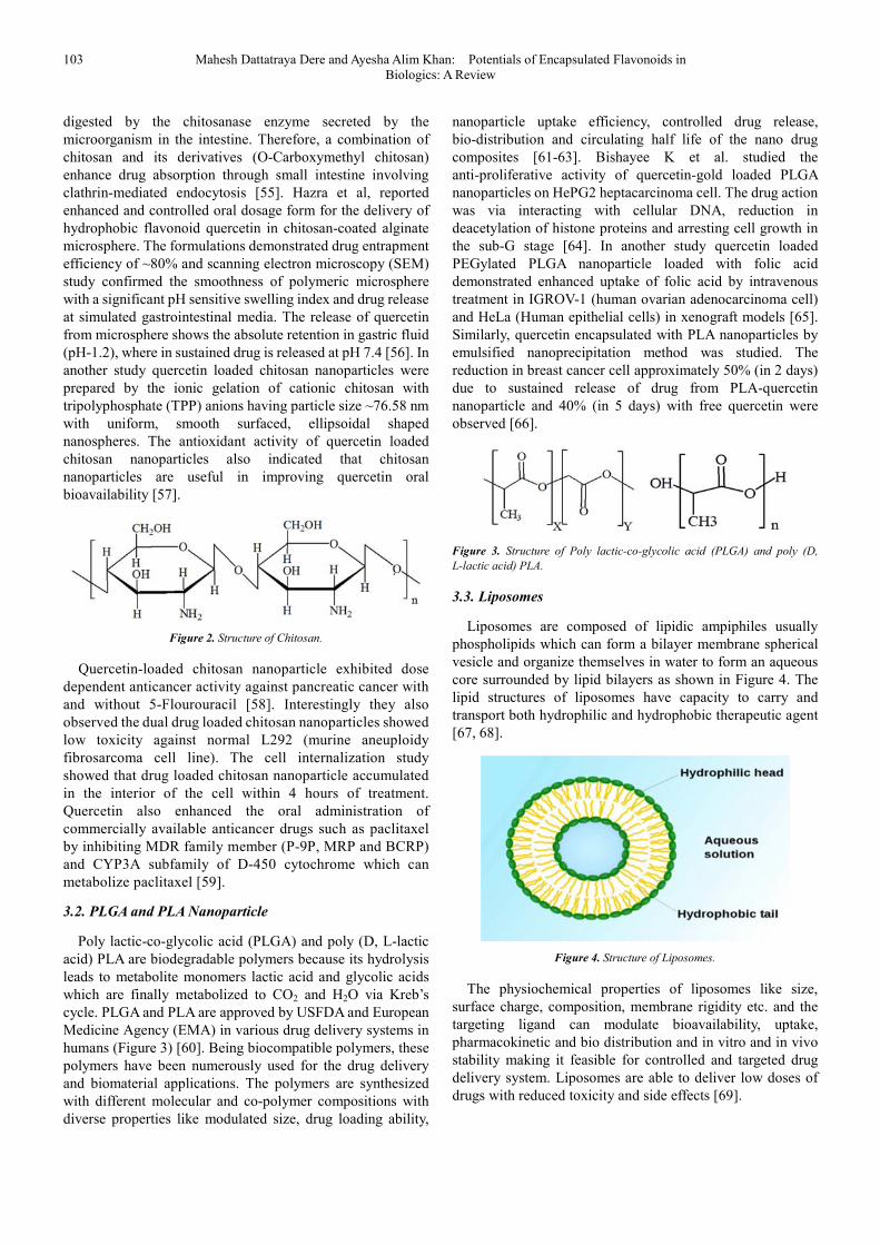
3.1. Chitosan
Chitosan is a natural N-deacetylated derivative of chitin polycationic polysaccharide consisting linear repeating unit of 2-acetamido-2-deoxy-D-glucose and β-(1-4)-2-amino-2- deoxy-D-glucose show in Figure 2 [45]. The presence of hydroxyl and amino functions show the modulatory effect on cellular-F actin, tight junction protein ZO-1 and protein kinase C and are rapidly internalized by the cell [46-48]. Chitosan is biodegradable, biocompatible, low toxic, non-immunogenic and mucoadhesive in nature. Therefore, chitosan favors wide range of biomedical applications including tissue engineering,
drug delivery, wound dressing antimicrobial activity, anti-inflammatory and antioxidant properties [49-52]. As a drug carrier, chitosan has the capacity to deliver drugs to various organs such as kidney, liver, lung and colon. Due to the polycationic characteristics chitosan can interact with the negatively charged molecules and form an efficient nanostructure drug delivery system of several bio molecules (drugs, flavonoids, proteins, DNA) [53, 54]. In addition being a known coadhesive polymer, chitosan helps to prolong mucus binding time of the drug molecules and transiently open the tight junction between the epithelial cells and help the drug transport in a sustained manner [52]. Chitosan are easily
103 Mahesh Dattatraya Dere and Ayesha Alim Khan: Potentials of Encapsulated Flavonoids in Biologics: A Review
digested by the chitosanase enzyme secreted by the microorganism in the intestine. Therefore, a combination of chitosan and its derivatives (O-Carboxymethyl chitosan) enhance drug absorption through small intestine involving clathrin-mediated endocytosis [55]. Hazra et al, reported enhanced and controlled oral dosage form for the delivery of hydrophobic flavonoid quercetin in chitosan-coated alginate microsphere. The formulations demonstrated drug entrapment efficiency of ~80% and scanning electron microscopy (SEM) study confirmed the smoothness of polymeric microsphere with a significant pH sensitive swelling index and drug release at simulated gastrointestinal media. The release of quercetin from microsphere shows the absolute retention in gastric fluid (pH-1.2), where in sustained drug is released at pH 7.4 [56]. In another study quercetin loaded chitosan nanoparticles were prepared by the ionic gelation of cationic chitosan with tripolyphosphate (TPP) anions having particle size ~76.58 nm with uniform, smooth surfaced, ellipsoidal shaped nanospheres. The antioxidant activity of quercetin loaded chitosan nanoparticles also indicated that chitosan nanoparticles are useful in improving quercetin oral bioavailability [57].
Figure 2. Structure of Chitosan.
Quercetin-loaded chitosan nanoparticle exhibited dose dependent anticancer activity against pancreatic cancer with and without 5-Flourouracil [58]. Interestingly they also observed the dual drug loaded chitosan nanoparticles showed low toxicity against normal L292 (murine aneuploidy fibrosarcoma cell line). The cell internalization study showed that drug loaded chitosan nanoparticle accumulated in the interior of the cell within 4 hours of treatment. Quercetin also enhanced the oral administration of commercially available anticancer drugs such as paclitaxel by inhibiting MDR family member (P-9P, MRP and BCRP) and CYP3A subfamily of D-450 cytochrome which can metabolize paclitaxel [59].
3.2. PLGA and PLA Nanoparticle
Poly lactic-co-glycolic acid (PLGA) and poly (D, L-lactic acid) PLA are biodegradable polymers because its hydrolysis leads to metabolite monomers lactic acid and glycolic acids which are finally metabolized to CO2 and H2O via Kreb’s cycle. PLGA and PLA are approved by USFDA and European Medicine Agency (EMA) in various drug delivery systems in humans (Figure 3) [60]. Being biocompatible polymers, these polymers have been numerously used for the drug delivery and biomaterial applications. The polymers are synthesized with different molecular and co-polymer compositions with diverse properties like modulated size, drug loading ability,
nanoparticle uptake efficiency, controlled drug release, bio-distribution and circulating half life of the nano drug composites [61-63]. Bishayee K et al. studied the anti-proliferative activity of quercetin-gold loaded PLGA nanoparticles on HePG2 heptacarcinoma cell. The drug action was via interacting with cellular DNA, reduction in deacetylation of histone proteins and arresting cell growth in the sub-G stage [64]. In another study quercetin loaded PEGylated PLGA nanoparticle loaded with folic acid demonstrated enhanced uptake of folic acid by intravenous treatment in IGROV-1 (human ovarian adenocarcinoma cell) and HeLa (Human epithelial cells) in xenograft models [65]. Similarly, quercetin encapsulated with PLA nanoparticles by emulsified nanoprecipitation method was studied. The reduction in breast cancer cell approximately 50% (in 2 days) due to sustained release of drug from PLA-quercetin nanoparticle and 40% (in 5 days) with free quercetin were observed [66].
Figure 3. Structure of Poly lactic-co-glycolic acid (PLGA) and poly (D,
L-lactic acid) PLA.
3.3. Liposomes
Liposomes are composed of lipidic ampiphiles usually phospholipids which can form a bilayer membrane spherical vesicle and organize themselves in water to form an aqueous core surrounded by lipid bilayers as shown in Figure 4. The lipid structures of liposomes have capacity to carry and transport both hydrophilic and hydrophobic therapeutic agent [67, 68].
Figure 4. Structure of Liposomes.
The physiochemical properties of liposomes like size, surface charge, composition, membrane rigidity etc. and the targeting ligand can modulate bioavailability, uptake, pharmacokinetic and bio distribution and in vitro and in vivo stability making it feasible for controlled and targeted drug delivery system. Liposomes are able to deliver low doses of drugs with reduced toxicity and side effects [69].

American Journal of Biomedical and Life Sciences 2020; 8(4): 97-113 104
3.4. Cyclodextrin (CDs)
Cyclodextrin (CDs) are natural macrocyclic oligosaccharides with toroidal structures having lipohilic cavities and a hydrophilic outer surface, thus capable to form inclusion complexes with hydrophobic molecule to significantly increase water solubility [70, 71]. Cyclodextrin inclusion complexes are capable to protect the active ingredients against oxidation, decomposition, light induced reactions, ocular disturbance, microbiological contamination, drug-additive interactions, hygroscopicity etc. There are three natural Cyclodextrin viz. α, β and γ- consisting of 6, 7 and 8 glucopyranose units linked by α-(1,4) bonds with internal diameter 0.5 to 0.8 nm (Figure 5) [72]. Moreover, a semi-synthetic derivative e.g. α-cyclodextrin and co-polymers of cyclodextrin can enhance the solubility in water, rate of release, inclusion capacity and reduce the side effects [73]. Cyclodextrin and its derivatives are reported as an attractive
candidate for biomedical applications including drug delivery; improve water solubility, stability, increase antioxidant activity, bioefficacy and bioavailability. Zheng et al. studied the chemical stability and water solubility of quercetin with three β-cyclodextrin derivatives such as unsubstituted β-cyclodextrin, hydroxylpropyl-β-cyclodextrin (HP-β-CD) and sulfobutyl ether-β-cyclodextrin (SBE-β-CD) at alkaline pH. The study revealed that β-cyclodextrin/ quercetin complex sustainably improved the solubility and stability due to formation of inclusion complex model studied by Nuclear Magnetic Resonance (NMR) spectroscopic analysis [74].
Carlotti et al. reported that the preparation of quercetin inclusion complex with methyl-β-cyclodextrin (Me-β-CD) improved the quercetin solubility without affecting the antioxidant activity and photostability, in vitro accumulation of quercetin in porcine skin studies with Franz diffusion cell [75].
Figure 5. Structure of α, β and γ-cyclodextrin (CD).
3.5. Miscellaneous Nanoparticles
To improve the delivery of drug in vitro and in vivo Wang et al. reported quercetin co-encapsulated fluorescent silicon quantum dots (SiQDS) in poly (ethylene glycol)–block–polylactide (PEG-PLA) by double emulsion method for simultaneous in vitro imaging and biocompatibility studies (Figure 6). The encapsulated nanoparticles effectively suppress human hepatoma HePG2 cell proliferation than free quercetin and significantly inhibit the hydrogen peroxide-induced DNA damage in HePG2 cells [76]. Barreto et al. proposed a new magnetic nanoparticle (Fe3O4) incorporated to a triblock
co-polymer of ethylene dioxide and oxyphenylethylene for quercetin delivery in cancer treatment. The magnetic nanoparticle demonstrated a targeted drug delivery and sustained release of drug (its peak at 14.5% after 96 h) [77]. Several researchers have reported the use of mesoporous silica nanoparticles as a promising drug delivery system due to low in vivo toxicity, stability, targeted drug delivery, high surface area and high drug loading efficiency with better release kinetics [78-83]. In another study Catauro et al. studied the silica-quercetin hybrid material using a sol-gel synthesis method for treatment of peri-implant diseases [84].
Figure 6. SEM micrographs of: (A) (SiO2/Quercetin hybrids) 5 Si/Que5, (B) Si/Que10, and (C) Si/Que15.

105 Mahesh Dattatraya Dere and Ayesha Alim Khan: Potentials of Encapsulated Flavonoids in Biologics: A Review
4. Biological Activity of Encapsulated
Flavonoids
Several biological and pharmacological activities of encapsulated flavonoids are widely known. A summary of the biological studies on encapsulated flavonoids are presented below.
4.1. Anticancer
4.1.1. Cytotoxicity Studies
Encapsulation of naturally occurring flavonoids such as Quercetin, Isoscutellarein, Rutin and Isoscutellarein glycoside into liposomes were tested against the cancer cell line SF268 (central nervous system), H460 (non-small cell lung) and MCF7 (breast) by M. Goniotaki et al. The result showed that the quercetin had growth inhibiting (GI50) activity against the cancer cell lines SF268 (31.75µM), MCF7 (24.19 µM) and H460 (80.0 µM). At higher concentration all the flavonoids were inactive against normal cells (peripheral blood mononuclear cells: resting or activated). The liposomal formulation of quercetin was less active than its free form. The liposomal formulation rutin proved to be more active and showed remarkable growth inhibition activity against H460 and SF268 cell lines and the liposomal formulation of isoscutellarein shows considerable growth inhibition activity for all cell lines and best among the all tested flavonoids. The free liposomes were inactive against all cell lines [85].
In another study Gao et al. stated that the encapsulation of quercetin with biodegradable monomethoxy poly(ethylene glycol)-poly(ε-caprolactone) MPEG-PCL micelles suppressed the growth of established xenograft A2780S ovarian tumors through cell apoptosis and inhibiting angiogenesis in vivo. The quercetin loaded micelles showed 36 nm of mean particle size with 6.9% drug loading [86]. Chitosan nanoparticles of Epigallocatechin-3-gallate(EGCG) showed anticancer effect on human melanoma Mell 928 cells by apoptosis via increase in Bax levels, increased poly (ADP-ribose), polymerase (PARP) cleavage, G2/M phase cell cycle arrest, inhibition of cyclin D and D3 induction of p21 and p27, decrease in Bcl-2, caspases-3 and caspases-9 protein expression, which resulted in reduction of cancer cell viability. The EGCG nanoformulated with chitosan showed anticancer effect on xenograft athymic mouse model of melanoma by suppression of tumor group and proliferation, inhibition of CDK4 and 6 and an increase in apoptosis [87]. In another study EGCG nanoformulated as Ca/Al-NO3 layered double hydroxide induced apoptosis, decreased the cell viability and inhibited colony formation in human prostate cancer PC-3 cells [88].
4.1.2. Colon Cancer
The natural flavonoid fisetin (3, 3’, 4’, 7’- tetrahydroxyflavone) encapsulated with monomethyl poly-(ethylene glycol)-poly (ε-caprolactone) (MPEG-PCL) was used to prepared nanoassemblies of fisetin by a self-assembly procedure. Yishan Chen et al studied the effectiveness of fisetin micelles and a promising source for
colon cancer therapy with high antitumor activity and low toxicity. The prepared fisetin micelles with particle size 22.4 ± 3.0 nm, polydisperse index 0.163 ± 0.032, the drug loading (DL) and encapsulation efficiency were 9.88±0.14% and 98.53 ± 0.02%. In vitro release study of fisetin micelles demonstrated a sustained and prolonged release than free fisetin and the cumulative release rate of fisetin micelles was 73.58 ± 3.99% and free fisetin was 92.95 ± 6.51% (P<0.05). The cytotoxicity of fisetin micelles by MTT assay indicate that cell viability of CT26 and L929 cells were upto 72.13 and 66.13 respectively. The results indicated that MPEG-PCL were biocompatible and exhibited low toxicity. Fisetin micelles cellular uptake and apoptosis in CT26 cells was higher than that of free fisetin. The in vivo studies were more efficient in suppressing growth and prolonging survival time of tumors than free fisetin (P<0.05). Tumor were analyzed using histological analysis (H & E), TUNEL assay, immunohistochemical detection of Ki-67 cell proliferation and immunofluorescence detection of micro vessel density (MVD). The fisetin micelles enhanced apoptosis induction, antiproliferation and antiangiogenesis effect than free fisetin in the animal model [89].
Multifunctional solid lipid nanoparticles loaded with a cyanine-type IR-780 acting as a diagnostic agent and a photosensitizer and a flavonoid derivative baicalein or fisetin as a therapeutic cargo were fabricated using a solvent diffusion method. The drug loaded lipid nanoparticle exhibited anticancer effect in colon adenocarcinoma cells with lower cytotoxicity and decrease in tumor growth on loVo and CHO-K1 cell lines. They also showed an increased p53 and MnSOD (Manganese superoxide dismutase) expressions after PDT-SLN-EP (photodynamic therapy-solid lipid nanoparticles-electroporation) [90]. Epigallocatechin-3-gallate (ECCG) nanoformulated by graphene nanosheet showed anticancer effect on colon cancer HT29 and SW48 cells, via photothermal destruction of cell assessed by high efficiency near-infrared photothermal therapy [91].
4.1.3. Lung Cancer
Baicalein nanoparticle with dual-targeted ligands of folate and hyaluronic acid showed the anticancer effect on human lung cancer A549 cells as well as paclitaxel-resistance lung cancer A549/PTX in xenograft mouse model of A549/PTX by decreasing cell viability and inhibiting tumor growth [92]. Luteolin nanoformulated with PLA-PEG polymer possesses anticancer effect against lung cancer H292 cells and TU212 head and neck squamous cell. The mode of anticancer effect was observed via inhibition of tumor growth, tumor size and colony formation in xenograft mouse model of head and neck cancer [93].
4.1.4. Breast Cancer
Ming sun et al. studied the quercetin-nanostructured lipid carrier (Q-NLC) synthesized using a phase inversion based process method. The size of NLC was 32 nm, the loading capacity and encapsulation efficiency of Q-NLC were 11% and 95% respectively. Q-NLC enhanced the cytotoxicity and

American Journal of Biomedical and Life Sciences 2020; 8(4): 97-113 106
apoptosis in MCF-7 and MDA-MB-231 breast cancer cells. The void NLC was found to be extremely less toxic for the breast cancer cells [94]. Kadari et. al studied the anticancer activity against MCF-7 breast cancer cells for fisetin (FST) encapsulated into PLGA (poly-lactide-co-glycolic acid) nanoparticles as a complex of HPβCD (Hydroxy propyl β cyclodextrin). In vitro studies with nanoformulation FHIC-PNP (FST-hydroxyl propyl β cyclodextrin inclusion complex into PLGA nanoparticles) showed 3.9 times higher toxicity than pure fisetin against MCF-7 human breast cancer cell lines and enhanced the FST-induced apoptosis and ROS generation. In vivo studies in C57BL6 mice revealed that incorporation of FHIC in FHIC-PNP improved the pharmacokinetics and oral bioavailability of fisetin [95].
EGCG core-shell PLGA-casein nanoparticles in combination with paclitaxel demonstrated the anticancer activity on MCF-7 cells and human MDA-MB-231 breast cancer cells by increasing apoptosis and decreasing NF-kB activation [96]. Luteolin in phytosomes possesses anticancer effect by decreasing the expression at NrF2 and its related downstream gene HOl on human MDA-MB-231 breast cancer cells [97]. Quercetin nanoformulated as phytosomes had anticancer effect on breast cancer MCF-7 cells by increasing apoptosis and decreasing mRNA expression of Nrf2 downstream genes NQO1 and MRP1 while no significant changes in Nrf2 expression was observed due to free quercetin [98].
4.1.5. Liver Cancer
Krishnan et al. studied the hesperetin conjugated gold nanoparticles (Au-mPEG(5000)-S-HP NPs) with an average size of 220 nm and exhibited sustained and slow release of hesperetin from Au-mPEG(5000)-S-HP NPs for 72 hours. Au-mPEG(5000)-S-HP NPs possessed anti-inflammatory, anti-proliferative, anti-carcinogen properties and modulated signaling pathways in male Wister albino rats by decreased levels of mast cell density in the liver, protein expression levels of TNF-α or NF-kB (Nuclear factor-kB) and β-actin, amount of glycoprotein level and protein expression levels of PCNA [99].
4.2. Anti-inflammatory
The quercetin loaded (β-CD)-dodecyl carbonate nanoparticle delivery for improved quercetin bioavailability, anti-inflammatory activity and treatment of Alzheimer’s disease (AD)-related neuropathalogical were studied. In vitro studies confirmed remarkable increase in anti-inflammatory effect of quercetin after encapsulation within the nanoparticles or nanoparticle were able to improve permeation across the blood brain barrier and produce enough bioavailability to reach target cells [100].
4.3. Antidiabetic
Naringenin loaded chitosan/alginate nanoparticles were prepared with weight ratio of alginate and chitosan 1: 3 and 1: 2 at pH 5.5. The in vivo hypoglycemic effect after oral delivery of the nanoparticles to streptozotocin-induced diabetic rats indicated that the nanoparticles were free from toxicity. The average hydrodynamic size of the nanoparticles ranged between
150 and 300 nm (approximately) and the surface charge varied from -26.3 mV to -38.21 mV. Naringenin encapsulation efficiency and loading capacity of CS/ALG (Chitosan/ Alginate) core shell nanoparticle at different weight ratios were varied between 57.34% to 98.36% and 7.41% to 19.87% respectively. Nanoparticles with weight ratio of 3: 1 (CS: ALG) having encapsulation efficiency 91.4% and loading capacity 15.9% were further used for in vitro and in vivo studies. At pH 1.2 maximum 15% and at pH 7.4 more than 90% Naringenin was released in a slow sustained manner from the core shell nanoparticle. The in-vivo toxicity assessment showed no significant difference between NC (Rats treated with normal saline orally), NTBN (Rats treated with blank nanoparticles orally, 50 mg/kg body weight) and NTNN (Rats treated with Naringenin loaded core shell nanoparticles orally, 50 mg/kg body weight) groups. Regarding the fasting blood glucose, cholesterol and triglyceride and almost no change were observed in serum ALT (Serum alanine transaminase), AST (Aspartate transaminase) and ALP (Alkaline phosphatase) levels in normal and treated group of rats. The encapsulated Naringenin within nanoparticles helps to normalize the pancreatic abnormalities in diabetes, better than free oral Naringenin. The revival and recovery of hepatic tissue architecture appeared to be better in ND (Diabetic rats fed orally with Naringenin-loaded core shell polymeric nanoparticle 50mg/kg body weight) group then in FD (Diabetic rats fed orally with free Naringenin 50 mg/kg body weight, dissolved in 60% ethanol) group [101].
Chitkara et al. studied the effect of quercetin loaded in PLGA nanoparticle by emulsion-diffusion-evaporation method. The average particle size of the nanoparticle was 179±11.2 nm, zeta potential -6.06±1-51 mV, polydispersity index 0-128 and ~86% quercetin entrapment efficiency. Surface morphology study confirmed the spherical shaped particles with smooth surface, ensuring the absence of unentrapped or adsorbed quercetin by scanning electron microscope (SEM). The nanoparticles retained the antioxidant property of quercetin due to easy lyophilization using D-trehalose (5%). In vitro release study confirmed a controlled release pattern of quercetin from the nanoparticles. In vivo, pharmacokinetics study revealed that the nanoformulation relatively increased the oral bioavailability (~52.3%) and the plasma quercetin concentration was sustained for 6 days, suggesting a reduced dosing frequency of the nanoformulations. An increased superoxide dismutase and catalase level in pancreas and kidneys after pre-oral treatment of nanoparticles were observed. Thus the nanocarries could be an effective oral therapy for diabetes and its related complications which reduces dose as well as dosing frequency [102].
4.4. Antimicrobial Activity
The antimicrobial activity of hesperetin-loaded PLGA (poly (d, l-lactic-co-glycolic acid) nanoparticle may be attributed to the following (i) the structural properties of hesperetin (flavonoids nature) [103-105]. (ii) The potential charge on PLGA nanoparticle causing cell membrane depolarization [106-108]. (iii) Solubility of hydrophobic hesperetin increase after encapsulation, and (iv) the sustained release of active

107 Mahesh Dattatraya Dere and Ayesha Alim Khan: Potentials of Encapsulated Flavonoids in Biologics: A Review
substance [60, 109-110]. Duranoglu et. al studied the effective encapsulation of hesperetin into PLGA nanoparticles by using experimental design method. The formed nanoparticles showed maximum encapsulation efficiency (80.5±4.9%) and minimum particle size (260.2 ± 16.5 nm). The process was optimized as follows; 0.5% polyvinyl alcohol (PVA) concentration, 5:13 water: organic phase ratio and 3.59 mL min-1 flow rate of the emulsified solution into 0.1% PVA. The cytotoxicity study or the biocompatibility of nanoparticles against the growth of L929 fibroblast cells was determined by the MTT method. The result revealed that the hesperetin and hesperetin-loaded nanoparticles were biocompatible with normal cell line L929 fibroblast cells up to 184.83 and190.88 µg ml-1 for 24 h and up to 133.24 and 134.80 µg ml-1 for 48 h. The antimicrobial activity studies were carried using two different methods against Staphylococcus aureus and Escherichia coli. The MIC (minimal inhibitory concentration) values were 125 µg ml-1 for Escherichia coli and 200 µg ml-1 for Staphylococcus aureus, while the free hesperetin did not demonstrate activity in both strains (MIC value >200 µg ml-1) [111].
4.5. Anti-quorum Sensing Activity
Sedef Ilk et al studied the kaempferol loaded chitosan nanoparticles by anti-quorum sensing mechanism against Chromobacterium violaceum CV026. The chitosan/ TPP nanoparticles were synthesized by ionic gelation method with nanoparticle size and zeta potential of 192.27± 13.6 and +35 mV. The loading and encapsulation efficiency of kaempferol
loaded chitosan nanoparticles were 78% and 93%. The antioxidant activity evaluation by DPPH assay method indicates that kaempferol loaded chitosan/TPP nanoparticles show scavenging activity of 37±2.5% than free kaempferol (Scavenging activity 22±1.8%). The anti-QS activity of kaempferol loaded chitosan nanoparticle and free kaempferol by disc diffusion method on Chromobacterium violaceum
CV026 at different storage time indicate that the nanoparticle inhibited violacein production up to 76%. Hence kaempferol loaded chitosan nanoparticles can act as strong and prolonged time stable quorum quenchers than corresponding pure kaempferol [112].
4.6. Antifungal Activity
Sedef Ilk et al studied the antifungal activity of kaempferol (KAE) loaded into lecithin/chitosan nanoparticles (Lc NPs) against the phytopathogenic fungus Fusarium oxysporium. The mean particle size, poly disperse index (PDI) and zeta potential were found to be 270 ± 10 nm, PDI≤0.2 and + 56 ± 4 mV respectively. KAE-LC NPs showed slow and sustained released for KAE in PBS + DMSO buffer at 37°C with encapsulation efficiency of 93.8 ± 4.28%. In vitro evaluation of KAE-LC NPs was studied by the release kinetics, antioxidant and antifungal activity in a time dependent manner against free KAE. The results demonstrate that nanoparticles had higher antioxidant and antifungal activity (67%) compared to free KAE (no inhibition) against Fusarium
oxysporium by the end of 60 day storage period [113].
Figure 7. In vitro release profiles of KAE from lecithin/ chitosan nanoparticles (A) at 37°C, (B) at 25°C temperature.
Figure 8. Antioxidant activity of KAE-LC NP and pure KAE evaluated by reducing power (FRAP).

American Journal of Biomedical and Life Sciences 2020; 8(4): 97-113 108
Figure 9. Antifungal activity of KAE-LC NPs and pure KAE against pathogenic fungi F. oxysporium. (A) Hyphal-extension growth inhibition.(B) Microscopic
analysis of hyphae treated with KAE-LC NPs.
Figure 10. Effect of KAE-LC NPs and pure KAE on the radial growth of Fusarium oxysporium in time dependent manner. All determinations were performed in
triplicate and the results expressed as mean±standard deviation.
4.7. Antioxidant Activity
Quercetin encapsulated nanoemulsions were produced using a low energy method-emulsion inversion point and two surfactant viz: Tween 80 and Brij 30. The average droplet diameters were 180-200 nm in the range. Quercetin loaded nanoemulsion incorporated in chicken pate was capable of
protection against lipid oxidation but not against protein oxidation. Inhibition of secondary lipid oxidation was about 60% after 24 week of storage. While the free quercetin exhibited 35.4% inhibition and about 8.4% of inhibition in pates added with synthetic antioxidant such as butylated hydroxytoluene-BHT and sodium nitrite after 24 weeks [114].

109 Mahesh Dattatraya Dere and Ayesha Alim Khan: Potentials of Encapsulated Flavonoids in Biologics: A Review
Juan Huang et. al studied the quercetin and linseed oil encapsulated into nanostructured lipid carrier (NLC) by high pressure homogenization technique. The sustained release pattern of quercetin from quercetin loaded NLCs and antioxidant study by DPPH assay showed that linseed oil could improve the free radical scavenging activity of quercetin loaded NLCs [115].
5. Conclusions
Flavonoids form an integral part of human and animal diet. Flavonoids exhibit diverse categories of pharmacological and biological activities such as Anti-oxidant, Anti-inflammatory, Antidiabetic, Antimicrobial activity, Anti-quorum sensing activity, Anticancer, Modulators of cell signaling etc. Flavonoids exhibit the low water solubility, low permeability, gastric stability etc. which are the major limiting factor for the potential health benefits. Flavonoids when encapsulated with natural, synthetic and semisynthetic materials such as Chitosan, PLGA and PLA, Liposomes, Cyclodextrins etc. shows much better stability, bioavailability, increase shelf life, controlled and sustained release, protect against oxidation, isomerization and degradation etc.
However, only a small portion has been investigated or studied in both flavonoid encapsulation and its biological activities. There are gaps in the research, which need to be bridged in order to exploit the full medicinal potential of flavonoids or encapsulated flavonoids.
Conflicts of Interest
The authors declare no conflicts of interest regarding the publication of this paper.
Acknowledgements
The authors acknowledge the Department of Chemistry, Savitribai Phule Pune University, Pune, India for providing research support to continue the research work.
References
[1] Mahomoodally, M. F., Gurib-Fakim, A., and Subratty, A. H. (2005). Antimicrobial Activities and Phytochemical Profiles of Endemic Medicinal Plants of Mauritius. Pharmaceutical Biology 43, 237-242.
[2] Pandey, A. K. (2007). Anti-staphylococcal activity of a pan-tropical aggressive and obnoxious weed Parthenium histerophorus: An in vitro study. National Academy Science Letters 30, 383-386.
[3] Agati, G., Azzarello, E., Pollastri, S., and Tattini, M. (2012). Flavonoids as antioxidants in plants: location and functional significance. Plant Sci 196, 67-76.
[4] Mitek, M., and Gasik, A. (2009). Polyphenols in food. The impact on organo leptic characteristics of food [in Polish]. PrzemSpoż 5, 34-39.
[5] J, O., and E, S. (2005). The biological activity of flavonoids [in Polish]. Post Fitoter 3, 71-79.
[6] Heim, K. E., Tagliaferro, A. R., and Bobilya, D. J. (2002). Flavonoid antioxidants: chemistry, metabolism and structure-activity relationships. The Journal of nutritional biochemistry 13, 572-584.
[7] Ross, J., and Kasum, C. (2002). Dietary flavonoids: Bioavailability, metabolic effects, and safety. Annual review of nutrition 22, 19-34.
[8] Yao, L. H., Jiang, Y. M., Shi, J., TomÁS-BarberÁN, F. A., Datta, N., Singanusong, R., and Chen, S. S. (2004). Flavonoids in Food and Their Health Benefits. Plant Foods for Human Nutrition 59, 113-122.
[9] Middleton, E. (1998). Effect of Plant Flavonoids on Immune and Inflammatory Cell Function. In Flavonoids in the Living System, J. A. Manthey and B. S. Buslig, eds. (Boston, MA: Springer US), pp. 175-182.
[10] Koes, R., Verweij, W., and Quattrocchio, F. (2005). Flavonoids: a colorful model for the regulation and evolution of biochemical pathways. Trends in Plant Science 10, 236-242.
[11] Hollman, P. C., Bijsman, M. N., van Gameren, Y., Cnossen, E. P., de Vries, J. H., and Katan, M. B. (1999). The sugar moiety is a major determinant of the absorption of dietary flavonoid glycosides in man. Free Radic Res 31, 569-573.
[12] Manach, C., Williamson, G., Morand, C., Scalbert, A., and Rémésy, C. (2005). Bioavailability and bioefficacy of polyphenols in humans. I. Review of 97 bioavailability studies. The American Journal of Clinical Nutrition 81, 230S-242S.
[13] Stahl, W., van den Berg, H., Arthur, J., Bast, A., Dainty, J., Faulks, R. M., Gartner, C., Haenen, G., Hollman, P., Holst, B., et al. (2002). Bioavailability and metabolism. Mol Aspects Med 23, 39-100.
[14] Bilia, A., Isacchi, B., Righeschi, C., Guccione, C., Maria, C., and Bergonzi, M. (2014). Flavonoids Loaded in Nanocarriers: An Opportunity to Increase Oral Bioavailability and Bioefficacy. Food and Nutrition Sciences 05.
[15] Kumar, S., and Pandey, A. K. (2013). Chemistry and Biological Activities of Flavonoids: An Overview. The Scientific World Journal 2013, 162750.
[16] Macheix, J.-J., Fleuriet, A., and Jay-Allemand, C. (2005). Les composés phénoliques des végétaux: un exemple de métabolites secondaires d'importance économique, (Lausanne: Presses polytechniques et universitaires romandes).
[17] Fang, Z., and Bhandari, B. (2010). Encapsulation of polyphenols – a review. Trends in Food Science & Technology 21, 510-523.
[18] El Gharras, H. (2009). Polyphenols: food sources, properties and applications – a review. International Journal of Food Science & Technology 44, 2512-2518.
[19] Manach, C., Scalbert, A., Morand, C., Remesy, C., and Jimenez, L. (2004). Polyphenols: food sources and bioavailability. Am J Clin Nutr 79, 727-747.
[20] Maatta-Riihinen, K. R., Kahkonen, M. P., Torronen, A. R., and Heinonen, I. M. (2005). Catechins and procyanidins in berries of vaccinium species and their antioxidant activity. J Agric Food Chem 53, 8485-8491.

American Journal of Biomedical and Life Sciences 2020; 8(4): 97-113 110
[21] Bagchi, D., Sen, C. K., Bagchi, M., and Atalay, M. (2004). Anti-angiogenic, antioxidant, and anti-carcinogenic properties of a novel anthocyanin-rich berry extract formula. Biochemistry (Mosc) 69, 75-80, 71 p preceding 75.
[22] Marin, F. R., Perez-Alvarez, J. A., and Soler-Rivas, C. (2005). Isoflavones as functional food components. In Stud. Nat. Prod. Chem., Volume 32. (Elsevier), pp. 1177-1207.
[23] Lin, Y., Shi, R., Wang, X., and Shen, H. M. (2008). Luteolin, a flavonoid with potential for cancer prevention and therapy. Curr Cancer Drug Targets 8, 634-646.
[24] Patel, D., Shukla, S., and Gupta, S. (2007). Apigenin and cancer chemoprevention: progress, potential and promise (review). Int J Oncol 30, 233-245.
[25] Lapidot, T., Walker, M. D., and Kanner, J. (2002). Antioxidant and prooxidant effects of phenolics on pancreatic beta-cells in vitro. J Agric Food Chem 50, 7220-7225.
[26] Galati, G., and O'Brien, P. J. (2004). Potential toxicity of flavonoids and other dietary phenolics: significance for their chemopreventive and anticancer properties. Free Radic Biol Med 37, 287-303.
[27] Munin, A., and Edwards-Levy, F. (2011). Encapsulation of natural polyphenolic compounds; a review. Pharmaceutics 3, 793-829.
[28] HJ, R., S, L., C, E., and B, K. (2009). Anthocyanins. Pennigton Nutrition Series.
[29] Sarni-Manchado, P., and Cheynier, V. (2005). Les polyphénolsenagroalimentaire, (Paris, France: Tec & Doc Lavoisier).
[30] Chiu, Y. T., Chiu, C. P., Chien, J. T., Ho, G. H., Yang, J., and Chen, B. H. (2007). Encapsulation of Lycopene Extract from Tomato Pulp Waste with Gelatin and Poly (γ-glutamic acid) as Carrier. Journal of Agricultural and Food Chemistry 55, 5123-5130.
[31] Jahanshahi, M., Najafpour, G., and Rahimnejad, M. (2008). Applying the Taguchi method for optimized fabrication of bovine serum albumin (BSA) nanoparticles as drug delivery vehicles. African Journal of Biotechnology (ISSN: 1684-5315) Vol 7 Num 4 7.
[32] Jin, J., Sklar, G. E., Min Sen Oh, V., and Chuen Li, S. (2008). Factors affecting therapeutic compliance: A review from the patient's perspective. Ther. Clin. Risk Manag. 4, 269-286.
[33] Solecki, R. S. (1975). Shanidar IV, a Neanderthal Flower Burial in Northern Iraq. Science 190, 880.
[34] Saklani, A., and Kutty, S. K. (2008). Plant-derived compounds in clinical trials. Drug Discov. Today 13, 161-171.
[35] Shahidi, F., and Han, X. Q. (1993). Encapsulation of food ingredients. Critical Reviews in Food Science and Nutrition 33, 501-547.
[36] Available from: http://www.apps.who.int/medicinedocs/ en/d/Js7916e/7.12.html., Volume 2015.
[37] Marin, E., Briceño, M. I., and Caballero-George, C. (2013). Critical evaluation of biodegradable polymers used in nanodrugs. Int J Nanomedicine 8, 3071-3090.
[38] Augustin, M. A., and Hemar, Y. (2009). Nano- and micro-structured assemblies for encapsulation of food
ingredients. Chem. Soc. Rev. 38, 902-912.
[39] Desai, K. G. H., and Jin Park, H. (2005). Recent Developments in Microencapsulation of Food Ingredients. Drying Technol. 23, 1361-1394.
[40] Gibbs, B. F., Kermasha, S., Alli, I., and Mulligan, C. N. (1999). Encapsulation in the food industry: a review. Int J Food Sci Nutr 50, 213-224.
[41] Mozafari, M. R., Khosravi-Darani, K., Borazan, G. G., Cui, J., Pardakhty, A., and Yurdugul, S. (2008). Encapsulation of Food Ingredients Using Nanoliposome Technology. Int. J. Food Prop. 11, 833-844.
[42] N., B. L. (2001). Stability testing of nutraceuticals and functional foods. In nutraceuticals and functional foods R. E. C. Wildman, ed. (New York: CRC Press), pp. 501-516.
[43] Chen, L., Remondetto, G., and Subirade, M. (2006). Food protein-based materials as nutraceutical delivery systems. Trends in Food Science & Technology - TRENDS FOOD SCI TECHNOL 17, 272-283.
[44] Mukhopadhyay, P., Mishra, R., Rana, D., and Kundu, P. P. (2012). Strategies for effective oral insulin delivery with modified chitosan nanoparticles: A review. Prog. Polym. Sci. 37, 1457-1475.
[45] Ravi Kumar, M. N. V. (2000). A review of chitin and chitosan applications. React. Funct. Polym. 46, 1-27.
[46] Huang, M., Khor, E., and Lim, L.-Y. (2004). Uptake and Cytotoxicity of Chitosan Molecules and Nanoparticles: Effects of Molecular Weight and Degree of Deacetylation. Pharm. Res. 21, 344-353.
[47] Ma, Z., Lim, T. M., and Lim, L. Y. (2005). Pharmacological activity of peroral chitosan-insulin nanoparticles in diabetic rats. Int J Pharm 293, 271-280.
[48] Smith, J. M., Dornish, M., and Wood, E. J. (2005). Involvement of protein kinase C in chitosan glutamate-mediated tight junction disruption. Biomaterials 26, 3269-3276.
[49] Qiao, Y., Bai, X. F., and Du, Y. G. (2011). Chitosan oligosaccharides protect mice from LPS challenge by attenuation of inflammation and oxidative stress. Int. Immunopharmacol. 11, 121-127.
[50] Liu, H. T., Li, W. M., Xu, G., Li, X. Y., Bai, X. F., Wei, P., Yu, C., and Du, Y. G. (2009). Chitosan oligosaccharides attenuate hydrogen peroxide-induced stress injury in human umbilical vein endothelial cells. Pharmacol. Res. 59, 167-175.
[51] Singla, A., and Chawla, M. J. (2001). Chitosan: Some pharmaceutical and biological aspects - An update. The Journal of pharmacy and pharmacology 53, 1047-1067.
[52] Mukhopadhyay, P., Sarkar, K., Bhattacharya, S., Bhattacharyya, A., Mishra, R., and Kundu, P. P. (2014). pH sensitive N-succinyl chitosan grafted polyacrylamide hydrogel for oral insulin delivery. Carbohydr. Polym. 112, 627-637.
[53] Sonvico, F., Cagnani, A., Rossi, A., Motta, S., Di Bari, M. T., Cavatorta, F., Alonso, M. J., Deriu, A., and Colombo, P. (2006). Formation of self-organized nanoparticles by lecithin/chitosan ionic interaction. Int. J. Pharm. 324, 67-73.

111 Mahesh Dattatraya Dere and Ayesha Alim Khan: Potentials of Encapsulated Flavonoids in Biologics: A Review
[54] Raj, L., Jonisha, R., Revathi, B., and Jayalakshmy, E. (2015). Preparation and characterization of BSA and chitosan nanopartices for sustainable delivery system for quercetin. Journal of Applied Pharmaceutical Science, 001-005.
[55] Feng, C., Wang, Z., Jiang, C., Kong, M., Zhou, X., Li, Y., Cheng, X., and Chen, X. (2013). Chitosan/o-carboxymethyl chitosan nanoparticles for efficient and safe oral anticancer drug delivery: in vitro and in vivo evaluation. Int J Pharm 457, 158-167.
[56] Hazra, M., Dasgupta Mandal, D., Mandal, T., Bhuniya, S., and Ghosh, M. (2015). Designing polymeric microparticulate drug delivery system for hydrophobic drug quercetin. Saudi Pharmaceutical Journal 23, 429-436.
[57] Zhang, Y., Yang, Y., Tang, K., Hu, X., and Zou, G. (2008). Physicochemical characterization and antioxidant activity of quercetin-loaded chitosan nanoparticles. J. Appl. Polym. Sci. 107, 891-897.
[58] David, K. I., Jaidev, L. R., Sethuraman, S., and Krishnan, U. M. (2015). Dual drug loaded chitosan nanoparticles-sugar--coated arsenal against pancreatic cancer. Colloids Surf. B. Biointerfaces 135, 689-698.
[59] Choi, J.-S., Jo, B.-W., and Kim, Y.-C. (2004). Enhanced paclitaxel bioavailability after oral administration of paclitaxel or prodrug to rats pretreated with quercetin. European journal of pharmaceutics and biopharmaceutics: official journal of Arbeitsgemeinschaft für Pharmazeutische Verfahrenstechnik e. V 57, 313-318.
[60] Kumari, A., Yadav, S. K., and Yadav, S. C. (2010). Biodegradable polymeric nanoparticles based drug delivery systems. Colloids Surf. B. Biointerfaces 75, 1-18.
[61] Asghar, W., Islam, M., Wadajkar, A., Wan, Y., Ilyas, A., Nguyen, K., and Iqbal, S. (2012). PLGA Micro and Nanoparticles Loaded Into Gelatin Scaffold for Controlled Drug Release. IEEE Transactions on Nanotechnology - IEEE TRANS NANOTECHNOL 11, 546-553.
[62] Hussein, A. S., Abdullah, N., and Ahmadun, F. R. (2013). In vitro degradation of poly (D, L-lactide-co-glycolide) nanoparticles loaded with linamarin. IET Nanobiotechnol 7, 33-41.
[63] Gratton, S. E. A., Ropp, P. A., Pohlhaus, P. D., Luft, J. C., Madden, V. J., Napier, M. E., and DeSimone, J. M. (2008). The effect of particle design on cellular internalization pathways. Proceedings of the National Academy of Sciences 105, 11613.
[64] Bishayee, K., Khuda-Bukhsh, A. R., and Huh, S. O. (2015). PLGA-Loaded Gold-Nanoparticles Precipitated with Quercetin Downregulate HDAC-Akt Activities Controlling Proliferation and Activate p53-ROS Crosstalk to Induce Apoptosis in Hepatocarcinoma Cells. Mol. Cells 38, 518-527.
[65] El-Gogary, R. I., Rubio, N., Wang, J. T.-W., Al-Jamal, W. T., Bourgognon, M., Kafa, H., Naeem, M., Klippstein, R., Abbate, V., Leroux, F., et al. (2014). Polyethylene Glycol Conjugated Polymeric Nanocapsules for Targeted Delivery of Quercetin to Folate-Expressing Cancer Cells in Vitro and in Vivo. ACS Nano 8, 1384-1401.
[66] Pandey, S. K., Patel, D. K., Thakur, R., Mishra, D. P., Maiti, P., and Haldar, C. (2015). Anti-cancer evaluation of quercetin embedded PLA nanoparticles synthesized by emulsified nanoprecipitation. Int. J. Biol. Macromol. 75, 521-529.
[67] Iao, V. (1993). Liposomes: from physics to applications.
[68] Stone, W. L., and Smith, M. (2004). Therapeutic uses of antioxidant liposomes. Mol. Biotechnol. 27, 217-230.
[69] Schnyder, A., and Huwyler, J. (2005). Drug transport to brain with targeted liposomes. NeuroRx 2, 99-107.
[70] Soica, C., Dehelean, C., Danciu, C., Wang, H. M., Wenz, G., Ambrus, R., Bojin, F., and Anghel, M. (2012). Betulin complex in gamma-cyclodextrin derivatives: properties and antineoplasic activities in in vitro and in vivo tumor models. Int. J. Mol. Sci. 13, 14992-15011.
[71] Crini, G. (2014). Review: A History of Cyclodextrins. Chem. Rev. 114, 10940-10975.
[72] Dodziuk, H. (2006). Cyclodextrins and their complexes: chemistry, analytical methods, applications, (John Wiley & Sons).
[73] Folch-Cano, C., Guerrero, J., Speisky, H., Jullian, C., and Olea-Azar, C. (2014). NMR and molecular fluorescence spectroscopic study of the structure and thermodynamic parameters of EGCG/β-cyclodextrin inclusion complexes with potential antioxidant activity. J. Incl. Phenom. Macrocycl. Chem. 78, 287-298.
[74] Zheng, Y., Haworth, I. S., Zuo, Z., Chow, M. S., and Chow, A. H. (2005). Physicochemical and structural characterization of quercetin-beta-cyclodextrin complexes. J. Pharm. Sci. 94, 1079-1089.
[75] Carlotti, M. E., Sapino, S., Ugazio, E., and Caron, G. (2011). On the complexation of quercetin with methyl-β-cyclodextrin: photostability and antioxidant studies. J. Incl. Phenom. Macrocycl. Chem. 70, 81-90.
[76] Wang, Q., Bao, Y., Ahire, J., and Chao, Y. (2013). Co-encapsulation of Biodegradable Nanoparticles with Silicon Quantum Dots and Quercetin for Monitored Delivery. Advanced Healthcare Materials 2, 459-466.
[77] Barreto, A. C. H., Santiago, V. R., Mazzetto, S. E., Denardin, J. C., Lavín, R., Mele, G., Ribeiro, M. E. N. P., Vieira, I. G. P., Gonçalves, T., Ricardo, N. M. P. S., et al. (2011). Magnetic nanoparticles for a new drug delivery system to control quercetin releasing for cancer chemotherapy. J. Nanopart. Res. 13, 6545-6553.
[78] Hudson, S. P., Padera, R. F., Langer, R., and Kohane, D. S. (2008). The biocompatibility of mesoporous silicates. Biomaterials 29, 4045-4055.
[79] Ambrogio, M. W., Thomas, C. R., Zhao, Y.-L., Zink, J. I., and Stoddart, J. F. (2011). Mechanized Silica Nanoparticles: A New Frontier in Theranostic Nanomedicine. Acc. Chem. Res. 44, 903-913.
[80] Fontecave, T., Sanchez, C., Azaïs, T., and Boissière, C. (2012). Chemical Modification As a Versatile Tool for Tuning Stability of Silica Based Mesoporous Carriers in Biologically Relevant Conditions. Chem. Mater. 24, 4326-4336.
[81] Wang, Y., Zhao, Q., Han, N., Bai, L., Li, J., Liu, J., Che, E., Hu, L., Zhang, Q., Jiang, T., et al. (2015). Mesoporous silica nanoparticles in drug delivery and biomedical applications. Nanomed. Nanotechnol. Biol. Med. 11, 313-327.
[82] Andersson, J., Rosenholm, J., Areva, S., and Lindén, M. (2004). Influences of Material Characteristics on Ibuprofen Drug Loading and Release Profiles from Ordered Micro- and Mesoporous Silica Matrices. Chem. Mater. 16, 4160-4167.

American Journal of Biomedical and Life Sciences 2020; 8(4): 97-113 112
[83] Hu, Y., Zhi, Z., Zhao, Q., Wu, C., Zhao, P., Jiang, H., Jiang, T., and Wang, S. (2012). 3D cubic mesoporous silica microsphere as a carrier for poorly soluble drug carvedilol. Microporous Mesoporous Mater. 147, 94–101.
[84] Catauro, M., Papale, F., Bollino, F., Piccolella, S., Marciano, S., Nocera, P., and Pacifico, S. (2015). Silica/quercetin sol-gel hybrids as antioxidant dental implant materials. Sci Technol Adv Mater 16, 035001-035001.
[85] Goniotaki, M., Hatziantoniou, S., Dimas, K., Wagner, M., and Demetzos, C. (2004). Encapsulation of naturally occurring flavonoids into liposomes: Physicochemical properties and biological activity against human cancer cell lines. The Journal of pharmacy and pharmacology 56, 1217-1224.
[86] Gao, X., Wang, B., Wei, X., Men, K., Zheng, F., Zhou, Y., Zheng, Y., Gou, M., Huang, M., and Guo, G. (2012). Anticancer effect and mechanism of polymer micelle-encapsulated quercetin on ovarian cancer. Nanoscale 4, 7021-7030.
[87] Siddiqui, I. A., Bharali, D. J., Nihal, M., Adhami, V. M., Khan, N., Chamcheu, J. C., Khan, M. I., Shabana, S., Mousa, S. A., and Mukhtar, H. (2014). Excellent anti-proliferative and pro-apoptotic effects of (-)-epigallocatechin-3-gallate encapsulated in chitosan nanoparticles on human melanoma cell growth both in vitro and in vivo. Nanomedicine 10, 1619-1626.
[88] Shafiei, S. S., Solati-Hashjin, M., Samadikuchaksaraei, A., Kalantarinejad, R., Asadi-Eydivand, M., and Abu Osman, N. A. (2015). Epigallocatechin Gallate/Layered Double Hydroxide Nanohybrids: Preparation, Characterization, and In Vitro Anti-Tumor Study. PLoS One 10, e0136530.
[89] Chen, Y., Wu, Q., Song, L., He, T., Li, Y., Li, L., Su, W., Liu, L., Qian, Z., and Gong, C. (2015). Polymeric micelles encapsulating fisetin improve the therapeutic effect in colon cancer. ACS Appl Mater Interfaces 7, 534-542.
[90] Kulbacka, J., Pucek, A., Kotulska, M., Dubinska-Magiera, M., Rossowska, J., Rols, M. P., and Wilk, K. A. (2016). Electroporation and lipid nanoparticles with cyanine IR-780 and flavonoids as efficient vectors to enhanced drug delivery in colon cancer. Bioelectrochemistry 110, 19-31.
[91] Abdolahad, M., Janmaleki, M., Mohajerzadeh, S., Akhavan, O., and Abbasi, S. (2013). Polyphenols attached graphene nanosheets for high efficiency NIR mediated photodestruction of cancer cells. Materials Science and Engineering: C 33, 1498-1505.
[92] Liu, A., Wang, W., Fang, H., Yang, Y., Jiang, X., Liu, S., Hu, J., Hu, Q., Dahmen, U., and Dirsch, O. (2015). Baicalein protects against polymicrobial sepsis-induced liver injury via inhibition of inflammation and apoptosis in mice. Eur. J. Pharmacol. 748, 45-53.
[93] Majumdar, D., Jung, K. H., Zhang, H., Nannapaneni, S., Wang, X., Amin, A. R., Chen, Z., Chen, Z. G., and Shin, D. M. (2014). Luteolin nanoparticle in chemoprevention: in vitro and in vivo anticancer activity. Cancer Prev. Res. (Phila.) 7, 65-73.
[94] Sun, M., Nie, S., Pan, X., Zhang, R., Fan, Z., and Wang, S. (2014). Quercetin-nanostructured lipid carriers: characteristics and anti-breast cancer activities in vitro. Colloids Surf. B. Biointerfaces 113, 15-24.
[95] Kadari, A., Gudem, S., Kulhari, H., Bhandi, M. M., Borkar, R. M., Kolapalli, V. R. M., and Sistla, R. (2017). Enhanced oral
bioavailability and anticancer efficacy of fisetin by encapsulating as inclusion complex with HPβCD in polymeric nanoparticles. Drug Deliv. 24, 224-232.
[96] Narayanan, S., Mony, U., Vijaykumar, D. K., Koyakutty, M., Paul-Prasanth, B., and Menon, D. (2015). Sequential release of epigallocatechin gallate and paclitaxel from PLGA-casein core/shell nanoparticles sensitizes drug-resistant breast cancer cells. Nanomedicine 11, 1399-1406.
[97] Sabzichi, M., Hamishehkar, H., Ramezani, F., Sharifi, S., Tabasinezhad, M., Pirouzpanah, M., Ghanbari, P., and Samadi, N. (2014). Luteolin-loaded phytosomes sensitize human breast carcinoma MDA-MB 231 cells to doxorubicin by suppressing Nrf2 mediated signalling. Asian Pac J Cancer Prev 15, 5311-5316.
[98] Minaei, A., Sabzichi, M., Ramezani, F., Hamishehkar, H., and Samadi, N. (2016). Co-delivery with nano-quercetin enhances doxorubicin-mediated cytotoxicity against MCF-7 cells. Mol. Biol. Rep. 43, 99-105.
[99] Krishnan, G., Subramaniyan, J., Chengalvarayan Subramani, P., Muralidharan, B., and Thiruvengadam, D. (2017). Hesperetin conjugated PEGylated gold nanoparticles exploring the potential role in anti-inflammation and anti-proliferation during diethylnitrosamine-induced hepatocarcinogenesis in rats. Asian J Pharm Sci 12, 442-455.
[100] Testa, G., Gamba, P., Badilli, U., Gargiulo, S., Maina, M., Guina, T., Calfapietra, S., Biasi, F., Cavalli, R., Poli, G., et al. (2014). Loading into Nanoparticles Improves Quercetin's Efficacy in Preventing Neuroinflammation Induced by Oxysterols. PLoS One 9, e96795.
[101] Maity, S., Mukhopadhyay, P., Kundu, P. P., and Chakraborti, A. S. (2017). Alginate coated chitosan core-shell nanoparticles for efficient oral delivery of naringenin in diabetic animals-An in vitro and in vivo approach. Carbohydr. Polym. 170, 124-132.
[102] Chitkara, D., Nikalaje, S. K., Mittal, A., Chand, M., and Kumar, N. (2012). Development of quercetin nanoformulation and in vivo evaluation using streptozotocin induced diabetic rat model. Drug Deliv Transl Res 2, 112-123.
[103] Ameer, B., Weintraub, R. A., Johnson, J. V., Yost, R. A., and Rouseff, R. L. (1996). Flavanone absorption after naringin, hesperidin, and citrus administration. Clin. Pharmacol. Ther. 60, 34-40.
[104] Cushnie, T. P., and Lamb, A. J. (2005). Antimicrobial activity of flavonoids. Int. J. Antimicrob. Agents 26, 343-356.
[105] Tripoli, E., La Guardia, M., Giammanco, S., Di Majo, D., and Giammanco, M. (2007). Citrus flavonoids: Molecular structure, biological activity and nutritional properties: A review. Food Chem. 104, 466-479.
[106] Song, X., Zhao, Y., Wu, W., Bi, Y., Cai, Z., Chen, Q., Li, Y., and Hou, S. (2008). PLGA nanoparticles simultaneously loaded with vincristine sulfate and verapamil hydrochloride: Systematic study of particle size and drug entrapment efficiency. Int. J. Pharm. 350, 320-329.
[107] Moon, S. H., Lee, J. H., Kim, K.-T., Park, Y.-S., Nah, S.-Y., Ahn, D. U., and Paik, H.-D. (2013). Antimicrobial effect of 7-O-butylnaringenin, a novel flavonoid, and various natural flavonoids against Helicobacter pylori strains. International journal of environmental research and public health 10, 5459-5469.

113 Mahesh Dattatraya Dere and Ayesha Alim Khan: Potentials of Encapsulated Flavonoids in Biologics: A Review
[108] Natan, M., and Banin, E. (2017). From Nano to Micro: using nanotechnology to combat microorganisms and their multidrug resistance. FEMS Microbiol Rev 41, 302-322.
[109] Danhier, F., Ansorena, E., Silva, J. M., Coco, R., Le Breton, A., and Préat, V. (2012). PLGA-based nanoparticles: an overview of biomedical applications. Journal of controlled release 161, 505-522.
[110] I, N., F, K., J, F. S., M, P. M., and T, A. (2017). Nutrient Delivery (Amsterdam: Elsevier).
[111] Duranoğlu, D., Uzunoglu, D., Mansuroglu, B., Arasoglu, T., and Derman, S. (2018). Synthesis of hesperetin-loaded PLGA nanoparticles by two different experimental design methods and biological evaluation of optimized nanoparticles. Nanotechnology 29, 395603.
[112] Ilk, S., Saglam, N., Ozgen, M., and Korkusuz, F. (2017).
Chitosan nanoparticles enhances the anti-quorum sensing activity of kaempferol. Int. J. Biol. Macromol. 94, 653-662.
[113] Ilk, S., Saglam, N., and Özgen, M. (2017). Kaempferol loaded lecithin/chitosan nanoparticles: Preparation, characterization, and their potential applications as a sustainable antifungal agent. Artificial cells, nanomedicine, and biotechnology 45, 907-916.
[114] De Carli, C., Moraes-Lovison, M., and Pinho, S. C. (2018). Production, physicochemical stability of quercetin-loaded nanoemulsions and evaluation of antioxidant activity in spreadable chicken pâtés. LWT 98, 154-161.
[115] Huang, J., Wang, Q., Li, T., Xia, N., and Xia, Q. (2017). Nanostructured lipid carrier (NLC) as a strategy for encapsulation of quercetin and linseed oil: Preparation and in vitro characterization studies. J. Food Eng. 215.