Embed Size (px)
Citation preview

MICROBIOLOGY AND MOLECULAR BIOLOGY REVIEWS,0146-0749/97/$04.0010
Mar. 1997, p. 65–89 Vol. 61, No. 1
Copyright q 1997, American Society for Microbiology
Potential Virulence Factors of Proteus BacilliANTONI ROZALSKI,* ZYGMUNT SIDORCZYK, AND KRYSTYNA KOTEŁKO
Institute of Microbiology and Immunology, University of Łodz, Banacha 12/16, 90-237 Łodz, Poland
INTRODUCTION .........................................................................................................................................................65GENERAL CHARACTERISTIC OF THE GENUS PROTEUS AND ITS ROLE IN INFECTIONS ................65POTENTIAL VIRULENCE FACTORS OF PROTEUS BACILLI..........................................................................67Surface Structures ....................................................................................................................................................67Fimbriae and adherence ability ..........................................................................................................................67(i) MR/P fimbriae .............................................................................................................................................68(ii) MR/K hemagglutinins ...............................................................................................................................68(iii) PMF fimbriae ............................................................................................................................................69(iv) Ambient-temperature fimbriae ................................................................................................................69(v) Uroepithelial cell adhesin..........................................................................................................................69(vi) P. mirabilis P-like fimbriae.......................................................................................................................70
Flagella and swarming motility ..........................................................................................................................70Outer membrane proteins ...................................................................................................................................72Lipopolysaccharide (O-antigen, endotoxin) ......................................................................................................73Capsule antigens ...................................................................................................................................................76
Natural resistance of Proteus rods to polymyxins ................................................................................................76Invasiveness ...............................................................................................................................................................77Urease.........................................................................................................................................................................78IgA and IgG Proteases .............................................................................................................................................79Hemolysins.................................................................................................................................................................80Siderophores ..............................................................................................................................................................82
CONCLUDING REMARKS........................................................................................................................................82ACKNOWLEDGMENTS .............................................................................................................................................83REFERENCES ..............................................................................................................................................................83
INTRODUCTION
Bacterial pathogens have developed many strategies for sur-vival in higher organisms, which during their evolution haveformed very sophisticated defense mechanisms. This defensesystem includes nonspecific reactions such as mechanical clear-ing of the mucosa, control of iron transfer, phagocytosis, elim-ination of bacteria by enzyme attack (e.g., by lysozyme), andactivation of complement, as well as specific reactions involv-ing antibodies and cells of the immune system. Pathogenicbacteria have worked out many different ways to overcome thehost defense system. A number of biological features known asvirulence factors are common to many bacterial species, al-though some of these are characteristic only for certain bacte-ria (76). Common bacterial properties involved in the infectionprocess include adhesion to epithelial surfaces, invasion (pen-etration) of host cells, intracellular multiplication of the patho-gen, colonization of the cell tissue or transmission to a newsusceptible host, production of enzymes which damage thehost defense system, and synthesis of toxins (91, 110).For a long time, scientists have been fascinated by the prob-
lem of particular virulence factors characteristic of certain bac-teria but have often neglected the complex role of the set ofvirulence factors developed by them, as well as the complexmechanisms of the host defense. More recently, investigatorshave paid more attention to detailed examination of bacterialpathogenic factors, which is made possible by the use of dif-ferent genetic and molecular techniques. These factors can
also be studied in animal models. The development of cell lineculture techniques enabled us to monitor host-pathogen inter-actions in vitro. Genetic cloning techniques allowed the iden-tification of clusters of genes which code for virulence factors(37, 41, 205, 273, 282). The results of these studies have madepossible a better understanding of the infection process on themolecular level.Below, we focus on particular pathogenic factors expressed
by gram-negative bacteria of the genus Proteus. Proteus bacillihave developed several morphological and biochemical fea-tures and factors such as fimbriae, flagella, enzymes (urease,proteases, and amino acid deaminases), and toxins (hemolysinsand endotoxin), which act individually or in concert duringinfection (3, 13, 23, 65, 156, 172, 179–181, 309).
GENERAL CHARACTERISTIC OF THE GENUSPROTEUS AND ITS ROLE IN INFECTIONS
The first description of Proteus rods as putrefying bacteriawas made by Hauser in 1885 (93). The author considered theirpossible role in pathological events in higher organisms. Thisrole was fully confirmed in subsequent decades (311). Proteusbacilli are now treated as well-known opportunistic pathogensthat cause infections in humans, most frequently in personswith anatomical and physiological defects. It should bestressed, however, that these bacteria are in general less viru-lent than the pathogenic Escherichia coli strains.The genus Proteus belongs to the family Enterobacteriaceae
(39, 209, 210). The most characteristic feature which distin-guishes Proteus rods from other members of this family is theswarming phenomenon. Bacteria belonging to Proteus spp.(and Serratia spp.) exhibit swarming growth (2, 21, 180, 313).
* Corresponding author. Phone: 48 42 354464 or 48 42 354463. Fax:48 42 784932. E-mail: [email protected].
65
on April 5, 2019 by guest
http://mm
br.asm.org/
Dow
nloaded from

According to Bergey’s Manual (209), Proteus consists of fourspecies: Proteus vulgaris, P. mirabilis, P. penneri, and P. myxo-faciens. The last of these is insignificant in infections of humansand has been isolated from living and dead larvae of the gypsymoth, Porthetria dispar (60). P. penneri has been proposed as anovel species by Hickman et al. (100) on the basis of DNA-DNA relatedness found among three P. vulgaris biogroups.Identification and typing of Proteus clinical isolates can be alsoperformed by computerized analysis of electrophoretic pat-terns of cell proteins (61, 105) or outer membrane proteins andmultilocus enzyme profiles (124). Table 1 shows the biochem-ical classification of these bacteria. Microorganisms belongingto Proteus spp. are widely distributed in the natural environ-ment. They can be found in polluted water and in soil andmanure, where they play an important role in decomposingorganic matter of animal origin. Proteus rods also exhibit pro-teolytic activity under aerobic and facultatively anaerobic con-ditions. The oxidative deamination of amino acids and theability to hydrolyze urea to ammonia and carbon dioxide arethe most representative biochemical properties of these bac-teria. Besides the saprophytic mode of life in the natural en-vironment and in the intestines of humans and wild and do-mestic animals, Proteus bacilli, under favorable conditions, areable to cause pathological events such as nosocomial infec-tions, mainly among geriatric, psychiatric, and paraplegic pa-tients and patients in intensive care units, who are often im-munocompromised (55, 69, 129, 247, 274). These kinds ofinfections are associated with all three opportunistic species, P.vulgaris, P. penneri, and P. mirabilis. However, the last of theseis the most common pathogen; this can be explained by its highcarriage rate (25%) in human intestines (55). This part of thebody is the major reservoir of these bacteria in humans, andthis can result in autoinfections or transmission of the bacteriafrom patient to patient in hospitals (55).Proteus bacilli play a particularly important role in urinary
tract infections (UTI), which can be subdivided into two cate-gories: hematogenous infections (also known as systemic in-fections) and ascending infections, in which bacteria colonize,step by step, the introitus, urethra, bladder, ureter, and, in theend, the kidneys (241). The second type of UTI is more com-mon to Proteus strains. P. mirabilis most frequently causes UTIin patients with urinary catheters in place (303, 304) or withstructural abnormalities (281), as well as after surgical inter-vention in the urinary tract. Warren (302) showed that P.mirabilis is the third most common (after Escherichia coli andKlebsiella pneumoniae) cause of complicated UTI (causing12% of infections) and the second most common (after Provi-dencia stuartii) cause of catheter-associated bacteriuria in thegroup of long-term catheterized patients (causing 15% of in-fections). UTI are also caused by the two other species, P.
vulgaris and P. penneri (210, 253). Proteus UTI are known to befrequently persistent, difficult to treat, and even fatal, depend-ing on the severity of illness in particular patients. The com-plications of infection in catheterized and noncatheterized pa-tients include the development of urolithiasis, urinary tractobstruction, obstruction of urinary catheters, bladder and kid-ney stone formation, and bacteriuria (145, 247, 258, 274, 304).Proteus bacilli, when present in the kidneys, can cause severehistological damage, characterized as acute pyelonephritis(72). It was found by Larsson et al. that Proteus rods cause UTIamong young boys whereas urinary tract infections caused byE. coli are more common in girls (150, 152).Besides UTI, P. mirabilis and P. vulgaris have been described
as opportunistic etiological agents in infections of the respira-tory tract and of wounds, burns, skin, eyes, ears, nose, andthroat, as well as in gastroenteritis resulting from the consump-tion of contaminated meat or other food (53, 59, 210). Thepossible role of Proteus in diarrheal diseases was studied in the1950s and was later investigated in studies of children (82, 149,278, 300), adults (96, 246), and birds (17). Epidemiological andserological investigation confirmed the presence of Proteusand/or Providencia-Morganella strains in the stools of the pa-tients. However, the problem remained unsolved, for two rea-sons. There was no proof that any of the isolated Proteusserotypes could initiate epidemic disease in infants, like E. colipathogenic strains did, and other possible etiological agents inthe feces were not always studied and ruled out. It is alsopostulated that these bacteria may play a role in rheumatoidarthritis (RA) since specific anti-Proteus antibodies were foundin patients with active RA (64, 70, 314). Recently, Senior et al.(256) analyzed by enzyme-linked immunosorbent assay 100rheumathoid factor (RF)-positive serum samples and 100 RF-negative serum samples from patients with various autoim-mune diseases. They found that sera from patients with clini-cally proven RA contain elevated levels of immunoglobulin M(IgM) but not IgG antibodies to P. mirabilis with respect toRF-negative sera. Sera from the RA patients contain greateramounts of IgM antibodies to P. mirabilis than to the othermicroorganisms. In these sera, significantly higher levels of IgAantibodies to P. mirabilis were also found. The authors showthat the IgM response in RA patients was associated with all 11different O serotypes of P. mirabilis tested and with those ofother Proteus spp.Since P. penneri has been created as a new species in the
genus Proteus, a short description is included. P. penneri hasthe general characteristics of the genus Proteus such as pro-duction of hemagglutinins and fimbriae (306) and synthesis ofhemolysins (159, 161, 236, 239), IgA proteases (156, 250), andurease (187). The biochemical properties of the last enzymeare unique, and it could be distinguished from the ureases of
TABLE 1. Tests useful for differentiating the species and biogroups of Proteusa
Test or descriptionP. vulgaris
P. penneri P. mirabilis P. myxofaciensBiogroup 2 Biogroup 3
Indole production 1 1 2 2 2Ornithine decarboxylase 2 2 2 1 2Maltose fermentation 1 1 1 2 1Salicin fermentation 1 2 2 2 2D-Xylose fermentation 1 1 1 1 2Esculin hydrolysis 1 2 2 2 2Human pathogen 1 1 1 1 2Insect pathogen 2 2 2 2 1
a See reference 100 for details.
66 ROZALSKI ET AL. MICROBIOL. MOL. BIOL. REV.
on April 5, 2019 by guest
http://mm
br.asm.org/
Dow
nloaded from

Providencia,Morganella, and other Proteus spp. The etiologicalrole of P. penneri in UTI was confirmed (72, 144, 145); more-over, the presence of this species in fecal specimens fromhealthy people and patients with gastrointestinal syndromesand from birds was also shown (17, 193, 246). Engler et al. (72)isolated P. penneri from patients with acute lymphocytic leu-kemia. The resistance of P. penneri strains to chloramphenicoldistinguished them from the other Proteus bacilli (74, 77, 100).The susceptibility of P. penneri strains to antimicrobial agentshas been investigated by many laboratories (77, 83, 95, 97,216). It seems that resistance of these bacteria to antibiotics isdetermined chromosomally rather than by plasmids (160).
POTENTIAL VIRULENCE FACTORS OFPROTEUS BACILLI
Potential virulence factors, as well as features of Proteus rodsimportant in infection processes, are specified in Table 2. Briefdescriptions are presented below.
Surface Structures
In interactions between gram-negative bacterial pathogensand higher organisms, a significant role is played by surfacestructures of bacteria (e.g., fimbriae, flagella, capsule antigens,and outer membrane components), as well as by host tissuefactors. It is believed that the cell surface of Proteus bacilli isvery important to their virulence, particularly their adherenceability, colonization of the urinary tract, and formation ofstones.Fimbriae and adherence ability. Bacterial adhesion to epi-
thelial surfaces is thought to be one of the most importantvirulence factors (227, 245), playing a significant role in theinitiation of UTI. Bacterial uropathogens (E. coli, Proteus spp.,and Streptococcus spp.) exist mostly in the intestinal tract, fromwhere they colonize the periurethral region and then ascendinto the bladder, causing symptomatic or asymptomatic bacte-riuria. The data obtained by several authors suggest that ad-hesion of bacterial cells to uroepithelial cells is very importantin this process in infections caused by Proteus spp. (47, 243,283).The bacterial adhesion capacity is most frequently associ-
ated with the presence of fimbriae on bacterial cells. It hasbeen shown that fimbriae are indeed responsible for the at-tachment of Proteus bacilli to uroepithelial cells. Silverblatt(270) observed in an experimental ascending infection that aheavily fimbriated P. mirabilis strain caused pyelonephritis withhigher efficacy than a lightly fimbriated one did. By electronmicroscopy, it was possible to see bacteria with fimbriae boundto renal pelvic mucosa. In contrast to this effect, fimbriaediminished the ability of P. mirabilis rods to infect the renal
parenchyma by the hematogenous route (272). Studies in vitrohave shown that fimbriae enhance the binding of bacterial cellsto uroepithelial cells but render the pathogen more susceptibleto phagocytosis. Quantitative assay showed that heavily fimbri-ated strains were more strongly bound to the exfoliated rabbitbladder cells than lightly fimbriated bacilli were. The oppositeresults were obtained when the phagocytosis of both kinds ofstrains was investigated. The bacteria with large numbers offimbriae were readily ingested by polymorphonuclear cellmonolayers, whereas the bacilli with small numbers of fimbriaewere resistant to phagocytosis. It was also shown that a Proteusstrain that was originally strongly fimbriated became resistantto phagocytosis after it had been defimbriated (272).Previous ultrastructural studies (40, 66, 272) of Proteus
strains have shown two types of fimbriae—thick (approximate-ly 7 nm in filament diameter) and thin (4 nm in diameter). Thefirst type, also known as type IV fimbriae, was found to bemannose resistant and was named Proteus-like fimbriae (MR/P). The second type is the type III fimbriae, which are man-nose-resistant Klebsiella-like fimbriae (MR/K). These types offimbriae are associated with their ability to hemagglutinateuntreated (MR/P) or tannic acid-treated (MR/K) erythrocytesfrom several animal species (1, 57, 78, 202, 203). The MR/Kphenomenon was associated only with the hemagglutinationpattern of Proteus and was loosely correlated with the expres-sion of fimbriae shown by electron microscopy analysis. Sinceno direct correlation with any of genetically characterized Pro-teus fimbriae was found, we will use the term “MR/K hemag-glutinins” rather than “MR/K fimbriae” in this review. Thepresence of mannose-binding type I fimbriae on Proteus cells isnot settled. Some authors have found them on these bacteriabut only rarely expressed by strains from UTI (2, 11, 67, 202,259). Hornick et al. (106) showed that these types of fimbriaeare absent in P. mirabilis isolates. It has also been found that inMorganella, Proteus, and Providencia spp., the different types offimbriae may exist simultaneously. Thus, it is very difficult toidentify and characterize them separately. Electron micro-scopic examination of bacilli from cultures giving hemaggluti-nation showed at least six distinct fimbrial types (202).Silverblatt and Ofek (272) demonstrated that MR/P fimbriae
are more important for the adherence of bacteria to epithelialcells than MR/K hemagglutinins. Strains possessing MR/Khemagglutinins adhere no better to these cells than do depili-ated bacteria. This adhesion capacity does not depend on thetype of host cell; the fimbriae were bound to human buccalepithelial cells, as well as to rabbit bladder cells. Moreover, itwas found that in rats infected transurethrally with P. mirabilisrods expressing either MR/P or MR/K hemagglutinins, ahigher frequency of cortical abscess was observed within 1week in the rats infected with strains having MR/P fimbriaethan in those infected with MR/K strains (271). Taken to-
TABLE 2. Virulence factors of Proteus bacilli
Virulence factor Contribution to the pathogenicity References
Fimbriae Adherence of bacteria to the epithelial tissue 13, 14, 34, 168, 242, 315Flagella (swarming phenomenon) Ascension of bacteria from ureter to the kidneys 3, 4, 21, 22, 180Urease Elevation of pH in the surrounding of bacterial growth resulting in
stone formation; cytotoxicity against HRPTEC109, 111, 162, 185, 187, 188
IgA proteases Disruption of IgA 155, 156, 250, 254, 255Amino acid deaminases Production of a-keto acids acting as siderophores 65Invasiveness Penetration and internalization of bacteria into host cells 54, 207, 238Hemolysins (HpmA and HlyA) Cytotoxicity 37, 181, 187, 239, 309CPS Formation of biofilm and stones 68, 218LPS (endotoxin) Endotoxicity; resistance to the human serum 151, 152, 153, 195, 229, 231
VOL. 61, 1997 POTENTIAL VIRULENCE FACTORS OF PROTEUS BACILLI 67
on April 5, 2019 by guest
http://mm
br.asm.org/
Dow
nloaded from

gether, the results of these studies suggest that MR/P fimbriaemay play a more important role in the development of pyelo-nephritis than do MR/K hemagglutinins. This suggestion wasconfirmed by others, who found that mice immunized withpurified fimbriae of uropathogenic P. mirabilis strains wereprotected against infection caused by homo- and heterologousstrains (154).Intensive studies carried out mainly by Mobley’s group have
enabled us to draw our conclusions about Proteus fimbriae andnonfimbrial adhesins (Table 3).(i) MR/P fimbriae. The MR/P fimbriae were isolated and
purified for the first time by Sareneva et al. (242) from P.mirabilis 3087 and appeared to be 21-kDa proteins. This fim-brial material had the best affinity to the urethral epithelium ofthe lower part of the urinary tract in vitro. The adhesionphenomenon could be completely inhibited by the Fab frag-ments of antibodies against the purified MR/P fimbriae (242).MR/P fimbriae were also characterized by Bahrani et al. (13,
15). The major subunit of these fimbriae, designated the MrpAprotein, has a molecular mass of 18.5 kDa and is encoded by a525-bp open reading frame (mrpA). MrpA was predicted to bea 175-amino-acid polypeptide with a 23-amino-acid hydropho-bic leader sequence. It was found that MrpA was similar touroepithelial cell adhesin (315) (these two proteins possessidentical fragments of 10 amino acids) but not to the MR/Pfimbriae isolated by Sareneva et al. (242). The mrpA gene wastransferred to E. coli HB101, after which these bacteria ex-pressed on their surface MR/P fimbriae that were recognizedby MR/P-specific monoclonal antibodies and agglutinatederythrocytes in a mannose-resistant manner. The isolatedMrpA polypeptide showed a strong similarity to SmfA protein,
the structural subunit of mannose-resistant fimbriae of Serratiamarcescens (57% amino acid sequence identity, particularly inthe N and C termini) (16). It is speculated that gene organi-zation of MR/P fimbrial sequence may be similar in somerespect to that of P fimbria operon in E. coli (13). The othergenes also involved in the determination of MR/P fimbriaehave been identified. The mrp gene cluster is organized simi-larly to that of other fimbrial operons and predicts eightpolypeptides (Fig. 1). The mrp1 gene is upstream of the mrpAgene and is transcribed in the direction opposite to the rest ofthe operon. ThemrpB lies downstream ofmrpA and is followedby mrpC, which determines the outer membrane “platform”polypeptide. Downstream of mrpC is mrpD, predicted to de-termine a chaperonin protein whose role is to carry the majorand minor fimbrial subunits to MrpC. mrpD is followed bythree genes, mrpE, mrpF, and mrpG, encoding minor fimbrialsubunits (16). By use of the Swiss Prot and PIR databases,Bahrani and Mobley (16) have shown that all eight predictedpolypeptides share $25% amino acid identity with at least oneof the other fimbrial proteins determined by the fim, pap, smf,fan, and mrk operons. No homology between MR/P fimbrialproteins and any nonfimbrial polypeptides has been found.MR/P fimbriae are strongly immunogenic. Mice infected
transurethrally with MR/P P. mirabilis strains produced specificantibodies whose level increased in the chronically infectedanimals (13). To investigate the contribution of MR/P fimbriaeto colonization of the urinary tract, the MR/P fimbrial mutantwas constructed (14). The facts show that the mrpA mutantfailed to express MR/P fimbriae and that it was not able toagglutinate erythrocytes, confirming the previous opinion thatMR/P fimbriae are MR/P hemagglutinins. Using the CBAmouse model of ascending UTI, it was shown that MR/P fim-briae contributed to the renal infections by facilitating coloni-zation of the upper part of the urinary tract. The mrpA mutantwas recovered in significantly smaller numbers from the urine,bladder, and kidneys. It caused less severe damage to theuroepithelium than did the parent strain, and it did not causepyelonephritis. The results of this study also showed that ad-ditional virulence factors contributed to the pathogenesis ofthe upper part of the urinary tract, since the loss of MR/Pfimbriae by bacterial cells did not completely abolish the col-onization of kidneys (14).(ii) MR/K hemagglutinins. The MR/K hemagglutinins are
completely different from the MR/P fimbriae in the tissue-binding pattern. MR/K hemadhesins bound strongly to Bow-man’s capsule of the glomeruli and to the tubular basementmembranes and did not adhere to epithelial cells of urinary
FIG. 1. MR/P fimbriae showing the genetic organization of the mrp operon and characteristics of the predicted MR/P fimbrial polypeptides. AA, amino acids. Pindicates putative consensus sequence promoters. Arrows show the predicted directions of transcription. A stem-loop structure is predicted to follow mrpA. Valuesshown are for unprocessed polypeptide. Reprinted from reference 16 with permission.
TABLE 3. Fimbriae of Proteus rods and theirimportance in pathogenicity
Type of fimbriae Contribution to the pathogenicity Reference(s)
MR/P Colonization of the upper partof the urinary tract
14
MR/K Association with the adhesionof strains to catheters
183, 316
PMF Colonization of bladder butnot kidneys
168
ATF Not studiedNAF Not studiedP. mirabilis P-likefimbriae
Not studied
68 ROZALSKI ET AL. MICROBIOL. MOL. BIOL. REV.
on April 5, 2019 by guest
http://mm
br.asm.org/
Dow
nloaded from

sediment. They do not appear to be homogeneous, and it is notknown if the peptides observed in sodium dodecyl sulfate-polyacrylamide gel electrophoresis correspond to a differentvariant of MR/K hemagglutinins or to different agglutinintypes or both (242). MR/K hemadhesins are more character-istic of P. penneri than of P. mirabilis strains (316). It was shownthat 12 of 18 P. penneri strains studied formed only MR/Khemagglutinins, which were associated with type III MR/Kfimbriae, while 2 strains synthesized both MS (mannose-sensi-tive) fimbriae and MR/K hemagglutinins and 4 strains pro-duced both MR/K hemadhesins and MR/P fimbriae simulta-neously. No P. penneri strains which produced MS or MR/Pfimbriae alone were found. MR/K hemagglutinins from P. pen-neri E180 were isolated and purified. They represented at leasttwo antigenic types; however, they were antigenically distinctfrom those of fimbriae from Klebsiella spp. (203).MR/K agglutinins are strongly associated with the adhesion
of strains to catheters and with persistence of catheter-associ-ated bacteriuria. It was detected previously in Providencia stu-artii infections in catheterized elderly patients (183). Robertset al. (232) observed that of all gram-negative bacteria, P.mirabilis bacilli showed the greatest adherence to catheters;the most marked attachment was noticed to red rubber cath-eters. A high affinity of P. mirabilis strains to the polymersubstrata used in catheter production e.g., ethylene, propylene,polystyrene, and sulfonated polystyrene, was observed (98).Significant adherence of P. mirabilis strains was also found topolymethyl methacrylate (an artificial material used in arthro-plasty) and gentamicin-containing polymethyl methacrylate,which were covered by biofilm produced by Staphylococcusepidermidis (48). In contrast, P. mirabilis bacilli do not adhereto the Tamm-Horsfall protein, which contains mannose glyco-proteins, binding the type I fimbriae. Tamm-Horsfall protein isproduced in the kidneys and then released into urine togetherwith the bacteria attached by MS fimbriae. The prevalence inthe formation of MR/K hemagglutinins in the absence of MSfimbriae, as is strongly manifested in P. penneri strains, is prob-ably common to all Proteus bacilli and is responsible for thepersistence of these bacteria in long-term chronic infections ofthe urinary tract (316). Further studies involving more sensitivetechniques could clear up this problem.(iii) PMF fimbriae. Previous observations suggested that the
MR/K fimbrial subunit was a 19.5-kDa protein, since it wasfound in material isolated from the surface of bacteria able toagglutinate tannic acid-treated erythrocytes (12). However, thelack of homology between the pmfA gene, encoding this pro-tein, and the Klebsiella mrkA gene, as well as the lack ofsignificant similarity of the predicted amino acid sequence ofMrkA protein (K. pneumoniae major fimbrial subunit, 21.5kDa) to the PmfA protein, indicated that this last one repre-sented the other type of fimbriae, which was termed PMF (P.mirabilis fimbriae).The gene encoding PMF was isolated and sequenced; a
552-bp open reading frame predicts a 184-amino-acid polypep-tide, including a 22-amino-acid hydrophobic leader sequence.The unprocessed and processed polypeptides are predicted tobe 18.9 and 16.7 kDa, respectively. The predicted amino acidsequence of PmfA protein demonstrated significant amino acidsimilarity to the S. marcescens SmfA fimbrial subunit (35%exact matches) and displayed only 15% exact matches with thepredicted amino acid sequences of K. pneumoniae MrkA fim-brial protein (12). The genetic organization of the pmf regionhas been shown. The pmf gene cluster predicts five polypep-tides: PmfA (18.9 kDa), PmfC (93.1 kDa), PmfD (28.2 kDa),PmfE (38.9 kDa), and PmfF (19.7 kDa) (179).To study the possible role of PMF in the virulence of P.
mirabilis, a pmfA mutant which does not express the 18.9-kDaprotein was constructed and used in the CBA mouse model ofascending UTI (168). This mutant colonized bladders of miceat an 83-fold-lower rate than that of the wild-type strain. Thisshows that PMF may recognize and adhere to a specific recep-tor on the bladder. Since the pmfA mutant colonizes the kid-neys in numbers similar to those of the wild-type strain, otherfimbriae or hemagglutinins may be important for adherence tokidney epithelium. It was also found that this mutant couldadhere to uroepithelial cells as well as the parent strain could.All these data suggest the important role of PMF in coloniza-tion of the bladder but not of the kidneys.(iv) Ambient-temperature fimbriae. Ambient-temperature
fimbriae (ATF) were purified and characterized by Massad etal. (167). These fimbriae possess as a major subunit a 24-kDapolypeptide whose N-terminal amino acid sequence of theprocessed form does not demonstrate similarity to that of anyother fimbriae or other protein in the Swiss Prot database. Byuse of electron microscopy and immunogold labelling of bac-terial cells, it was shown that ATF were recognized, by aspecific antiserum, as rod-like organelles on the surface ofbacteria. The expression of ATF was not correlated with hem-agglutination in the manner of MR/P or MR/K. ATF werefound on all eight strains tested, and their expression wasaffected by bacterial growth conditions. The optimal expres-sion of this type of fimbriae was observed in static growthcultures in Luria broth for 48 h at 238C. The synthesis of ATFwas also seen at 378C in shaking or static Luria broth cultures,at 428C in liquid minimal medium shaken at 378C, or on solidmedium. The ATF genes were isolated, and a partial DNAsequence was determined. The polypeptides predicted areAtfA (19.0 kDa, a fimbrial subunit) and AtfB (a chaperonin-like polypeptide) (179). ATF may play a role in the growth ofProteus bacilli in the natural environment.(v) Uroepithelial cell adhesin.Wray et al. (315) isolated and
identified a protein, termed uroepithelial cell adhesin (UCA),from a uropathogenic isolate of P. mirabilis HU 1069. Thisadhesin was found to be responsible for the attachment ofbacteria to uroepithelial cells. The morphological organizationof UCA resembles a rod-shaped structure, typical of fimbriaethat are also visible on intact organisms. Recently, Bijlsma etal. (34) have shown by electron microscopy that UCA synthe-sized by UTI-associated P. mirabilis strains isolated from dogscorresponds to thin fimbriae with a diameter of 4 nm. Thegreatest expression of this adhesin took place during the max-imal adherence capability of bacteria to epithelial cells, whichalso suggests that the investigated protein acts as fimbriae,responsible for bacterial attachment to the uroepithelium. Itsamino acid composition proved to be similar to that describedfor E. coli fimbriae, and the major fragment of this proteinresponsible for adhesion had a molecular mass of 17.5 kDa.The N-terminal amino acid sequence of UCA revealed limitedhomology to K99 fimbriae of E. coli. The reason for this is notclear, since these two adhesins attach to different types oftissue: K99 attaches to the intestinal epithelium of calves andsheep erythrocytes, and UCA attaches to desquamated epithe-lium from the human urinary tract. Therefore, less homologywas observed between UCA and P fimbriae, as well as betweenUCA and type I fimbriae of E. coli. These findings may suggestthat UCA was formerly an adhesin for the intestinal epithelium(315).Recently, P. mirabilis UCA genes (ucaA) have been cloned
into E. coli K-12 and the DNA sequence of the ucaA gene hasbeen determined (58). The UcaA protein deduced from theDNA sequence displays attributes common to many pilins: asignal sequence at the N terminus, a cysteine loop in the amino
VOL. 61, 1997 POTENTIAL VIRULENCE FACTORS OF PROTEUS BACILLI 69
on April 5, 2019 by guest
http://mm
br.asm.org/
Dow
nloaded from

half of the polypeptide, and a penultimate tyrosine at the Cterminus. The nucleotide and deduced amino acid sequencesof UcaA are similar to those of E. coli major-subunit fimbriaeF17A and F111 (56 and 58% identity, respectively), whichsuggests that UcaA and these fimbriae have a common ances-tor. The amino acid sequence homology has also been foundbetween the UcaA protein and the amino end of the G hem-agglutinin of pyelonephritogenic E. coli. Significant amino acidsimilarities have also been demonstrated between the UcaAprotein and pilin proteins of Haemophilus influenzae, Borde-tella pertussis, Klebsiella spp., and E. coli type Ic (34, 33, 31 and28% identity respectively). However, this has not been re-flected in DNA sequence homology. These results suggestfunctional similarity but less genetic relatedness than betweenUcaA and the fimbriae mentioned above.In the genetic studies performed by Bijlsma et al. (34), it was
demonstrated that the complete ucaA gene of 540 bp encodesa polypeptide of 180 amino acids, including a 22-amino-acidsignal sequence peptide, and the molecular mass of the pro-cessed polypeptide containing 158 amino acids was calculatedto be 16 kDa. The ucaA gene was found in all 26 P. mirabilisstrains tested. None of the isolates other than P. mirabilis wasable to react with the ucaA probe.Recently, the name “nonagglutinating fimbriae” (NAF) has
been proposed for UCA to distinguish it from the other P.mirabilis fimbriae (287). It was also shown that the N-terminalsequence of P. mirabilis NAF is distinct from MR/P fimbriae,as well as from ATF and PMF. Moreover, it was demonstratedin vitro that the adherence of P. mirabilis bacilli to the HEp-2cell line could be inhibited by preincubation of bacteria withmonoclonal antibodies against NAF. This suggests an impor-tant role of that type of fimbriae in infections caused by thesemicroorganisms.(vi) P. mirabilis P-like fimbriae. The presence of P-like fim-
briae on P. mirabilis canine isolates was reported by Bijlsma etal. (34), who identified the pmpA gene as a 549-bp fragment ofDNA encoding a polypeptide containing 183 amino acids, in-cluding a 23-amino-acid signal sequence. This gene was foundin 24 of 26 P. mirabilis strains but in none of the P. vulgaris,Morganella morganii, and Providencia rettgeri strains used inthese studies. The PmpA protein showed a close similarity tothe pyelonephritis-associated fimbria (Pap)-like major proteinsubunit of uropathogenic E. coli strains.The contributions of particular fimbriae or hemagglutinins
to the pathogenicity of Proteus bacilli are listed in Table 3.Flagella and swarming motility. In general, the presence of
flagella on the surface of pathogenic and opportunistic bacteriahas been thought to facilitate the colonization and dissemina-tion from the initial site. The association of motility with thevirulence of flagellated gram-negative bacilli like Vibrio chol-erae (87) and Pseudomonas aeruginosa (174, 191) has alreadybeen demonstrated.Proteus bacilli are dimorphic bacteria. When grown in a
liquid medium, these cells display swimming behavior and havea distinct morphology; i.e., they are motile, peritrichously flag-ellated (6 to 10 flagella per cell) rods, 1.0 to 2.0 mm in length.These bacilli, referred to as swimmer cells, are similar in manyaspects of their physiology to other members of the familyEnterobacteriaceae. When transferred to a solid medium, Pro-teus bacilli undergo morphogenesis to swarmer cells and swarmover the surface of solid medium. This kind of growth ofProteus rods on solidified nutrient medium is termed theswarming phenomenon (10, 21, 139, 180, 313). Swarming as aform of bacterial translocation across the solid surface of ar-tificial or natural media is characteristic not only for Proteusspp. but also for the gram-negative bacteria Vibrio spp. and
Serratia spp. as well as the gram-positive microorganisms Ba-cillus spp. and Clostridium spp. (2, 99, 170).The swarming growth can be simply described as a differen-
tiation of short rods into nonseptate, multinucleate swarmers20 to 80 mm in length, which is accompanied by 50- to 500-foldincrease in the number of flagella, depending on the size of theindividual swarmer cell (Fig. 2). The newly synthesized flagellain swarmers are composed of the same protein (36.7-kDaflagellin) as are the flagella in the swimmer cells (25). Themorphological conversion of swimmers to swarmers coincideswith some marked structural and biochemical changes. Forexample, the number of nucleoids in swarmers is proportionalto the increase in length, and it was found that a 40-mmswarmer cell has about 20 chromosomes (21). In swarmer cells,lipopolysaccharides (LPS) with long O-antigenic side chainspredominate, whereas short bacilli synthesized both kinds ofLPS with a long and a short O chain. However, the majorfraction of this LPS is composed of low-molecular-weight ma-terial (269). The outer membrane (OM) of swarmers exhibitshigher fluidity than does the OM of the swimmers. There arealso differences in the levels of some proteins and expression ofsome enzymes, e.g., tryptophanase, phenylalanine deaminase,and urease, as well as HpmA hemolysin (6, 24).The swarming phenomenon is periodic in nature and con-
sists of three phases. Besides the differentiation mentionedabove, migration of bacterial mass and consolidation also oc-cur (21). Quite recently, it has been proven that Proteus swarm-ing represents a strictly multicellular character. An individualcell, separated from the bacterial population, is unable to moveon the agar plate unless it is engulfed by other swarmers. Thus,swarming can be considered a surface-induced, coordinatedmulticellular differentiation process. This phenomenon beginswhen a group of swarmer cells migrate rapidly and coordi-nately away from the colony on solid medium, and it continuesuntil the number of swarmers is reduced either by a loss of cellson the way or when the mass of bacteria changes the directionof motion (21). The cessation of movement is accompanied bydivision into the short, vegetative swimmer cells. This phase istermed consolidation. The process of differentiation of swim-mers to swarmers, migration of the swarmer population, andconsolidation is then repeated several times until the surface ofthe agar medium is covered by concentric rings formed byswarmers during their movement from the central colony tothe periphery of the plate. Swarmer cells are formed only whenbacteria grow on solid medium; when removed from this andtransferred to liquid medium, swarmers dedifferentiate toswimmers by formation of septa, cell division, and reduction ofsynthesis of flagella (21).An extracellular slime material was postulated by Stahl et al.
(277) as a factor which facilitates swarmer cell translocation,since it was made preferentially by these type of cells. Recently,Gygi et al. (90) showed that translocation of a swarmer cellpopulation of P. mirabilis on a solid medium is facilitated bycell surface polysaccharide, enriched in galacturonic acid andN-acetylgalactosamine, probably by the reduction of surfacefriction. This capsular polysaccharide (CPS) seems to be dif-ferent from the O-specific part of LPS, since the latter does notcontain the sugars mentioned above. Genetic analysis of mu-tants unable to produce CPS on the bacterial surface and of thewild-type strain showed that the mutation is located within a1,112-bp gene encoding enzymes important for LPS synthesisand assembly of a surface polysaccharide. The gene orf2, whichencodes a predicted 41-kDa protein, has been named cmfA(for capsular migration factor). The Cmf CPS is probably atype of II CPS linked to the phospholipid anchor present in theOM of bacteria.
70 ROZALSKI ET AL. MICROBIOL. MOL. BIOL. REV.
on April 5, 2019 by guest
http://mm
br.asm.org/
Dow
nloaded from

The genetic basis of the P. mirabilis swarming phenomenonwas studied by transposon mutagenesis. The transposon Tn5was used by Belas et al. (25) to obtain mutants defective inswarming motility. By using this approach, two classes of mu-tants were revealed. The first class (Srw2 mutants) was com-pletely unable to swarm because of defects in the synthesis offlagella, in their rotation, or in surface-induced elongation. Thesecond class consisted of flagellated mutants defective in con-trol and coordination of multicellular motility and was termedSwrcr (swarm-crippled mutants); these mutants had an aberra-tion in their swarming motility. Analysis of mutants defective inswarm cell differentiation and swarming motility provided re-sults which suggested that approximately 40 to 60 genes wereinvolved in P. mirabilis multicellular swarming behavior. Themarked differentiation of mutants of a broad group, Swrcr, intophenotypic classes can be a base for speculation on the partic-ular mechanisms which resulted in the observed differences(21). Recently, Belas et al. (27) published the results of ananalysis of 12 Tn5 transposon mutants defective in swarmerelongation. They found that a disorder in the function of genesdetermined flagellar synthesis, LPS and peptidoglycan produc-tion, and cell division, as well as synthesis of proline peptidasereflected during swarmer cell elongation.Analysis of transposon Tn5 phoA mutants of a uropatho-
genic strain, P. mirabilis U 6450, showed that at least 45 geneswere involved in controlling motility, cell differentiation, andmulticellular behavior of swarmer cells (21, 24, 25). The mu-tants obtained in this way were flagellated and motile butunable to elongate, or, when forming swarmer cells, they dis-played aberrant multicellular migration or consolidation. Inthe detailed genetic study, eight different mutated genetic lociwere identified within the phenotypic classes of mutants. It wasdemonstrated that a distinct phenotype resulted from muta-tions in different genetic loci.
Allison et al. (6) have demonstrated that in contrast to thevegetative swimmers, P. mirabilis swarmer cells show a sub-stantial increase in the activity of intracellular urease, extra-cellular hemolysin (HpmA [hemolysin Proteus mirabilis]), andmetalloproteases. Expression of the genes encoding the syn-thesis of these pathogenic factors is coordinated in swarmercells. The ability to invade human uroepithelial cells by P.mirabilis swarmers, described for the first time (3), is discussedbelow. Quite recently, Allison et al. (4) showed that swarmingdifferentiation occurs in vivo and that the differentiated cellsare the virulent forms of P. mirabilis.A very important development in the understanding of the
swarming phenomenon was the discovery of a signal moleculewhich initiates cell differentiation and migration. Allison et al.(5) convincingly demonstrated that of the 20 common aminoacids, only 1, glutamine, was able to initiate the process on aminimal growth medium. After supplementation with glu-tamine, which did not support swarming migration, the typicalfilamentous forms synthesizing high levels of flagellin and he-molysin were isolated from the edges of colonies. The glu-tamine analog gamma-glutamyl hydroxamate inhibited swarm-ing but not growth of P. mirabilis on glutamine-containingminimal growth medium. Transposon mutants defective in glu-tamine uptake exhibited a response to signal molecule andinhibition by gamma-glutamyl hydroxamate. This can suggestthat differentiation created by glutamine signaling can betransduced independently of the well-known glutamine trans-port system. As compared with swimmer cells, levels of mRNAtranscribed from the hemolysin (hpmA) and flagellin (fliC)genes were markedly increased in swarmers during glutamine-dependent differentiation. The authors consider glutamine tobe a specific chemoattractant for swarming cells.The high viscosity of the bacterial environment and the pos-
sible presence of antiflagellar antibodies in the growth medium
FIG. 2. Swarming cycle of Proteus bacilli growing on solid media (6, 21, 23, 180). Swarmer cell differentiation and swarming motility are cyclic. A short, vegetativebacillus (swimmer cell), as observed in liquid culture, differentiates on solid media into the highly flagellated, polyploid, elongated swarmer cell. This process iscontrolled through the environmental signals, e.g., reduction of flagellum rotation and/or glutamine, a specific chemical stimulus. Then, the population of swarmer cellsmigrates rapidly and coordinately away from the initial point of inoculation until the number of swarmer cells is reduced. At the consolidation stage, swarming motilityis stopped and swarmer cells, after dedifferentiation, revert to a swimmer cells, i.e., short rods.
VOL. 61, 1997 POTENTIAL VIRULENCE FACTORS OF PROTEUS BACILLI 71
on April 5, 2019 by guest
http://mm
br.asm.org/
Dow
nloaded from

are other types of signals involved in swarmer cell differentia-tion. Both inhibit normal rotation of the flagellar filament,resulting in tethering of flagella and abnormal expression ofswarmer cell differentiation (23, 89). From this observation, itcan be assumed that the flagella function as tactile sensors ofthe external condition of growth which directly transfer thesignals from the outside into the cells. Gygi et al. (89) analyzedP. mirabilismutants defective in flhA synthesis and showed thatthe mutation affects the function of FlhA (a protein belongingto a family of proteins that are required for flagellar biosyn-thesis and are implicated in different cellular processes, includ-ing the synthesis of virulence factors), leading not only to thelack of motility of bacteria due to the loss of fliC transcriptionbut also to the reduction of expression of the hpmA hemolysingenes. Reintroduction of flhA restored cell elongation andmigration and resulted in differentiation-specific hyperexpres-sion of flagellin and hemolysin genes to the levels above thoseseen in the parent strain (89). This could, of course, be relatedto the contribution of swarmer cells to the virulence, which wasstudied by the use of the CBA mouse model of ascending UTIand a nonmotile, nonswarming flagellar mutant, P. mirabilisWPM111 (an HpmA hemolysin mutant in which the assemblyof intact flagellum was affected but not the synthesis of flagella,i.e., a hpmA flaD mutant). Results of these examinationsshowed that flagella themselves or the swarming differentiationwhich requires the synthesis of these cell surface structurescontributes significantly to the virulence of P. mirabilis (179).Besides the questions concerning the signal molecules which
initiate cell differentiation and migration, the mechanism ofoverproduction of flagella by swarmer cells and the significanceof this phenomenon in pathogenicity are also closer to beingunderstood. Belas and Flaherty (26) have shown that the syn-thesis of swimmer and swarmer cell flagella is encoded by thegenes flaA, flaB, flaC, and flaD. flaA and flaB are tandemlylinked and are nearly identical copies of flagellin-determininggenes, flaC is located an undetermined distance downstreamfrom flaB, and flaD is responsible for flagellin assembly. It hasbeen found that flaA alone, but not flaB and flaC, is expressedin wild-type P. mirabilis. Thus, flaB and flaC are silent copies offlagellin-encoding genes. Moreover, it has also been demon-strated that during differentiation of swimmers to swarmers, flatranscription increases eightfold and flaA is required for theinduction of swarmer cell differentiation, since mutations inthis region of DNA resulting in the loss of FlaA completelyabolished this process.Another aspect of this problem is associated with the im-
portant role of flagellin as a bacterial surface antigen (H an-tigen). Since flagellin is strongly immunogenic, it can be as-sumed that at least part of the immunoresponse of the hostduring the infection is directed against this antigen. Thus, thepossible changes in flagellin antigenicity may enable bacteria toescape the immunoresponse of the infected macroorganisms.Belas (22) showed that P. mirabilis flagellin variation can bedemonstrated in vitro (Fig. 3). By insertion of the cam gene, hedisrupted the flaA reading frame and prevented the expressionof flaA, which resulted in FlaA2 mutants. However, it ap-peared that the spontaneous mutation within flaA can lead toremoval not only of the cam gene insertion but also of the 39end of flaA and sequence in the flaB-flaC region. Such deletioncan be followed by genetic fusion of the 59-end region of flaAwith the 39-end region of previously silent copies of either flaBor flaC, which leads to the synthesis of hybrid flagellum (FlaR[Fig. 3]) containing the N-terminal portion of FlaA and theC-terminal part of FlaB or FlaC. This process enables bacteriato produce flagella antigenically distinct from those encoded byflaA. Indeed, Belas (22) has demonstrated that FlaR is larger
(FlaR, 40.3 kDa; FlaA, 39.4 kDa), that FlaA antiserum haslimited cross-reactivity with FlaR, showing that this flagellumcontains only part of the FlaA epitopes, and that flaA Mot1
revertants are able to move through the semisolid agar con-taining anti-FlaA polyvalent antiserum. The last observation isin contrast to what happens when anti-FlaA antibodies areadded to cultures of a wild-type P. mirabilis strain. In this case,bacteria cannot move, because their swimming or swarmingmotility is inhibited by tethering of flagella due to binding ofantibodies. There is still an open question if such flagellinantigenic variation described in vitro can occur in vivo duringthe infection caused by P. mirabilis. In this context, Belas (22)points out that such antigenic changes could potentially in-crease the survival of Proteus rods colonizing the urinary tract.This phenomenon can possibly protect bacteria against theaction of secreted IgA during the invasion of the bladder. It isvery likely that IgA antibodies are directed against bacterialcell surface antigens, e.g., flagella. Binding of flagella by suchantibodies results in tethering of bacteria and inhibition oftheir movement in host organisms. A spontaneous deletion, asdescribed above, could allow bacteria to produce antigenicallydistinct flagella and escape their immobilization by antibodies.Outer membrane proteins. In general, OM proteins (OMP)
possess immunogenic properties and mitogenic activity for Bcells (31, 49, 175, 279); furthermore, OM lipoproteins and theirsynthetic analogs function as adjuvants (32) and can also acti-vate macrophages to produce tumor necrosis factor (TNF)(101). The OM of P. mirabilis contains three major proteins of39.0, 36.0, and 17.0 kDa (42, 200). These proteins correspondto the ones previously shown by Hasin et al. (92), Datta et al.(63), and Lugtenberg et al. (158) to be OMP of Proteus spp.Hofstra et al. (102–104) showed that OMP from P. mirabilis, P.vulgaris, and Providencia stuartii demonstrate antigenic rela-tionship with OMP of the other members of the Enterobacte-riaceae. The 39-kDa protein was identified as the OmpA pro-tein (127), and the 36-kDa protein appeared to be apeptidoglycan-associated matrix protein (158).
FIG. 3. Schematic model of antigenic variation P. mirabilis flagellin (22). (A)The flaA locus consists of three copies of flagellin-encoding genes; two of them,flaA and flaB, are physically adjacent to one to another on the bacterial chro-mosome, whereas the third is located at an undetermined distance downstreamfrom flaB. flaB and flaC are silent copies of fla genes. Transcription of flaA in thewild-type strain results in the synthesis of flagellin built up from FlaA. (B)Introduction of the cam gene into the flaA locus disrupts the flaA reading frameleading to the FlaA2 cells. (C) Spontaneous mutations within the flaA generemove the cam gene insertion together with the 39 end of flaA and part of the59 end in the flaB-flaC region. The genetic fusion of the 59 region of flaA and the39 region of either flaB or flaC followed some deletions in this part of chromo-some, resulting in the synthesis of hybrid FlaR flagellin antigenically differentfrom FlaA. Reprinted from reference 22 with permission.
72 ROZALSKI ET AL. MICROBIOL. MOL. BIOL. REV.
on April 5, 2019 by guest
http://mm
br.asm.org/
Dow
nloaded from

The OMP of P. mirabilis were able to mediate the penetra-tion of hydrophilic molecules through the artificial modelmembranes and protect them from disaggregation by deter-gents (200). In particular, OmpA is effective as an immuno-modulator of the immune response to LPS, greatly enhancingthe level of O-specific IgG (125, 127, 199). This protein is alsoa mitogen for murine B cells in vitro and displays strong ad-juvant activity. These three properties of the P. mirabilisOmpA protein could indicate that it belongs to the proteosomeclass of adjuvants (157). The 39-kDa protein inhibits oxygenradicals, as well as interleukin-1 (IL-1) production, and en-hances TNF secretion by LPS-stimulated macrophages (306,307). Moayeri et al. (178) have studied the ability of P. mirabilisK7 OMP and LPS vaccines to prevent experimental pyelone-phritis in the mouse BALB/c model. They have shown thatmice with P. mirabilis pyelonephritis produce IgG antibodiesagainst OMP. The possible protecting effects of OMP and LPSwere investigated by administering these proteins intramuscu-larly in Freund’s complete adjuvant. After 2 weeks, the micewere challenged with bacteria of homologous K7 and heterol-ogous P. mirabilis strains. It was found that mice immunizedwith OMP were protected from renal colonization and inva-sion and death by both homologous and heterologous strainswhereas animals immunized with LPS were not protected frompyelonephritis. The negative results in the case of LPS werenot related to specific antibody levels in vaccinated mice. Theresults of structural, immunochemical, and biological studieson Proteus LPS are presented in the next section.Lipopolysaccharide (O-antigen, endotoxin). Proteus is an
antigenically heterogeneous genus, principally because ofstructural differences of its O-specific polysaccharide chain ofLPS (O antigen), as well as its H antigen. The serologicalclassification scheme of Kauffman and Perch includes 49 dif-ferent P. mirabilis and P. vulgaris O serogroups and 19 sero-logically distinct H antigens (128, 139, 213). Also, Penner andHennessy (211) have introduced a separate O-groupingscheme to classify clinical isolates of P. vulgaris and P. mirabilis.In spite of these efforts, a number of S forms remains unclas-sified, including strains of P. penneri (151, 317). Serologicalstudies of 69 P. penneri clinical isolates showed that only ninestrains could be classified into a definite serogroup (317).The chemical classification of P. mirabilis and P. vulgaris LPS
into 16 chemotypes (262) and P. penneri into 7 chemotypes(317) also remains to be completed. Results of immunochemi-cal and structural studies on all three regions of the P. mirabilisand P. vulgaris LPS O-specific part, core, and lipid A have beenreviewed by Kotełko (139) and Knirel et al. (134). The com-position and structure of 17 O-specific polysaccharides of P.mirabilis, P. vulgaris, and P. penneri strains were reported (33,45, 46, 116, 130, 131, 133, 134, 214, 265–267, 288, 293–298).The common structural feature of Proteus O antigens is thepresence of uronic acids sometimes substituted by amino acids.Besides the typical sugar constituents widespread in nature,like hexoses, hexosoamines, and uronic acids, they also contain6-deoxyamino sugars like L-fucosamine, L-quinovosamine,D-quinovoso-3-amine, and D-fucoso-3-amine. From variousnonsugar constituents, amino acids (L- and D-alanine, L-serine,L-threonine, and L-lysine) were attached to the carboxyl groupof uronic acids. In Proteus O antigens, other unusual acidiccomponents like (R)- and (S)-lactic acid ethers and (R)-hy-droxybutyryl, pyruvic, and phosphate groups were found. InTable 4, selected Proteus O-antigen structures are presented.By use of polyclonal rabbit antisera, the degraded polysac-
charides and their partial structures, and the synthetic antigens(51, 52) corresponding to Proteus O antigens, we investigatedthe epitopes that play an important role in their specificity. It
is worth noting that the unusual LPS components mentionedabove do not always play an immunodominant role. The im-munodeterminant oligosaccharide characteristic for P. mirabi-lis S1959 (OXK serogroup) was found to be D-galacturonyl-1,4-D-galactosamine disaccharide substituted by lysine (85, 86,116, 117). The immunodominant role of the lateral N-acetyl-D-glucosamine linked to the b-D-GlcA-L-Lys, as well as phos-phoethanolamine linked to the additional residue of b-D-Glc-NAc in the O-specific polysaccharide from P. mirabilis O27(Table 4), was also described (146, 296). Serological studieswith the synthetic antigens showed the importance of a-D-GalA-(L-Lys) and a-D-GalA-(L-Thr) in the specificity of P.mirabilisO28 (223) and P. penneri 12 (264), respectively (Table4).Special attention was paid to O-specific polysaccharides
from P. vulgaris OX19 and OX2, as well as P. mirabilis OXK,since strains belonging to these serogroups cross-react withantibodies from patients with different rickettsial infectionsand are commonly used in the diagnostic Weil-Felix test (8, 9,177, 226, 294). It was shown that LPS from spotted fever grouprickettsial strains (Thai tick typhus and Katayama strains) hadsome components in common with LPS from P. vulgaris OX2,e.g., Glc, GlcNAc, and QuiNAc (quinovosamine). By use of anenzyme-linked immunosorbent assay, it was demonstrated thatsera from 10 patients with Japanese spotted fever reacted withthe LPS of Katayama strains and LPS from P. vulgaris OX2.This strongly suggested that the antigen common to spottedfever group rickettsiae and P. vulgaris OX2 is LPS (7).The core region of Proteus mirabilis LPS was studied by
using R mutants of different chemotypes, synthesizing LPSwithout O-specific repeating units (141), or by performingchemical analyses of core oligosaccharides isolated from Sform LPS of P. mirabilis and P. vulgaris (221). Six types of coreregions have been identified so far in Proteus LPS (224) (Table5). However, the structures of core type II chemotype Ra (142,222), Rc (219, 220), and Re (262, 299) have now been estab-lished (Fig. 4). The characteristic feature which distinguishesthe Proteus core region from E. coli and Salmonella cores is thepresence of D-galacturonic acid, which, in the case of core typesI to III, was found both in terminal and in chain-linked posi-tions. The terminal and chain-linked units of D-galacturonicacid were also identified in the closely related species Provi-dencia rettgeri and M. morganii, as well as in S. marcescens,which demonstrated some similarities to Proteus spp. (140,221). The presence of uronic acid in the core region of K.pneumoniae R20/O1 was shown by Susskind et al. (280). In-tensive structural investigations performed by Sidorczyk et al.(261, 268) and Vinogradov et al. (299) led to the full structureof P. mirabilis R45 LPS (Re chemotype), containing lipid Aand 3-deoxy-D-manno-octulosonic acid (Kdo) disaccharidesubstituted by AraN (Fig. 4). Chemical and structural analysisof LPS from P. mirabilis R14/1959 showed that this mutantsynthesized high-molecular-weight polysaccharide differing inits composition from the repeating units of the parental strainP. mirabilis S1959, linked to the core oligosaccharide. Becauseof some similarities to the chain in Salmonella friedenau T1LPS, the name “T chain” for this high-molecular-weight part ofLPS R14/1959 was proposed. Methylation analysis and nuclearmagnetic resonance spectroscopy studies revealed that T anti-gen from P. mirabilis R14/1959 was identical to O antigen fromP. penneri 42 (18, 19, 132).The knowledge of the chemical structure of the core region
of Proteus LPS led us to initiate studies on the epitope speci-ficity of antisera against P. mirabilis R mutants. We found thatgalacturonic acid plays an important role in the specificity of P.mirabilis R110 LPS, as well as T antigen from P. mirabilis R14
VOL. 61, 1997 POTENTIAL VIRULENCE FACTORS OF PROTEUS BACILLI 73
on April 5, 2019 by guest
http://mm
br.asm.org/
Dow
nloaded from

(18). It was also demonstrated that the majority of antibodiespresent in P. mirabilis R45 antiserum recognized an epitopewhich comprised both the Kdo and lipid A domains. Anti-Kdoor anti-lipid A antibodies were not found in this antiserum(237).
Figure 5 shows the structure of P. mirabilis lipid A (263,268). It differs from lipid A of E. coli and Salmonella minnesotain the composition of fatty acids and the presence of 4-amino-4-deoxy-L-arabinosyl residue, which quantitatively substitutesthe ester-linked phosphate residue of its glucosamine back-
TABLE 4. Structures of O-specific polysaccharides from some Proteus lipopolysaccharides
O serogroup or strain Structure of repeating unit Reference
P. mirabilis
O27 296
O28 223
1959 116
P. vulgarisO19 214
P. penneri
12 264
14 297
16 298
62 131
74 ROZALSKI ET AL. MICROBIOL. MOL. BIOL. REV.
on April 5, 2019 by guest
http://mm
br.asm.org/
Dow
nloaded from

bone. Based on the result of chemical analysis of lipid A fromP. mirabilis R45 mutant, Proteus lipid A was synthesized andsome of its biological effects (mitogenic activity, lethal toxicity,and the local Shwartzman reaction) were found to be compa-rable to those of E. coli and Salmonella synthetic analogs (260).Biologically, LPS are endotoxins, well-known pathogenic
factors of gram-negative bacteria, which cause a broad spec-trum of pathophysiological effects such as fever, hypotension,disseminated intravascular coagulation, and lethal shock (231).Endotoxin can be released from cell surfaces of bacteria duringtheir multiplication, lysis, and death. Such a free LPS is abioactive molecule and acts through its biological center (lipidA component) on various cell types, of which macrophages andmonocytes are the most important. We already understand themechanisms of biological activity of LPS. It binds to the LPSbinding protein in the blood; this complex then activates aCD14 receptor on macrophages. LPS-induced activation ofthese cells results in the production of biologically active lipids
(prostaglandins, thromboxane A2, and platelet-activating fac-tor), oxygen free radicals (O2
2, H2O2, and NO), and peptidemediators (TNF-a, IL-1, IL-6, IL-8, and IL-10). These medi-ators act independently or in concert, and, depending on theirlevel in the macroorganisms, they elicit beneficial (e.g., adju-vant activity) or detrimental (e.g., shock syndrome) effects(229). On the other hand, LPS as a bacterial surface antigen isrecognized by specific antibodies produced by the host defensesystem. LPS from the S form of pathogenic bacteria contrib-utes to their resistance against bactericidal action of serum andintracellular killing by phagocytes (230).The biological properties of endotoxin described above also
refer to Proteus LPS. It was demonstrated that LPS from the Sand R forms of Proteus showed inhibitory activity towardmouse liver cytochrome P-450 (118). It is also worth notingthat human hemoglobin decreases complement fixation by dif-ferent Proteus LPS (120). In contrast, some other biologicaleffects of Proteus LPS were increased by the addition of human
FIG. 4. Structure of core oligosaccharides of P. mirabilis LPS (219, 222, 268, 299).
TABLE 5. Core types of Proteus LPSa
Core type LPS
Constituents
Common Characteristic
GalA LD-Hep Kdo EtN Glc DD-Hep GlcN GalN Gal
I P. mirabilis O28, O23, O32; F F F F F EP. vulgaris O39
II P. mirabilis 1959 F F F F F E EIII P. mirabilis O28 F F F F F E E
IV P. mirabilis O38 F F F F F E EV P. vulgaris O23 F F F F F E E EVI P. mirabilis O11, O13, O30, O31, O35, O36 F F F F F E E E E
a For details, see reference 224.
VOL. 61, 1997 POTENTIAL VIRULENCE FACTORS OF PROTEUS BACILLI 75
on April 5, 2019 by guest
http://mm
br.asm.org/
Dow
nloaded from

hemoglobin, as measured in the Limulus assay (121, 122). Thechain length of LPS plays an important role in its interactionwith the bactericidal/permeability-increasing protein in such away that long polysaccharide chains inhibit this interaction(43). Proteus infections may result in bacteremia and septicshock (166). Epidemiological investigations showed thatamong Proteus spp., strains classified into the O3, O27, O10,and O28 serogroups occurred most often in clinical isolates(151). However, this observation was not correlated with theresistance of these strains to the bactericidal effect of anti-serum (115). It was found that Proteus, Morganella, and Provi-dencia strains isolated from urine of patients with UTI weremore frequently resistant to the action of normal human serumthan were strains isolated from feces (195). The role of Proteusstrains in glycocalyx formation was described previously (56)and is discussed below. Larsson and Olling (153) reported onthe isolation of spontaneously agglutinating phenotypically R-like forms from the urine of patients with chronic UTI. One ofthese strains—P. mirabilis 17301, which synthesized LPS of theRc chemotype—was shown to survive in the kidneys whentested in a hematogenous infection model in mice. P. mirabilis17301 was characterized as the most invasive compared to theother Proteus S forms and R mutants (148). The data men-tioned above indicate that P. mirabilis strains may be patho-genic even if they synthesize a very incomplete LPS. The roleof endotoxin in UTI is not clear at this time. The possibility ofdevelopment of local nephritis in dogs was demonstrated afterdirect injection of lipid A into the renal pelvis. Such adminis-tration of lipid A results in an immunopathophysiological pro-cess in which complement activated by lipid A–anti-lipid Acomplexes causes inflammation (312).Capsule antigens. For a long time, it was believed that Pro-
teus strains did not produce the typical capsule antigens char-acteristic of some gram-negative bacteria, e.g., Klebsiella spp.or particular E. coli strains. The capsule structure, also termedas slime material or glycocalyx (highly hydrated polymerspresent on the surface of bacteria), was demonstrated to be apotential pathogenic factor of Proteus strains because of its
positive effect on struvite crystal growth and stone formation(33, 68). Detailed studies showed that P. mirabilis O6 and O57and P. vulgaris O19 could synthesize a capsule antigen struc-ture identical to the O-specific chain of their LPS (33, 214,288). The acidic character of Proteus CPS, due to the presenceof uronic acids, pyruvic acid, or phosphate groups, enabledthem to bind metal cations (e.g., Mg21) via electrostatic inter-actions. Dumanski et al. (68) compared struvite growth in vitroin urine in the presence of purified CPS of known structure andvarying in charge. It was found that of the tested capsuleantigens, CPS of P. mirabilis ATCC 49965 enhances the for-mation of struvite crystals. This effect was correlated with thelow affinity of P. mirabilis CPS for Mg21. The authors havespeculated that the weak binding of Mg21 by this CPS facili-tates the growth of struvite crystals, because magnesium ionsbeing accumulated can be readily released from LPS for cal-culus formation (68). Other aspects of CPS formation by P.mirabilis rods are reported in the section on flagella andswarming motility, above.
Natural Resistance of ProteusRods to Polymyxins
Proteus spp. are naturally resistant to polycationic, cyclicantibiotics, i.e., polymyxins. These antibiotics, due to the pos-itive charges in the structure, bind negatively charged surfacesof bacterial cell envelopes, e.g., LPS, capsule antigens, andphospholipids. This results in a disorganization of the outerand inner membranes of gram-negative bacteria (290, 292).Polymyxin B (PmxB) is a good inhibitor of LPS-induced syn-thesis of TNF-a by macrophages and release of IL-1 fromhuman macrophages (44, 62). Moreover, PmxB interacts withnegatively charged Kdo-lipid A region of LPS (189, 244). Therelationship between the composition of LPS and the ability tobind polycationic antibiotics by Enterobacteriaceae was demon-strated by Vaara et al. (291), who showed that complete sub-stitution of the ester-linked phosphate group of lipid A byL-arabinoso-4-amine (in S. typhimurium pmrA mutants) led to
FIG. 5. Chemical structure of the lipid A component of Proteus LPS (268).
76 ROZALSKI ET AL. MICROBIOL. MOL. BIOL. REV.
on April 5, 2019 by guest
http://mm
br.asm.org/
Dow
nloaded from

the resistance of bacteria to PmxB action. This also explainsthe natural resistance to this antibiotic of several P. mirabilis,M. morganii, Providencia rettgeri, and Serratia marcescensstrains, since LPS of these bacteria contain L-arabinoso-4-amine (20). In LPS of P. mirabilis R45 (mutant Re), this com-pound not only is present in lipid A but also is attached to theKdo residue by a (13 8) linkage (Fig. 4) (261, 299). Moreover,it was shown that P. mirabilis mutant R4 (Rc), which lackedarabinoso-4-amine residues, appeared to be sensitive to PmxBactivity (119). Recently Boll et al. (35) have demonstrated thatthe L-Arap4N present in the inner core region of LPS (at-tached to Kdo residue) can play a decisive role in the decreas-ing of binding of PmxB, which results in the resistance ofbacteria to this antibiotic.
Invasiveness
Cell invasiveness, also termed cell penetration, is an impor-tant step in infection and has been investigated, in relation toProteus rods, by several groups of authors (75, 76, 207, 238).Braude and Siemienski (36) found that the invasion of mam-malian cells by P. mirabilis rods in vivo and in vitro was stim-ulated by urea. It was shown by others that all P. mirabilis andP. vulgaris strains investigated, including those from patientswith UTI as well as those from healthy persons, were able topenetrate Vero cells (the African green monkey kidney cells)(207, 208). A correlation of cell-associated hemolytic activitywith penetration was also observed (207). A statistically higherVero cell invasiveness by strongly hemolytic strains of P. mira-bilis, as compared with P. vulgaris strains with low hemolyticactivity, was found (208).The penetration ability of P. mirabilis, P. vulgaris, and P.
penneri strains has also been studied by our group (148, 238,239). We have shown that all the cell lines used (Vero, HeLa,L-929 mouse fibroblasts, and human blood lymphocytes) werepenetrated by these bacteria. The maximal invasiveness wasobserved between 3 and 5 h after incubation of the tested cellswith bacteria. The invasiveness was markedly higher for P.mirabilis strains producing cell-associated hemolysin (238,239), which was in agreement with data obtained by Peer-booms et al. (207, 208). In the course of a prolonged observa-tion time, survival and subsequent multiplication of P. mirabilisrods in invaded cells were noted, not only in the stable L-929cell line but also in fresh human blood lymphocytes (207). Ofthe Proteus S forms and R mutants investigated in our labora-tory, P. mirabilis 17301 was the most invasive and colonized theL-929 mouse fibroblasts in a very short time (148). This strainwas isolated from the urine of a patient with asymptomaticbacteriuria (153) and was a phenotypically rough form similarto the P. mirabilis mutant class Rc chemotype (147, 148). Itspenetration ability was higher than that of P. mirabilis S forms,which suggested that the hydrophobicity of its cell wall cancontribute to the invasiveness phenomenon. It cannot be ex-cluded either that P. mirabilis 17301 synthesized a proteininvasiveness factor(s), like Yersinia pseudotuberculosis invasin(107) or Listeria monocytogenes internalin (79). Such an inva-sion-related surface protein has not been identified on Proteusrods so far. However, Moayeri et al. (178) have found thatOMP from P. mirabilis could be useful as antigens to raiseantibodies preventing pyelonephritis in the BALB/c mousemodel.The ability of P. penneri strains producing cell-free HlyA
hemolysin to penetrate Vero and L-929 cell lines was signifi-cantly different from that of P. mirabilis and P. penneri strainssynthesizing cell-associated HpmA hemolysin (239). Maximalpenetration of P. penneri strains not producing HlyA hemolysin
occurred at 2 h and was not accompanied by a cytotoxic effect.The penetration of Vero cells by HlyA P. penneri strains wasmarkedly different; bacterial invasion was observed until 3 h,and after that it was dramatically decreased, due to the cyto-toxic effect of extracellular hemolysin (239). These results wereconfirmed in separate studies which compared the abilities ofP. penneri HlyA1, HpmA1 and HlyA1 HpmA1 strains topenetrate the mouse L-929 fibroblasts (236). Strains synthesiz-ing HlyA cytolysin were strongly cytotoxic, in contrast to strainsexpressing HpmA hemolytic activity, which evoked a weakcytotoxicity. The results of these studies showed that hemolyticP. penneri strains were able to penetrate tested cells; however,the cytotoxic activity of HlyA hemolysin produced by themdestroyed the L-929 fibroblast monolayer. It is not clear if thisdestructive effect was due to the action of hemolysin fromoutside the monolayer or to the cytolysin released from bac-teria which had been previously internalized. These bacteriawere killed by the gentamicin used in this assay. Such obser-vations have been made recently by Chippendale et al. (54),who studied the internalization process of P. mirabilis strainsby human renal epithelial cells. For this investigation, conflu-ent monolayers of primary cultures of human renal proximaltubular epithelial cells (HRPTEC) were used. It was demon-strated by light and electron microscopy that internalized bac-teria were present within membrane-bound vacuoles of kidneycells. Cytochalasin D does not inhibit this process, suggestingthat microfilament formation due to the actin polymerization isnot involved in the mechanism of internalization of P. mirabilisbacilli by HRPTEC. Moreover, it was shown that the nonhe-molytic P. mirabilis WPM 111, which was not able to disruptthe monolayer, was 10- to 100-fold more invasive than a he-molytic parent strain, BA 6163. Chippendale et al. (54) alsofound that short rod-shaped vegetative forms of P. mirabilisinternalized within membrane-bound vacuoles were not able toreplicate, which was in contrast to the results obtained byAllison et al. (3) and Rozalski et al. (238). These discrepanciesbetween all the studies may be due to the different cell culturesused.As mentioned above, Allison et al. (3) also showed that the
ability to invade Vero cells and two human uroepithelial celllines, EJ 128 and 5637, was closely coupled to the swarmingphenomenon. It was shown that invasion by swarmer cellsoccurred within 30 min and was about 15- to 20-fold greaterafter 2 h when compared with invasion by vegetative cells,which were internalized more slowly. The cell penetration byswarmers was complemented by overproduction of proteases,urease, and cell surface-bound hemolysin (HpmA) (3). By useof different transposon (Tn5) mutants with specific defects inmotility and multicellular behavior, it was possible to study therole of the swarming cycle in the invasion process. The non-flagellated mutants, which were nonmotile and nonswarming,were completely noninvasive. Mutants which were motile butdefective in swarmer formation (motile, nonswarming mu-tants) had very limited invasion ability. Similar results wereobtained when using mutants with defects in coordination ofmulticellular migration and control of consolidation. The ad-dition of flagellin to the environment did not significantly in-fluence the invasiveness of any of the investigated mutants. Inthis study, it was also shown that nonhemolytic mutants wereonly slightly less able to invade tested cells than was the wild-type form. This is in contrast to the above-mentioned results ofPeerbooms et al. (207) and Rozalski et al. (238). The dataobtained by Allison et al. (3) indicated that differentiatedswarmer cell filaments of P. mirabilis were the most invasiveforms of these bacteria and may play a major role in thecolonization of human epithelium during UTI. The mecha-
VOL. 61, 1997 POTENTIAL VIRULENCE FACTORS OF PROTEUS BACILLI 77
on April 5, 2019 by guest
http://mm
br.asm.org/
Dow
nloaded from

nisms of this process are not clear. High activity in swarmercells of two important virulence factors, HpmA hemolysin andurease (due to specific transcriptional activation) and possiblysynthesis of invasins (cell surface proteins [not yet proved]) arethought to be crucial to the invasion process and thereby to thepathogenicity of Proteus rods. Until now, the results of studiesof Allison et al. (3) have not been confirmed by other labora-tories. Chippendale et al. (54), who also studied the penetra-tion of P. mirabilis swarmer cells by HPRTEC, found that whenadded to monolayers, these cells rapidly reverted to short rodsand then were internalized in this form. A similar observationwas made in our laboratory when testing the invasiveness of P.mirabilis swarmer cells for L-929 mouse fibroblasts (235).
Urease
Urea represents the main nitrogenous excretory product inhumans and the majority of animals. Urease (urea amidohy-drolase; EC 5.1.5) catalyzes the hydrolysis of this compound toyield ammonia and carbon dioxide, which results in an increasein the urine pH (56, 172, 184, 185). Urease activity has beenfound in over 200 species of gram-negative and gram-positivebacteria (84, 172, 180, 185). This enzyme has also been impli-cated as a factor contributing to the pathogenicity of manybacteria including Proteus, Providencia, and Morganella species(88, 112, 164, 165, 212, 234, 251). The urease activity of thesebacteria is used to distinguish them from other Enterobacteri-aceae family members. This activity was constitutive in most P.mirabilis strains (184), plasmid mediated (e.g., Providencia stu-artii) (172, 184), and inducible (in some P. mirabilis, P. vulgaris,and P. penneri strains [187], as well as in Providencia rettgeri[184]). The unique feature, characteristic of bacterial ureases,is their association with nickel and its large number of cystineresidues (11, 181, 225, 275).In cell fractionation studies, it was shown that the majority of
urease from P. mirabilis is present in the soluble cytoplasmicfraction (113). The opposite results were obtained when elec-tron microscopic methods were used; it was found that P.mirabilis urease was associated with the periplasm and OM(171). However it is important to note that detection was doneby monitoring the product of hydrolysis, that is, ammonia butnot urease itself. P. mirabilis urease in its native form is a 212-to 280-kDa protein containing a, b, and g subunits (38, 112,184). P. mirabilis urease was stable at 08C in 50 mM HEPES(N-2-hydroxyethylpiperazine-N9-2-ethanesulfonic acid) in buffercontaining 1 mMEDTA and 1mM 2-mercaptoethanol (pH 7.5).It was also stable for 24 h at pH 7 to 10 at 08C (184), whereasat 378C the stability was observed only in the range of pH 8 to9 (38). At pH 7.5, P. mirabilis urease was stable for 10 min at608C and completely inactivated at higher temperatures (184).The Km value of this enzyme varied from 13 to 60 mM urea(38, 112). The activity of Proteus ureases can be inhibited byseveral substances, e.g., structural analogs of urea (hydroxyu-
rea, thiourea, and methylurea), acetohydroxamic and hippuricacids, phosphoramides, and boric and boronic acids (38, 184).An increase in the urease activity was caused by posttransla-tional insertion of nickel into performed apourease (225).The operon of P. mirabilis urease (6.45 kb) shows homology
to the urease operon of the related species Providencia stuartii(113, 186, 301). The sequence of the P. mirabilis urease operonshows that it contains seven open reading frames, ureA to ureG(Table 6). The proteins determined by ureA, ureB, and ureC area (UreA), b (UreB), and g (UreC) structural subunits ofurease with predicted molecular masses of 11.0, 12.2 and 61.0kDa, respectively. The additional polypeptides, UreD, UreE,and UreF (31.0, 17.9, and 23.0 kDa, respectively), were en-coded by accessory genes and were involved in nickel ioninsertion into the apoenzyme (114). For the full enzymaticactivity of P. mirabilis, urease polypeptide UreG (22.4 kDa),determined by sequence downstream from ureF (Table 6), wasalso required (276). Disruption of ureD, ureF, or ureG led toproduction of inactive enzyme without nickel ions, whereasdisruption of ureE reduced only the level of urease activity. Itis speculated that UreD plays the role of a urease-specificchaperone protein important for the formation of active en-zyme (180).Transcription of the enzyme structural genes ureA, ureB, and
ureC is repressed by a trans-acting protein encoded by ureR. Itwas found that derepression of transcription, probably due tothe binding of urea to ureR, led to the expression of structuralgenes which determined the synthesis of urease (197). Re-cently, it has been found that ureR is transcribed from its ownpromoter in a direction opposite to that of the transcription ofthe rest of the operon. The UreR is predicted to be a 33.4-kDaprotein, characteristic for the DNA-binding regulatory proteinfamily, with 18 Glu and 16 Lys residues, which probably takepart in the binding of the DNA sequence. It is probable thatthis protein contains a helix turn which binds the helix DNA-binding domain. This part of UreR is similar to the DNA-binding proteins (AraC family, regulating acid phosphatasesynthesis [AppY], porin synthesis [EnvY], and rhamnose utili-zation [RhaR]) (191). UreR activates the transcription of ure-ase structural and accessory genes ureD and ureA from a pro-moter upstream of ureD (108, 190).The role of urease in infections has been studied by several
authors (36, 81, 162, 163, 194), and this enzyme has beendemonstrated to be a significant virulence factor in P. mirabilisstrains. Mobley et al. (182) have shown in vitro in culturedhuman renal proximal tubular epithelial cells that its cytotoxiceffect is of minor importance in comparison with that of HpmAhemolysin. The constructed urease-negative mutant of P. mira-bilis containing an insertion mutation within ureC was used todemonstrate the significant role of urease in a mouse model ofascending UTI (109, 111). Particularly, it was found that theurease-negative mutant had a 50% infective dose (ID50) over1,000-fold greater than that of the parent strain. This mutant
TABLE 6. Comparison of ure clusters and ure-encoded polypeptides of P. mirabilis, P. vulgaris, and other selected gram-negative bacteriaa
Species ure genessequenced
Predicted mol mass of ure-encoded polypeptides (kDa)
A B C D E F G R
P. mirabilis RDABCEFG 11.0 12.2 61.0 31.0 17.9 23.0 22.4 33.4K. aerogenes DABCEFG 11.1 11.7 60.3 29.8 17.6 25.2 21.9Y. enterocolitica ABCEFGD 11.1, 11.3 17.9 61.0, 61.5 36.4 29.5 25.0 24.1P. vulgaris ABC 11.0 12.1 61.0K. pneumoniae DA 11.1 30.0
a For details, see reference 184.
78 ROZALSKI ET AL. MICROBIOL. MOL. BIOL. REV.
on April 5, 2019 by guest
http://mm
br.asm.org/
Dow
nloaded from

was cleared from the bladder, whereas the urease-positivestrain persisted in the bladder and kidney and caused signifi-cantly more severe renal lesions.P. mirabilis and P. penneri are the most common microor-
ganisms implicated in kidney and bladder stone formation(144, 233). Urease plays the major role in this phenomenon(56, 172, 184). It is well known that hydrolysis of urea leads toelevation of the urine pH, which results in precipitation ofurinary components such as Mg21 and Ca21, which are solubleat the slightly acidic or neutral pH characteristic in normalurine. As a result of this effect, struvite (MgNH4PO4 z 6H2O)or carbonate apatite [C10(PO4)6 z CO3] stones or both areformed (56, 172, 184). This phenomenon does not take placeduring UTI due to urease-negative E. coli (184).It is also known that in addition to urease activity, the or-
ganic gel-like surrounding contributes to the stone formationmechanism. This organic medium, also called a capsule orglycocalyx, consists of exopolysaccharide, which facilitates theadhesion of bacterial cells to the tissue surface. Bacteria candivide in the glycocalyx, which finally leads them to form iso-lated clumps or microcolonies as well as continuous biofilm(50, 56, 172). Biofilm formation is common not only to theurinary tract but also to the other tissues, artificial surfaces(e.g., catheters), and natural and industrial water systems. Itprotects bacteria against antimicrobial agents and leukocytes,and facilitates the binding of metal ions from the surroundings.In light of the knowledge of bacterial glycocalyx and biofilmformation, Clapham et al. (56), in in vitro studies, revealed astrong influence of bacterial capsule on the crystal growth ofstones. From this experiment, it can be concluded that struvitecrystals may grow within P. mirabilis biofilms. Taking this intoaccount, McLean et al. (172) have formed an etiological con-cept of stone formation which can be summarized as follows:(i) P. mirabilis ascends the urinary tract, colonizes uroepithe-lium, and then forms glycocalyx-enclosed microcolonies andfacilitates bacterial adhesion; (ii) urease production by bacteriaelevates urine pH, resulting in the precipitation of struvite andcarbonate apatite crystals, which settle down in glycocalyx, alsoprotecting uropathogens from antibiotics, antibodies, and ure-ase inhibitors, as well as host defense mechanisms (glycocalyxis also a good ground for incorporation of host mucoproteinsand polysaccharides into the matrix); and (iii) encrusted, sub-dividing bacteria continue production of glycocalyx, synthesisof urease, and incorporation into the matrix elements of hosttissue; in this microenvironment, nucleation and growth ofstruvite and carbonate apatite crystals stones occur, and finallythe mature stone looks like an enlarged “fossilized” bacterialcolony (56, 172, 173, 198).
IgA and IgG Proteases
IgA in the form of secretory protein (sIgA) is a dimer of IgAheld together by the J chain and contains the secretory com-ponent used to transport the antibody molecule. The sIgA isthe predominant Ig in mucus secretion. Its function is to pro-tect mucous membrane and underlying tissue from bacteriaand their products. sIgA is markedly resistant to degradationby proteolytic enzymes of many microorganisms examined;only a few microbes have been found to synthesize extracellu-lar proteolytic enzymes capable of degrading IgA. These in-clude Neisseria meningitidis, N. gonorrhoeae, H. influenzae, andStreptococcus pneumoniae, which are associated with diseasesat mucosal surfaces, as well as some periodontal pathogens(192, 217). Because other related but not pathogenic species ofthe same bacterial genera do not synthesize IgA proteases,production of these enzymes may be associated with the viru-
lence. Bacterial IgA proteases are distinguished from otherproteolytic enzymes by their extremely narrow substrate spec-ificity, which is restricted to the IgA1 subclass of Igs producedby humans, chimpanzees, and gorillas. IgA proteases cleavethe heavy chain of the IgA1 isotype at a specific site within a13-amino-acid proline-rich polypeptide segment in the hingeregion. Since this sequence is not present in IgA2, this subclassof Ig is resistant to IgA protease action (135).It was reported that P. mirabilis strains of different origins
(250, 255), as well as P. vulgaris and P. penneri strains (156),produce IgA proteases. The most characteristic feature of P.mirabilis IgA proteases is the cleavage of Ig heavy chain out-side the hinge region (249). It was also found that about 50%of the investigated P. vulgaris strains exhibit this activity. How-ever, the frequency of IgA protease production by strains be-longing to biotypes 2 and 3 was different (250). A P. mirabilisprotease which hydrolyzed myeloma IgA to smaller fragmentswas also reported (176).An extracellular proteolytic enzyme which cleaves two
classes of antibodies, IgA and IgG, as well as non-Ig proteinssuch as gelatin, secretory component, casein, and bovine serumalbumin, was isolated from a strain of P. mirabilis isolated frompatients with chronic UTI (155). The enzymatic activity wasdemonstrated to be due to a 50-kDa polypeptide, which prob-ably cleaved the a chain of IgA mainly between the CH2 andCH3 domains to produce the 47-kDa fragment, with extensivebreakdown of the released CH3 domain. Cleavage of the IgGby P. mirabilis protease was a two-stage process. The first stepled to the elimination of F(ab9)2 and pFc fragments, whereasthe second step generated Fab and Fc fragments (155).Proteus proteinases are metalloenzymes that are similar in
some respect to metalloproteinases of Pseudomonas aeruginosaand Serratia marcescens (156, 255). The optimum pH for theiraction was 8, which is not surprising because of the alkalinesurroundings in which enzymes “work” in vivo (156). It seemsthat during infection, P. mirabilis strains synthesize urease,which degrades urea, resulting in the production of alkalineconditions optimal for the action of IgA (and IgG) proteases.Both types of Igs, IgA and IgG, are present in urine, in a ratioof about 1:3. Moreover, the urine of patients with P. mirabilisUTI contained a-chain fragments identical in size to thoseformed as a result of in vitro action of Proteus proteases. Suchfragments were not detectable when the infecting strain wasnot proteolytic (248). Some of the urine specimens also con-tained proteases of the same size as the above-mentionedisolated and characterized enzymes, which provides direct ev-idence of the synthesis of these kinds of enzymes in vivo.Recently, Wassif et al. (305) reported on the biochemical
characterization and the genetic and sequence analysis of arecombinant extracellular metalloprotease (Zap A; 55 kDa,488-amino-acid polypeptide) from P. mirabilis. This enzyme isencoded by the zapA structural gene (1.4 kb long), which is partof the gene cluster. It is able to digest both serum and secretoryforms of IgA1 and IgA2 from both humans and mice, as wellas IgG, and its activity is stimulated by the divalent cationsCa21 and Mg21. Zap A metalloprotease is very similar to theIgA and IgG proteases of Pseudomonas aeruginosa, Serratiamarcescens, and Erwinia chrysanthemi. All these proteins be-long to the serralysin family of zinc metalloproteases, whichare members of the ABC superfamily of prokaryotic and eu-karyotic transporters. The serralysin family is a branch of alarger group of virulence proteins from pathogenic Actinoba-cillus spp., Bordetella pertussis, enterohemorrhagic E. coli, Neis-seria meningitidis, and Pseudomonas fluorescens.Allison et al. (3, 4) have demonstrated that the differentia-
tion of P. mirabilis short vegetative rods into filamentous,
VOL. 61, 1997 POTENTIAL VIRULENCE FACTORS OF PROTEUS BACILLI 79
on April 5, 2019 by guest
http://mm
br.asm.org/
Dow
nloaded from
multinucleate, and hyperflagellate swarmer cells is accompa-nied by substantial increases in the activities of virulence fac-tors, including proteases. Since the ability of P. mirabilis toinvade human urothelial cells is primarily characteristic ofswarmer cells but not vegetative bacilli, the protease activityhas been hypothesized to be relevant to differentiation andpossibly to the pathogenicity of P. mirabilis.The role of P. mirabilis IgA and IgG proteases as virulence
factors in UTI seems to be important. However, the absoluteamount of specific antibody isotypes in the urinary tract is notknown. Since complement is not secreted in large amounts, itis possible that host defense against the infection process oc-curring in the urinary tract includes antibody-mediated opso-nization of bacteria and, afterward, binding of the Fc fragmentof antibodies to phagocytic cells. This defense mechanism mayplay a major role, since IgA and IgG antibodies can be opsonicand the Fc receptor of IgA has been identified on humanpolymorphonuclear cells. Thus, the synthesis and action of theabove-described proteinases in vivo in the urinary tract mightdiminish or even eliminate phagocytosis due to hydrolysis ofopsonic antibodies to ineffective fragments (156).
Hemolysins
The synthesis of cytotoxic hemolysins is common amongboth gram-negative and gram-positive bacteria. The history ofinvestigation of Proteus hemolytic activity is very long andstarts from the beginning the 20th century, when Wenner andRettger (311) were not able to find this activity whereas Taylorobserved lysis of erythrocytes in young broth cultures of Pro-teus bacilli (286). More detailed studies by Philips (215)showed the differences in lysis of erythrocytes of different or-igin by Proteus strains. The relationships between the produc-tion of extracellular hemolysin and virulence of Proteus mor-ganii, now designated Morganella morganii, were described byEmody et al. (71). Systematic studies of hemolytic activity of P.mirabilis and P. vulgaris strains were conducted in our labora-tory. It was found that most of the 84 strains isolated frompatients with UTI were able to degrade erythrocytes, mani-fested as greenish discoloration of blood agar plates. None ofthese strains exhibited extracellular hemolytic activity (142,143). A similar picture of hemolysis on blood agar was de-scribed by Senior and Hughes (252), who found that 32 P.mirabilis strains, including isolates from patients with UTI andfrom feces, were nonhemolytic within 16 h at 378C. However,after incubation, discoloration of medium was observed. Of 31P. vulgaris strains representing biotypes 2 and 3 also examinedunder these conditions, 48% displayed hemolysis, which wasvisible as large, narrow or wide zones around the colonies.There was no association between the biotype of the investi-gated P. vulgaris strains and hemolysis.In other studies, it was found that all 126 P. mirabilis and P.
vulgaris strains, representing urinary isolates, laboratory collec-tions, and isolates from soil, were able to hemolyze human andsheep erythrocytes in gently aerated cultures in nutrient broth(142, 143, 148). Moreover, 10 P. mirabilis strains tested underthe same conditions also hemolyzed guinea pig, rabbit, mouse,horse, cattle, and hen erythrocytes. Similar results were ob-tained only when Serratia marcescens strains were used (240).This hemolytic phenomenon was shown to be cell associatedand could be demonstrated only by actively growing and mul-tiplying cells in the presence of erythrocytes. Cell-associatedhemolytic activity also interested Peerbooms et al. (206, 208),who showed that these properties occurred at a significantlylower level in P. vulgaris strains than in P. mirabilis ones. Sig-nificant differences were not observed in the expression of
production of cell-bound hemolysin by virulent P. mirabilisstrains isolated from patients with pyelonephritis and catheter-associated or fecal isolates, except for fecal isolates from hos-pitalized children, which exhibited higher activity than did iso-lates from chronically catheterized elderly patients (181).Comparable results were reported by Senior and Hughes(252), who were also interested in cell-associated hemolyticactivity of the family Proteeae. They observed that P. mirabilisand P. vulgaris strains exhibit this kind of hemolysis in detect-able amounts in early log phase and that hemolysis reached itsmaximum in the mid-log to late log phase; thereafter, theactivity rapidly declined. This phenomenon was calcium inde-pendent (252). Similar results were obtained by our group(123, 142). The production of cell-associated hemolysin by P.mirabilis S1959 was comparable in nutrient broth, Luria broth,and semisynthetic M9 medium supplemented with 0.4% glu-cose and 0.3% yeast extract. The cell-bound hemolysin wasalso produced in semiliquid and semisynthetic M9 mediumsupplemented as above, containing 0.1 to 0.5% agar. Sheeperythrocytes were lysed not only in aerated cultures but alsounder anaerobic conditions and in cultures with reduced redoxpotential achieved by adding cysteine. This hemolytic activitywas observed at 28, 37, and 428C but not at 4 or 108C. Themaximal hemolysis was expressed by bacterial cultures in thelogarithmic and stationary phases of growth. The hemolyticfactor seems to be strongly cell associated and not stable, sinceno hemolytic activity was observed in cell-free supernatantsand filtrates of bacterial cultures growing under the conditionsmentioned above, as well as in material after the disintegrationof bacterial mass. Hemolytic activity was found in cell-freefiltrates of the cultures of one strain of P. mirabilis and onestrain of P. vulgaris in the death growth phase. The manifesta-tion of this activity was considered to be a result of autolysis ofbacteria in old, decaying cultures and “natural secretion” offormerly cell-bound hemolytic factor. This hemolysin was Ca21
independent and negatively charged (123).A more complex investigation of the hemolytic activity of
Proteus rods, also from the genetic point of view, was per-formed by Koronakis et al. (136–138) and Welch (308). Thefirst authors distinguished three types of hemolytic activity:intracellular and cell-associated hemolysis, characteristic of allinvestigated strains of P. mirabilis, P. vulgaris, andM. morganii,as well as cell-free hemolysis, found in P. vulgaris and M.morganii but not in P. mirabilis (136). The production of activeintracellular hemolysin was similar in all investigated strains,although its level was considerably higher than that seen in theE. coli strain used as a control. The dynamics of synthesis ofintracellular hemolysin were comparable in these strains; max-imal activity was observed in the mid-logarithmic phase ofgrowth and rapidly decreased as culture growth ended. Extra-cellular hemolysin was produced by all tested M. morganiistrains and about 40% of P. vulgaris strains but none of the P.mirabilis strains, which synthesized cell-bound hemolysin. Thestudies of hybridization of DNA from hemolytic isolates of P.mirabilis, M. morganii, and P. vulgaris to the cloned E. coli hlydeterminant showed incomplete homology among the genesencoding production of hemolysins in all four tested strains.DNA from P. mirabilis, P. vulgaris, and M. morganii hybridizedwith E. coli hlyA and hlyB genes. One of the two E. coli secre-tion genes—hlyD—hybridized only with DNA from P. vulgarisand M. morganii (strains producing cell-free hemolysin) butnot with DNA from P. mirabilis exhibiting cell-associated he-molytic activity. Moreover, the molecular cloning of the totalcellular DNA from P. vulgaris and M. morganii showed therelationship between the hly genes of these strains and those ofE. coli. It was also interesting that elimination of secretion of P.
80 ROZALSKI ET AL. MICROBIOL. MOL. BIOL. REV.
on April 5, 2019 by guest
http://mm
br.asm.org/
Dow
nloaded from

vulgaris and M. morganii hemolysin by transposon mutationcould be specifically complemented by addition of the E. colisecretion genes hlyB and hlyD. These data led the authors tothe conclusion that the secreted hemolysins of P. vulgaris andM. morganii are strongly genetically related to each other andto E. coli alpha-hemolysin (136–138).The results of the above-described studies were confirmed
by Welch et al. (308) and Swihart and Welch (284), who foundthat hemolysins from M. morganii, E. coli, P. vulgaris, andPasteurella haemolytica represented a closely related familydesignated MEPP hemolysins. By use of immunoblotting tech-niques, it was shown that P. vulgaris and M. morganii isolatesproduce a polypeptide similar in molecular size (110 kDa) andantigenically to E. coli HlyA hemolysin. These calcium-depen-dent pore-forming cytolysins are also known as RTX proteins,because of the presence of the 9-amino-acid repeat L-X-G-G-X-G-(N/D)-D-X in each of the toxins (RTX [repeat in toxins])(309, 310).The second family shares two strongly related P. mirabilis
HpmA hemolysins (formerly described as cell-associated orcell-bound hemolysin) (123, 136, 142, 206, 252) and Serratiamarcescens hemolysin (37, 309). The HpmA hemolysin, unre-lated to E. coli HlyA cytolysin, was identified as a 166-kDapolypeptide common to P. mirabilis and P. vulgaris strains. Byusing immunoblots and DNA-DNA hybridization, it wasshown that all 63 tested P. mirabilis strains and 23 of 24 testedP. vulgaris strains were able to produce calcium-independenthemolysin. These strains also hybridized with the hpmA geneprobe and, in addition, the 166-kDa protein produced by themreacted with anti-HpmA antiserum. Moreover, the HpmA2
mutant, which did not contain hpmA gene, had lost this hemo-lytic activity. The HpmA hemolysin was isolated from the su-pernatant of intensively aerated P. mirabilis cultures. This he-molysin appeared not to be stable, and it was detectable only ifthe hemolysin assay was immediately performed on fresh sam-ples (284). Some P. vulgaris strains simultaneously expressedboth calcium-dependent and calcium-independent hemolyticactivities. Several investigated Proteus strains strongly hybrid-ized to Proteus hly probes but did not produce calcium-depen-dent hemolytic ability, which shows that the hly operon ispresent but not expressed in these strains or that only smallportions of the hly operon, not enough to express this type ofhemolytic activity, are present.The calcium-independent hemolysin determinant from a
clinical isolate of P. mirabilis 477-12 was cloned, the DNAsequence of the 7,191-bp region was determined, and a func-tional characterization of products of the hpm genes was per-formed (161). It was found that two polypeptides, HpmA (166kDa) and HpmB (63 kDa), are encoded by these loci and thatthe hemolysin genes are similar to the S. marcescens hemolysingenes shlA and shlB. Significant nucleotide identity (52.1%)was observed between the hpm and shl gene sequences. Strongsimilarities between the amino acid sequences of proteins Aand B from these two bacteria were also observed (the respec-tive sequence identities were 55.4 and 46.7%). The data men-tioned above, as well as the presence of many conserved hy-drophobic and amphipathic domains and predicted cysteineresidues, indicate that P. mirabilis HpmA and S. marcescensShlA hemolysins form a new hemolysin family among gram-negative opportunistic pathogens (289).The studies of a biological function of these genes and cor-
responding proteins showed that HpmA did not requireCOOH-terminal domains for secretion. The HpmB proteinsare responsible for transport and activation of hemolyticHpmA protein. HpmB is probably located in OM, whereasHpmA is located in the periplasm. In contrast to RTX pro-
teins, HpmA is released with cleavage of the 29-amino-acidNH2-terminal leader peptide sequence. HpmA hemolysin wasproduced at maximal level by E. coli DH1 harboring plasmidpWPM140 containing hpmA and hpmB genes, as well as by P.mirabilis strains, during the late logarithmic phase of growth(285). By construction of HpmA2 mutants of two P. mirabilisstrains, the distinct role of the amino-terminal part of theprotein in the activity of HpmA was studied. It was shown thatthe in-frame deletion of the amino-terminal 140-kDa trun-cated HpmA led to the loss of its hemolytic activity. Moreover,it was also found that the first 355 amino acids of HpmA wererequired for secretion (285).The role of HpmA hemolysin in UTI was studied by several
authors. Peerbooms et al. (206, 208) showed that P. mirabilisstrains synthesizing cell-associated hemolysin had a lower 50%lethal dose (LD50) than nonhemolytic strains when injectedintravenously into mice. It was also shown that hemolytic ac-tivity is correlated with cell invasiveness of Proteus strains (207,238). Mobley and Chippendale (181) have found that strainsisolated from patients with pyelonephritis or catheter-associ-ated bacteriuria are not more active in the production ofHpmA hemolysin than are fecal isolates, which suggested thatproduction of this type of hemolysin is not a reliable index ofvirulence of P. mirabilis. HpmA was also cytotoxic against avariety of target cell lines: Daudi and Raji (human b-cell lym-phoma), U937 (human monocytes), Vero, and cultured humanrenal proximal tubular epithelial cells (182, 285). In ascendingmurine UTI, in which Proteus strains were introduced into thebladder by urethral catheterization, mutant HpmA2 behavedsimilarly to its parent strain in respect to kidney colonizationand histopathological changes. The differences were found inthe LD50 dose; mutant HpmA
2 had a sixfold higher LD50 thandid the parent wild-type strains (285).Quite a lot is known about the hemolytic activity of P. pen-
neri. A collection of 45 strains of this genus were tested withrespect to their cell-associated and cell-free hemolytic activi-ties. The P. penneri strains synthesized extracellular and/orcell-bound hemolysins (239, 252). The cell-free hemolytic fac-tor proved to be calcium dependent, and the kinetics of itsproduction was similar to that of E. coli HlyA hemolysin. Themaximal production of this hemolysin was observed in thelogarithmic phase of growth, and thereafter its level was de-creased (236, 252). The extracellular hemolysin of P. penneristrains was characterized as a polypeptide of approximately110 kDa, characteristic of HlyA hemolysin. Senior (248) hasfound that P. penneri HlyA hemolysin was degradable by anEDTA-sensitive protease (probably the IgA protease pro-duced by the same strain) to biologically inactive fragments.The results of genetic studies led to the suggestion that in P.penneri strains the hly determinant was localized on the chro-mosome, and, moreover, it showed partial homology betweenthe hly determinants in P. penneri and E. coli (159). By usingthe colony hybridization technique, it was found that the alpha-hemolysin-like determinant was widely distributed among P.penneri strains (161). Only 1 of 71 strains of the P. pennericollection did not possess an hlyA locus (257). Cell-free P.penneri hemolysin reacted with polyclonal antiserum but notwith monoclonal antibodies against E. coli HlyA hemolysin.These results indicate that extracellular hemolysin of P. pen-neri, although similar, differs in some epitopes from thatpresent in E. coli a-cytolysin (161).The presence of the hpmA genetic determinant and the
production of the corresponding hemolysin were also exam-ined in a collection of P. penneri strains (257). By use of hpmAand hpmB genetic probes, as well as HpmA-specific antiserum,it was shown that most P. penneri strains expressed HpmA
VOL. 61, 1997 POTENTIAL VIRULENCE FACTORS OF PROTEUS BACILLI 81
on April 5, 2019 by guest
http://mm
br.asm.org/
Dow
nloaded from

hemolytic activity. A 70% homology was found between P.mirabilis and P. penneri hpm determinants. Its localization inthe P. penneri strains is similar (except for two strains) to thatin P. mirabilis and P. vulgaris (257).Synthesis of cell-free hemolysin by P. penneri strains is cor-
related with cytotoxic activity tested in vitro on Vero cell lineand L-929 mouse fibroblasts (236, 239). The highest cytotoxiceffect occurred in the most hemolytic HlyA1 P. penneri 44;strain P. penneri 5 (HpmA1 and HlyA1) was less cytotoxic,whereas strain P. penneri 42 (HpmA1) exhibited the weakestactivity (236).Proteus hemolysins belong to the family of pore-forming
toxins (37). Recently, Benz et al. (28) have studied pore for-mation by P. vulgaris and M. morganii HlyA hemolysins andshowed that both acted similarly to E. coliHlyA cytolysin. Theyformed transient ion-permeable, water-filled channels whichare cation selective at neutral pH. The minimal diameter ofthese channels was estimated to be about 1 nm. It is most likelythat HlyA hemolysins from all three species, P. vulgaris, M.morganii, and E. coli, oligomerize to form pores in lipid bilayerof membranes.
Siderophores
For a long time, it has been known that virtually all bacteriarequire soluble iron as an important nutritive compound. It isindispensable for growth and metabolism, mainly for mostredox processes in all ecological systems: in the natural envi-ronment (soil, water), in artificial media, and in such niches asliving organisms. In the presence of a deficiency of iron, bac-teria produce iron chelators, named siderophores, which areexcreted to the surroundings; they bind iron and transport itinto the bacterial cells by using suitable receptor proteins andappropriate transport mechanisms. The synthesis of sid-erophores is under the control of chromosomal or plasmidgenes.In all kinds of the host-bacterium relationships (commensals
and conventional and opportunistic pathogens), the bacteriaare in competition with their host for iron. Eukaryotic proteinslike transferrin and lactoferrin, with high iron affinity, renderprokaryotic cells iron deficient. An efficient production of sid-erophores may seal the fate of an invader. From this point ofview, they can be considered one of the virulence (invasive-ness) factors.In the family Enterobacteriaceae, most intensively studied for
the production of siderophores, the situation is very differen-tiated. Certain species produce nearly exclusively the cat-echolate type (enterobactins) (201, 218), some produce thehydroxamate type (aerobactins) (80, 204), and others producethe recently described ferrioxamine type (30, 228) sid-erophores. In contrast to other members of the family Enter-obacteriaceae, none of the Proteus-Morganella-Providencia(PPM) group produced the siderophores enumerated above.a-Hydroxyisovaleric acid has been described as a siderophoreproduced by P. mirabilis (73). Its general significance in amino-acid rich media is, however, disputable. The siderophore ac-tivity of a-keto acids, products of deamination of amino acidsby these bacteria, was studied by Drechsel et al. (65). Theproduction of different a-keto acids was monitored in a largecollection of strains belonging to the PPM group. Their sid-erophore activity was studied by growth promotion and irontransport assays. The most significant siderophore activity waspossessed by a-keto acids with aromatic or heteroaromatic sidechains (phenylpyruvic acid from phenylalanine, or indolylpyru-vic acid from tryptophan). Other a-keto acids with longer non-polar side chains (a-ketoisocaproic or a-ketoisovaleric acid)
also displayed the activity in question when smaller a-ketoacids were lacking (like pyruvic, a-ketobutyric, or a-ketoglu-taric acids). It must be stressed that a-keto acids do not formsuch stable complexes with ferric iron as hydroxamate sid-erophores do. Nevertheless, they are stable enough to functionas iron chelators in the PPM group in the permanent absenceof “classic,” highly active siderophores. Deaminase-negativebacilli (E. coli and Salmonella spp.), used as control, are notable to use a-keto acids, which supports the view that a par-ticular iron transport system has evolved in the PPM group.Especially, a-keto acids are easily obtained by L-amino aciddeamination. The possibility of iron uptake in the course ofone particular and unique metabolic reaction may support thecolonization of host organisms by such opportunistic patho-gens as P. mirabilis, P. vulgaris, and P. penneri, and in conse-quence it could be considered an important factor of thepathogenicity of these bacilli; however, it should be stressedthat deaminases involved in this process are indispensable en-zymes of bacterial fundamental metabolism.Recently, Massad et al. (169) identified the aad gene encod-
ing an amino acid deaminase (51 kDa; 473 amino acids) of auropathogenic P. mirabilis strain. Its expression is not regu-lated by iron availability, since the nucleotide sequence up-stream of aad does not contain the consensus Fur-binding site(iron box). Moreover, the activity of the amino acid deaminasewas not affected by iron restriction in both P. mirabilis, as wellas in E. coli carrying aad on a plasmid. It was also found thatthe amino acid deaminase activity was reduced by adding glu-cose to the bacterial growth medium, but this effect was notconsistent with catabolite repression. Surprisingly, the aadgene probe did not hybridize with genomic DNA from Provi-dencia or Morganella species, suggesting that these distinctgenera produce deaminases encoded by the gene sequencesunrelated to the aad gene of P. mirabilis or that these enzymesare conserved only at the level of the amino acid sequence.
CONCLUDING REMARKS
Bacteria belonging to the genus Proteus represent a partic-ular group of microorganisms in the family Enterobacteriaceae.The name of the genus was given by Hauser (96) because theorganisms reminded him of the Greek deity Proteus, who wasable to change his shape, as these bacteria do. Now we knowthat the variable appearance of Proteus bacilli is related to theswarming phenomenon expressed by them and, moreover, thatthis ability seems to play a very important role not only in theircolonization of solid surfaces but also in the pathogenicity ofthese bacteria. Proteus rods—the opportunistic pathogens—have been documented as virulent when certain conditionsexist in humans and animals and are able to evoke infections indifferent regions of the body. Depending on the type of at-tacked tissue, Proteus bacilli mobilize virulence factors that areeffective in adhesion to and penetration into the particularepithelial cells and in development of the infection syndrome.In general, P. mirabilis strains, which are less virulent than E.coli, are most often isolated from patients with recurrent UTI,from the urine of elderly, long-term catheterized patients (188,304), and from men with anatomic abnormalities or with localdamage of the urinary tract (281), and they are more commonin young boys (29). However, it is well known that P. mirabiliswill not cause bacteriuria unless an underlying defect is presentin the host. Their virulence factors described above seem to actin a well-defined sequence (Fig. 6). It can be assumed thatfimbriae and flagella are involved in the attachment of thesebacilli to the epithelial tissue and colonization of the upperpart of the urinary tract, respectively. The important role of
82 ROZALSKI ET AL. MICROBIOL. MOL. BIOL. REV.
on April 5, 2019 by guest
http://mm
br.asm.org/
Dow
nloaded from
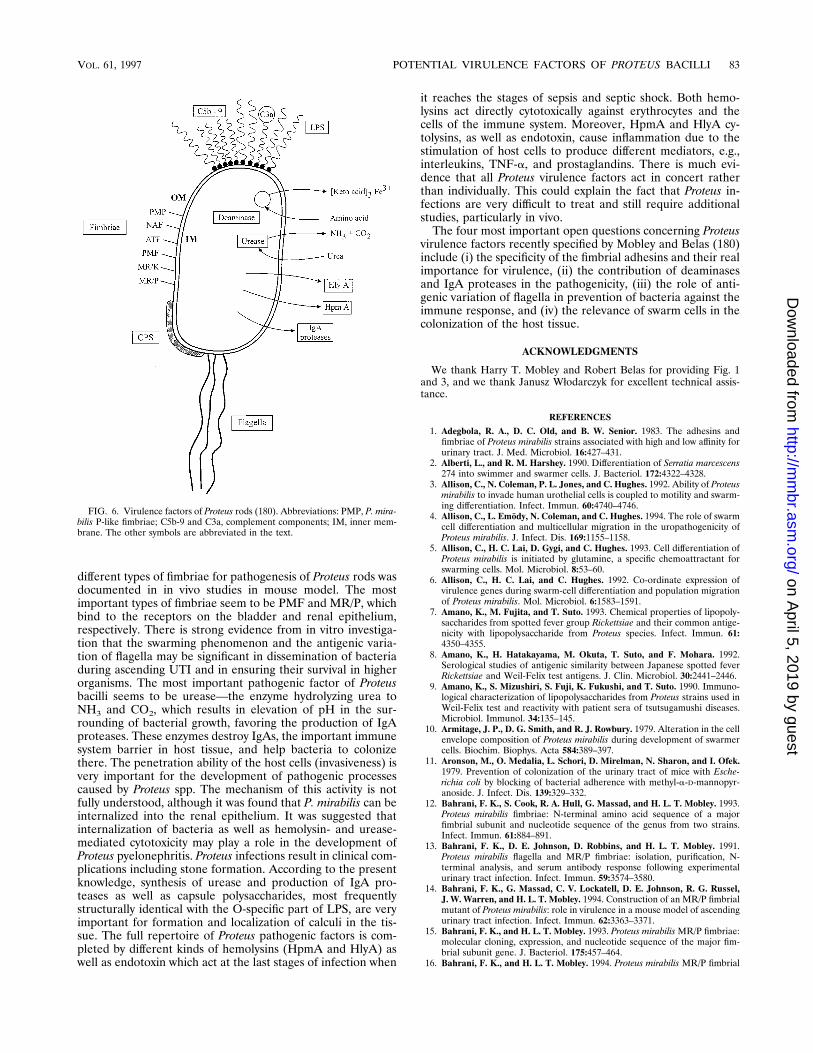
different types of fimbriae for pathogenesis of Proteus rods wasdocumented in in vivo studies in mouse model. The mostimportant types of fimbriae seem to be PMF and MR/P, whichbind to the receptors on the bladder and renal epithelium,respectively. There is strong evidence from in vitro investiga-tion that the swarming phenomenon and the antigenic varia-tion of flagella may be significant in dissemination of bacteriaduring ascending UTI and in ensuring their survival in higherorganisms. The most important pathogenic factor of Proteusbacilli seems to be urease—the enzyme hydrolyzing urea toNH3 and CO2, which results in elevation of pH in the sur-rounding of bacterial growth, favoring the production of IgAproteases. These enzymes destroy IgAs, the important immunesystem barrier in host tissue, and help bacteria to colonizethere. The penetration ability of the host cells (invasiveness) isvery important for the development of pathogenic processescaused by Proteus spp. The mechanism of this activity is notfully understood, although it was found that P. mirabilis can beinternalized into the renal epithelium. It was suggested thatinternalization of bacteria as well as hemolysin- and urease-mediated cytotoxicity may play a role in the development ofProteus pyelonephritis. Proteus infections result in clinical com-plications including stone formation. According to the presentknowledge, synthesis of urease and production of IgA pro-teases as well as capsule polysaccharides, most frequentlystructurally identical with the O-specific part of LPS, are veryimportant for formation and localization of calculi in the tis-sue. The full repertoire of Proteus pathogenic factors is com-pleted by different kinds of hemolysins (HpmA and HlyA) aswell as endotoxin which act at the last stages of infection when
it reaches the stages of sepsis and septic shock. Both hemo-lysins act directly cytotoxically against erythrocytes and thecells of the immune system. Moreover, HpmA and HlyA cy-tolysins, as well as endotoxin, cause inflammation due to thestimulation of host cells to produce different mediators, e.g.,interleukins, TNF-a, and prostaglandins. There is much evi-dence that all Proteus virulence factors act in concert ratherthan individually. This could explain the fact that Proteus in-fections are very difficult to treat and still require additionalstudies, particularly in vivo.The four most important open questions concerning Proteus
virulence factors recently specified by Mobley and Belas (180)include (i) the specificity of the fimbrial adhesins and their realimportance for virulence, (ii) the contribution of deaminasesand IgA proteases in the pathogenicity, (iii) the role of anti-genic variation of flagella in prevention of bacteria against theimmune response, and (iv) the relevance of swarm cells in thecolonization of the host tissue.
ACKNOWLEDGMENTS
We thank Harry T. Mobley and Robert Belas for providing Fig. 1and 3, and we thank Janusz Włodarczyk for excellent technical assis-tance.
REFERENCES
1. Adegbola, R. A., D. C. Old, and B. W. Senior. 1983. The adhesins andfimbriae of Proteus mirabilis strains associated with high and low affinity forurinary tract. J. Med. Microbiol. 16:427–431.
2. Alberti, L., and R. M. Harshey. 1990. Differentiation of Serratia marcescens274 into swimmer and swarmer cells. J. Bacteriol. 172:4322–4328.
3. Allison, C., N. Coleman, P. L. Jones, and C. Hughes. 1992. Ability of Proteusmirabilis to invade human urothelial cells is coupled to motility and swarm-ing differentiation. Infect. Immun. 60:4740–4746.
4. Allison, C., L. Emody, N. Coleman, and C. Hughes. 1994. The role of swarmcell differentiation and multicellular migration in the uropathogenicity ofProteus mirabilis. J. Infect. Dis. 169:1155–1158.
5. Allison, C., H. C. Lai, D. Gygi, and C. Hughes. 1993. Cell differentiation ofProteus mirabilis is initiated by glutamine, a specific chemoattractant forswarming cells. Mol. Microbiol. 8:53–60.
6. Allison, C., H. C. Lai, and C. Hughes. 1992. Co-ordinate expression ofvirulence genes during swarm-cell differentiation and population migrationof Proteus mirabilis. Mol. Microbiol. 6:1583–1591.
7. Amano, K., M. Fujita, and T. Suto. 1993. Chemical properties of lipopoly-saccharides from spotted fever group Rickettsiae and their common antige-nicity with lipopolysaccharide from Proteus species. Infect. Immun. 61:4350–4355.
8. Amano, K., H. Hatakayama, M. Okuta, T. Suto, and F. Mohara. 1992.Serological studies of antigenic similarity between Japanese spotted feverRickettsiae and Weil-Felix test antigens. J. Clin. Microbiol. 30:2441–2446.
9. Amano, K., S. Mizushiri, S. Fuji, K. Fukushi, and T. Suto. 1990. Immuno-logical characterization of lipopolysaccharides from Proteus strains used inWeil-Felix test and reactivity with patient sera of tsutsugamushi diseases.Microbiol. Immunol. 34:135–145.
10. Armitage, J. P., D. G. Smith, and R. J. Rowbury. 1979. Alteration in the cellenvelope composition of Proteus mirabilis during development of swarmercells. Biochim. Biophys. Acta 584:389–397.
11. Aronson, M., O. Medalia, L. Schori, D. Mirelman, N. Sharon, and I. Ofek.1979. Prevention of colonization of the urinary tract of mice with Esche-richia coli by blocking of bacterial adherence with methyl-a-D-mannopyr-anoside. J. Infect. Dis. 139:329–332.
12. Bahrani, F. K., S. Cook, R. A. Hull, G. Massad, and H. L. T. Mobley. 1993.Proteus mirabilis fimbriae: N-terminal amino acid sequence of a majorfimbrial subunit and nucleotide sequence of the genus from two strains.Infect. Immun. 61:884–891.
13. Bahrani, F. K., D. E. Johnson, D. Robbins, and H. L. T. Mobley. 1991.Proteus mirabilis flagella and MR/P fimbriae: isolation, purification, N-terminal analysis, and serum antibody response following experimentalurinary tract infection. Infect. Immun. 59:3574–3580.
14. Bahrani, F. K., G. Massad, C. V. Lockatell, D. E. Johnson, R. G. Russel,J. W. Warren, and H. L. T. Mobley. 1994. Construction of an MR/P fimbrialmutant of Proteus mirabilis: role in virulence in a mouse model of ascendingurinary tract infection. Infect. Immun. 62:3363–3371.
15. Bahrani, F. K., and H. L. T. Mobley. 1993. Proteus mirabilisMR/P fimbriae:molecular cloning, expression, and nucleotide sequence of the major fim-brial subunit gene. J. Bacteriol. 175:457–464.
16. Bahrani, F. K., and H. L. T. Mobley. 1994. Proteus mirabilis MR/P fimbrial
FIG. 6. Virulence factors of Proteus rods (180). Abbreviations: PMP, P. mira-bilis P-like fimbriae; C5b-9 and C3a, complement components; IM, inner mem-brane. The other symbols are abbreviated in the text.
VOL. 61, 1997 POTENTIAL VIRULENCE FACTORS OF PROTEUS BACILLI 83
on April 5, 2019 by guest
http://mm
br.asm.org/
Dow
nloaded from

operon: genetic organization, nucleotide sequence, and conditions for ex-pression. J. Bacteriol. 176:3412–3419.
17. Bangert, R. L., A. C. S. Ward, E. H. Stauber, B. R. Cho, and P. R. Widders.1988. A survey of the aerobic bacteria in the feces of captive raptors. AvianDis. 32:53–62.
18. Bartodziejska, B., J. Radziejewska-Lebrecht, M. Lipinska, L. O. Kononov,A. Y. Chernyak, H. Mayer, and A. Rozalski. 1996. Structural and immuno-chemical studies on lipopolysaccharide of the “T-antigen” containing mu-tant of P. mirabilisR14/1959. FEMS Immunol. Med. Microbiol. 13:113–121.
19. Bartodziejska, B., J. Radziejewska-Lebrecht, M. Lipinska, A. Ziołkowski,and A. Rozalski. 1993. Epitope specificity of polyclonal antibodies againstProteus mirabilis R mutants. Med. Dosw. Mikrobiol. 45:99–102. (In Polish.)
20. Basu, S., J. Radziejewska-Lebrecht, and H. Mayer. 1986. Lipopolysaccha-ride of Providencia rettgeri. Chemical studies and taxonomical implications.Arch. Microbiol. 144:213–218.
21. Belas, R. 1992. The swarming phenomenon of Proteus mirabilis. ASM News58:15–22.
22. Belas, R. 1994. Expression of multiple flagellin-encoding genes of Proteusmirabilis. J. Bacteriol. 176:7169–7181.
23. Belas, R. 1996. Proteus mirabilis swarmer cell differentiation and urinarytract infection, p. 271–298. In H. L. T. Mobley and J. W. Warren (ed.),Urinary tract infections. Molecular pathogenesis and clinical management.ASM Press, Washington, D.C.
24. Belas, R., D. Erskine, and D. Flaherty. 1991. Proteus mirabilis mutantsdefective in swarmer cell differentiation and multicellular behavior. J. Bac-teriol. 173:6279–6288.
25. Belas, R., D. Erskine, and D. Flaherty. 1991. Transposon mutagenesis inProteus mirabilis. J. Bacteriol. 173:6289–6293.
26. Belas, R., and D. Flaherty. 1994. Sequence and genetic analysis of multipleflagellin-encoding genes from Proteus mirabilis. Gene 148:33–41.
27. Belas, R., M. Goldman, and K. Ashliman. 1995. Genetic analysis of Proteusmirabilis mutants defective in swarm cell elongation. J. Bacteriol. 177:823–828.
28. Benz, R., K. R. Hardie, and C. Hughes. 1994. Pore formation in artificialmembranes by the selected hemolysins of Proteus vulgaris and Morganellamorganii. Eur. J. Biochem. 230:339–347.
29. Bergstrom, T. 1972. Sex differences in childhood urinary tract infection.Arch. Dis. Child. 47:227–232.
30. Berner, I., S. Konetschny-Rapp, G. Jung, and G. Winkelmann. 1988. Char-acterization of ferrioxamine E as the principal siderophore of Erwiniaherbicida (Enterobacter agglomerans). Bio-Metals 1:51–56.
31. Bessler, W. G., and U. Henning. 1979. Protein I and protein II from theouter membrane of Escherichia coli are mouse b-lymphocyte mitogen. Z.Immunitatsforsch. 155:387–398.
32. Bessler, W. G., A. Lex, B. Suhr, A. Ortmann, S. Schlecht, H. Buhring, C.Muller, J. Metzger, K. H. Wiesmuller, and G. Jung. 1986. The syntheticanalogs of bacterial lipoprotein are protect immunoadjuvants in combina-tion with or covalently linked to antigen, p. 337–348. In J. Oppenheim andD. Jacobs (ed.), Leukocytes and host defense, vol. 5. Alan R. Liss, Inc., NewYork, N.Y.
33. Beynon, L. M., D. J. Dumanski, R. J. C. McLean, L. L. MacLean, J. C.Richards, and M. B. Perry. 1992. Capsule structure of Proteus mirabilis(ATCC 49565). J. Bacteriol. 174:2172–2177.
34. Bijlsma, I. G. W., L. van Dijk, J. G. Kusters, and W. Gaastra. 1995.Nucleotide sequence of two fimbrial major subunit genes, pmpA and ucaAfrom canine-uropathogenic Proteus mirabilis strains. Microbiology141:1349–1357.
35. Boll, M., J. Radziejewska-Lebrecht, C. Warth, D. Krajewska-Pietrasik, andH. Mayer. 1994. 4-Amino-4-deoxy-L-arabinose in LPS of enterobacterial Rmutants and its possible role for their polymyxin reactivity. FEMS Immu-nol. Med. Microbiol. 8:329–342.
36. Braude, A. J., and J. Siemienski. 1960. Role of bacterial urease in experi-mental pyelonephritis. J. Bacteriol. 80:171–179.
37. Braun, V., and T. Focareta. 1991. Pore-forming bacterial protein hemo-lysins (cytolysins). Crit. Rev. Microbiol. 18:115–158.
38. Breitenbach, J. M., and R. P. Hausinger. 1988. Proteus mirabilis ureasepartial purification and inhibition by boric acid and boronic acid. Biochem.J. 250:917–920.
39. Brenner, D. J., J. J. Farmer III, G. R. Fanning, A. G. Steigerwalt, P.Klykken, H. G. Wathert, F. W. Hickman, and H. W. Ewing. 1978. Deoxyri-bonucleic acid relatedness of Proteus and Providencia species. Int. J. Syst.Bacteriol. 28:269–282.
40. Brinton, C. C. 1965. The structure, function, synthesis and genetic controlof bacterial pili and a molecular model of DNA and RNA transport ingram-negative bacteria. Trans. N. Y. Acad. Sci. 27:1003–1054.
41. Brubaker, R. R. 1985. Mechanisms of bacterial virulence. Annu. Rev. Mi-crobiol. 39:21–50.
42. Bub, F., P. Bieker, H. H. Martin, and K. Nixdorff. 1980. Immunologicalcharacterization of two major proteins isolated from the outer membrane ofProteus mirabilis. Infect. Immun. 27:315–321.
43. Caprodici, C., S. Chen, Z. Sidorczyk, P. Elsbach, and J. Weiss. 1994. Effectsof lipopolysaccharide (LPS) chain length on interactions of bactericidal/
permeability-increasing protein and its bioactive 23-kilodalton NH2-termi-nal fragment with isolated LPS and intact Proteus mirabilis and Escherichiacoli. Infect. Immun. 62:259–265.
44. Cavaillon, J. M., and N. Haeffner-Cavaillon. 1986. Polymyxin B inhibitionof LPS-induced interleukin-1 secretion by human monocytes is dependenton the LPS origin. Mol. Immun. 23:965–969.
45. Cedzynski, M., Y. A. Knirel, A. Rozalski, A. S. Shashkov, E. V. Vinogradov,and W. Kaca. 1995. The structure and serological specificity of Proteusmirabilis O43 O antigen. Eur. J. Biochem. 232:558–562.
46. Cedzynski, M., A. Rozalski, K. Kotełko, W. Kaca, E. V. Vinogradov, Y. A.Knirel, and N. K. Kochetkov. 1993. Structural and immunological studies ofProteus mirabilis O33 of O-specific polysaccharide. Med. Dosw. Mikrobiol.45:93–97. (In Polish.)
47. Cellini, L., R. Piccolomini, N. Allocati, and G. Ravagnan. 1987. Adhesiveproperties of Proteus genus related to antimicrobial agents resistance. Mi-crobiologica 10:291–299.
48. Chang, C. C., and K. Merritt. 1991. Effect of Staphylococcus epidermidis onadherence of Pseudomonas aeruginosa and Proteus mirabilis to polymethylmetacrylate (PMMA) and gentamicin-containing PMMA. J. Orthop. Res.9:284–288.
49. Chen, Y. H. U., R. E. W. Hancock, and R. I. Mishell. 1980. Mitogenic effectof purified outer membrane proteins from Pseudomonas aeruginosa. Infect.Immun. 27:178–184.
50. Cheng, K. J., R. T. Irwin, and J. W. Costerton. 1981. Autochthonous andpathogenic colonization of animal tissues by bacteria. Can. J. Microbiol.27:461–490.
51. Chernyak, A. Y., L. O. Kononov, and N. K. Kochetkov. 1994. Glycopolymersfrom synthetic fragments (amides of a-D-galacturonic acid with amino ac-ids) of Proteus O-antigens. J. Carbohydr. Chem. 13:386–396.
52. Chernyak, A. Y., L. O. Kononov, P. R. Krishna, N. K. Kochetkov, andA. V. R. Rao. 1992. Synthesis of lysine-containing fragments of Proteusmirabilis O27 O-specific polysaccharide and neoglycoconjugates therefrom.Carbohydr. Res. 225:279–289.
53. Cherry, W. B., P. L. Lentz, and L. A. Barnes. 1946. Implication of Proteusmirabilis in an outbreak of gastroenteritis. Am. J. Public Health 36:484–488.
54. Chippendale, G. R., J. W. Warren, A. L. Trifillis, and H. L. T. Mobley. 1994.Internalization of Proteus mirabilis by human renal epithelial cells. Infect.Immun. 62:3115–3121.
55. Chow, A. W., P. R. Taylor, T. T. Yoshikawa, and L. B. Guze. 1979. Anosocomial outbreak of infection due to multiply resistant Proteus mirabilis:role of intestinal colonization as a major reservoir. J. Infect. Dis. 130:621–627.
56. Clapham, L., R. J. C. McLean, J. C. Nickel, J. Downey, and J. W. Costerton.1990. The influence of bacteria on struvite crystal habit and its importancein urinary stone formation. J. Crystal Growth 104:475–484.
57. Clegg, S., and G. F. Gerlach. 1987. Enterobacterial fimbriae. J. Bacteriol.169:934–938.
58. Cook, S. W., N. Mody, J. Valle, and R. Hull. 1995. Molecular cloning ofProteus mirabilis uroepithelial cell adherence (uca) genes. Infect. Immun.63:2082–2086.
59. Cooper, K. E., J. Davies, and J. Wieseman. 1971. An investigation of anoutbreak of food poisoning associated with organisms of the Proteus group.J. Pathol. Bacteriol. 52:91–98.
60. Cosenza, B. J., and J. D. Podgwaite. 1966. A new species of Proteus isolatedfrom the larvae of the gypsy moth Porthetria dispar (L). J. Microbiol. Serol.32:187–191.
61. Costas, M., B. Holmes, K. A. Frith, C. Riddle, and P. M. Hawkey. 1993.Identification and typing of Proteus penneri and Proteus vulgaris biogroups 2and 3, from clinical sources, by computerized analysis of electrophoreticprotein patterns. J. Appl. Bacteriol. 75:489–498.
62. Coyne, C. P., and B. W. Fenwich. 1993. Inhibition of lipopolysaccharide-induced macrophage tumor necrosis factor a synthesis by polymyxin Bsulfate. Am. J. Vet. Res. 54:305–314.
63. Datta, D. B., C. Kramer, and U. Henning. 1976. Diploidy for a structuralgene specifying a major protein of the outer cell envelope membrane fromEscherichia coli K-12. J. Bacteriol. 128:834–841.
64. Deighton, C. H., J. W. Gray, A. M. Bint, and D. J. Walker. 1992. Anti-Proteus antibodies in rheumatoid arthritis same-sexed sibships. Br. J. Rheu-matol. 31:241–245.
65. Drechsel, H., A. Thieken, R. Reissbrodt, G. Jung, and G. Winkelmann.1993. a-Keto acids are novel siderophores in the genera Proteus, Providen-cia, and Morganella and are produced by amino acid deaminases. J. Bacte-riol. 175:2727–2733.
66. Duguid, J. P., and P. R. Gillies. 1957. Fimbrial and adhesive properties indysentery bacilli. J. Pathol. Bacteriol. 74:397–411.
67. Duguid, J. P., and D. C. Old. 1980. Adhesive properties of Enterobacteri-aceae, p. 185–217. In E. H. Beachey (ed.), Bacterial adherence. Receptorsand recognition, ser. B, vol. 6. Chapman & Hall Ltd., London, UnitedKingdom.
68. Dumanski, A. J., H. Hedelin, A. Edin-Liljegren, D. Beauchemin, andR. J. C. McLean. 1994. Unique ability of the Proteus mirabilis capsule to
84 ROZALSKI ET AL. MICROBIOL. MOL. BIOL. REV.
on April 5, 2019 by guest
http://mm
br.asm.org/
Dow
nloaded from

enhance mineral growth in infectious urinary calculi. Infect. Immun. 62:2998–3003.
69. Dutton, A. A. C., and M. Ralston. 1957. Urinary tract infection in a maleurological ward with special reference to the mode of infection. Lanceti:115–119.
70. Ebringer, A., S. Khalafpour, and C. Wilson. 1989. Rheumathoid arthritisand Proteus: a possible aetiological association. Rheumatol. Int. 9:223–228.
71. Emody, L., S. Voros, and T. Pal. 1982. Alpha-hemolysin, a possible factor inProteus morganii. FEMS Microbiol. Lett. 13:329–331.
72. Engler, H. D., K. Troy, and E. J. Bottone. 1990. Bacteremia and subcuta-neous abscess caused by Proteus penneri in a neutropenic host. J. Clin.Microbiol. 28:1645–1646.
73. Evanylo, L. P., S. Kadis, and J. R. Maudsley. 1984. Siderophore productionby Proteus mirabilis. Can. J. Microbiol. 30:1046–1051.
74. Farmer, J. J., III, B. R. Davis, F. W. Hickman-Brenner, A. Wharter, C. P.Huntley-Carter, M. A. Asbury, C. Ridle, H. G. Watheney-Grady, C. Elias,G. R. Fanning, A. G. Steigerwalt, C. M. O’Hara, G. K. Morris, P. B. Smith,and D. J. Brenner. 1985. Biochemical identification of new species andbiogroups of Enterobacteriaceae isolated from clinical specimens. J. Clin.Microbiol. 21:46–76.
75. Finlay, B. B. 1990. Cell adhesion and invasion mechanisms in microbialpathogenesis. Curr. Opin. Cell Biol. 2:815–820.
76. Finlay, B. B., and S. Falkow. 1989. Common themes in microbial pathoge-nicity. Microbiol. Rev. 53:210–230.
77. Fuksa, M., S. Krajden, and A. Lee. 1984. Susceptibility of 45 clinical isolatesof Proteus penneri. Antimicrob. Agents Chemother. 26:419–420.
78. Gabidullin, Z. G., and I. B. Ishkildin. 1989. Morphological properties andadhesiveness of bacteria of the genus Proteus. J. Microbiol. Epidemiol.Immunol. 6:83–86.
79. Gaillard, L. J., B. Berche, C. Frehel, E. Gonin, and P. Cossart. 1991. Entryof L. monocytogenes into cells is mediated by internalin, a repeat proteinreminiscent of surface antigens from gram-positive bacteria. Cell 65:1127–1141.
80. Gibson, F., and D. I. Magrath. 1969. The isolation and characterization ofa hydroxamic acid (aerobactin) formed by Aerobacter aerogenes 62-1. Bio-chim. Biophys. Acta 192:175–184.
81. Gorvil, R. H. 1965. The fate of Pseudomonas aeruginosa, Proteus mirabilisand Escherichia coli in the mouse kidney. J. Pathol. Bacteriol. 89:81–88.
82. Graber, C. D., and A. F. Lincoln. 1955. Infantile diarrhoea in the Denverarea: significance of the Proteus-Providencia organisms. Pediatrics 16:585–591.
83. Grace, M. E., F. J. Gregory, P. P. Hung, and K. P. Fu. 1986. Amplificationand properties of a beta-lactamase from Proteus penneri. J. Antibiot. 39:938–942.
84. Griffith, D. P., D. M. Musher, and C. Itin. 1976. Urease: the primary causeof infection-induced urinary stones. Invest. Urol. 13:346–350.
85. Gromska, W., W. Kaca, and K. Kotełko. 1978. The role of lysine in theserological specificity of some Proteus mirabilis lipopolysaccharides. Bull.Acad. Pol. Sci. Ser. Sci. Biol. 26:7–13.
86. Gromska, W., and H. Mayer. 1976. The linkage of lysine in the O-specificchains of Proteus mirabilis 1959. Eur. J. Biochem. 62:391–399.
87. Guentzel, M. N., and L. J. Berry. 1975. Motility as a virulence factor forVibrio cholerae. Infect. Immun. 11:890–897.
88. Guo, M. M. S., and P. V. Lin. 1965. Serological specificities of ureases ofProteus species. J. Gen. Microbiol. 38:417–422.
89. Gygi, D., M. J. Bailey, C. Allison, and C. Hughes. 1995. Requirement forFlhA in flagella assembly and swarm-cell differentiation by Proteus mirabilis.Mol. Microbiol. 15:761–769.
90. Gygi, D., M. M. Rahman, H.-C. Lai, R. Carlson, J. Guard-Petter, and C.Hughes. 1995. A cell-surface polysaccharide that facilitates rapid popula-tion migration by differentiated swarm cell of Proteus mirabilis. Mol. Mi-crobiol. 17:1167–1175.
91. Hacker, J., and W. Goebel. 1987. Mechanism and methods for analysingphatogenicity. Swiss Biotechnol. 5:21–31.
92. Hasin, M., S. Rottem, and S. Razin. 1975. The outer membrane of Proteusmirabilis. I. Isolation and characterization of the outer and cytoplasmicmembrane fractions. Biochim. Biophys. Acta 375:381–394.
93. Hauser, G. 1885. Uber Faulnisbakterien und deren Beziehung zur Septi-camia, p. 107. Vogel, Leipzig, Germany.
94. Hausinger, R. P. 1987. Nickel utilization by microorganisms. Microbiol.Rev. 51:22–42.
95. Hawkey, P. M., and C. A. Hawkey. 1984. Comparative in vitro activity ofquinolone carboxylic acids against Proteus. J. Antimicrob. Chemother. 14:485–489.
96. Hawkey, P. M., J. Haynes, J. Heritage, and L. Penner. 1986. Role ofProteeae in diarrheal disease. J. Clin. Microbiol. 24:504–505. (Letter.)
97. Hawkey, P. M., S. J. Pedler, and A. Turner. 1983. Comparative in vitroactivity of semisynthetic penicillins against Proteeae. Antimicrob. AgentsChemother. 23:619–621.
98. Hawthorn, L., and G. Reid. 1990. The effect of protein and urine onuropathogen adhesion to polymer substrata. J. Biomed. Mater. Res. 24:1325–1332.
99. Henriksen, J. 1972. Bacterial surface translocation: a survey and a classifi-cation. Bacteriol. Rev. 36:478–503.
100. Hickman, F. W., A. G. Steigerwalt, J. J. Farmer III, and D. J. Brenner.1982. Identification of Proteus penneri sp. nov., formerly known as Proteusvulgaris indole negative or as Proteus vulgaris biogroup 1. J. Clin. Microbiol.15:1097–1102.
101. Hoffman, P., and W. G. Bessler. 1988. Tumor cytotoxicity involving TNFinduced in bone marrow-derived macrophages by two lipopeptide ana-logues of bacterial lipoprotein. Immunobiology 178:94–101.
102. Hofstra, H., and J. Dankert. 1979. Antigenic cross-reactivity of major outermembrane proteins in Enterobacteriaceae species. J. Gen. Microbiol. 111:293–302.
103. Hofstra, H., and J. Dankert. 1980. Antigenic cross-reactivity of outer mem-brane proteins of E. coli and Proteus species. FEMS Microbiol. Lett. 7:171–174.
104. Hofstra, H., M. J. D. Van Tol, and J. Dankert. 1980. Cross-reactivity ofmajor outer membrane proteins of Enterobacteriaceae studied by crossedimmunoelectrophoresis. J. Bacteriol. 143:328–337.
105. Holmes, B., M. Costas, and A. C. Wood. 1991. Typing of Proteus mirabilisfrom clinical sources by computerized analysis of electrophoretic proteinpatterns. J. Appl. Bacteriol. 71:467–476.
106. Hornick, D. B., B. L. Allen, M. A. Horn, and S. Clegg. 1991. Fimbrial typesamong respiratory isolates belonging to the family Enterobacteriaceae.J. Clin. Microbiol. 29:1795–1800.
107. Isberg, R. R., S. D. L. Voorhis, and S. Falkow. 1987. Identification ofinvasin: a protein that allows enteric bacteria to penetrate cultured mam-malian cells. Cell 50:769–778.
108. Island, M. D., and H. L. T. Mobley. 1995. Proteus mirabilis urease: operonfusion and linker insertion analysis of ure gene organization, regulation, andfunction. J. Bacteriol. 177:5653–5660.
109. Johnson, D. E., R. G. Russell, C. V. Lockatell, J. C. Zulty, J. W. Warren,and H. L. T. Mobley. 1993. Contribution of Proteus mirabilis urease topersistent urolithiasis and acute pyelonephritis in a mouse model of as-cending urinary tract infection. Infect. Immun. 61:2748–2754.
110. Johnson, J. R. 1991. Virulence factors in Escherichia coli urinary tractinfection. Clin. Microbiol. Rev. 4:80–121.
111. Jones, B. D., C. V. Lockatell, D. E. Johnson, J. W. Warren, and H. L. T.Mobley. 1990. Construction of urease-negative mutant of Proteus mirabilis:analysis of virulence in a mouse model of ascending urinary tract infection.Infect. Immun. 58:1120–1123.
112. Jones, B. D., and H. L. T. Mobley. 1987. Genetic and biochemical diversityof ureases of Proteus, Providencia, and Morganella species isolated fromurinary tract infection. Infect. Immun. 55:2198–2203.
113. Jones, B. D., and H. L. T. Mobley. 1988. Proteus mirabilis urease: geneticorganization, regulation, and expression of structural genes. J. Bacteriol.170:3342–3349.
114. Jones, B. D., and H. L. T. Mobley. 1989. Proteus mirabilis urease: nucleotidesequence determination and comparison with Jack bean urease. J. Bacte-riol. 171:6414–6422.
115. Kaca, W., and M. Cedzynski. Unpublished data.116. Kaca, W., Y. A. Knirel, E. V. Vinogradov, and K. Kotełko. 1987. Structure
of the O-specific polysaccharide of Proteus mirabilis S1959. Arch. Immunol.Ther. Exp. 35:431–437.
117. Kaca, W., and K. Kotełko. 1983. Lipopolysaccharide phage receptor ofProteus mirabilis 1959 strain. Site of phage mediated hydrolysis on O-specific polysaccharide. Arch. Immunol. Ther. Exp. 31:691–700.
118. Kaca, W., M. Mara, and J. Ocenaskova. 1996. Inhibition of mouse livercytochrome P-450 by gram-negative bacteria lipopolysaccharides. Arch.Immunol. Ther. Exp. 44:39–44.
119. Kaca, W., J. Radziejewska-Lebrecht, and U. R. Bhat. 1990. Effect of poly-myxins on the lipopolysaccharide defective mutants of P. mirabilis. Micro-bios 61:23–32.
120. Kaca, W., and R. I. Roth. 1995. Activation of complement by humanhemoglobin and by mixtures of hemoglobin and bacterial endotoxin. Bio-chim. Biophys. Acta 1245:49–56.
121. Kaca, W., R. I. Roth, and J. Levin. 1994. Hemoglobin, a newly recognizedlipopolysaccharide (LPS)-binding protein that enhances LPS biological ac-tivity. J. Biol. Chem. 269:25078–25084.
122. Kaca, W., R. I. Roth, A. Ziołkowski, and J. Levin. 1994. Human hemoglobinincreases the biological activity of bacterial lipopolysaccharides in activa-tion of Limulus amebocyte lysate and stimulation of tissue factor produc-tion by endothelial cells in vitro. J. Endotoxin Res. 1:243–252.
123. Kaca, W., and A. Rozalski. 1991. Characterization of cell-bound and cell-free hemolytic activity of Proteus strains. Eur. J. Epidemiol. 7:159–165.
124. Kappos, T., M. A. John, Z. Hussain, and M. A. Valvano. 1992. Outermembrane protein profiles and multilocus enzyme electrophoresis analysisfor differentiation of clinical isolates of Proteus mirabilis and Proteus vul-garis. J. Clin. Microbiol. 30:2632–2637.
125. Karch, H., J. Gmeiner, and K. Nixdorff. 1983. Alteration of the immuno-globulin G subclass responses in mice to lipopolysaccharide effects of non-bacterial proteins and bacterial membrane phospholipids or outer mem-brane proteins of Proteus mirabilis. Infect. Immun. 40:157–165.
VOL. 61, 1997 POTENTIAL VIRULENCE FACTORS OF PROTEUS BACILLI 85
on April 5, 2019 by guest
http://mm
br.asm.org/
Dow
nloaded from

126. Karch, H., and K. Nixdorff. 1981. Antibody producing cell responses to anisolated outer membrane protein and to complexes of this antigen withlipopolysaccharide or with vesicles of phospholipids from Proteus mirabilis.Infect. Immun. 31:862–867.
127. Karch, H., and K. Nixdorff. 1983. Modulation of the IgG subclass responsesto lipopolysaccharide by bacterial membrane components: differential ad-juvant effects produced by primary and secondary stimulation. J. Immunol.131:6–8.
128. Kauffmann, F. 1966. The bacteriology of Enterobacteriaceae, 3rd ed., p. 333.The Williams & Wilkins, Co., Baltimore, Md.
129. Kippax, P. W. 1957. A study of Proteus infections in a male urological ward.J. Clin. Pathol. 10:211–214.
130. Knirel, Y. A., N. A. Paramonov, E. V. Vinogradov, N. K. Kochetkov, Z.Sidorczyk, and K. Zych. 1994. 2-Acetamido-4-O-[(S)-1-carboxyethyl]-2-de-oxy-D-glucose: a new natural isomer of N-acetylmuramic acid from theO-specific polysaccharide of Proteus penneri 35. Carbohydr. Res. 259:C1–C3.
131. Knirel, Y. A., N. A. Paramonov, E. V. Vinogradov, A. S. Shashkov, N. K.Kochetkov, Z. Sidorczyk, and A. Swierzko. 1992. Structure of the O-specificpolysaccharide of Proteus penneri 62 containing 2-acetamido-3-O[(S)-1-car-boxyethyl]-2-deoxy-D-glucose (N-acetylisomuramic acid). Carbohydr. Res.235:C19–C23.
132. Knirel, Y. A., A. S. Shashkov, E. V. Vinogradov, A. Swierzko, and Z.Sidorczyk. 1995. The structure of the O-specific polysaccharide chain ofProteus penneri strain 42 lipopolysaccharide. Carbohydr. Res. 155:C1–C6.
133. Knirel, Y. A., E. V. Vinogradov, A. S. Shashkov, Z. Sidorczyk, W. Kaca, A.Rozalski, K. Kotełko, and N. K. Kochetkov. 1992. Structure of ProteusO-specific polysaccharides. Dokl. Ross. Akad. Nauk 324:333–338. (In Rus-sian.)
134. Knirel, Y. A., E. V. Vinogradov, A. S. Shashkov, Z. Sidorczyk, A. Rozalski,J. Radziejewska-Lebrecht, and W. Kaca. 1993. Structural study of O-spe-cific polysaccharides of Proteus. J. Carbohydr. Chem. 12:379–414.
135. Kornfeld, S. J., and A. G. Plant. 1981. Secretory immunity and the bacterialIgA proteases. Rev. Infect. Dis. 3:521–534.
136. Koronakis, V., M. Cross, B. Senior, E. Koronakis, and C. Hughes. 1987.The secreted hemolysins of Proteus mirabilis, Proteus vulgaris, and Mor-ganella morganii are genetically related to each other and to the alpha-hemolysin of Escherichia coli. J. Bacteriol. 169:1509–1515.
137. Koronakis, V., and C. Hughes. 1988. Identification of the promoters direct-ing in vivo expression of hemolysin genes in Proteus vulgaris and Escherichiacoli. Mol. Gen. Genet. 213:99–104.
138. Koronakis, V., E. Koronakis, and C. Hughes. 1988. Comparison of thehemolysin secretion protein HlyB from Proteus vulgaris and Escherichia coli:site directed mutagenesis causing impairment of export function. Mol. Gen.Genet. 213:551–555.
139. Kotełko, K. 1986. Proteus mirabilis: taxonomic position, peculiarities ofgrowth, components of the cell envelope. Curr. Top. Microbiol. Immunol.129:181–215.
140. Kotełko, K., M. Deka, W. Gromska, W. Kaca, J. Radziejewska-Lebrecht,and A. Rozalski. 1983. Galacturonic acid as the terminal constituent in theR-core polysaccharide of Proteus R110 (Ra) mutant. Arch. Immunol. Ther.Exp. 31:619–623.
141. Kotełko, K., W. Gromska, M. Papierz, Z. Sidorczyk, D. Krajewska, and K.Szer. 1977. Core region in Proteus mirabilis lipopolysaccharide. J. Hyg.Epidemiol. Microbiol. Immunol. 21:271–284.
142. Kotełko, K., W. Kaca, A. Rozalski, and M. Deka. 1983. Some biologicalfeatures of Proteus bacilli. 2. Haemolytic activities of Proteus mirabilis andProteus vulgaris strains. Acta Microbiol. Pol. 32:345–351.
143. Kotełko, K., A. Rozalski, M. Deka, Z. Sidorczyk, W. Gromska, and K. Zych.1983. Some biological features of Proteus bacilli. 1. Comparison of Proteusmirabilis strains provided from various sources. Acta Microbiol. Pol. 32:334–339.
144. Krajden, S., M. Fuksa, W. Lizewski, L. Barton, and A. Lee. 1984. Proteuspenneri and urinary calculi formation. J. Clin. Microbiol. 19:541–542.
145. Krajden, S., M. Fuksa, C. Petrea, L. J. Crisp, and J. L. Penner. 1987.Expanded clinical spectrum of infections caused by Proteus penneri. J. Clin.Microbiol. 25:578–579.
146. Krajewska-Pietrasik, D., A. Y. Chernyak, and A. Rozalski. 1995. Use ofsynthetic antigens in studies of antigen specificity of Proteus mirabilis O27lipopolysaccharide Med. Dosw. Mikrobiol. 47:169–176. (In Polish.)
147. Krajewska-Pietrasik, D., P. Larsson, K. Zych, J. Włodarczyk, and W.Gromska. 1991. Characteristics of spontaneously agglutinating Proteusmirabilis strains from bacteriuric patients. APMIS 99:956–960.
148. Krajewska-Pietrasik, D., A. Rozalski, B. Bartodziejska, J. Radziejewska-Lebrecht, H. Mayer, and K. Kotełko. 1991. Properties of a deep Proteus Rmutant isolated from clinical material. APMIS 99:499–506.
149. Lanyi, B. 1956. Serological typing of Proteus strains from infantile enteri-tides and other sources. Acta Microbiol. Acad. Sci. Hung. 3:417–428.
150. Larsson, P. 1978. A comparison of the host-parasite relationship in urinarytract infections caused by Proteus and Escherichia coli in animals and chil-dren. Ph.D. thesis. University of Goteborg, Goteborg, Sweden.
151. Larsson, P. 1984. Serology of Proteus mirabilis and Proteus vulgaris. Meth-ods Microbiol. 14:187–214.
152. Larsson, P., A. Fasth, U. Jodal, A. Sohl Akerlund, and C. Svanborg Eden.1978. Urinary tract infections caused by Proteus mirabilis in children. Theantibody response to O and H antigens and Tamm Horsfall protein andbacterial adherence to uroepithelium. Acta Paediatr. Scand. 67:591–596.
153. Larsson, P., and S. Olling. 1977. O-Antigen distribution and sensitivity tothe bactericidal effect of normal human serum of Proteus strains fromclinical specimens. Med. Microbiol. Immunol. 163:77–82.
154. Legnani-Fajardo, C., and P. Zunino. 1990. Antigenic and immunogenicactivity of flagella and fimbriae of preparation from uropathogenic Proteusmirabilis. Can. J. Microbiol. 37:325–328.
155. Loomes, L. M., B. W. Senior, and M. A. Kerr. 1990. A proteolytic enzymesecreted by Proteus mirabilis degrades immunoglobulins of the immuno-globulin A1 (IgA1), IgA2, and IgG isotypes. Infect. Immun. 58:1979–1985.
156. Loomes, L. M., B. W. Senior, and M. A. Kerr. 1992. Proteinases of Proteusspp.: purification, properties, and detection in urine of infected patients.Infect. Immun. 60:2267–2273.
157. Lowell, G. H. 1990. Proteosomes, hydrophobic anchors, incoms and lipo-somes for improved presentation of peptide and protein vaccines, p. 141–160. In G. C. Woodrow and M. M. Levine (ed.), New generation of vac-cines. Marcel Dekker, Inc., New York, N.Y.
158. Lugtenberg, B., H. Bronstein, N. Van Selm, and R. Peters. 1977. Pepti-doglycan-associated outer membrane proteins in gram-negative bacteria.Biochim. Biophys. Acta 465:571–578.
159. Łukomski, S., B. Muller, and G. Schmidt. 1990. Extracellular hemolysin ofProteus penneri coded by chromosomal hly genes is similar to the a-hemo-lysin of Escherichia coli. Zentralbl. Bakteriol. 273:150–155.
160. Łukomski, S., M. Pytlos, L. Serwecinska, Z. Sidorczyk, and A. Jaworski.1993. Analysis of antibiotic resistance determinants in Proteus penneri. Eur.J. Clin. Microbiol. Infect. Dis. 12:467–470.
161. Łukomski, S., L. Serwecinska, A. Rozalski, J. Dziadek, P. Staczek, and A.Jaworski. 1991. Cell-free and cell-bound hemolytic activities of Proteuspenneri determination by different Hly determinants. Can. J. Microbiol.37:419–424.
162. MacLaren, D. M. 1968. The significance of urease in Proteus pyelonephritis:a bacteriological study. J. Pathol. Bacteriol. 96:45–56.
163. MacLaren, D. M. 1969. The significance of urease in Proteus pyelonephritisa histological and biochemical study. J. Pathol. Bacteriol. 97:43–49.
164. Magana-Plaza, I., C. Montes, and J. Ruiz-Herrera. 1971. Purification andbiochemical characteristics of urease from Proteus rettgeri. Biochim. Bio-phys. Acta 242:230–237.
165. Magana-Plaza, I., and J. Ruiz-Herrera. 1967. Mechanisms of regulation ofurease biosynthesis in Proteus rettgeri. J. Bacteriol. 93:1294–1301.
166. Martin, M. A. 1991. Epidemiology and clinical impact of gram-negativesepsis. Infect. Dis. Clin. North Am. 5:739–752.
167. Massad, G., F. K. Bahrani, and H. L. T. Mobley. 1994. Proteus mirabilisfimbriae: identification, isolation, and characterization of a new ambient-temperature fimbria. Infect. Immun. 62:1989–1994.
168. Massad, G., C. V. Lockatell, D. E. Johnson, and H. L. T. Mobley. 1994.Proteus mirabilis fimbriae: construction of an isogenic pmfA mutant andanalysis of virulence in a CBA mouse model of ascending urinary tractinfection. Infect. Immun. 62:536–542.
169. Massad, G., H. Zhao, and H. L. T. Mobley. 1995. Proteus mirabilis aminoacid deaminase: cloning, nucleotide sequence, and characterization of aad.J. Bacteriol. 177:5878–5883.
170. McCarter, L., and M. Silverman. 1989. Iron regulation of swarmer celldifferentiation of Vibrio parahaemolyticus. J. Bacteriol. 171:731–736.
171. McLean, R. J. C., K.-J. Cheng, W. D. Gould, and J. W. Costerton. 1986.Histochemical and biochemical urease localization in the periplasm andouter membrane of two Proteus mirabilis strains. Can. J. Microbiol. 32:772–778.
172. McLean, R. J. C., J. C. Nickel, K.-J. Cheng, and J. W. Costerton. 1988. Theecology and pathogenicity of urease-producing bacteria in the urinary tract.Crit. Rev. Microbiol. 16:37–79.
173. McLean, R. J. C., J. C. Nickel, V. C. Noakes, and J. W. Costerton. 1985. Anin vitro ultrastructural study of infectious kidney stone genesis. Infect.Immun. 49:805–811.
174. McManus, A. T., E. E. Moody, and A. D. Mason. 1980. Bacterial motility:a component in experimental Pseudomonas aeruginosa burn wound sepsis.Burns 6:235–239.
175. Melchers, F., V. Braun, and C. Galanos. 1975. The lipoprotein of the outermembrane of Escherichia coli: a b-lymphocyte mitogen. J. Exp. Med. 142:473–478.
176. Milazzo, F. H., and G. J. Delisle. 1984. Immunoglobulin A proteases ingram-negative bacteria isolated from human urinary tract infections. Infect.Immun. 43:11–13.
177. Mizushiri, S., K. Amano, S. Fujii, K. Fukushi, and M. Watanabe. 1990.Chemical characterization of lipopolysaccharides from Proteus strains usedin Weil-Felix test. Microbiol. Immunol. 34:121–133.
178. Moayeri, N., C. M. Collins, and P. O’Hanley. 1991. Efficacy of a Proteusmirabilis outer membrane protein vaccine in preventing experimental Pro-
86 ROZALSKI ET AL. MICROBIOL. MOL. BIOL. REV.
on April 5, 2019 by guest
http://mm
br.asm.org/
Dow
nloaded from

teus pyelonephritis in a BALB/c mouse model. Infect. Immun. 59:3778–3786.
179. Mobley, H. L. T. 1996. Virulence of Proteus mirabilis, p. 245–269. InH. L. T.Mobley and J. W. Warren (ed.), Urinary tract infections. Molecular patho-genesis and clinical management. ASM Press, Washington, D.C.
180. Mobley, H. L. T., and R. Belas. 1995. Swarming and pathogenicity of Proteusmirabilis in the urinary tract. Trends Microbiol. 3:280–284.
181. Mobley, H. L. T., and G. R. Chippendale. 1990. Hemagglutinin, urease andhemolysin production by Proteus mirabilis from clinical sources. J. Infect.Dis. 161:525–530.
182. Mobley, H. L. T., G. R. Chippendale, K. Swihart, and R. Welch. 1991.Cytotoxicity of the HpmA hemolysin and urease of Proteus mirabilis andProteus vulgaris against cultured human renal proximal tubular cells. Infect.Immun. 59:2036–2042.
183. Mobley, H. L. T., G. R. Chippendale, J. H. Tenney, A. R. Mayer, L. J. Crisp,J. L. Penner, and J. W. Warren. 1988. MR/K hemagglutination of Provi-dencia stuartii correlates with adherence to catheters and with persistence incatheter-associated bacteriuria. J. Infect. Dis. 157:264–271.
184. Mobley, H. L. T., and R. P. Hausinger. 1989. Microbial ureases: signifi-cance, regulation, and molecular characterization. Microbiol. Rev. 53:85–108.
185. Mobley, H. L. T., M. D. Island, and R. P. Hausinger. 1995. Molecularbiology of microbial ureases. Microbiol. Rev. 59:451–480.
186. Mobley, H. L. T., B. D. Jones, and A. E. Jerse. 1986. Cloning of urease genesequence from Providencia stuartii. Infect. Immun. 54:161–169.
187. Mobley, H. L. T., B. D. Jones, and J. L. Penner. 1987. Urease activity ofProteus penneri. J. Clin. Microbiol. 25:2302–2305.
188. Mobley, H. L. T., and J. W. Warren. 1987. Urease-positive bacteriuria andobstruction of long-term urinary catheters. J. Clin. Microbiol. 25:2216–2217.
189. Morrison, D. C., and D. M. Jacobs. 1976. Binding of polymyxin B to thelipid A portion of bacterial lipopolysaccharide. Immunochemistry 13:813–818.
190. Morsdorf, G., and H. Kaltwasser. 1990. Cloning of the genes encodingurease from Proteus vulgaris and sequencing of the structural genes. FEMSMicrobiol. Lett. 66:67–74.
191. Mortie, T. C., D. Doyle-Huntzinger, R. C. Craven, and I. A. Holder. 1982.Loss of virulence associated with absence of flagellum in an isogenic mutantof Pseudomonas aeruginosa in the burned-mouse model. Infect. Immun.38:1296–1298.
192. Mulks, M. H. 1985. Microbial IgA proteases, p. 81–104. In I. A. Holder(ed.), Bacterial enzymes and virulence. CRC Press, Inc., Boca Raton, Fla.
193. Muller, H. E. 1986. Occurrence and pathogenic role of Morganella-Proteus-Providencia group bacteria in human feces. J. Clin. Microbiol. 23:404–405.
194. Musher, D. M., D. P. Griffith, D. Yawn, and R. D. Rossen. 1975. Role ofurease in pyelonephritis resulting from urinary tract infection with Proteus.J. Infect. Dis. 131:177–181.
195. Nawrot, U., G. Mokracka-Latajka, J. Grzybek-Hryncewicz, B. Krzyza-nowska, and S. Jankowski. 1995. Bactericidal activity of normal humanserum against Morganella, Proteus and Providencia strains. Acta Microbiol.Pol. 44:55–61.
196. Nicholson, E. B., E. A. Concaugh, P. A. Foxall, M. D. Island, and H. L. T.Mobley. 1993. Proteus mirabilis urease transcriptional regulation byUreR. J. Bacteriol. 175:465–473.
197. Nicholson, E. B., E. A. Concaugh, and H. L. T. Mobley. 1991. Proteusmirabilis urease: use of a UreA-LacZ fusion demonstrates that induction ishighly specific for urea. Infect. Immun. 59:3360–3365.
198. Nickel, J. C., J. Eutag, and J. W. Costerton. 1985. Ultrastructural microbialecology of infection induced urinary stones. J. Urol. 133:622–627.
199. Nikaido, H., and M. Vaara. 1985. Molecular basis of bacterial outer mem-brane permeability. Microbiol. Rev. 49:1–32.
200. Nixdorff, K., H. Fitzer, J. Gmeiner, and K. H. Martin. 1977. Reconstructionof model membranes from phospholipid and outer membrane proteins ofProteus mirabilis. Eur. J. Biochem. 81:63–69.
201. O’Brien, I. G., G. B. Cox, and F. Gibson. 1970. Biologically active com-pound containing 2,3,dihydroxybenzoic acid and serine formed by Esche-richia coli. Biochim. Biophys. Acta 201:453–460.
202. Old, D. C., and R. Adegbola. 1982. Hemagglutinins and fimbriae of Mor-ganella, Proteus and Providencia. J. Med. Microbiol. 15:551–564.
203. Old, D. C., and R. A. Adegbola. 1985. Antigenic relationships among type-3fimbriae of Enterobacteriaceae revealed by immunoelectronmicroscopy.J. Med. Microbiol. 20:113–121.
204. Payne, S. M. 1988. Iron and virulence in the family Enterobacteriaceae. Crit.Rev. Microbiol. 16:81–111.
205. Peerbooms, P. G. H., A. M. J. J. Verweij, and P. M. MacLaren. 1982.Urinary virulence of Proteus mirabilis in two experimental mouse models.Infect. Immun. 36:1246–1248.
206. Peerbooms, P. G. M., A. M. J. J. Verweij, and D. M. MacLaren. 1983.Investigation of the haemolytic activity of Proteus mirabilis strains. AntonieLeeuwenhoek 49:1–11.
207. Peerbooms, P. G. M., A. M. J. J. Verweij, and D. M. MacLaren. 1984. Verocell invasiveness of Proteus mirabilis. Infect. Immun. 43:1068–1071.
208. Peerbooms, P. G. M., A. M. J. J. Verweij, and D. M. MacLaren. 1985.Uropathogenic properties of Proteus mirabilis and Proteus vulgaris. J. Med.Microbiol. 19:55–60.
209. Penner, J. L. 1984. Genus XI. Proteus, p. 491–494. In N. R. Krieg and J. G.Holt (ed.), Bergey’s manual of systematic bacteriology, vol. 1. The Williams& Wilkins Co., Baltimore, Md.
210. Penner, J. L. 1992. The genera Proteus, Providencia and Morganella, p.2849–2853. In A. Balows, H. G. Truper, W. Harder, and K. H. Schleifer(ed.), The prokaryotes, vol. III. Springer-Verlag KG, Berlin, Germany.
211. Penner, J. L., and J. N. Hennessy. 1980. Separate O-grouping schemes forserotyping clinical isolates of Proteus vulgaris and Proteus mirabilis. J. Clin.Microbiol. 12:77–82.
212. Penner, J. L., N. A. Hinton, G. R. Whiteley, and J. N. Hennessy. 1976.Variation in urease activity of endemic hospital strains of Proteus rettgeriand Providencia stuartii. J. Infect. Dis. 134:370–376.
213. Perch, B. 1948. On the serology of the Proteus group. Acta Pathol. Micro-biol. Scand. 25:703–714.
214. Perry, M. B., and L. L. MacLean. 1994. The structure of the polysaccharideproduced by Proteus vulgaris (ATCC 49990). Carbohydr. Res. 253:257–263.
215. Philips, J. F. 1955. In vitro studies on Proteus organisms of animal origin. J.Hyg. 53:26–31.
216. Piccolomini, R., L. Cellini, N. Allocati, A. Di Girolamo, and G. Ravagnan.1987. Comparative in vitro activities of 13 antimicrobial agents againstMorganella-Proteus-Providencia group bacteria from urinary tract infec-tions. Antimicrob. Agents Chemother. 31:1644–1647.
217. Plant, A. G. 1983. The IgA1 proteases of pathogenic bacteria. Annu. Rev.Microbiol. 37:603–622.
218. Pollack, J. R., and J. B. Neilands. 1970. Enterobactin, an iron transportcompound from Salmonella typhimurium. Biochem. Biophys. Res. Com-mun. 38:989–992.
219. Radziejewska-Lebrecht, J., U. R. Bhat, H. Brade, and H. Mayer. 1988.Structural studies on the core and lipid A region of a 4-amino-L-arabinose-lacking Rc type mutant of Proteus mirabilis. Eur. J. Biochem. 172:532–542.
220. Radziejewska-Lebrecht, J., U. Feige, M. Jensen, K. Kotełko, U. Friebolin,and H. Mayer. 1980. Structural studies on the glucose-heptose region of theProteus mirabilis R core. Eur. J. Biochem. 107:31–38.
221. Radziejewska-Lebrecht, J., D. Krajewska-Pietrasik, and H. Mayer. 1990.Terminal and chain-linked residues of D-galacturonic acid: characteristicconstituents of the R-core regions of Proteeae and Serratia marcescens. Syst.Appl. Microbiol. 13:214–219.
222. Radziejewska-Lebrecht, J., and H. Mayer. 1989. The core region of Proteusmirabilis R110/1959 lipopolysaccharide. Eur. J. Biochem. 183:573–581.
223. Radziejewska-Lebrecht, J., A. S. Shashkov, E. V. Vinogradov, U.Grosskurth, B. Bartodziejska, A. Rozalski, W. Kaca, L. O. Kononov, A. Y.Chernyak, H. Mayer, Y. A. Knirel, and N. K. Kochetkov. 1995. Structureand epitope characterization of the O-specific polysaccharide of Proteusmirabilis O28 containing amides of D-galacturonic acid with L-serine andL-lysine. Eur. J. Biochem. 230:705–712.
224. Radziejewska-Lebrecht, J., K. Zych, M. Lipinska, and Z. Sidorczyk. 1993.Further core types of Proteus lipopolysaccharides. Med. Dosw. Mikrobiol.45:65–68. (In Polish.)
225. Rando, D., U. Steglitz, G. Morsdorf, and H. Kaltwasser. 1990. Nickelavailability and urease expression in Proteus mirabilis. Arch. Microbiol.154:428–432.
226. Raoult, D., and G. A. Dasch. 1995. Immunoblot cross-reactions amongRickettsia, Proteus sp. and Legionella sp. in patients with Mediterraneanspotted fever. FEMS Immunol. Med. Microbiol. 11:13–18.
227. Reid, G., and J. D. Sobel. 1987. Bacterial adherence in the pathogenesis ofurinary tract infections: a review. Rev. Infect. Dis. 9:470–487.
228. Reissbrodt, R., W. Rabsch, A. Chapeaurouge, G. Jung, and G. Winkel-mann. 1990. Isolation and identification of ferroxamine G and E in Hafniaalvei. Bio-Metals 3:54–60.
229. Rietschel, E. T., and H. Brade. 1992. Bacterial endotoxins. Sci. Am. 267:26–33.
230. Rietschel, E. T., L. Brade, B. Lindner, and U. Zahringer. 1992. Biochem-istry of lipopolysaccharides. p. 3–41. InD. C. Morrison and J. L. Ryan (ed.),Bacterial endotoxic lipopolysaccharides. CRC Press, Inc., Boca Raton, Fla.
231. Rietschel, E. T., T. Kirikae, F. U. Schade, U. Mamat, G. Schmidt, H.Loppnow, A. J. Ulmer, U. Zahringer, U. Seydel, F. Di Padova, M. Schreier,and H. Brade. 1994. Bacterial endotoxin: molecular relationships of struc-ture to activity and function. FASEB J. 8:217–225.
232. Roberts, J. A., E. N. Fussell, and M. B. Kaach. 1990. Bacterial adherence tourethial catheters. J. Urol. 144:264–269.
233. Rosenstein, I. J. M. 1986. Urinary calculi: microbiological and crystallo-graphic studies. Crit. Rev. Clin. Lab. Sci. 22:245–277.
234. Rosenstein, I. J., J. M. Hamilton-Miller, and W. Brumfitt. 1981. Role ofurease in the formation of infection stones: comparison of ureases fromdifferent sources. Infect. Immun. 32:32–37.
235. Rozalski, A. Unpublished data.236. Rozalski, A., B. Bartodziejska, M. Wykrota, L. Serwecinska, S. Łukomski,
and K. Kotełko. 1993. Characterization of hemolytic activity of Proteuspenneri. Med. Dosw. Mikrobiol. 45:79–83. (In Polish.)
VOL. 61, 1997 POTENTIAL VIRULENCE FACTORS OF PROTEUS BACILLI 87
on April 5, 2019 by guest
http://mm
br.asm.org/
Dow
nloaded from

237. Rozalski, A., L. Brade, P. Kosma, B. J. Appelmelk, C. Krogman, and H.Brade. 1989. Epitope specificity of murine monoclonal and rabbit poly-clonal antibodies against enterobacterial lipopolysaccharides of the Re che-motype. Infect. Immun. 57:2645–2652.
238. Rozalski, A., H. Długonska, and K. Kotełko. 1986. Cell invasiveness ofProteus mirabilis and Proteus vulgaris strains. Arch. Immunol. Ther. Exp.34:505–511.
239. Rozalski, A., and K. Kotełko. 1987. Hemolytic activity and invasiveness instrains of Proteus penneri. J. Clin. Microbiol. 25:1094–1096.
240. Rozalski, A., and M. Wykrota. 1986. Some biological features of Proteusbacilli. 3. Comparison of haemolytic activity of Proteus and Serratia strains.Acta Microbiol. Pol. 35:57–59.
241. Rubin, R. H., N. E. Tolkoff-Rubin, and R. S. Cotran. 1986. Urinary tractinfection, pyelonephritis and reflux nephropathy, p. 1085–1143. In B. M.Brenner and F. C. Rector (ed.), The kidney. The W. B. Saunders Co.,Philadelphia, Pa.
242. Sareneva, T., H. Holthofer, and T. K. Korhonen. 1990. Tissue-bindingaffinity of Proteus mirabilis fimbriae in the human urinary tract. Infect.Immun. 58:3330–3336.
243. Savoia, D., P. Martineto, A. Achino, and A. Pugliese. 1983. Adhesion ofProteus species to various cell types. Eur. J. Clin. Microbiol. 2:571–576.
244. Schindler, M., and M. J. Osborn. 1979. Interaction of divalent cations andpolymyxin B with lipopolysaccharide. Biochemistry 18:4425–4430.
245. Schoolnik, G. K., D. Lark, and P. O’Hanley. 1980. Bacterial adherence andanticolonization vaccines, p. 85–102. In J. S. Remington, and M. N. Swartz(ed.), Current clinical topics in infectious diseases. McGraw-Hill Book Co.,New York, N.Y.
246. Schwalbe, R. S., P. R. Verma, and W. N. Campbell. 1991. Disseminatedinfection caused by Proteus penneri. Clin. Microbiol. Newsl. 13:93–95.
247. Scott, T. G. 1960. The bacteriology of urinary infections in paraplegia.J. Clin. Pathol. 13:54–56.
248. Senior, B. W. 1993. The production of HlyA toxin by Proteus penneri strains.J. Med. Microbiol. 39:282–289.
249. Senior, B. W., M. Albrechtsen, and M. A. Kerr. 1987. Proteus mirabilisstrains of diverse type have IgA protease activity. J. Med. Microbiol. 24:175–180.
250. Senior, B. W., M. Albrechtsen, and M. A. Kerr. 1988. A survey of IgAprotease production among clinical isolates of Proteeae. J. Med. Microbiol.25:27–31.
251. Senior, B. W., N. C. Bradford, and D. S. Simpson. 1980. The ureases ofProteus strains in relation to virulence for the urinary tract. J. Med. Micro-biol. 13:507–512.
252. Senior, B. W., and C. Hughes. 1987. Production and properties of haemo-lysins from clinical isolates of the Proteeae. J. Med. Microbiol. 24:17–25.
253. Senior, B. W., and D. L. Leslie. 1986. Rare occurrence of Proteus vulgaris infeces: a reason for its rare association with urinary tract infections. J. Med.Microbiol. 21:139–144.
254. Senior, B. W., L. M. Loomes, and M. A. Kerr. 1991. The production andactivity in vivo of Proteus mirabilis IgA protease in infections of the urinarytract. J. Med. Microbiol. 35:203–207.
255. Senior, B. W., L. M. Loomes, andM. A. Kerr. 1991. Microbial IgA proteasesand virulence. Rev. Med. Microbiol. 2:200–207.
256. Senior, B. W., P. D. P. McBride, K. D. Morley, and M. A. Kerr. 1995. Thedetection of raised levels of IgM to Proteus mirabilis in sera from patientswith rheumatoid arthritis. J. Med. Microbiol. 43:176–184.
257. Serwecinska, L. 1992. Ph.D. thesis. University of Łodz, Łodz, Poland. (InPolish.)
258. Setia, U., I. Serventi, and P. Lorenz. 1984. Bacteremia in a long term carefacility: spectrum and mortality. Arch. Intern. Med. 144:1633–1635.
259. Sharon, N., Y. Eshdat, F. J. Silverblat, and I. Ofek. 1981. Bacterial adher-ence to cell surface sugars. Ciba Found. Symp. 80:119–141.
260. Shimizu, T., S.-I. Akiyama, T. Masuzawa, Y. Yanagihara, K. Ikeda, T.Takahashi, H. Kondo, and K. Achiwa. 1987. Biological activities of chem-ically synthesized Proteus-type lipid A. Microbiol. Immunol. 31:381–386.
261. Sidorczyk, Z., W. Kaca, H. Brade, E. T. Rietschel, U. Sinwell, and U.Zahringer. 1987. Isolation and structural characterization of an 8-O-(4-amino-4-deoxy-b-L-arabinopyranosyl)-3-deoxy-D-manno-octulosonic aciddisaccharide in the lipopolysaccharide of Proteus mirabilis deep rough mu-tant. Eur. J. Biochem. 168:269–273.
262. Sidorczyk, Z., W. Kaca, and K. Kotełko. 1975. Studies on lipopolysaccha-rides of Proteus vulgaris serogroups. Chemotypes of genus Proteus lipopoly-saccharides. Bull. Acad. Pol. Sci. Ser. Sci. Biol. 32:603–609.
263. Sidorczyk, Z., A. Rozalski, M. Deka, and K. Kotełko. 1978. Immunochemi-cal studies on free lipid A from Proteus mirabilis. Arch. Immunol. Ther.Exp. 26:239–243.
264. Sidorczyk, Z., A. Swierzko, Y. A. Knirel, E. V. Vinogradov, A. Y. Chernyak,L. O. Kononov, M. Cedzynski, A. Rozalski, W. Kaca, A. S. Shashkov, andN. K. Kochetkov. 1995. Structure and epitope specificity of the O-specificpolysaccharide of Proteus penneri 12 (ATCC33519) containing the amide ofD-galacturonic acid with L-threonine. Eur. J. Biochem. 230:713–721.
265. Sidorczyk, Z., A. Swierzko, E. V. Vinogradov, Y. A. Knirel, and A. S.Shashkov. 1994. Structural and immunochemical studies of O-specific poly-
saccharide of Proteus penneri strain 14. Arch. Immunol. Ther. Exp. 42:209–215.
266. Sidorczyk, Z., A. Swierzko, K. Zych, A. S. Shashkov, E. V. Vinogradov, andY. A. Knirel. 1993. Immunochemical studies of O-specific polysaccharide ofProteus penneri 14 lipopolysaccharide. Med. Dosw. Mikrobiol. 45:85–87. (InPolish.)
267. Sidorczyk, Z., A. Swierzko, K. Zych, A. S. Shashkov, E. V. Vinogradov, andY. A. Knirel. 1993. Immunochemical studies of O-specific polysaccharide ofProteus penneri 42 lipopolysaccharide. Med. Dosw. Mikrobiol. 45:89–92. (InPolish.)
268. Sidorczyk, Z., U. Zahringer, and E. T. Rietschel. 1983. Chemical structureof the lipid A component of the lipopolisacharide of Proteus mirabilis Remutant. Eur. J. Biochem. 137:15–22.
269. Sidorczyk, Z., and K. Zych. 1986. Lipopolysaccharides of flagellated andnonflagellated Proteus vulgaris strains. Arch. Immunol. Ther. Exp. 34:461–469.
270. Silverblatt, F. J. 1974. Host-parasite interaction in the rat renal pelvis: apossible role of pili in the pathogenesis of pyelonephritis. J. Exp. Med.140:1696–1711.
271. Silverblatt, F. J., and I. Ofek. 1978. Influence of pili on the virulence ofProteus mirabilis in experimental hematogenous pyelonephritis. J. Infect.Dis. 138:664–667.
272. Silverblatt, F. J., and I. Ofek. 1978. Effects of pili on susceptibility of Proteusmirabilis to phagocytosis and on adherence to bladder cells, p. 49–59. InE. H. Kass and W. Brumfitt (ed.), Infections of the urinary tract. Universityof Chicago Press, Chicago, Ill.
273. Smith, H. 1990. Pathogenicity and the microbe in vivo. J. Gen. Microbiol.136:377–383.
274. Sogaard, H. C., Zimmermann-Nielsen, and K. Siboni. 1974. Antibiotic-resistant gram-negative bacilli in a urological ward for male patients duringa nine-year period: relationship to antibiotic consumption. J. Infect. Dis.130:646–650.
275. Sriwanthana, B., M. D., Island, D. Maneval, and H. L. T. Mobley. 1994.Single-step purification of Proteus mirabilis urease accessory protein UreE,a protein with a naturally occurring histidine tail, by nickel chelate affinitychromatography. J. Bacteriol. 176:6836–6841.
276. Sriwanthana, B., M. D. Island, and H. L. T. Mobley. 1993. Sequence ofProteus mirabilis urease accessory gene ure G. Gene 129:103–106.
277. Stahl, S. J., K. R. Stewart, and F. D. Williams. 1983. Extracellular slimeassociated with Proteus mirabilis during swarming. J. Bacteriol. 154:930–937.
278. Stintzing, G., B. Tufvesson, D. Habte, E. Back, T. Johnsson, and T. Wad-strom. 1977. Aetiology of acute diarrhoeal disease in infancy and childhoodduring the peak season in Addis-Ababa. Ethiop. Med. J. 15:141–146.
279. Sugawa, E., and H. Nikaido. 1992. Pore-forming activity of OmpA proteinof Escherichia coli. J. Biol. Chem. 267:2507–2511.
280. Susskind, M., S. Muller-Loennies, W. Nimmich, H. Brade, and O. Holst.1995. Structural investigation on the carbohydrate backbone of the lipo-polysaccharide from Klebsiella pneumoniae rough mutant 1220/O12. Car-bohydr. Res. 269:C1–C7.
281. Svanborg Eden, C., L. Hagberg, L. A. Hanson, T. Korhonen, H. Leffler, andS. Olling. 1981. Adhesion of Escherichia coli in urinary tract infection. CibaFound. Symp. 80:161–187.
282. Svanborg Eden, C., S. Hansson, U. Jodal, G. Lidin-Janson, K. Lincoln, H.Linder, H. Lomberg, P. de Man, S. Marild, J. Martinell, K. Plos, T.Sandberg, and K. Stenquvist. 1988. Host-parasite interaction in the urinarytract. J. Infect. Dis. 167:421–426.
283. Svanborg Eden, C., P. Larsson, and H. Lomberg. 1980. Attachment ofProteus mirabilis to human urinary sediment epithelial cells in vitro isdifferent from that of Escherichia coli. Infect. Immun. 27:804–807.
284. Swihart, K. G., and R. A. Welch. 1990. The HpmA hemolysin is morecommon than HlyA among Proteus isolates. Infect. Immun. 58:1853–1860.
285. Swihart, K. G., and R. A. Welch. 1990. Cytotoxic activity of the Proteushemolysin HpmA. Infect. Immun. 58:1861–1869.
286. Taylor, J. F. 1928. B. proteus infections. J. Pathol. Bacteriol. 31:897–915.287. Tolson, D. L., D. L. Barrigar, R. J. C. McLean, and E. Altman. 1995.
Expression of a nonagglutinating fimbriae by Proteus mirabilis. Infect. Im-mun. 63:1127–1129.
288. Uhrin, D., J. R. Brisson, L. L. MacLean, J. C. Richards, and M. B. Perry.1994. Application of 1D and 2D NMR techniques to the structure eluci-dation of the O-polysaccharide from Proteus mirabilisO57. J. Biomol. NMR4:615–630.
289. Uphoff, T. S., and R. A. Welch. 1990. Nucleotide sequencing of the Proteusmirabilis calcium independent hemolysin genes (hpmA and hpmB) revealssequence similarity with Serratia marcescens hemolysin genes (shlA andshlB). J. Bacteriol. 172:1206–1216.
290. Vaara, M. 1982. Agents that increase the permeability of the outer mem-brane. Microbiol. Rev. 56:395–411.
291. Vaara, M., T. Vaara, M. Jensen, I. Helander, M. Nurminen, E. T. Rietschel,and P. H. Makala. 1981. Characterization of the lipopolysaccharide fromthe polymyxin-resistant pmrA mutants of Salmonella typhimurium. FEBSLett. 129:145–149.
88 ROZALSKI ET AL. MICROBIOL. MOL. BIOL. REV.
on April 5, 2019 by guest
http://mm
br.asm.org/
Dow
nloaded from

292. Vaara, M., and P. Viljanen. 1985. Binding of polymyxin B nonapeptide togram-negative bacteria. Antimicrob. Agents Chemother. 27:548–554.
293. Vinogradov, E. V., W. Kaca, Y. A. Knirel, A. Rozalski, and N. K. Kochetkov.1989. Structural studies of the fucose-containing O-specific polysaccharideof Proteus vulgaris O19. Eur. J. Biochem. 180:95–99.
294. Vinogradov, E. V., W. Kaca, A. Rozalski, A. S. Shashkov, M. Cedzynski,Y. A. Knirel, and N. K. Kochetkov. 1991. Structural and immunochemicalstudies of O-specific polysaccharide of Proteus vulgaris 5/43 belonging toOX19 group (O-variants). Eur. J. Biochem. 200:195–201.
295. Vinogradov, E. V., W. Kaca, A. S. Shashkov, D. Krajewska-Pietrasik, A.Rozalski, Y. A. Knirel, and N. K. Kochetkov. 1990. The structure of ProteusmirabilisO3 O-specific polysaccharide containing N-(2-hydroxyethyl)-D-ala-nine. Eur. J. Biochem. 188:645–651.
296. Vinogradov, E. V., D. Krajewska-Pietrasik, W. Kaca, A. S. Shashkov, Y. A.Knirel, and N. K. Kochetkov. 1989. Structure of Proteus mirabilis O27O-specific polysaccharide containing amino acids and phosphoetha-nolamine. Eur. J. Biochem. 185:645–650.
297. Vinogradov, E. V., A. S. Shashkov, Y. A. Knirel, N. K. Kochetkov, A.Swierzko, and Z. Sidorczyk. 1991. The structure of Proteus penneri strain 14O-specific polysaccharide containing D- and L-alanine. Carbohydr. Res.219:C1–C3.
298. Vinogradov, E. V., Z. Sidorczyk, A. Swierzko, A. Rozalski, E. D. Daeva, A. S.Shashkov, Y. A. Knirel, and N. K. Kochetkov. 1991. The structure of theO-specific polysaccharide chain of Proteus penneri strain 16 lipopolysaccha-ride. Eur. J. Biochem. 197:93–103.
299. Vinogradov, E. V., E. Thomas-Oates, H. Brade, and O. Holst. 1994. Struc-tural investigation of the lipopolysaccharide from Proteus mirabilis R45(Re-chemotype). J. Endotoxin Res. 1:199–206.
300. Wadstrom, T., A. Aust-Kettis, D. Habte, J. Holmgren, G. Meenwisse, R.Mollby, and O. Soderlind. 1976. Endotoxin-producing bacteria and para-sites in stools of Ethiopian children with diarrhoeal disease. Arch. Dis.Child. 51:865–870.
301. Walz, S. E., S. K. Wray, S. I. Hull, and R. F. Hul. 1988. Multiple proteinsencoded within the urease gene complex of Proteus mirabilis. J. Bacteriol.170:1027–1033.
302. Warren, J. W. 1996. Clinical presentations and epidemiology of urinarytract infections, p. 3–27. InH. L. T. Mobley and J. W. Warren (ed.), Urinarytract infections. Molecular pathogenesis and clinical management. ASMPress, Washington, D.C.
303. Warren, J. W., D. Damron, J. H. Tenney, I. M. Hoopes, B. Deforge, andH. L. Muncie, Jr. 1987. Fever, bacteremia and death as complications of
bacteriuria in women with long-term urethral catheters. J. Infect. Dis.155:1151–1158.
304. Warren, J. W., J. H. Tenney, J. M. Hoopes, H. J. Muncie, and W. C.Anthony. 1982. A prospective microbiologic study of bacteriuria in patientswith chronic indwelling urethral catheters. J. Infect. Dis. 146:719–723.
305. Wassif, C., D. Cheek, and R. Belas. 1995. Molecular analysis of metallo-protease from Proteus mirabilis. J. Bacteriol. 177:5790–5798.
306. Weber, G., D. Heck, R. R. Bartlett, and K. Nixdorff. 1992. Modulation ofeffects of lipopolysaccharide on macrophages by a major outer membraneprotein of Proteus mirabilis as measured in a chemiluminescence assay.Infect. Immun. 60:1069–1075.
307. Weber, G., F. Link, E. Farber, P. G. Munder, D. Zeitter, R. R. Bartlett, andK. Nixdorff. 1993. Differential modulation of the effects of lipopolysaccha-ride on macrophages by a major outer membrane protein of Proteus mira-bilis. J. Immunol. 151:415–424.
308. Welch, R. A. 1987. Identification of two different hemolysin determinants inuropathogenic Proteus isolates. Infect. Immun. 55:2183–2190.
309. Welch, R. A. 1990. Pore forming cytolysins of gram-negative bacteria. Mol.Microbiol. 5:521–528.
310. Welch, R. A., C. Forestier, A. Lobo, S. Pellett, W. Thomas, Jr., and G. Rowe.1992. The synthesis and function of the Escherichia coli hemolysin andrelated RTX exotoxins. FEMS Microbiol. Immunol. 105:29–36.
311. Wenner, J. J., and L. F. Rettger. 1919. A systematic study of the Proteusgroup of bacteria. J. Bacteriol. 4:331–353.
312. Westenfelder, M., C. Galanos, and W. Marget. 1977. Role of antibodies tolipid A in nephritis induced by lipid A: a possible mechanism in the patho-genesis of chronic pyelonephritis, p. 100–104. In E. H. Kass and W. B.Brumfitt (ed.), Infection of the urinary tract. University of Chicago Press,Chicago, Ill.
313. Williams, F. D., and R. H. Schwarzhof. 1978. Nature of the swarmingphenomenon in Proteus. Annu. Rev. Microbiol. 32:101–122.
314. Wilson, C., M. Corbett, and A. Ebringer. 1990. Increased isolation ofProteus mirabilis species from rheumatoid arthritis patients and healthycontrols. Br. J. Rheumatol. 29(Suppl. II):99. (Abstract.)
315. Wray, S. K., S. I. Hull, R. G. Cook, J. Barrish, and R. A. Hull. 1986.Identification and characterization of a uroepithelial cell adhesion from auropathogenic isolate of Proteus mirabilis. Infect. Immun. 54:43–49.
316. Yakubu, D. E., D. C. Old, and B. W. Senior. 1989. The haemagglutinins andfimbriae of Proteus penneri. J. Med. Microbiol. 30:279–284.
317. Zych, K., and Z. Sidorczyk. 1989. Lipopolysaccharides of Proteus pennerispecies novum. Carbohydr. Res. 188:105–111.
VOL. 61, 1997 POTENTIAL VIRULENCE FACTORS OF PROTEUS BACILLI 89
on April 5, 2019 by guest
http://mm
br.asm.org/
Dow
nloaded from