Embed Size (px)
Citation preview
PowerPoint® Lecture
Presentations prepared by
Bradley W. Christian,
McLennan Community
College
C H A P T E R
© 2016 Pearson Education, Ltd.
Practical
Applications of
Immunology
18
© 2016 Pearson Education, Ltd.
Vaccines
• Variolation: inoculation of smallpox into the skin
• Jenner inoculated cowpox to prevent smallpox
• Termed vaccination by Pasteur
• vacca = cow
• Vaccine: suspension of organisms or fractions of
organisms that induce immunity

© 2016 Pearson Education, Ltd.
PLAY Animation: Vaccines: Function
Vaccines: Function
© 2016 Pearson Education, Ltd.
Principles and Effects of Vaccination
• Provokes a primary immune response
• Leads to the formation of antibodies and memory cells
• Produces a rapid, intense secondary response
• Herd immunity: immunity in most of the
population
• Outbreaks are sporadic due to the lack of susceptible
individuals

© 2016 Pearson Education, Ltd.
Table 18.1 Principal Vaccines Used in the United States to Prevent Bacterial Diseases in Humans
© 2016 Pearson Education, Ltd.
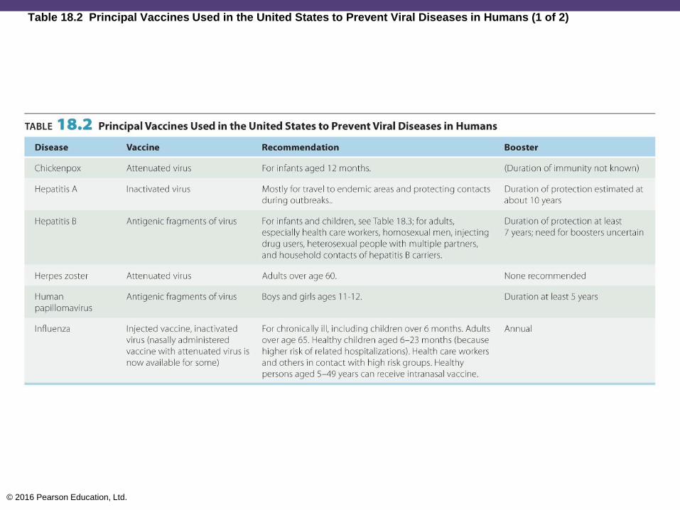
Table 18.2 Principal Vaccines Used in the United States to Prevent Viral Diseases in Humans (1 of 2)

© 2016 Pearson Education, Ltd.
Table 18.2 Principal Vaccines Used in the United States to Prevent Viral Diseases in Humans (2 of 2)
© 2016 Pearson Education, Ltd.
Types of Vaccines and Their Characteristics
• Live attenuated vaccines
• Weakened pathogen
• Closely mimic an actual infection
• Confers lifelong cellular and humoral immunity
• Inactivated killed vaccines
• Safer than live vaccines
• Require repeated booster doses
• Induce mostly humoral immunity

© 2016 Pearson Education, Ltd.
Types of Vaccines and Their Characteristics
• Subunit vaccines use antigenic fragments to
stimulate an immune response
• Recombinant vaccines: subunit vaccines produced by
genetic modification
• Virus-like particle (VLP) vaccines: resemble intact
viruses but do not contain viral genetic material
• Toxoids: inactivated toxins
• Antitoxins: serums containing antibodies against the
toxin
© 2016 Pearson Education, Ltd.
Types of Vaccines and Their Characteristics
• Conjugated vaccines
• Used for diseases in children with poor immune
response to capsular polysaccharides
• Nucleic acid (DNA) vaccines
• Injected naked DNA produces the protein antigen
encoded in the DNA
• Protein antigens carried to the red bone marrow
stimulate humoral and cellular immunity

© 2016 Pearson Education, Ltd.
The Development of New Vaccines
• Less profitable than medicines
• Develop vaccines without the use of animals
• Use of plants as source for vaccines
• More oral vaccines
• Vaccines for chronic diseases
• "Reverse vaccinology" for the development of
cellular immunity
© 2016 Pearson Education, Ltd.
Vaccination Technologies
• Nanopatch: delivers a dry formulation of a vaccine
to the skin
• Skin contains high numbers of APCs
• Requires no refrigeration
• Multiple-combination vaccines
© 2016 Pearson Education, Ltd.
Adjuvants
• Adjuvants are chemical additives added to
vaccines to improve effectiveness
• Alum is the only approved adjuvant in the United States
• Improve the innate immune response

© 2016 Pearson Education, Ltd.
Safety of Vaccines
• On rare occasions, vaccines can cause the
disease
• No medical or scientific proof of MMR vaccines
being linked to autism
• Safest and most effective means of preventing
infectious disease in children

© 2016 Pearson Education, Ltd.
Diagnostic Immunology
• Sensitivity: probability that the test is reactive if
the specimen is a true positive
• Specificity: probability that a positive test will not
be reactive if a specimen is a true negative
• Immunologic-based diagnostic tests
• Interactions of humoral antibodies with antigens
• Known antibody can identify an unknown pathogen
• Known pathogen can identify an unknown antibody

© 2016 Pearson Education, Ltd.
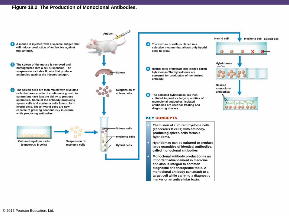
Monoclonal Antibodies
• Hybridoma: "immortal" cancerous B cell
(myeloma) combined with an antibody-producing
normal B cell
• Hybridoma produces monoclonal antibodies (Mabs)
© 2016 Pearson Education, Ltd.
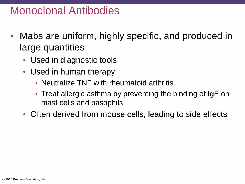
Monoclonal Antibodies
• Mabs are uniform, highly specific, and produced in
large quantities
• Used in diagnostic tools
• Used in human therapy
• Neutralize TNF with rheumatoid arthritis
• Treat allergic asthma by preventing the binding of IgE on
mast cells and basophils
• Often derived from mouse cells, leading to side effects
© 2016 Pearson Education, Ltd.
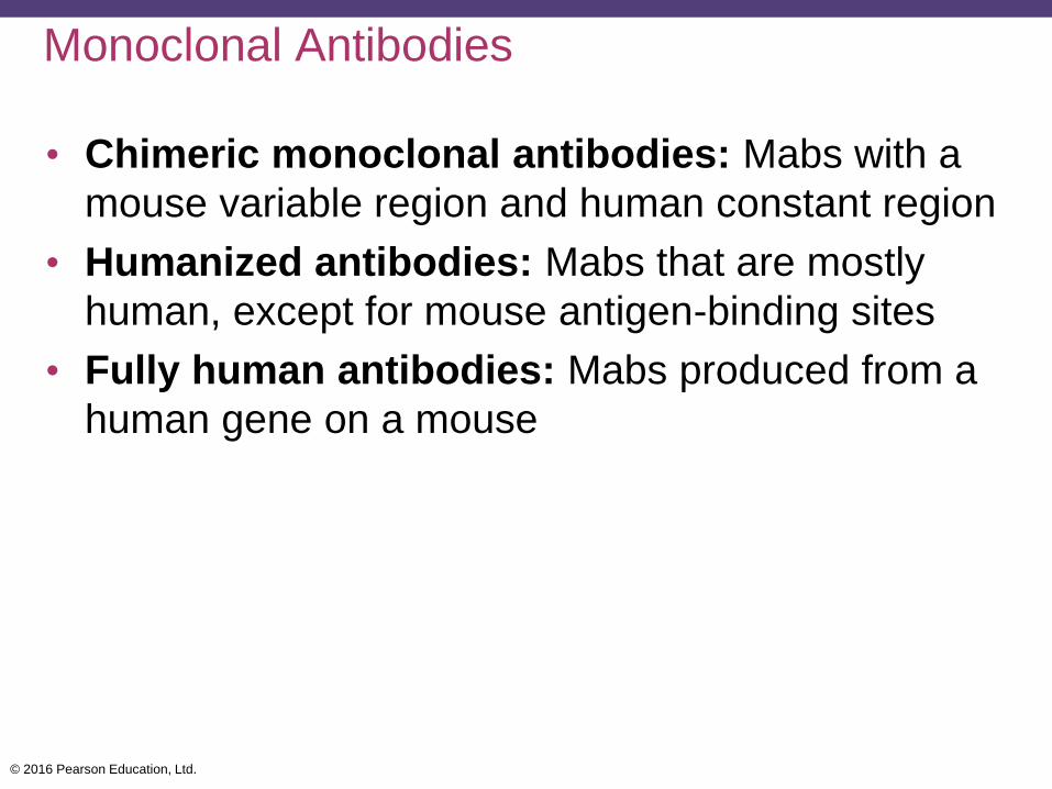
Monoclonal Antibodies
• Chimeric monoclonal antibodies: Mabs with a
mouse variable region and human constant region
• Humanized antibodies: Mabs that are mostly
human, except for mouse antigen-binding sites
• Fully human antibodies: Mabs produced from a
human gene on a mouse
© 2016 Pearson Education, Ltd.
Figure 18.2 The Production of Monoclonal Antibodies.
A mouse is injected with a specific antigen that
will induce production of antibodies against
that antigen.
The spleen of the mouse is removed and
homogenized into a cell suspension. The
suspension includes B cells that produce
antibodies against the injected antigen.
The spleen cells are then mixed with myeloma
cells that are capable of continuous growth in
culture but have lost the ability to produce
antibodies. Some of the antibody-producing
spleen cells and myeloma cells fuse to form
hybrid cells. These hybrid cells are now
capable of growing continuously in culture
while producing antibodies.
Cultured myeloma cells
(cancerous B cells)
Suspension of
myeloma cells
Spleen cells
Myeloma cells
Hybrid cells
Suspension of
spleen cells
Spleen
Antigen
The mixture of cells is placed in a
selective medium that allows only hybrid
cells to grow.
Hybrid cell Myeloma cell Spleen cell
Hybridomas
Hybrid cells proliferate into clones called
hybridomas.The hybridomas are
screened for production of the desired
antibody.
The selected hybridomas are then
cultured to produce large quantities of
monoclonal antibodies. Isolated
antibodies are used for treating and
diagnosing disease.
Desired
monoclonal
antibodies
•
•
•
The fusion of cultured myeloma cells
(cancerous B cells) with antibody-
producing spleen cells forms a
hybridoma.
Hybridomas can be cultured to produce
large quantities of identical antibodies,
called monoclonal antibodies
Monoclonal antibody production is an
important advancement in medicine
and also is integral to common
diagnostic and therapeutic tools. A
monoclonal antibody can attach to a
target cell while carrying a diagnostic
marker or an anticellular toxin.
KEY CONCEPTS

© 2016 Pearson Education, Ltd.
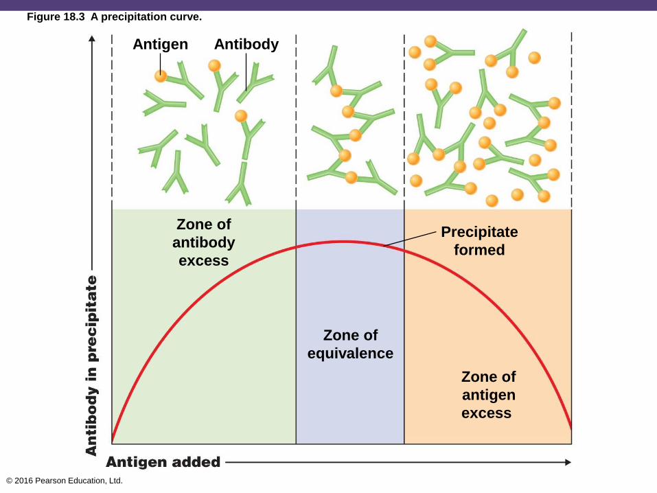
Precipitation Reactions
• Reaction of soluble antigens with antibodies to
form large, interlocking aggregates called lattices
• Antigen-antibody complex forms, followed by the
formation of a lattice that precipitates from solution
• Precipitin ring test: a cloudy line forms where
there is the optimal ratio of antigen and antibody
© 2016 Pearson Education, Ltd.
Figure 18.3 A precipitation curve.
AntibodyAntigen
Zone of
antibody
excess
Zone of
equivalence
Precipitate
formed
Zone of
antigen
excess

© 2016 Pearson Education, Ltd.
Figure 18.4 The precipitin ring test.
Antigens
(soluble)
Zone of equivalence:
visible precipitate
AntibodiesPrecipitation
band
© 2016 Pearson Education, Ltd.
Precipitation Reactions
• Immunodiffusion tests are precipitation reactions
carried out in an agar gel medium
• Precipitate develops at the point where the optimal
antigen-antibody ratio occurs
• Immunoelectrophoresis combines
electrophoresis with immunodiffusion
• Separates proteins in human serum
© 2016 Pearson Education, Ltd.
Agglutination Reactions
• Particulate antigens binding to antibodies to form
visible aggregates
• Direct agglutination tests
• Detect antibodies against large cellular antigens
• Measure concentration of serum antibody (known as
titer)
• Rise in titer indicates a greater immunity to disease
• Seroconversion is a significant change in titer as a
disease progresses

© 2016 Pearson Education, Ltd.
Figure 18.5 An agglutination reaction.
Epitopes
IgM
Bacterium
© 2016 Pearson Education, Ltd.
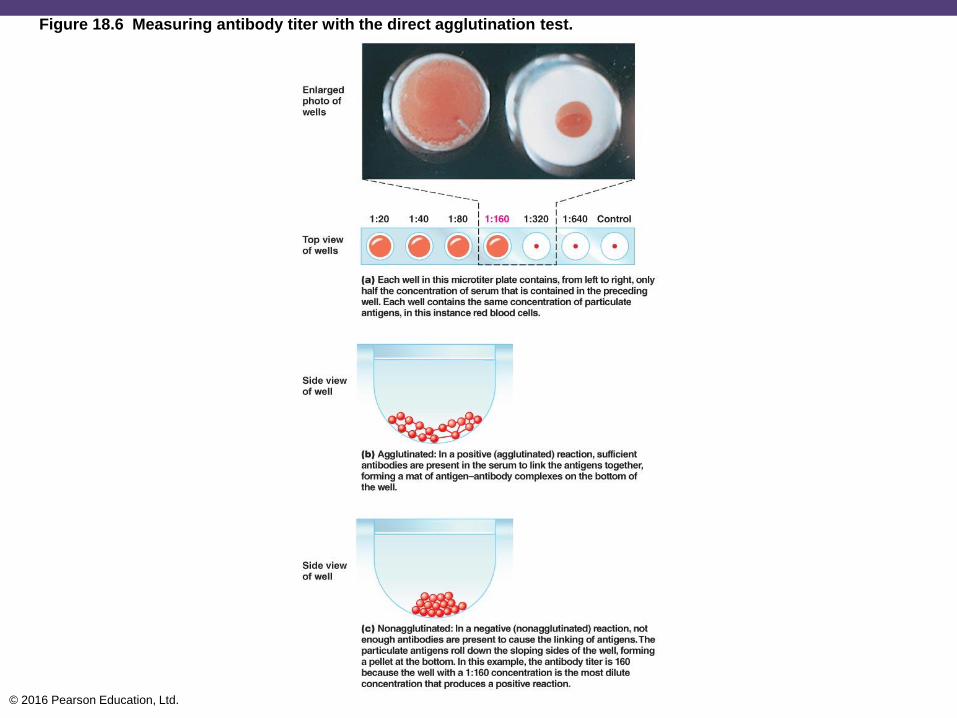
Figure 18.6 Measuring antibody titer with the direct agglutination test.

© 2016 Pearson Education, Ltd.
Agglutination Reactions
• Indirect (passive) agglutination tests
• Antibody reacts with the soluble antigen adhering to the
particles or vice versa
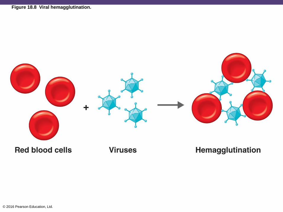
• Hemagglutination
• Agglutination of RBC surface antigens and
complementary antibodies; used in blood typing
• Viral hemagglutination occurs when viruses
agglutinate RBCs without an antigen-antibody reaction
• Mumps, measles, influenza
© 2016 Pearson Education, Ltd.
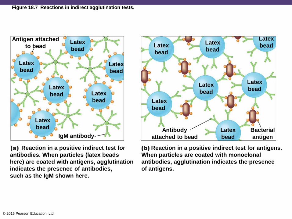
Figure 18.7 Reactions in indirect agglutination tests.
Antigen attached
to beadLatex
bead
Latex
bead
Latex
bead
Latex
bead
Latex
bead
Latex
bead
Latex
bead
Latex
bead
Latex
bead
Latex
bead
Latex
bead
Latex
bead
Latex
beadIgM antibodyBacterial
antigen
Reaction in a positive indirect test for
antibodies. When particles (latex beads
here) are coated with antigens, agglutination
indicates the presence of antibodies,
such as the IgM shown here.
Reaction in a positive indirect test for antigens.
When particles are coated with monoclonal
antibodies, agglutination indicates the presence
of antigens.
Antibody
attached to bead
© 2016 Pearson Education, Ltd.
Figure 18.8 Viral hemagglutination.
© 2016 Pearson Education, Ltd.
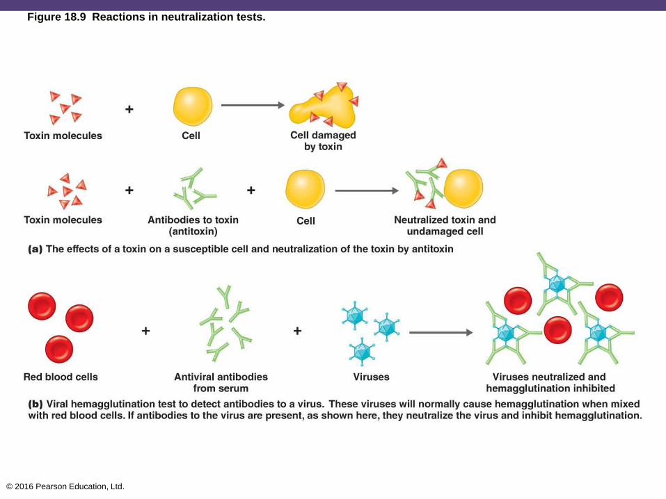
Neutralization Reactions
• Antigen-antibody reaction where the harmful
effects of an exotoxin or a virus are blocked by
antibodies to the toxin (antitoxin)
• Viral hemagglutination inhibition test is used for
subtyping viruses
• Viruses and RBCs are mixed with the patient's serum
• If the serum contains antibodies to a virus, they neutralize
the virus and inhibit hemagglutination
© 2016 Pearson Education, Ltd.
Figure 18.9 Reactions in neutralization tests.
© 2016 Pearson Education, Ltd.
Complement-Fixation Reactions
• Complement fixation: complement serum protein
binds to and is fixed to the antigen–antibody
complex
• Detects small amount of antibody
• Works for antibodies that do not work with precipitation
or agglutination reactions

© 2016 Pearson Education, Ltd.
Figure 18.10 The complement-fixation test.

© 2016 Pearson Education, Ltd.
Fluorescent-Antibody Techniques
• Combine fluorescent dyes with antibodies
• Direct FA tests
• Identify a microorganism in a clinical specimen
• Indirect FA tests
• Detect a specific antibody in serum
• Anti-human immune serum globulin (anti-HISG) is
added and will react with any antibody in serum if the
result is positive

© 2016 Pearson Education, Ltd.
Figure 18.11 Fluorescent-antibody (FA) techniques.
© 2016 Pearson Education, Ltd.
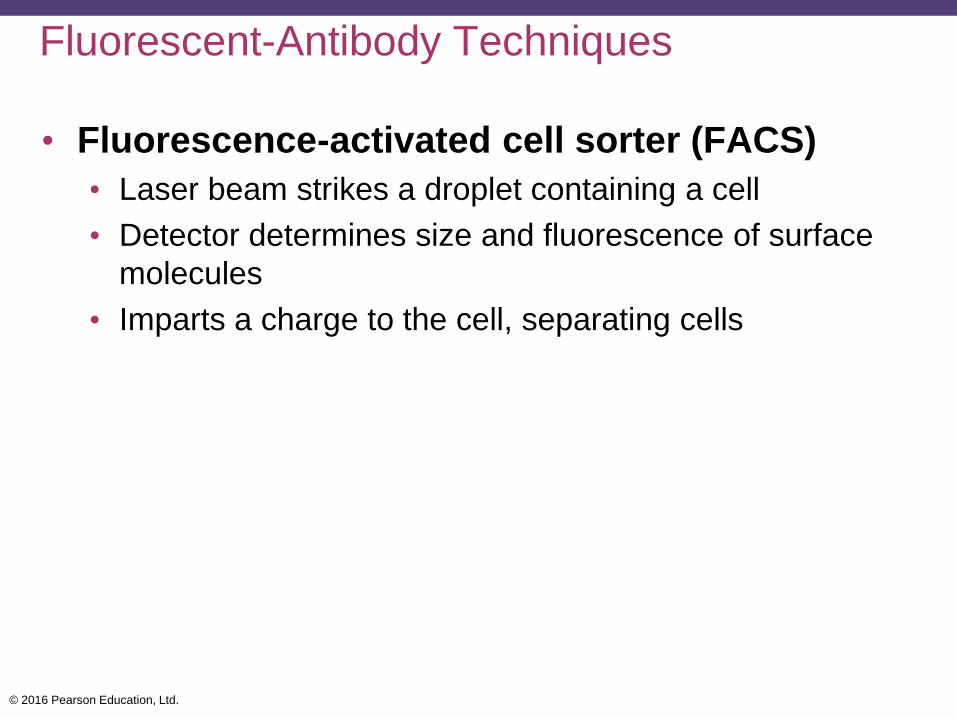
Fluorescent-Antibody Techniques
• Fluorescence-activated cell sorter (FACS)
• Laser beam strikes a droplet containing a cell
• Detector determines size and fluorescence of surface
molecules
• Imparts a charge to the cell, separating cells
© 2016 Pearson Education, Ltd.
Figure 18.12 The fluorescence-activated cell sorter (FACS).
Fluorescently
labeled cells
Laser beam
Laser
Fluorescence
detector
Electrically
charged
metal plates
Collection
tubes
The separated cells
fall into different
collection tubes.
As cells drop between
electrically charged
plates, the cells with
a positive charge
move closer to the
negative plate.
Electrode gives
positive charge to
identified cells.
Detector of
scattered light
Laser beam strikes
each droplet.
Cell mixture leaves
nozzle in droplets.
A mixture of cells is
treated to label cells
that have certain
antigens with
fluorescent-antibody
markers.
ElectrodeFluorescence detector
identifies fluorescent
cells by fluorescent
light emitted by cell.

© 2016 Pearson Education, Ltd.
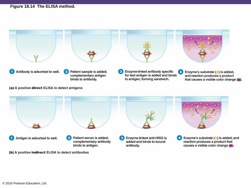
Enzyme-Linked Immunosorbent Assay (ELISA)
• Direct ELISA
• Detects antigens
• Sample containing antigens is mixed with antibody
• Enzyme-linked antibodies react with the antigen
• Detected by adding a substrate for the linked enzyme; a
color is produced
• Indirect ELISA
• Detects antibodies
• Western blotting
• Identifies proteins via electrophoresis and a blotter

© 2016 Pearson Education, Ltd.
Figure 18.13 The use of monoclonal antibodies in a home pregnancy test.
Control
windows
Test
windows
Not pregnant Pregnant
Free monoclonal antibody specific
for hCG, a hormone produced during
pregnancy.
Capture monoclonal antibody bound
to substrate.
Sandwich formed by combination of
capture antibody and free antibody when
hCG is present, creating a color change.
© 2016 Pearson Education, Ltd.
Figure 18.14 The ELISA method.

© 2016 Pearson Education, Ltd.
The Future of Diagnostic and Therapeutic
Immunology
• Tests that require less human judgment and
personnel
• PCR and DNA probes
• Inexpensive and simple tests for developing
countries
• Disease prevention and therapy