Embed Size (px)
Citation preview

The Human Eye and Vision
• The structure of the eye– Iris– Cornea– Lens
• Focusing– Cornea– Accommodation
• The Retina– Photoreceptors– Processing time– Sensitivity

Structure of the Eye
The iris is roughly equivalent to the diaphragm in a camera, the cornea and the lens are both lens-like objects, and the retina is where the image is recorded, similar to a CCD sensor or film.

Structure of the Eye: Iris• The iris is similar to the diaphragm
in a camera
• Your iris widens in dim light and narrows in bright light
• The f-number of your eye varies from f/2 (large opening) to f/8 (small opening)
• Compare this to the range of an average camera lens, which may have f-numbers from f/2.8 to f/22.

Structure of the Eye: Iris• With a range of only f/2 – f/8, your iris
can only reduce the light coming into your eye by a factor of 20.
• The range of intensities that your eye can respond to is a factor of 1013
• The main function of the iris is not to control the intensity of light coming into your eye
• Main functions of iris– Reduce aberrations, sharpen image– Increase depth of field

Structure of the Eye: Cornea and LensCornea
Eyelens
Retina
• There are two lenses in your eye, the cornea and the eyelens.• The cornea, the front surface of the eye, does most of the
focusing in your eye• The eyelens provides adjustable fine-tuning of the focus

Structure of the Eye: Cornea and Lens
eyelens: n ≈ 1.4
Retina
• This is because the cornea-air surface has a large change in the index of refraction, so light bends a lot
• The power of the cornea lens is ~43 diopters (focal length 2.3 cm)• The eyelens is surrounded by the humors, which have a very
similar index of refraction as the lens itself.
air: n = 1
cornea: n ≈ 1.4
humors: n ≈ 1.3

How a Camera Lens Focuses• A camera is focused by changing the distance, xi, from
the lens to the image at the back on the film or CCD as the distance to the object, xo, changes.
Focal length (fixed)Object distance (varies)
Image distance(changes to satisfy equation when xo changes)

How Your Eye Focuses
• The eyelens is a fixed, unchanging distance, xi, from the retina at the back of the eyeball where the image is created
Focal length (changed to satisfy the equation when xo changes)
Object distance (varies)
Image distance (fixed)

The Eyelens: Accommodation• The eyelens changes its focal length by changing
its shape. Ligaments pull on the lens to change the amount of “bulge”
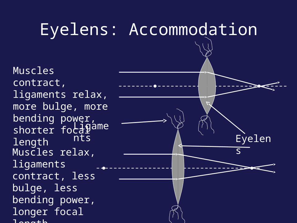
Eyelens: Accommodation
Muscles contract, ligaments relax, more bulge, more bending power, shorter focal length
Muscles relax, ligaments contract, less bulge, less bending power, longer focal length
Eyelens Ligaments

How Your Eyelens Focuses• Your eyelens has a small depth of field
– You can't see something close and far with both objects in focus at the same time
• Hold out your thumb about a foot away from your eye– Then, alternately focus on thumb and me (right above your
thumb)• Note that you cannot see both me and your thumb
sharply (in focus) at the same time– You focus on one or the other by changing the bulge of your
eyelens

Accommodation
less bulgy, longer f
professor is in focus
thumb is out of focus
more bulgy, shorter f
thumb is in focus
professor is out of focus

Concept Questions on Focusing
You can't see the Flatirons and your thumb clearly at the same time
a) because your pupil is too smallb) because your iris can't change fast enoughc) because your eye cannot accommodate d) because your eye does not have enough depth of field

Concept Questions on Focusing
When you see someone out-of-focusa) There is no image anywhereb) There is an in-focus image on your foveac) There is an in-focus image on your retinad) There is an image in-focus either in front or in back of
your retina

Concept Questions on Focusing
In order to focus on close objectsa) your eyelens needs to bulgeb) your eyelens needs to flattenc) your cornea needs to bulged) your cornea needs to flattene) the distance (xi) between your eyelens and retina
needs to change

Structure of the Eye: Retina• The retina is the sensor or film of your eye.
• Its layers do three things– Provide blood and nutrients (choroid)– Absorb light and convert to an electrical signal
(photoreceptors)– Transfer the signal to the brain (nerve cells)
Plexiform layer (nerve cells)
Rods and Cones (photoreceptors)
Choroid (blood vessels)
Light

Structure of the Retina
← Nerve cells
← Photoreceptors
← Choroid
Light

Photoreceptors: Rods and Cones• Light is detected and
converted to an electrical signal by the photoreceptors in the retina. There are two main kinds of receptors, rods and cones
• This is a false color image, rods and cones are not actually different colors
rod
cone

Photoreceptors: Cones
• Cones are responsible for our fine detailed and color vision
• Cones are clustered near the center of your retina, called the fovea
• There are 5 million cones in the average retina

Photoreceptors: Rods
• Rods are responsible for low light and peripheral vision
• They are present everywhere in the retina except the fovea
• There are 125 million rods in the average retina

Rods and Cones• Because of their different functions, rods and cones are
present in varying densities in the retina. The blind spot is due to the connection of the optic nerve

Processing Time: Shutter Speed?• The closest thing your eye has to a shutter speed
is called “persistance of response”
• This is the time during which the photoreceptor is active and responding to light
• It varies between 1/25 s in low light to 1/50 s in bright light
• Compare this to a camera, which has shutter speeds from more than 1 s to 1/1000 s

Light Sensitivity
• We said earlier that your eye can respond to a range of light intensities (basically “brightnesses”) that vary by a factor of 1013
• Clearly the iris and the response time of the photoreceptors is not enough to allow this
• How then do our eyes respond to such an enormous range of light intensities?

Light Sensitivity: Analogy to Film Speed
We didn’t talk about film, but for those of you who know about it:•In low light, you can use “fast” film, which has lower resolution (coarser grain) and is often black and white•In bright light, you can use “slow” film, which is fine grain and color•In it is not practical to change the film in your camera every time you walk from a dark building out into the sun, but this is what your retina does!

Light Sensitivity• Remember we talked about rods and cones• Cones:
– Sensitive to bright light, photopic conditions– Densely packed in the fovea– Only a few cones per nerve fiber
• Rods:– Sensitive to low light, scotopic conditions– Widely distributed across the retina– Up to 1000s of rods per nerve fiber (think of this as
many many drops falling into the same pipe, one drop can’t be detected, but many drops generate some water flow that can be measured)

Dark Adaption• Even within the cone and rod system, your retina
adjusts its sensitivity in response to the overall light level
• When you walk into a dark room, you can’t see anything, but after a few minutes, you adapt and can start to see things
• When you walk out into the bright sunlight, everything is blindingly white, but gradually things look “normal” again

Dark Adaptation
Object must be very bright to be seen
Dim objects can be seen

Dark Adaptation
• After about thirty minutes, your eyes are completely dark adapted and can see an amount of light equivalent to a candle 10 miles away.
• Dark adaptation is a slow process, but allows us to see in a huge range of light levels
10 miles!