Embed Size (px)
Citation preview
2/7/2016
1
Tuula Salo1, Susanna Teppo1, Sini Nurmenniemi1, Marilena Vered2, Dan Dayan2,
Carolina Bitu1 and Pia Nyberg1.
1Department of Diagnostics and Oral Medicine, Institute of Dentistry, University of Oulu, Oulu, Finland, and2 Institute of Pathology, Chaim Sheba Medical Center Center, Tel Hashomer, Ramat Gan, Israel.
Tuula Salo Professor of Oral Pathology
Universities of Oulu and Helsinki
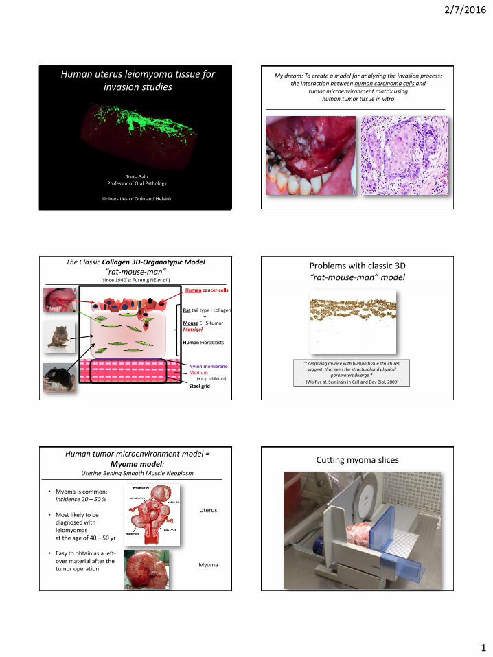
Human uterus leiomyoma tissue for invasion studies
My dream: To create a model for analyzing the invasion process:the interaction between human carcinoma cells and
tumor microenvironment matrix using human tumor tissue in vitro
Human cancer cells
Rat tail type I collagen+
Mouse EHS-tumorMatrigel
+Human Fibroblasts
Nylon membraneMedium
(+ e.g. inhibitors)
Steel grid
The Classic Collagen 3D-Organotypic Model
”rat-mouse-man” (since 1980´s; Fusenig NE et al.)
Problems with classic 3D”rat-mouse-man” model
”Comparing murine with human tissue structures suggest, that even the structural and physical
parameters diverge “
(Wolf et al. Seminars in Cell and Dev Biol, 2009)
Human tumor microenvironment model = Myoma model:
Uterine Bening Smooth Muscle Neoplasm
• Myoma is common: incidence 20 – 50 %
• Most likely to be diagnosed with leiomyomas at the age of 40 – 50 yr
• Easy to obtain as a left-over material after thetumor operation
Uterus
Myoma
Cutting myoma slices
2/7/2016
2
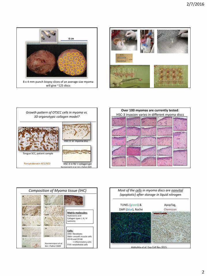
8 x 4 mm punch biopsy slices of an average size myoma will give ~125 discs
6 cm
Myoma disks have been cut out from the original tissue with 8 m biopsy punch.
4 mm
8 mm
Figure 1.
HSC-3 in fbl + collagen gelNurmenniemi et al. Am J Pathol 2009
Tongue SCC, patient sample
HSC-3 in myoma disc
Pancytokeratin AE1/AE3
Highly invasive tongue carcinoma cells (HSC-3) invade in myoma similar to OTSCC in vivo=
(mesenchymal and collective or chain migration)
Growth pattern of OTSCC cells in myoma vs. 3D organotypic collagen model?
Over 100 myomas are currently tested:HSC-3 invasion varies in different myoma discs
7A 28 16 34A
32C54
5026B49B47
32A 41C
39B 54 26A 53
35A235148
37
27
53
Cells:VIM= fibroblastsSMA= smooth muscle cells CD 45 and CD 68
= inflammatory cellsFVIII =endothelial cells
Nurmenniemi et al. Am J Pathol 2009
Composition of Myoma tissue (IHC)
Matrix molecules:Hyaluronic acidCollagen types I, III, IVLaminins
Hyaluronic acid
Most of the cells in myoma discs are nonvital (apoptotic) after storage in liquid nitrogen
TUNEL (green) &
DAPI (blue), Roche
ApopTag,
Chemicon
Alahuhta et al. Exp Cell Res 2015
2/7/2016
3
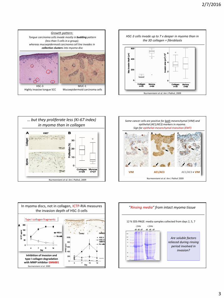
Growth pattern:Tongue carcinoma cells invade mostly in budding pattern
(less than 5 cells in a group); whereas mucoepidermoid carcinoma cell line invades in
collective clusters into myoma disc
HSC-3 Highly invasive tongue SCC
MUC-1 Mucoepidermoid carcinoma cells
HSC-3 cells invade up to 7 x deeper in myoma than in the 3D collagen + fibroblasts
Nurmenniemi et al. Am J Pathol, 2009
… but they proliferate less (Ki-67 index) in myoma than in collagen
Nurmenniemi et al. Am J Pathol, 2009
Some cancer cells are positive for both mesenchymal (VIM) and epithelial (AE1/AE3) markers in myoma.
Sign for epithelial-mesenchymal-transition (EMT)
Nurmenniemi et al. 2009 Nurmenniemi et al. Am J Pathol 2009
VIM AE1/AE3 AE1/AE3 + VIM
Nurmenniemi et al. 2009
Inhibition of invasion and type I collagen degradation
with MMP inhibitor GM6001
Analyses of collagen degradation products (RIA) from media samples reflect carcinoma cell invasion depth
In myoma discs, not in collagen, ICTP-RIA measures the invasion depth of HSC-3 cells
Type I collagen fragments12 % SDS-PAGE: media samples collected from days 2, 5, 7
- bMe + bMe
28
36
55
72
95
130
250
d2 d5 d7 d2 d5 d7
”Rinsing media” from intact myoma tissue
Are soluble factors relieced during rinsing
period involved in invasion?
2/7/2016
4
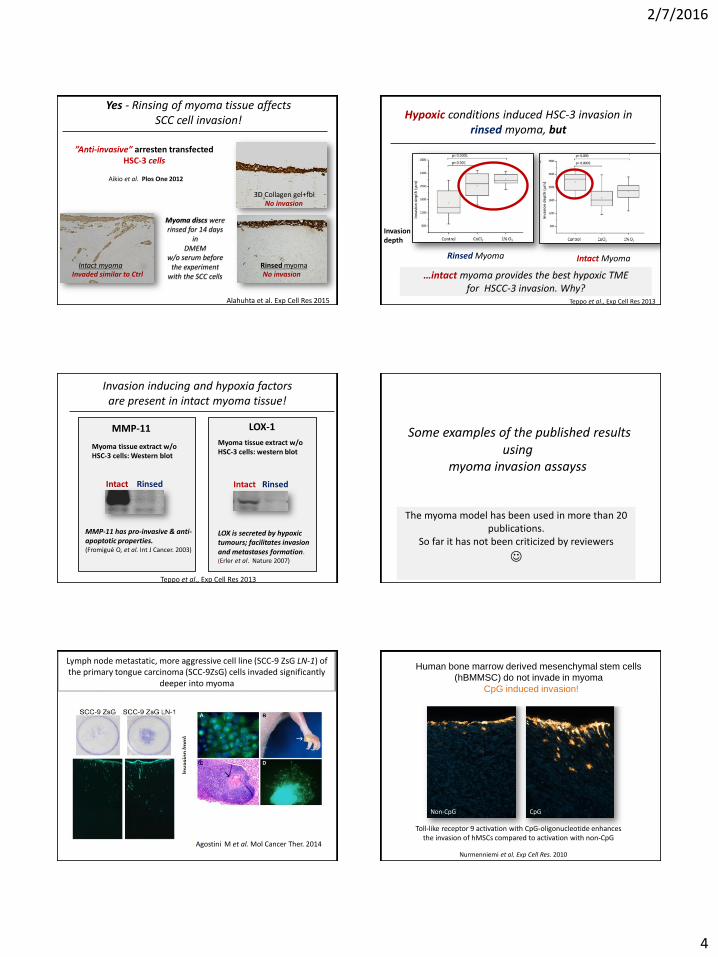
Yes - Rinsing of myoma tissue affects SCC cell invasion!
Alahuhta et al. Exp Cell Res 2015
”Anti-invasive” arresten transfected HSC-3 cells
Aikio et al. Plos One 2012
3D Collagen gel+fbl
Intact myoma
Myoma discs were rinsed for 14 days
in DMEM
w/o serum before the experiment
with the SCC cells Invaded similar to Ctrl
No invasion
Rinsed myomaNo invasion
Rinsed Myoma
Invasion depth
Teppo et al., Exp Cell Res 2013
Hypoxic conditions induced HSC-3 invasion in rinsed myoma, but
…intact myoma provides the best hypoxic TME for HSCC-3 invasion. Why?
Intact Myoma
LOX is secreted by hypoxic tumours; facilitates invasion and metastases formation. (Erler et al. Nature 2007)
Invasion inducing and hypoxia factorsare present in intact myoma tissue!
Myoma tissue extract w/o HSC-3 cells: Western blot
Intact Rinsed
Teppo et al., Exp Cell Res 2013
MMP-11 has pro-invasive & anti-apoptotic properties.(Fromigué O, et al. Int J Cancer. 2003)
MMP-11 LOX-1
Intact Rinsed
Myoma tissue extract w/o HSC-3 cells: western blot
Some examples of the published resultsusing
myoma invasion assayss
The myoma model has been used in more than 20 publications.
So far it has not been criticized by reviewers
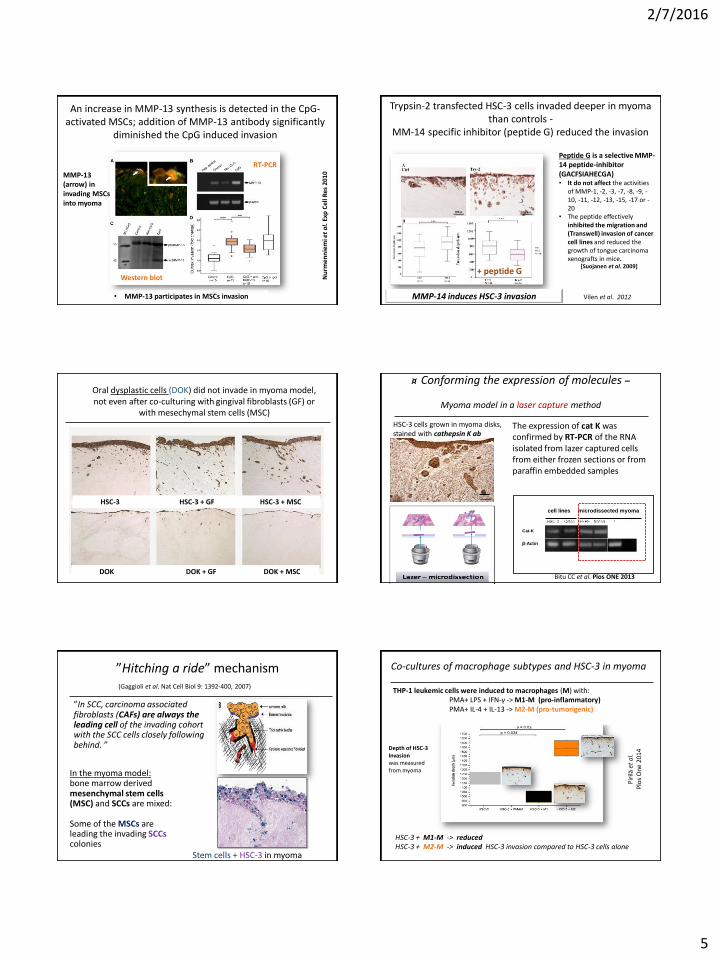
Lymph node metastatic, more aggressive cell line (SCC-9 ZsG LN-1) of the primary tongue carcinoma (SCC-9ZsG) cells invaded significantly
deeper into myoma
Agostini M et al. Mol Cancer Ther. 2014
Toll-like receptor 9 activation with CpG-oligonucleotide enhancesthe invasion of hMSCs compared to activation with non-CpG
Non-CpG CpG
Nurmenniemi et al. Exp Cell Res. 2010
Human bone marrow derived mesenchymal stem cells
(hBMMSC) do not invade in myoma
CpG induced invasion!
2/7/2016
5
An increase in MMP-13 synthesis is detected in the CpG-activated MSCs; addition of MMP-13 antibody significantly
diminished the CpG induced invasion
RT-PCR
Western blot
MMP-13 (arrow) in invading MSCs into myoma
Nu
rmen
nie
mi e
t a
l. E
xpC
ellR
es20
10
• MMP-13 participates in MSCs invasion
Trypsin-2 transfected HSC-3 cells invaded deeper in myoma than controls -
MM-14 specific inhibitor (peptide G) reduced the invasion
Peptide G is a selective MMP-14 peptide-inhibitor (GACFSIAHECGA)• It do not affect the activities
of MMP-1, -2, -3, -7, -8, -9, -10, -11, -12, -13, -15, -17 or -20
• The peptide effectively inhibited the migration and (Transwell) invasion of cancer cell lines and reduced the growth of tongue carcinoma xenografts in mice.
[Suojanen et al. 2009]
MMP-14 induces HSC-3 invasion
+ peptide G
Vilen et al. 2012
HSC-3 HSC-3 + GF HSC-3 + MSC
DOK DOK + GF DOK + MSC
Oral dysplastic cells (DOK) did not invade in myoma model, not even after co-culturing with gingival fibroblasts (GF) or
with mesechymal stem cells (MSC)
¤ Conforming the expression of molecules –
Myoma model in a laser capture method
A
B
HSC-3 cells grown in myoma disks, stained with cathepsin K ab
Laser capture of HSC-3 cells
β-Actin
cell lines microdissected myoma
Cat-K
The expression of cat K was confirmed by RT-PCR of the RNA isolated from lazer captured cells from either frozen sections or from paraffin embedded samples
Bitu CC et al. Plos ONE 2013
”In SCC, carcinoma associatedfibroblasts (CAFs) are always the leading cell of the invading cohortwith the SCC cells closely followingbehind. ”
”Hitching a ride” mechanism(Gaggioli et al. Nat Cell Biol 9: 1392-400, 2007)
In the myoma model: bone marrow derived mesenchymal stem cells (MSC) and SCCs are mixed:
Some of the MSCs are leading the invading SCCscolonies
Stem cells + HSC-3 in myoma
HSC-3 + M1-M -> reducedHSC-3 + M2-M -> induced HSC-3 invasion compared to HSC-3 cells alone
Pir
ilä e
t a
l. P
los
On
e 20
14
p<0,02
p=0,002
p=0,034
THP-1 leukemic cells were induced to macrophages (M) with:PMA+ LPS + IFN-γ -> M1-M (pro-inflammatory)PMA+ IL-4 + IL-13 -> M2-M (pro-tumorigenic)
Co-cultures of macrophage subtypes and HSC-3 in myoma
Depth of HSC-3 Invasionwas measuredfrom myoma
2/7/2016
6
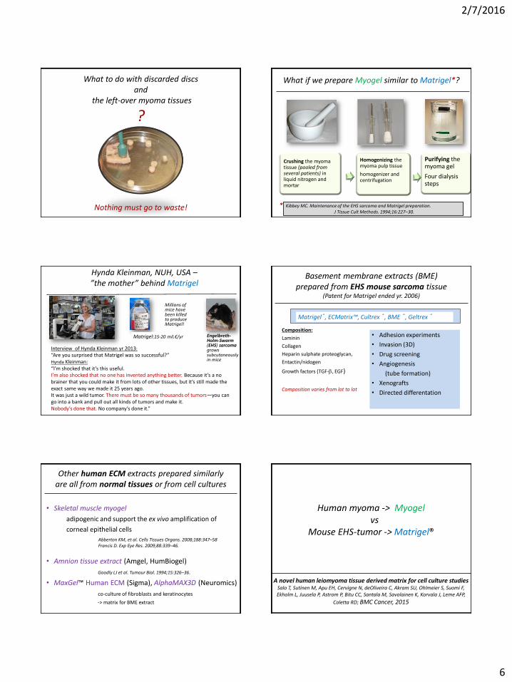
What to do with discarded discsand
the left-over myoma tissues
?
Nothing must go to waste!
What if we prepare Myogel similar to Matrigel*?
Crushing the myomatissue (pooled fromseveral patients) in liquid nitrogen and mortar
Homogenizing the myoma pulp tissue
homogenizer and centrifugation
Purifying the myoma gel
Four dialysissteps
Kibbey MC. Maintenance of the EHS sarcoma and Matrigel preparation.J Tissue Cult Methods. 1994;16:227–30.
*
Hynda Kleinman, NUH, USA –”the mother” behind Matrigel
Interview of Hynda Kleinman yr 2013:
“Are you surprised that Matrigel was so successful?”Hynda Kleinman:“I’m shocked that it’s this useful. I’m also shocked that no one has invented anything better. Because it’s a no brainer that you could make it from lots of other tissues, but it’s still made the exact same way we made it 25 years ago. It was just a wild tumor. There must be so many thousands of tumors—you can go into a bank and pull out all kinds of tumors and make it. Nobody’s done that. No company’s done it.”
Matrigel:15-20 mil.€/yr Engelbreth-Holm-Swarm(EHS) sarcomagrowssubcutaneouslyin mice
Millions of mice havebeen killedto produceMatrigel!
Basement membrane extracts (BME)prepared from EHS mouse sarcoma tissue
(Patent for Matrigel ended yr. 2006)
Composition:
Laminin
Collagen
Heparin sulphate proteoglycan,
Entactin/nidogen
Growth factors (TGF-b, EGF)
Composition varies from lot to lot
Matrigel®, ECMatrix™, Cultrex ®, BME ®, Geltrex ®
• Adhesion experiments
• Invasion (3D)
• Drug screening
• Angiogenesis
(tube formation)
• Xenografts
• Directed differentation
Other human ECM extracts prepared similarlyare all from normal tissues or from cell cultures
• Skeletal muscle myogel
adipogenic and support the ex vivo amplification of
corneal epithelial cells
• Amnion tissue extract (Amgel, HumBiogel)
Goodly LJ et al. Tumour Biol. 1994;15:326–36.
• MaxGel™ Human ECM (Sigma), AlphaMAX3D (Neuromics)
co-culture of fibroblasts and keratinocytes
-> matrix for BME extract
Abberton KM, et al. Cells Tissues Organs. 2008;188:347–58Francis D. Exp Eye Res. 2009;88:339–46.
Crushingthe myomadiscs
• Using liquid nitrogen and mortar
Homogenizingthe myoma pulp
• Using tissue homogenizer and centrifugation
Purifying the myoma gel
• Four dialysissteps
MyogelPreparation of the gel:
Human myoma -> Myogelvs
Mouse EHS-tumor -> Matrigel®
A novel human leiomyoma tissue derived matrix for cell culture studiesSalo T, Sutinen M, Apu EH, Cervigne N, deOliveira C, Akram SU, Ohlmeier S, Suomi F, Ekholm L, Juusela P, Astrom P, Bitu CC, Santala M, Savolainen K, Korvala J, Leme AFP,
Coletta RD; BMC Cancer, 2015
2/7/2016
7
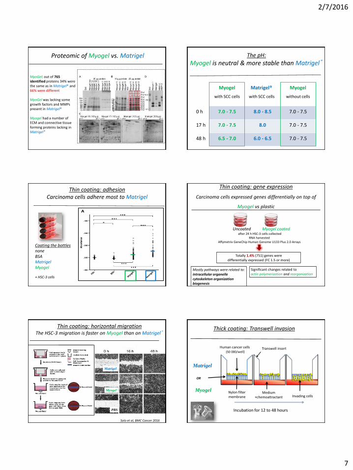
Proteomic of Myogel vs. Matrigel
MyoGel: out of 765 identified proteins 34% were the same as in Matrigel® and 66% were different
MyoGel was lacking some growth factors and MMPs present in Matrigel®
Myogel had a number of ECM and connective tissue forming proteins lacking in Matrigel®
Myogel
with SCC cells
Matrigel®
with SCC cells
Myogel
without cells
0 h 7.0 - 7.5 8.0 - 8.5 7.0 - 7.5
17 h 7.0 - 7.5 8.0 7.0 - 7.5
48 h 6.5 - 7.0 6.0 - 6.5 7.0 - 7.5
The pH:
Myogel is neutral & more stable than Matrigel®
Thin coating: adhesionCarcinoma cells adhere most to Matrigel
Coating the bottlesnoneBSAMatrigelMyogel
+ HSC-3 cells
Thin coating: gene expression
Carcinoma cells expressed genes differentially on top of
Myogel vs plastic
Mostly pathways were related to: intracellular organelle cytoskeleton organization biogenesis
Totally 1.4% (751) genes were differentially expressed (FC 1.5 or more)
For microarray analysis, 90,000 HSCseeded into uncoated or Myogel coated 6each). The next day the cells were harvested for RNA extraction using a Qiagen RNA kit. Three samples of each group (on top of plastic or Myogel coating) were pooled; the pools contained an equal amount of RNA from each sample. AffymetrixPlus 2.0 Arrays were used for microarray analysis and experimental procedures were performed according to the Expression Analysis Technical Manual. Briefly, 1used as a template to synthesize biotinylated GeneChip 3’IVT Express kit (manufacturer’s instructions. The prior to hybridization to Affymetrixarrays containing approximately 55,000 human transcripts. The array was washed and stained with streptavidinProbes). Finally, biotinylated antiwas used to amplify the staining signal and a second staining was performed with streptavidinon a GeneChip Scanner 3000 (Kingdom). The expression data was analyzed to find genes with fold changes (FC) of 1.5 or more using
FC 1.2 or more were divided into Gene Ontology (GO) categories using
a dChip enrichment analysis tool
Uncoated Myogel coated after 24 h HSC-3 cells collected
RNA harvested Affymetrix GeneChip Human Genome U133 Plus 2.0 Arrays
Significant changes related to actin polymerization and reorganization
PBS
Thin coating: horizontal migrationThe HSC-3 migration is faster on Myogel than on Matrigel®
Myogel
Matrigel
PBS
Salo et al, BMC Cancer 2016
Matrigel
Myogel
Incubation for 12 to 48 hours
Nylon filter membrane
Human cancer cells(50 000/well)
Medium +chemoattractant
Transwell insert
Thick coating: Transwell invasion
Invading cells
OR
2/7/2016
8
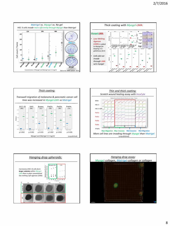
Cel
l co
un
t/
fram
e
12h 24h 48h
Concentration of Myogel and Matrigel was 2.4 mg/ml
Matrigel vs. Myogel vs. No gel HSC-3 cells invade more efficiently through Myogel than Matrigel
Matrigel
Myogel
No gel
Salo et al. BMC cancer 2015
Thick coating with Myogel-LMA:
Myogel-LMA:
• Low Melting Agarose (LMA) is added to Myogel for keeping it in gelatinous form
• Cells did notinvadethrough LMA w/o myogel
SCC-25 SAS Bowes Pa02c Pa03c Pa04c
p 0.002 p 0.002 p 0.015 p 0.002 p 0.002 p 0.026
** ********
Thick coating:
Transwell migration of melanoma & pancreatic cancer celllines was increased in Myogel-LMA vs Matrigel
Myogel and Matrigel 2.4 mg/ml (unpublished)
Thin and thick coating:Scratch wound healing assay with IncuCyte
MDA
HSC-3
HSC-3 GFP
Pa01c
Pa02c
Pa03c
Pa04c
empty
Myo Migration Myo Invasion Mat invasion Mat Migration
More cell lines are invading through Myogel than Matrigel(unpublished)
Hanging drop spheroids:
Carcinoma (HSC-3) cells form larger colonies within Myogel-LMA than in plain conventional low melting soft agarose (LMA)
1
2
4
3
Inner border
Plate lead
HangingDrop
Flipping the covered plate upside downIn a humadifying chamber
Plate containing drops in 4compartments from eachnumbered cell embeddedsample mixtures
Keep in incubator for 5 minutes
Keep in incubator for ≥ 3 hours, after that add 1ml 2% FBS containing media/compartment inthe microscopy unit.
Hanging-drop assay:Myogel-collagen, Matrigel-collagen or collagen
2/7/2016
9
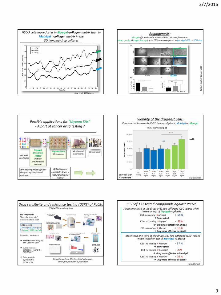
HSC-3 cells move faster in Myogel-collagen matrix than inMatrigel®-collagen matrix in the
3D hanging-drop cultures
Salo et al, BMC Cancer
Angiogenesis:Myogel efficiently induces endothelial cell tube formation:
more, smaller & longer lasting (up to 72h) tubes compared to Matrigel-GFR or ECMatrix
Salo
et
al,
BM
C C
an
cer,
201
6
100-1000candidates
Myogelthin/thick
coatedviability
proliferation, invasion
A) Analyzing most efficient drugs using 2D /3D cell cultures
Myoma Discs3D Invasion
B) Testing best candidate drugs in “natural 3D tumor
matrix”
Mice/animal experiments
Clinical trials
Possible applications for ”Myoma Kits” - A part of cancer drug testing ?
Viability of the drug-test cells:Pancreas carcinoma cells (Pa02c) on top of plastic, Matrigel or Myogel
CellTiter-Glo® ATP amount
Matri0.15
mg/ml
Matri0.31
mg/ml
Matri0.62
mg/ml
Myo0.15
mg/ml
Myo0.31
mg/ml
Myo0.62
mg/ml
FIMM-Wennerberg lab
No coating
(unpublished)
Drug sensitivity and resistance testing (DSRT) of Pa02c (FIMM-Wennerberg lab)
https://www.fimm.fi/en/services/technology-centre/htb/instructions/workflows
132 compounds”drugs for leukemia”5 concentrations each
1. No coating2. Matrigel (0,62 mg/ml) 3. Myogel (0,62 mg/ml)
Three days incubation
Viability measuring byThe CellTiter-Glo®
Luminescencedetection using thePHERAstar®
Data analysisby Dotmatics(EC50, IC50)
Name Mechanism/Targets IC50 GRAPH IC502 GRAPH3 IC503 GRAPH5
Panobinostat HDAC inhibitor 70,42 54,3 48,4
Dinaciclib CDK inhibitor 18,32 18 17,1
Selumetinib MEK inhibitor 97,31 342,5 91
GDC-0994 ERK inhibitor 679,58 175 1879,2
Mercaptopurine Antimetabolite 1031,3 295,2 717,8
Palbociclib Cdk inhibitor (Cdk4/6) 8409,1 3839 680,1
Clofarabine Anti-metabolite:Purine analog 3873,3 516,5 988,5
No gel Myogel Matrigel
IC50 of 132 tested compounds against Pa02cAbout one third of the drugs (48) had differend IC50 values when
tested on top of Myogel vs plasticIC50: no coating ≈ Myogel = 64 %
Same effect
IC50: no coating > Myogel = 20% Drug more effective in Myogel
IC50: no coating < Myogel = 16 % Drug more effective on plastic
More than one third of the drugs (56) had differend IC50 values when tested on top of Matrigel vs plastic
IC50: no coating ≈ Matrigel = 57 % Same effect
IC50: no coating > Matrigel = 27% Drug more effective in Matrigel
IC50: no coating < Matrigel = 16 % Drug more effective on plastic
(unpublished)
2/7/2016
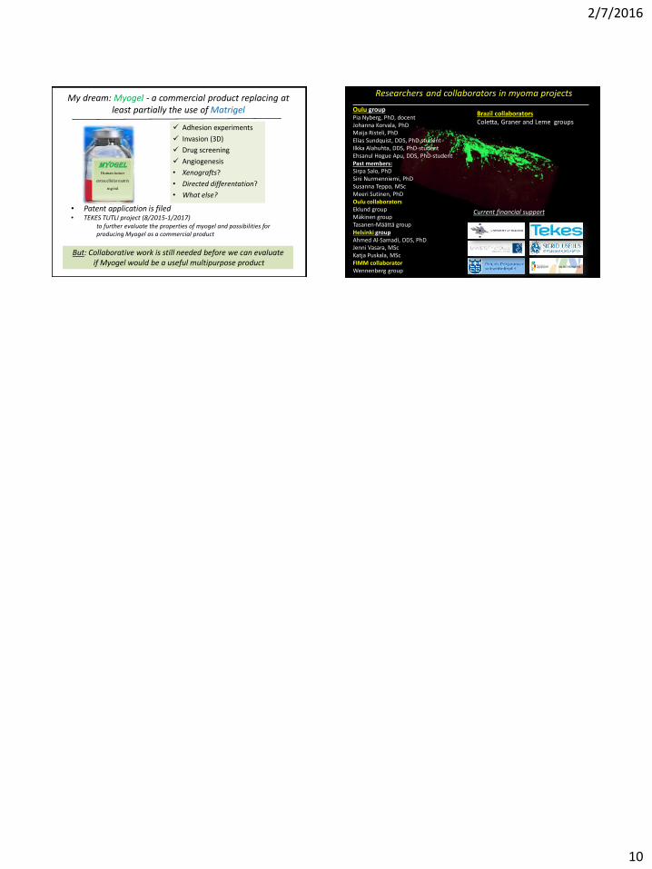
10
MyogelHuman tumor
extracellular matrixmg/ml
Adhesion experiments
Invasion (3D)
Drug screening
Angiogenesis
• Xenografts?
• Directed differentation?
• What else?
My dream: Myogel - a commercial product replacing at least partially the use of Matrigel
• Patent application is filed• TEKES TUTLI project (8/2015-1/2017)
to further evaluate the properties of myogel and possibilities for producing Myogel as a commercial product
But: Collaborative work is still needed before we can evaluateif Myogel would be a useful multipurpose product
Tuula Salo1, Susanna Teppo1, Sini Nurmenniemi1, Marilena Vered2, Dan Dayan2,
Carolina Bitu1 and Pia Nyberg1.
1Department of Diagnostics and Oral Medicine, Institute of Dentistry, University of Oulu, Oulu, Finland, and2 Institute of Pathology, Chaim Sheba Medical Center Center, Tel Hashomer, Ramat Gan, Israel.
Researchers and collaborators in myoma projects
Oulu groupPia Nyberg, PhD, docentJohanna Korvala, PhDMaija Risteli, PhDElias Sundquist, DDS, PhD-studentIlkka Alahuhta, DDS, PhD-studentEhsanul Hogue Apu, DDS, PhD-studentPast members:Sirpa Salo, PhDSini Nurmenniemi, PhDSusanna Teppo, MScMeeri Sutinen, PhDOulu collaboratorsEklund groupMäkinen groupTasanen-Määttä groupHelsinki groupAhmed Al-Samadi, DDS, PhDJenni Vasara, MScKatja Puskala, MScFIMM collaboratorWennenberg group
Brazil:Coletta, Graner and Leme groups
Current main financial support
Brazil collaboratorsColetta, Graner and Leme groups
Current financial support