Embed Size (px)
Citation preview
6/2/2017
1
DIAGNOSIS AND MANAGEMENT OF
MUCOCUTANEOUS DISEASES
H. C. Tenenbaum DDS, Dip Perio, PhD, FRCD(C)
Dentist-in-Chief, Mount Sinai Hospital
Professor of Periodontology, Faculty of Dentistry
Professor of Laboratory Medicine and Pathobiology,
Faculty of Medicine
University of Toronto
Some images thanks to Dr. D. Mock, and W. B. Saunders (Carranza©)
Diagnosis & Management of Oral
Mucocutaneous Disease
One of the most important considerations
with some of these conditions is that they
are often confused with the inflammatory
gingival diseases such as ANUG, gingivitis
or even periodontitis!
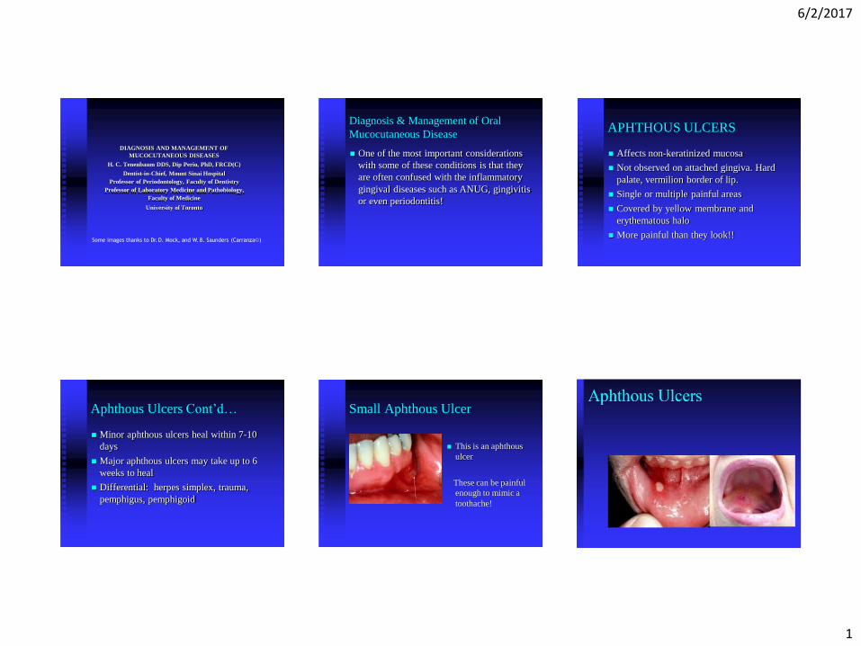
APHTHOUS ULCERS
Affects non-keratinized mucosa
Not observed on attached gingiva. Hard
palate, vermilion border of lip.
Single or multiple painful areas
Covered by yellow membrane and
erythematous halo
More painful than they look!!
Aphthous Ulcers Cont’d…
Minor aphthous ulcers heal within 7-10
days
Major aphthous ulcers may take up to 6
weeks to heal
Differential: herpes simplex, trauma,
pemphigus, pemphigoid
Small Aphthous Ulcer
This is an aphthous
ulcer
These can be painful
enough to mimic a
toothache!
6/2/2017
2
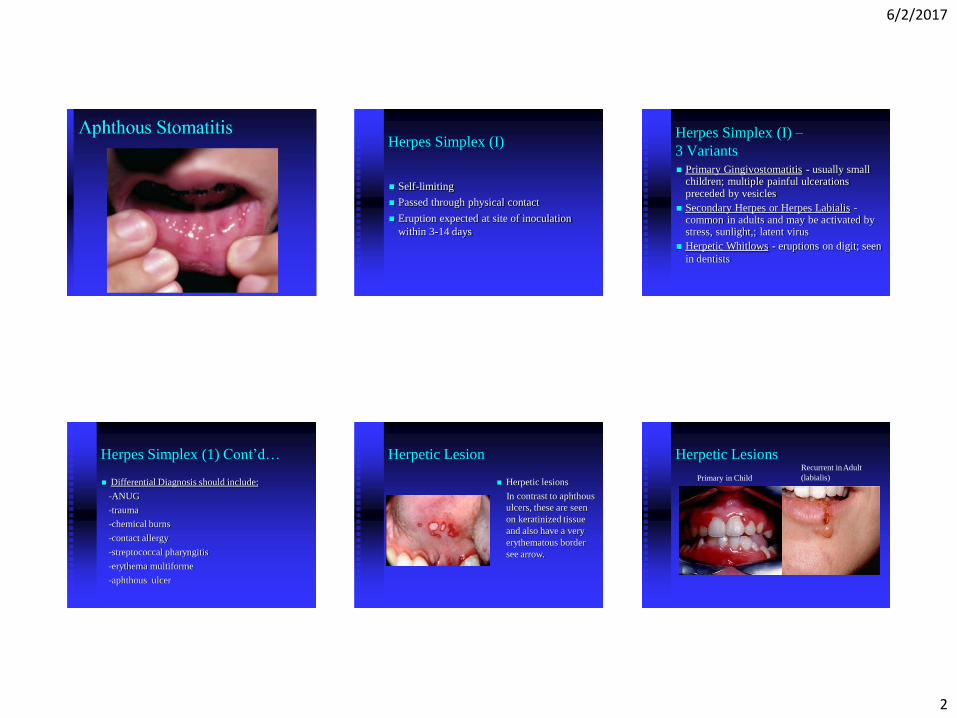
Herpes Simplex (I)
Self-limiting
Passed through physical contact
Eruption expected at site of inoculation
within 3-14 days
Herpes Simplex (I) –
3 Variants
Primary Gingivostomatitis - usually small children; multiple painful ulcerations preceded by vesicles
Secondary Herpes or Herpes Labialis -common in adults and may be activated by stress, sunlight,; latent virus
Herpetic Whitlows - eruptions on digit; seen
in dentists
Herpes Simplex (1) Cont’d…
Differential Diagnosis should include:
-ANUG
-trauma
-chemical burns
-contact allergy
-streptococcal pharyngitis
-erythema multiforme
-aphthous ulcer
Herpetic Lesion
Herpetic lesions
In contrast to aphthous
ulcers, these are seen
on keratinized tissue
and also have a very
erythematous border
see arrow.
Herpetic Lesions
Primary in Child
Recurrent inAdult
(labialis)
6/2/2017
3
Varicella Zoster Virus
Varicella Zoster primary infections:
common in children (chicken pox), painful,
pruritis vesicles on trunk, face and oral
mucosa
Varicella Zoster Virus Cont’d…
Herpes Zoster: adult (shingles). Seen commonly
in immunosuppressed and elderly
Due to reactivation of latent virus causing
unilateral painful vesicles and ulcers along V-2
and V-3 distribution
Prolonged and painful course and may also lead to
post-herpetic neuralgia and ophthalmologic
disorders
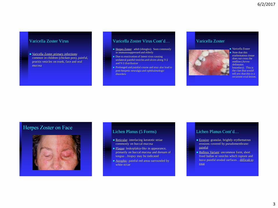
Varicella Zoster
Varicella Zoster
Note that this erythematous tissue does not cross the midline (Arrow delineates boundary). This is the clue that would tell you that this is a recurrent viral lesion.
Lichen Planus (5 Forms)
Reticular: interlacing keratotic striae
commonly on buccal mucosa
Plaque: leukoplakia-like in appearance,
primarily on buccal mucosa and dorsum of
tongue…biopsy may be indicated
Atrophic: painful red areas surrounded by
white striae
Lichen Planus Cont’d…
Erosive: granular, brightly erythematous
erosions covered by pseudomembrane:
painful
Bullous Variant: uncommon form, short
lived bullae or vesicles which rupture and
leave painful eroded surfaces…difficult to
treat
6/2/2017
4
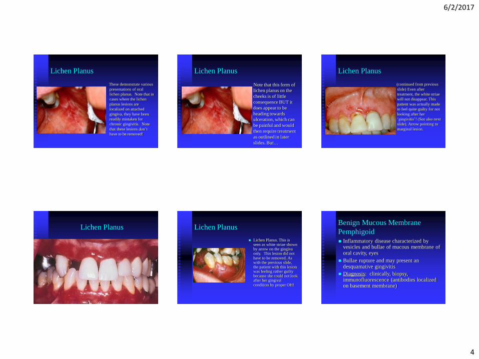
Lichen Planus
These demonstrate various
presentations of oral
lichen planus. Note that in
cases where the lichen
planus lesions are
localized on attached
gingiva, they have been
readily mistaken for
chronic gingivitis. Note
that these lesions don’t
have to be removed!
Lichen Planus
Note that this form of
lichen planus on the
cheeks is of little
consequence BUT it
does appear to be
heading towards
ulceration, which can
be painful and would
then require treatment
as outlined in later
slides. But…
Lichen Planus
(continued from previous
slide) Even after
treatment, the white striae
will not disappear. This
patient was actually made
to feel quite guilty for not
looking after her
‘gingivitis’! (See also next
slide). Arrow pointing to
marginal lesion.
Lichen Planus Lichen Planus
Lichen Planus. This is seen as white striae shown by arrow on the gingiva only. This lesion did not have to be removed. As with the previous slide, the patient with this lesion was feeling rather guilty because she could not look after her gingival condition by proper OH!
Benign Mucous Membrane
Pemphigoid
Inflammatory disease characterized by vesicles and bullae of mucous membrane of oral cavity, eyes
Bullae rupture and may present an desquamative gingivitis
Diagnosis: clinically, biopsy, immunofluorescence (antibodies localized on basement membrane)
6/2/2017
5
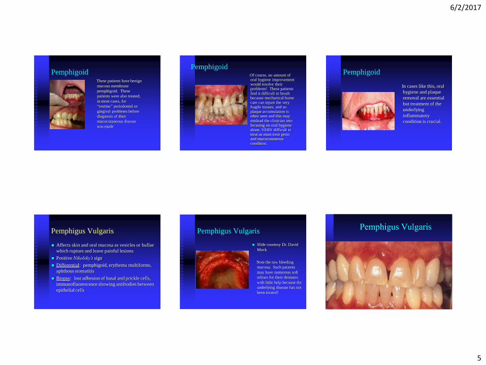
PemphigoidThese patients have benign
mucous membrane
pemphigoid. These
patients were also treated,
in most cases, for
“routine” periodontal or
gingival problems before
diagnosis of their
mucocutaneous disease
was made
PemphigoidOf course, no amount of oral hygiene improvement would resolve their problems! These patients find it difficult to brush because mechanical home care can injure the very fragile tissues, and so plaque accumulation is often seen and this may mislead the clinician into focusing on oral hygiene alone. VERY difficult to treat as must treat perio and mucocutaneous condition.
Pemphigoid
In cases like this, oral
hygiene and plaque
removal are essential
but treatment of the
underlying
inflammatory
condition is crucial.
Pemphigus Vulgaris
Affects skin and oral mucosa as vesicles or bullae
which rupture and leave painful lesions
Positive Nikolsky’s sign
Differential: pemphigoid, erythema multiforme,
aphthous stomatitis
Biopsy: lost adhesion of basal and prickle cells,
immunofluorescence showing antibodies between
epithelial cells
Pemphigus Vulgaris
Slide courtesy Dr. David
Mock
Note the raw bleeding
mucosa. Such patients
may have numerous soft
relines for their dentures
with little help because the
underlying disease has not
been treated!
6/2/2017
6
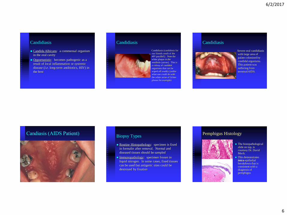
Candidiasis
Candida Albicans: a commensal organism
in the oral cavity
Opportunistic: becomes pathogenic as a
result of local inflammation or systemic
disease (i.e. long-term antibiotics, HIV) in
the host
Candidiasis
Candidiasis (candidosis for
our friends south of the
49th parallel!). Note the
white plaque in the
vestibule (arrow). This is
a colony of candidal
organisms that can be
wiped off readily (unlike
what one could do with
the white striae of lichen
planus for example)
Candidiasis
Severe oral candidiasis
with large area of
palate colonized by
candidal organisms.
This patient was
suffering from
terminal AIDS.
Biopsy Types
Routine Histopathology: specimen is fixed
in formalin after removal. Normal and
diseased tissues should be sampled
Immunopathology: specimen frozen in
liquid nitrogen. In some cases, fixed tissues
can be used but antigenic sites could be
destroyed by fixation
Pemphigus Histology
The histopathological slide on top, is courtesy Dr. David Mock.
This demonstrates intra-epithelial breakdown that is consistent with a diagnosis of pemphigus
6/2/2017
7
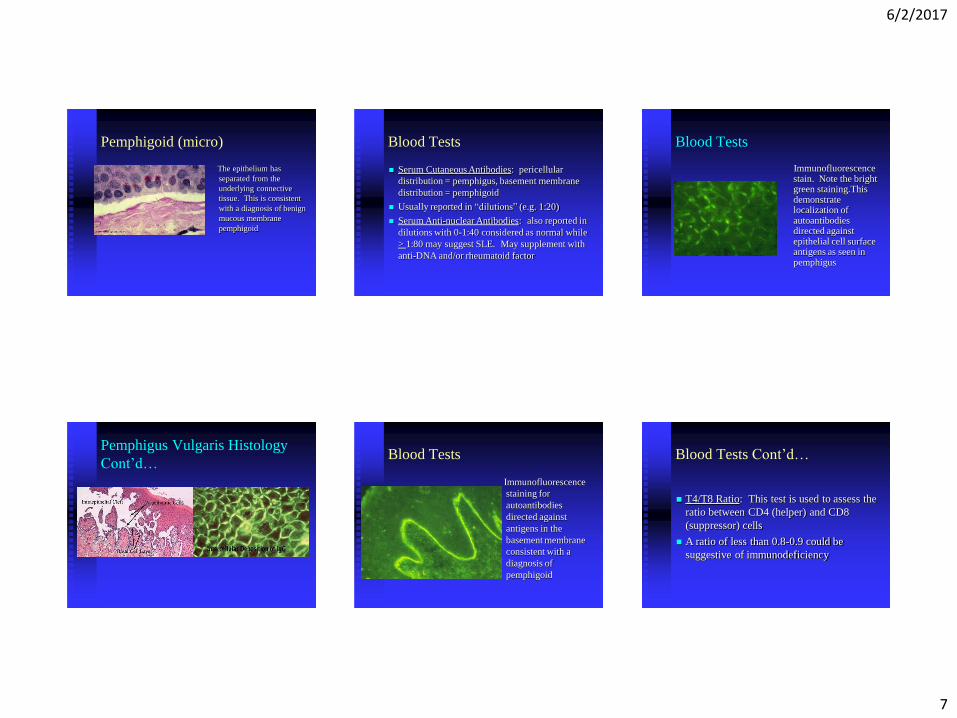
Pemphigoid (micro)
The epithelium has
separated from the
underlying connective
tissue. This is consistent
with a diagnosis of benign
mucous membrane
pemphigoid
Blood Tests
Serum Cutaneous Antibodies: pericellular
distribution = pemphigus, basement membrane
distribution = pemphigoid
Usually reported in “dilutions” (e.g. 1:20)
Serum Anti-nuclear Antibodies: also reported in
dilutions with 0-1:40 considered as normal while
> 1:80 may suggest SLE. May supplement with
anti-DNA and/or rheumatoid factor
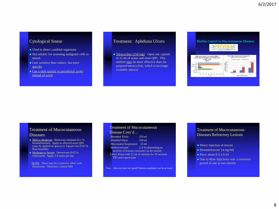
Immunofluorescence stain. Note the bright green staining.This demonstrate localization of autoantibodies directed against epithelial cell surface antigens as seen in pemphigus
Blood Tests
Pemphigus Vulgaris Histology
Cont’d…Blood Tests
Immunofluorescence
staining for
autoantibodies
directed against
antigens in the
basement membrane
consistent with a
diagnosis of
pemphigoid
Blood Tests Cont’d…
T4/T8 Ratio: This test is used to assess the
ratio between CD4 (helper) and CD8
(suppressor) cells
A ratio of less than 0.8-0.9 could be
suggestive of immunodeficiency
6/2/2017
8
Cytological Smear
Used to detect candidal organisms
Not reliable for assessing malignant cells in
mouth
Less sensitive than culture, but more
specific
Use a steel spatula or periodontal probe
instead of wood
Treatment: Aphthous Ulcers
Tetracycline (250 mg): Open one capsule
in 15 ml of water and rinse QID. This
method may be more effective than the
prepared tetracycline, which is no longer
available anyway
Biofilm Control in Mucocutaneous Diseases
Treatment of Mucocutaneous
Diseases Mild to Moderate: Betnovate ointment (0.1 %
betamethasone). Apply to affected areas QID (may be applied on gauze) or Topsyn Gel (0.05 % fluocinomide)
Moderate to Severe: Dermovate (0.05 %clobetasol). Apply 2-4 times per day
NOTE: There may be a systemic effect with
Dermovate. Therefore, consult MD
Treatment of Mucocutaneous
Disease Cont’d…-Benadryl Elixir
-Distilled Water
-Mycostatin Suspension
250 ml
250 ml
25 ml
-Hydrocortisone 2-4 % (depending onseverity of lesions, cortisone can be varied)
Label: Rinse with 15 ml of solution for 30 seconds
TID and expectorate
Note… does not taste very good! Patient-compliance can be an issue!
Treatment of Mucocutaneous
Diseases Refractory Lesions
Direct injection of lesions
Dexamethasone (4 mg/ml)
Place about 0.5-1.0 ml
One to three injections over a treatment
period of one to two months
6/2/2017
9
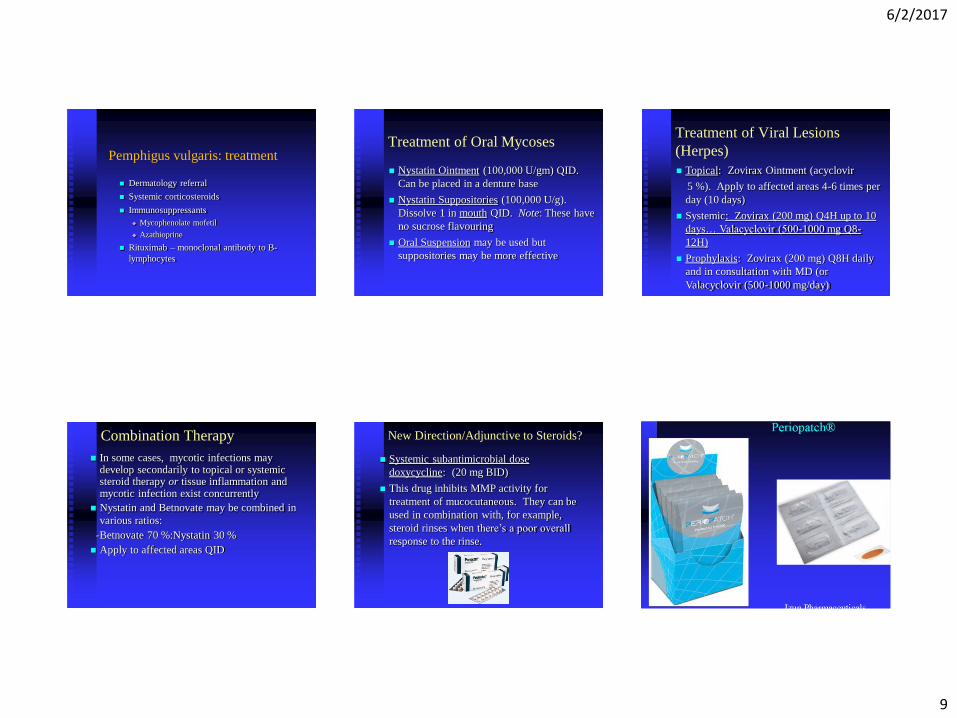
Pemphigus vulgaris: treatment
Dermatology referral
Systemic corticosteroids
Immunosuppressants
Mycophenolate mofetil
Azathioprine
Rituximab – monoclonal antibody to B-
lymphocytes
Treatment of Oral Mycoses
Nystatin Ointment (100,000 U/gm) QID.
Can be placed in a denture base
Nystatin Suppositories (100,000 U/g).
Dissolve 1 in mouth QID. Note: These have
no sucrose flavouring
Oral Suspension may be used but
suppositories may be more effective
Treatment of Viral Lesions
(Herpes)
Topical: Zovirax Ointment (acyclovir
5 %). Apply to affected areas 4-6 times per
day (10 days)
Systemic: Zovirax (200 mg) Q4H up to 10
days… Valacyclovir (500-1000 mg Q8-
12H)
Prophylaxis: Zovirax (200 mg) Q8H daily
and in consultation with MD (or
Valacyclovir (500-1000 mg/day)
Combination Therapy
In some cases, mycotic infections may develop secondarily to topical or systemic steroid therapy or tissue inflammation and mycotic infection exist concurrently
Nystatin and Betnovate may be combined in
various ratios:
-Betnovate 70 %:Nystatin 30 %
Apply to affected areas QID
New Direction/Adjunctive to Steroids?
Systemic subantimicrobial dose
doxycycline: (20 mg BID)
This drug inhibits MMP activity for
treatment of mucocutaneous. They can be
used in combination with, for example,
steroid rinses when there’s a poor overall
response to the rinse.
6/2/2017
10
PerioPatch Adheres to Mucosa;
The Components Draw Out Inflammatory
Cytokines
Evidently Leads to Long-Term Regulation of
Inflammation by Returning the Inflammatory
Response to a Constitutive Level
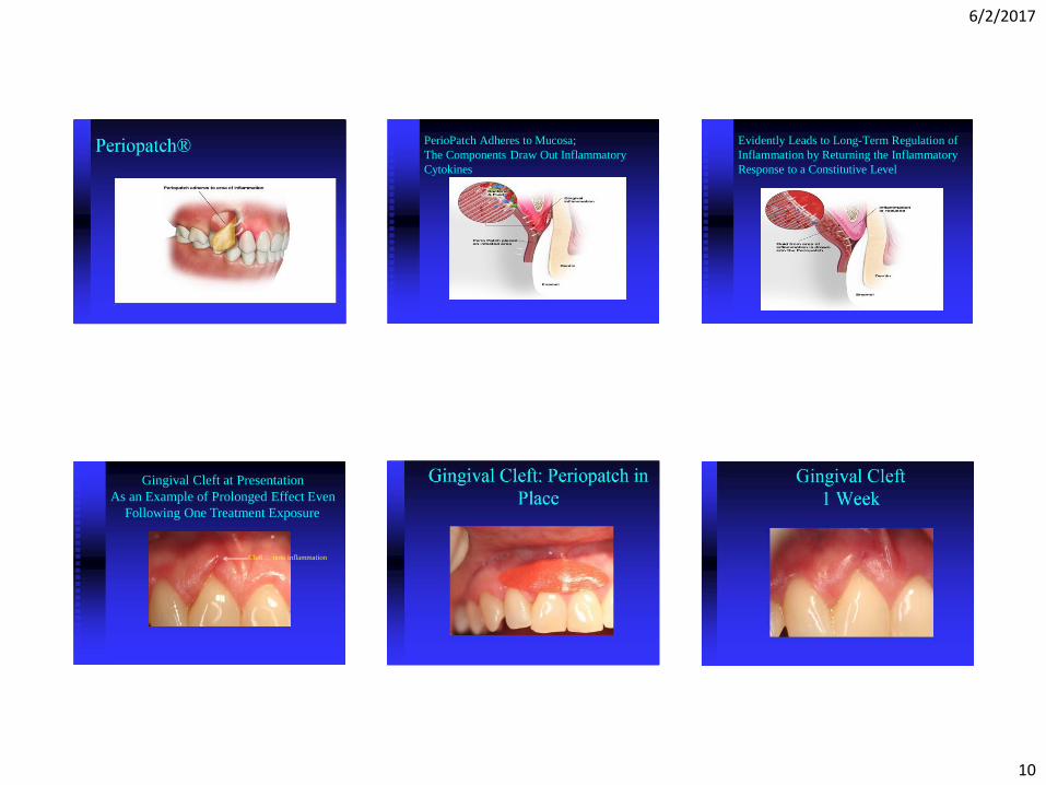
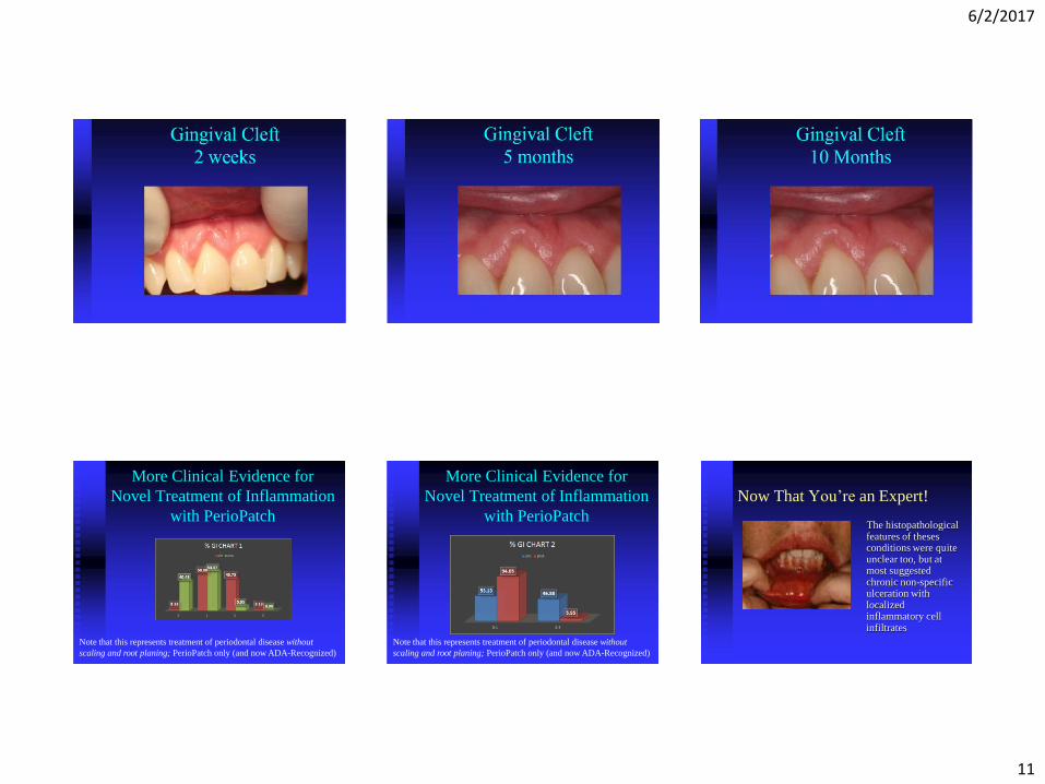
Gingival Cleft at Presentation
As an Example of Prolonged Effect Even
Following One Treatment Exposure
Cleft… note inflammation
6/2/2017
11
More Clinical Evidence for
Novel Treatment of Inflammation
with PerioPatch
Note that this represents treatment of periodontal disease without
scaling and root planing; PerioPatch only (and now ADA-Recognized)
More Clinical Evidence for
Novel Treatment of Inflammation
with PerioPatch
Note that this represents treatment of periodontal disease without
scaling and root planing; PerioPatch only (and now ADA-Recognized)
Now That You’re an Expert!
The histopathological features of theses conditions were quite unclear too, but at most suggested chronic non-specific ulceration with localized inflammatory cell infiltrates
6/2/2017
12
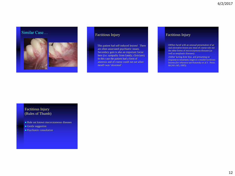
Factitious Injury
This patient had self-induced lesions! There
are often associated psychiatric issues.
Secondary gain is also an important factor
here (i.e. sympathy from family, clinicians).
In this case the patient had a form of
anorexia and of course could not eat when
mouth was ‘ulcerated’.
Factitious Injury
1)When faced with an unusual presentation of an
oral ulcerative lesion you must of course rule out
the other forms of mucocutaneous diseases (as
well as neoplastic diseases).
2)After having done that, and presuming no
response to treatment, begin to consider factitious
lesions (for reference see Kotansky et al J. Perio.
66:241-245, 1995).
Factitious Injury
(Rules of Thumb)
Rule out known mucocutaneous diseases
Gentle suggestion
Psychiatric consultation