Embed Size (px)
Citation preview

Poznan University of Medical SciencesPoland
2015Vol. 84, No. 4
eISSN 2353-9801ISSN 2353-9798
previously Nowiny Lekarskie
Founded in 1889
QUARTERLY
Indexed in:Polish Medical Bibliography, Index Copernicus,Ministry of Science and Higher Education, Ebsco, Google Scholar
www.jms.ump.edu.pl

EDITOR-IN-CHIEFMarian Grzymisławski
VICE EDITOR-IN-CHIEFJarosław Walkowiak
EDITORIAL BOARDDavid H. Adamkin (USA)Adrian Baranchuk (Canada)Grzegorz Bręborowicz (Poland)Paolo Castiglioni (Italy)Wolfgang Dick (Germany)Leon Drobnik (Poland)Janusz Gadzinowski (Poland)Michael Gekle (Germany)Karl-Heinz Herzig (Germany)Mihai Ionac (Romania)Lucian Petru Jiga (Germany)Berthold Koletzko (USA)Stan Kutcher (Canada)Odded Langer (USA)Tadeusz Maliński (USA)Leszek Paradowski (Poland)Antoni Pruszewicz (Poland)Georg Schmidt (Munich, Germany)Mitsuko Seki (Japan)Ewa Stępień (Poland)Jerzy Szaflarski (USA)Bruno Szczygieł (Poland)Kai Taeger (Germany)Marcos A. Sanchez-Gonzalez (Florida, USA)Krzysztof Wiktorowicz (Poland)Witold Woźniak (Poland)
ASSOCIATE EDITORSAgnieszka BienertMaria IskraEwa MojsAdrianna Mostowska
SECTION EDITORSJaromir Budzianowski – Pharmaceutical SciencesPaweł Jagodziński – Basic Sciences Joanna Twarowska-Hauser – Clinical Sciences
Publishing Manager: Grażyna DromireckaTechnical Editor: Bartłomiej Wąsiel
WYDAWNICTWO NAUKOWE UNIWERSYTETU MEDYCZNEGOIM. KAROLA MARCINKOWSKIEGO W POZNANIU60-812 Poznań, ul. Bukowska 70tel./fax: +48 61 854 71 51
Ark. wyd. 9,8. Ark. druk. 8,3.Zam. nr 10/16.
LANGUAGE EDITORSMargarita Lianeri (Canada)Jacek Żywiczka (Poland)
STATISTICAL EDITORMagdalena Roszak (Poland)
SECRETARIAT ADDRESS70 Bukowska Street, room 10460-812 Poznan, Polandphone/fax: +48 61 854 72 74email: jms @ump.edu.plwww.jms.ump.edu.pl
DISTRIBUTION AND SUBSCRIPTIONS37a Przybyszewskiego Street60-356 Poznan, Polandphone/fax: +48 61 854 64 87email: [email protected]
PUBLISHERPoznan University of Medical Sciences
© 2015 by respective Author(s). Production and hosting by Journal of Medical Science (JMS)
This is an open access journal distributed under the terms and conditions of the Creative Commons Attribution (CC BY-NC) licencse
eISSN 2353-9801ISSN 2353-9798

The Editorial Board kindly informs that since 2014 Nowiny Lekarskie has been renamed to Journal of Medical Science.
The renaming was caused by using English as the language of publications and by a wide range of other organisational changes. They were necessary to follow dynamic transformations on the publish-ing market. The Editors also wanted to improve the factual and publishing standard of the journal. We wish to assure our readers that we will contin-ue the good tradition of Nowiny Lekarskie.
You are welcome to publish your basic, medical and pharmaceutical science articles in Journal of Medical Science.

Ethical guidelinesThe Journal of Medical Science applies the ethical principles and procedures recommended by COPE (Committee on Conduct Ethics), contained in the Code of Conduct and Best Practice Guidelines for Journal Editors, Peer Reviewers and Authors available on the COPE website: https://publicationethics.org/resources/guidelines

Journal of Medical Science 4 (84) 2015
CO N T E N T S
ORIGINAL PAPERS
Artur Cieślewicz, Katarzyna Korzeniowska, Paweł Bogdański, Anna Jabłecka
Increased neopterin concentration in patients with primary arterial hypertension . . . . . . . . 213
Marta Holeyko, Volodymyr Zubachyk
Assessment of effectiveness of complex treatment of apical and marginal periodontitis with thiotriazoline and chloramphenicol ointment . . . . . . . . . . . . . . . . . . . . 218
Bartosz Bilski, Paweł Rzymski, Katarzyna Tomczyk, Izabela Rzymska
The impact of factors in work environment (especially shift and night work) on neoplasia of female reproductive organs . . . . . . . . . . . . . . . . . . . . . . 223
Julia Jajor, Marta Rosołek, Elżbieta Skorupska, Agnieszka Krawczyk-Wasielewska,
Przemysław Lisiński, Ewa Mojs, Włodzimierz Samborski
„UnderstAID – a platform that helps informal caregivers to understand and aid their demented relatives” – assessment of informal caregivers – a pilot study . . . . . . . 229
Łucja Czyżewska-Majchrzak, Roma Krzymińska-Siemaszko, Marta Pelczyńska, Henryk Witmanowski
The benefits and risks of short‑term diet changes on the example of the use a 5‑week long lactoovovegetarian diet. Analysis of 7‑day nutritional surveys of women – preliminary study . . . . 235
Monika Urbaniak
Legal aspects of a healthy diet for children . Comments on the grounds of the directive on foodstuffs in schools . . . . . . . . . . . . . . . . . . . . . . . . 244
REVIEW PAPER
Magdalena Gibas-Dorna, Piotr Turkowski, Małgorzata Bernatek, Kinga Mikrut, Justyna Kupsz, Jacek Piątek
Liposuction‑induced metabolic alterations – the effect on insulin sensitivity, adiponectin, leptin and resistin . . . . . . . . . . . . . . . . . . . . . . . . . . . 249
Piotr Stępniak
Institutions of health’s care . Aspects European and judicial . . . . . . . . . . . . . . . . . 257
CASE STUDY
Roman Jankowski, Jeremi Kościński, Bartosz Sokół, Stanisław Malinger, Janusz Szymaś
Presacral schwannoma . Case description . . . . . . . . . . . . . . . . . . . . . . . 264
Instructions for Authors . . . . . . . . . . . . . . . . . . . . . . . . . . . . . . 271

Journal of Medical Science 4 (84) 2015

213Journal of Medical Science 4 (84) 2015
© 2015 by the author(s). This is an open access article distributed under the terms and conditions of the Creative Commons Attribution (CC BY-NC) licencse. Published by Poznan University of Medical Sciences
DOI: https://doi.org/10.20883/medical.e1
Increased neopterin concentration in patients with primary arterial hypertensionArtur Cieślewicz1, Katarzyna Korzeniowska1, Paweł Bogdański2, Anna Jabłecka1
1 Deprtment of Clinical Pharmacology, Poznan University of Medical Sciences, Poland2 Department of Internal Medicine, Metabolic Disorders and Hypertension, Poznan University of Medical Sciences, Poland
Introduction
Neopterin (2-amino-4-hydroxy-6-(D-erythro-1',2',3'-tri- hydroxypropyl)-pteridine) is a pteridine derivative produced from guanosine triphosphate by activated monocytes, macrophages, dendritic cells, and endothe-lial cells and to a lesser extent in renal epithelial cells, fibroblasts, and vascular smooth muscle stimulated by interferon gamma. Because it is released in response to cytokines produced by T-lymphocytes and natu-ral killer cells, neopterin is an indicator of activation of cell mediated immunity [1–2]. Neopterin can be assessed in blood serum, plasma, urine, cerebrospinal fluid, pancreatic juice, saliva and gastric juices. Neop-terin is a light-sensitive substance, so probes collected for measurement must be protected from light. Physi-ological serum concentration is lesser than 10 nmol/L and is different in various age groups (Table 1) [21]. Other factors that can influence neopterin level include gender, race, BMI, and percentage of body fat. [3].Changes in neopterin level reflect the stage of activa-tion of cellular immune system and can be associated with various diseases. For example, increase neopterin
level was observed in patients with coronary artery dis-eases and was associated with the progression of the disease [4–5]. Therefore, the inflammation system, in association with other cardiovascular pathways, can be the central pathway in the development and progres-sion of cardiovascular diseases [6].
Essential hypertension can be characterized by increased peripheral vascular resistance to blood flow and is one of important risk factors for developing car-diovascular disease [7]. Most of this resistance results from resistance arteries, which are vessels with lumen diameters < 400 μm [8]. These arteries undergo struc-tural, mechanical or functional vascular remodeling in hypertensive patients – a process that involves extra-cellular matrix deposition and inflammation. In case of chronic vasoconstriction the vessels may become embedded in the remodeled extracellular matrix and
ABSTRACT
Introduction. The data on the safety profile of anti-tumor necrosis factor (anti-TNF) therapy in real-life patients cohorts with inflammatory bowel disease (IBD) still are lacking.Neopterin is a pteridine derivative produced from guanosine triphosphate mainly by activated monocytes and macrophages in response to cytokines produced by T-lymphocytes and natural killer cells. Changes in neopterin level reflect the stage of activation of cellular immune system and can be associated with various diseases. Low-grade inflammation is also an important factor in the pathophysiology of hypertension. In presented study we assessed neopterin concentration in 63 patients with primary arterial hypertension compared to 14 control healthy volunteers. Obtained results confirmed increased neopterin level in patients group.
Keywords: neopterin, hypertension, inflammation.
O R I G I N A L PA PE R
Table 1. Average neopterin concentration in various age groups
Age (years) Average neopterin concentration [nmol/L]< 18 6 .8 ± 3 .619–75 5.3 ± 2.7> 75 9.7 ± 5.0

214 Journal of Medical Science 4 (84) 2015
may not return to their vasodilated state. Moreover, endothelial dysfunction – an early determinant in the development of hypertension may also participate to the increased vascular tone in hypertension with reduced vasodilation associated with proinflammatory and prothrombotic state [9]. Chronic low-grade inflam-mation has been recently mentioned to be an integral part in the pathogenesis of vascular disease [10]. Sever-al clinical studies have revealed that pro-inflammatory markers, such as IL-6, ICAM-1 or CRP may be indepen-dent risk factors for the development of hypertension [9]. Neopterin is one of inflammatory mediators, which role in hypertension has not yet been sufficiently stud-ied. Numerous studies have confirmed the usefulness of neopterin level measurement in such cases as trans-plant rejection, viral infections, intracellular bacteria infections, coronary artery disease, angina pectoris and some autoimmune diseases (arthritis, type 1 diabetes, Crohn’s disease, autoimmune thyroiditis) [11]. As men-tioned earlier, low-grade inflammation is an important factor in the pathophysiology of hypertension. There-fore, the aim of the study was to assess neopterin level in patients with primary arterial hypertension.
Material and methods
63 patients (31 men, 32 women) with primary arterial hypertension, aged from 25 to 67 years (mean: 50.37;
standard deviation: 10.58) were enrolled to the study. Patients’ weight ranged from 59 to 167 kg (mean: 98.15; standard deviation: 21.50). Patients with acute coronary syndrome, cancer, heart failure, severe renal failure, severe hepatic insufficiency or pregnancy were excluded from the study. The control group consist-ed of 14 healthy volunteers (6 men, 8 women), aged from 25 to 59 years (mean: 42.57, standard devia-tion: 11.80), weighed from 50 to 83 kg (mean: 65.21; standard deviation: 9.18). Arterial blood pressure was measured in both groups. Blood samples were col-lected from elbow vein for biochemical measurements. Neopterin level was assessed using ELISA immunoas-say (DRG International Inc., USA). Statistical analyses were carried out using Statsoft Statistica 10.0 software. Normality of distribution was tested with Shapiro-Wilk Test. Statistical significance was assessed using Mann-Whitney U test.
Results
Estimated sample size for statistical power = 0.95 was 56 individuals. Shapiro-Wilk test of normality revealed that most of studied parameters (including neopterin) did not have normal distribution (Table 2). As a result, non-parametric Mann-Whitney U test was chosen as a measure of statistical significance (p < 0.05).
Table 2. Anthropometric and biochemical characteristic of the participants
Patients ControlsNormality of distribution
PN Mean
Standard deviation
N MeanStandard deviation
Age [years] 63 50.37 10.58 14 42.57 11.80 no 0.029Weight [kg] 63 98.15 21.50 14 65.21 9 .18 no 0.000Height [cm] 63 169 .94 9 .16 14 168.50 9.09 no 0.644BMI [kg/m2] 63 33.95 6.90 14 22 .91 2.03 no 0.000Waist [cm] 63 111.40 16.04 14 73 .43 3.50 yes 0.000Hips [cm] 63 114.56 14 .19 14 93 .71 5.53 yes 0.000SBP [mmHg] 63 158.49 29 .16 14 110.36 6 .34 no 0.000DBP [mmHg] 63 91 .98 11.20 14 72.50 5.46 no 0.000Creatinine [μmol/L] 63 81 .68 16 .31 14 70.50 10.38 no 0.015Tchol [mmol/L] 63 5.84 1 .36 14 5.11 0.61 yes 0.019LDL [mmol/L] 63 3.70 1 .13 14 2 .76 0.61 yes 0.002HDL [mmol/L] 63 1 .19 0.32 14 1.65 0.32 no 0.000TG [mmol/L] 63 2 .13 0.88 14 1.02 0.39 no 0.000Glucose [mmol/L] 63 5.09 0.42 14 4 .99 0.47 no 0.373CRP [mg/L] 63 5.73 4 .44 14 2.51 1.80 no 0.001ESR [mm/h] 63 9 .24 5.65 14 6 .64 4 .97 no 0.104ALAT [U/L] 63 34.05 16 .38 14 26.57 9 .19 no 0.109ASPAT [U/L] 63 29 .41 15.35 14 20.64 3 .37 no 0.002Neopterin [nmol/L] 63 6.50 2.510 14 5.17 0.72 no 0.001

215Increased neopterin concentration in patients with primary arterial hypertension
Anthropometric and biochemical characteristic of the participants is presented in Table 2. Patients and control groups did not differ in height, glucose, ESR and ALAT. Statistically significant differences between these groups were observed for such parameters as: age, weight, BMI, waist and hips circumference, blood pressure, creatinine, total cholesterol, LDL and HDL fraction, triglycerides, CRP, ASPAT and neopterin level. Higher level of neopterin was observed in patients with hypertension compared to healthy controls (Figure 1).
Discussion
According to WHO data, approximately 20% of adults (1 billion people in the world) are estimated to have hypertension, defined as blood pressure > 140/90 mm Hg. In the elderly, the prevalence of hypertension can be up to 50% [12]. For example, in the United States 1 per 3 adults have hypertension, while the prevalence increases to 50% for people aged 60 – 69 years and to 75% for patients older than 70 years [13].
Essential hypertension is a multifactorial disease caused by combined action of genetic, environmental,
and behavioral factors. A pro-hypertensive change in a single factor can be probably compensated by oth-er control mechanisms. However, any significant dis-turbance in the balance between the factors which increase and normalize the blood pressure can result in development of essential hypertension [14]. One of the factors which can contribute to the develop-ment of hypertension is inflammation [15]. Inflamma-tory cells accumulate in kidneys and vasculature of patients with hypertension. It was observed in animal models that loss of adaptive immune cells decreases the blood pressure response to such stimuli as ANG II, high salt, and norepinephrine. Moreover, agonistic antibodies to ANG II receptor (produced by B-cells) contribute to hypertension in experimental models of preeclampsia. Also, production of cytokines, such as TNF-α, interleukin-17, and interleukin-6 influences hypertension, possibly due to effects on both the kid-ney and vasculature. The innate immune system also appears to contribute to hypertension. Therefore, stud-ies concerning immune cell activation could be helpful in understanding this disease [16]. There are only few studies trying to evaluate neopterin level in hyperten-
Figure 1. Mean neopterin concentration observed in patients and controls
216 Journal of Medical Science 4 (84) 2015
sive patients. Avanzas et al. [17] assessed prognostic value of neopterin in the group of treated patients with hypertension, typical exertional chest pain and coro-nary artery stenosis of < 50% but without obstructive coronary artery disease, revealing that patients who developed adverse events during one year follow-up had significantly higher neopterin levels than patients without events (7.6 nmol/L vs. 5.4 nmol/L). Asci et al. [18] evaluated neopterin level in patients undergoing hemodialysis. The control group of that study consisted of three subgroups: healthy, diabetic and hypertensive subjects. Hypertensive control group had significantly higher serum neopterin level (16 +/- 1 nmol/L) than healthy control group (11 +/- 1 nmol/L). A recent study of Wang et al. [19] showed that plasma neopterin and hsCRP levels were increased in hypertensive patients with obstructive sleep apnea syndrome (OSAS) and correlated with severity of OSAS.
A similar tendency has been observed in our study: neopterin concentration was higher in hyperten-sive patients than in healthy controls (6.89 +/- 2.793 vs. 5.08 +/- 0.438); this result is consistent with the hypothesis on the role of inflammation processes in hypertension.
It should be also noted that assessed neopterin level seems to fall within the normal range, which is considered 8.7 nmol/L for 95th percentile of healthy population, according to Werner et al. [20]. However, it is usually recommended to estimate neopterin referen-tial values for each study as they can differ significantly due to measurement method or population diversity.
Acknowledgements
Conflict of interest statementThe authors declare that there is no conflict of interest in the authorship or publication of contribution.
Funding sourcesThere are no sources of funding to declare.
ReferencesEisenhut M. Neopterin in Diagnosis and Monitoring of 1. Infectious Diseases. Journal of Biomarkers Volume 2013, Article ID 196432, Hindawi Publishing Corporation. http://dx.doi.org/10.1155/2013/196432.De Rosa S, Cirillo P, Pacileo M, Petrillo G, D'Ascoli GL, 2. Maresca F et al. Neopterin: from forgotten biomarker to leading actor in cardiovascular pathophysiology. Curr Vasc Pharmacol. 2011;9(2):188–199.Spencer ME, Jain A, Matteini A, Beamer BA, Wang NY, 3. Leng SX et al. Serum levels of the immune activation marker neopterin change with age and gender and are
modified by race, BMI, and percentage of body fat. J Gerontol A Biol Sci Med Sci. 2010;65(8):858–865. Signorelli SS, Anzaldi M, Fiore V, Candido S, Di Mar-4. co R, Mangano K et al. Neopterin: a potential mar-ker in chronic peripheral arterial disease. Mol Med Rep. 2013;7(6):1855–1858. Avanzas P, Arroyo-Espliguero R, Kaski JC. Role of Neo-5. pterin in Cardiovascular Medicine. Rev Esp Cardiol. 2009;62(11):1341–1342.Visvikis-Siest S, Marteau JB, Samara A, Berrahmoune H, 6. Marie B, Pfister M. Peripheral blood mononuclear cells (PBMCs): a possible model for studying cardiovascular biology systems. Clin Chem Lab Med. 2007;45(9):1154–1168.Lund-Johansen P. Haemodynamics in early essential 7. hypertension-still an area of controversy. J Hypertens. 1983;1(3):209–213.Intengan HD, Schiffrin EL. Structure and mechanical 8. properties of resistance arteries in hypertension: role of adhesion molecules and extracellular matrix determi-nants. Hypertension. 2000;36(3):312–318.Savoia C, Sada L, Zezza L, Pucci L, Lauri FM, Befani 9. A et al. Vascular inflammation and endothelial dysfun-ction in experimental hypertension. Int J Hypertens. 2011;2011:281240. doi: 10.4061/2011/281240. Epub 2011 Sep 11.Boos CJ, Lip GY. Is hypertension an inflammatory pro-10. cess? Curr Pharm Des. 2006;12(13):1623–1635.Kozlowska-Murawska J, Obuchowicz A. Clinical usefulne-11. ss of neopterin. Wiad Lek. 2008;LXI:10–12.The World Health Report 2002-Reducing Risks, Promo-12. ting Healthy Life. Geneva, Switzerland: World Health Organization; 2002. Available at http://www.who.int/whr/2002/en.High Blood Pressure. American Heart Association. Ame-13. rican Stroke Association. Statistical Fact Sheet 2013 Update. http://www.heart.org/idc/groups/heart-pub-lic/@wcm/@sop/@smd/documents/downloadable/ucm_319587.pdf.Bolívar JJ. Essential Hypertension: An Approach to Its 14. Etiology and Neurogenic Pathophysiology. Int J Hyper-tens. 2013;2013:547809. doi: 10.1155/2013/547809. Epub 2013 Dec 9.Schiffrin EL. Immune mechanisms in hypertension and 15. vascular injury. Clin Sci (Lond). 2014;126(4):267–274. Trott DW, Harrison DG. The immune system in hyperten-16. sion. Adv Physiol Educ. 2014;38(1):20–24. Avanzas P, Arroyo-Espliguero R, Cosin-Sales J, Quiles J, 17. Zouridakis E, Kaski JC. Prognostic value of neopterin levels in treated patients with hypertension and chest pain but without obstructive coronary artery disease. Am J Cardiol. 2004;93(5):627–629.Asci A, Baydar T, Cetinkaya R, Dolgun A, Sahin G. Evalu-18. ation of neopterin levels in patients undergoing hemo-dialysis. Hemodial Int. 2010;14(2):240–6. doi: 10.1111/j.1542–4758.2010.00439.x. Epub 2010 Mar 10.Wang H, Cheng Y, Liu Z, Liu Q, Tong H, Wang X et al. 19. Relationship Between Plasma Neopterin and High Sens-itivity C-Reactive Protein Levels and Circadian Rhythm of Blood Pressure in Hypertensive Patients with Obstructi-

217Increased neopterin concentration in patients with primary arterial hypertension
ve Sleep Apnea Syndrome. Am J Hypertens 2012;25(7): 833.Werner ER, Bichler A, Daxenbichler G, Fuchs D, Fuith LC, 20. Hausen A, Hetzel H, Reibnegger G, Wachter H. Deter-mination of neopterin in serum and urine. Clin Chem. 1987;33(1):62–66.http://www.neopterin.net/neopterin.21.
Acceptance for editing: 2015-11-10 Acceptance for publication: 2015-12-31
Correspondence address:Artur Cieślewicz
Deprtment of Clinical PharmacologyPoznan University of Medical Sciences
1/2 Dluga Str., 61–848 Poznan, Polandphone: +48 61 8549216
email: [email protected]

218 Journal of Medical Science 4 (84) 2015
© 2015 by the author(s). This is an open access article distributed under the terms and conditions of the Creative Commons Attribution (CC BY-NC) licencse. Published by Poznan University of Medical Sciences
DOI: https://doi.org/10.20883/medical.e2
Assessment of effectiveness of complex treatment of apical and marginal periodontitis with thiotriazoline and chloramphenicol ointmentMarta Holeyko, Volodymyr Zubachyk
Department of Therapeutic Dentistry, Danylo Halytsky Lviv National Medical University, Lviv, Ukraine
Introduction
Anatomical and functional interrelationship between the endodontium and periodontium causes a high probability of their simultaneous involvement in the pathological process [1]. Diseases of the periodontal tissues may have a damaging effect on the pulp or the apical periodontium through the system of apical and lateral canals, dentinal tubules [2, 3]. Finally, peri-odontal surgery procedures with removal of damaged cement of the tooth root can contribute to the opening of the lateral canals and dentinal tubules, resulting in the pulp destruction. Progressive periodontal patholo-gy can lead to the pulp necrosis [4]. Chronic combined
periodontal and periapical lesions have a negative impact on the human organism, since they are a source of intoxication and sensitization causing progression of infection, development of focal sequelae and second-ary immunodeficiency [5, 6].
Problem of successful treatment of apical and marginal periodontitis is associated with a significant prevalence of these diseases, a complexity of medical procedures, a substantial proportion of failures and complications, and with a lack of long-term stability of gained results [7].
Endodontic treatment in patients with pathology of periodontal tissues is of particular importance: it is impossible to achieve a long-lasting remission and sta-
ABSTRACT
Introduction. Chronic combined periodontal and periapical lesions have a negative impact on the human organism, since they are a source of intoxication and sensitization causing progression of infection, development of focal sequelae and secondary immunodeficiency. Problem of successful treatment of apical and marginal periodontitis is associated with a significant prevalence of these diseases, a complexity of medical procedures, a substantial proportion of failures and complications, and with a lack of long-term stability of gained results.Aim. The aim of this work was to investigate clinical effectiveness of drug formulation with thiotriazoline and chloramphenicol in the integrated treatment of combined apical and marginal periodontitis.Meterial and methods. The condition of oral cavity of the 65 patients with combined lesions of periodontal and endodontic tissues before and after treatment was studied. Outcomes measured were X-ray examination, probing depth, OHI-S, PMA, PI, SBI indices. The complex treatment has been worked out in the patients of main group and the ointment with thiotriazoline and chloramphenicol was introduced in the scheme of periodontal treatment Results and conclusions. Results of applied treatment indicated to the acceleration of healing process, reduction of exudation period and decrease of exacerbations frequency in the patients of main group. Clinical experience also demonstrated positive dynamic in changes of periodontal indices after the conducted treatment.
Keywords: chronic generalized periodontitis, apical periodontitis, treatment, ointment with thiotriazoline and chloramphenicol.
O R I G I N A L PA PE R
219Assessment of effectiveness of complex treatment of apical and marginal periodontitis with thiotriazoline and chloramphenicol...
bilization of pathological process without an appropri-ate endodontic treatment; on the other hand, incom-plete endodontic therapy may result in the aggravation of periodontal status at the early stages of periodonti-tis [8, 9].
Aim
The aim of this work was to investigate clinical effec-tiveness of drug formulation with thiotriazoline and chloramphenicol [10] in the integrated treatment of combined apical and marginal periodontitis.
Material and methods
Status of oral cavity of 105 patients with apical and mar-ginal periodontitis, who were given complex treatment and a dynamic supervision, was studied. 46 patients were diagnosed with chronic generalized periodontitis (GP) of the I stage of severity; the rest 59 patients had the GP of the II stage of severity. All patients were also diagnosed as carrying lesions of chronic apical infec-tions, among which were granulating (45%), fibrous (37%) and granulomatous (28%) periodontitis.
Clinical examination was performed according to the standard scheme. The diagnosis was made based on the complaints, anamnesis, and assessment of gen-eral and local health of patients. The anatomical fea-tures of the oral vestibule, the condition of the gin-gival mucous membrane, the depth of the periodon-tal pockets, the extent of the gingival recession and pathological tooth mobility were considered during the physical examination of the periodontal tissues. Local irritating factors such as trauma, tooth extrac-tion, incorrect fillings, unstable dental prosthesis, plaque, calculus were also registered. The simplified oral hygiene index (OHI-S) [11], the sulcus bleeding index (SBI) [12], the papillary-marginal-alveolar index (PMA) [13], the periodontal index (PI) [14], and prob-ing depth [15] were evaluated during the index assess-ment. Among the complaints of the patients with peri-odontitis a special attention was paid to the spontane-ous pain in the causal tooth, the pain on biting, the presence of a fistula.
The duration of the disease, the occurrence of exac-erbations, the previous treatment and its effective-ness were determined from the anamnesis. During the physical examination the tooth percussion sensitivity was evaluated, the alveolar bone in the root apex pro-jection was palpated, and symptoms of angioparesis, edema and mucous membrane hyperemia were also
determined. The status of the alveolar process and the periapical tissues was assessed using the X-rays exami-nation. Patient’s examination was performed before and after treatment, and in the later periods – after 6 months.
All patients received endodontic and periodontal treatment. The patients were divided into two groups according to the given therapy. 52 patients of the main group were treated with thiotriazoline and chloram-phenicol ointment after the basic therapy. 53 patients of the control group were given standard treatment.
Providing the endodontic care for patients with combined marginal and apical periodontitis was a pri-ority task. Complex treatment of patients with different clinical forms of apical periodontits, besides mechani-cal tooth cleaning, disinfection and reliable obturation of the root canals, should include pharmacotherapy of the apical periodontal lesions for pharmacological cor-rection of inflammatory process. Scheme of the endo-dontic treatment included the following procedures: the creation of an optimal access to the root canals, the evacuation of the degradation products, the mechani-cal and antiseptic root canal treatment with sodium hypochlorite 5.25%. Apical therapy was finished by additional administration of the ointment with thio-triazoline and chloramphenicol into the root canal of patients from the main group, followed by closing with temporary filling; in patients from control group 0.2% chlorhexidine digluconate solution was introduced into root canal and teeth were temporarily closed.
After 2–3 days the drug preparation inside the root canal was changed into the fresh one, which was repeated 2–3 times. After the drug therapy patients without complications were exposed to obturation of root canals by sealer “Apexit” (Vivadent) and technique of cold lateral gutapercha condensation. Quality of obturation was radiographically assessed.
Periodontal treatment was performed according to the standard principles with consideration of the severi-ty of the periodontitis and the patients’ individual char-acteristics. Periodontal treatment included professional hygiene of oral cavity, scaling, root planning, open and closed curettage, medicamental treatment. Treatment of the control group was finished with conventional therapy. Patients in the main group were given the applications with thiotriazoline and chloramphenicol preparation in the periodontal pockets and marginal gums for 15–20 min. Number of visits depended on the severity of the inflammation.
Results of the study were statistically processed using Wilcoxon signed-rank test.

220 Journal of Medical Science 4 (84) 2015
Ethical Committee Approval: 29.10.2007, protocol number 8.
Results
After the started complex treatment patients in the main group subjectively noticed an improvement in the periapical tissues and gums, and the inflammation reduction was determined objectively. In the next visit the situation was markedly better: pain was gradually disappearing, the exudation was decreasing, the verti-cal percussion was negative and the paper points used for the root canal were clean. Only 5 patients within the first 2–3 days suffered a discomfort while eating solid foods. 65% patients in the control group who were treated by traditional endodontic treatment, felt the pain when biting on the causative tooth within 3–4 days. The pain syndrome in patients with GP of the II stage lasted for 5–6 days.
The proposed method of medicated influence on lesions of apical periodontal tissues enabled us to reduce the number of visits in the main group from 4.2 to 2.4 and decrease the treatment time 1.8 times.
This was confirmed by more rapid disappearance of clinical symptoms such as spontaneous pain, pain to palpation of the gums or percussion of the teeth, symptoms of angioparesis, edema and hyperemia of the gums in projection of root apex of affected tooth; if the fistula had been present, it was quickly closed. The number of exacerbations that occurred during the treat-ment and after the root canal dressing in patients of the main group was much less than in the control group.
6 months after root canal treatment the mild ten-derness to percussion was detected in 5 patients of control group. Control roentgenograms showed widen-
ing of periodontal slit and increasing of bone loss in the apical part of the tooth root in 8 patients. Clinical picture in main group was better: complaints on dis-comfort and pain were absent; destructive lesions in the apical part of bone had tendency of decrease.
A positive dynamics in patients of the main group was also observed in periodontal tissues: pain, gingival hyperemia and hemorrhage were gradually decreased. Objective examinations showed marginal gingiva indu-ration, decrease of periodontal pocket’s depth and tooth mobility, color of gums was gradually changing into light pink.
We determined a positive dynamics of clinical indi-ces. All indices, which describe the status of periodon-tal tissues of GP of the I stage of severity are shown in Table 1. For example, OHI-S index by Green-Vermillion in patients of the main group with chronic GP of the I stage decreased from 1.69 ± 0.04 points (before treat-ment) to 0.34 ± 0.02 points (after treatment). After the ointment with thiotriazoline and chloramphenicol the parameters decreased 5 times, in the patients of con-trol group – 3 times. 6 months later OHI-S increased a little both in main group (to 0.51 ± 0.03 points), and in control group (to 0.84 ± 0.03 points) that may be explained by reduction in hygienic skills of patients.
Similar changes were also found for the SBI. Right after the treatment the SBI in patients of the main group significantly decreased from 1.73 ± 0.06 to 0.51 ± 0.03 points. In the control group the SBI was higher – 0.75 ± 0.03 points. But within 6 months it was observed the increasing of the SBI and at the moment of repeated examination the SBI in patients of the main group was 0.66 ± 0.03 points, in patients of control group – 0.98 ± 0.04 points.
Table 1. Periodontal indices in the patients with combined lesions with GP of I stage of heaviness in dynamics
Indices Before treatment After treatment 6 months after treatment
Main groupn = 24
Control groupn = 22
Main groupn = 24
Control group n = 22
Main groupn = 22
Control groupn = 21
OHI‑S ( points) 1.69 ± 0.04 1.74 ± 0.010.34 ± 0.02
††† 0.57 ± 0.02
***†††0.51 ± 0.03
†††0.84 ± 0.03
***†††
PI ( points) 1.85 ± 0.09 1.87 ± 0.080.81 ± 0.02
†††0.99 ± 0.02
***†††0.94 ± 0.02
†††1.03 ± 0.02
**†††
PMA (%) 34.46 ± 0.35 35.09 ± 0.213.68 ± 0.16
†††7.10 ± 0.25
***††† 4.53 ± 0.13
†††9.63 ± 0.16
***†††
SBI (points) 1.73 ± 0.06 1.70 ± 0.060.51 ± 0.03
†††0.75 ± 0.03
**†††0.66 ± 0.02
†††0.98 ± 0.04
**†††
Probing depth (mm) 2.78 ± 0.04 2.68 ± 0.051.65 ± 0.03
†††2.04 ± 0.03
***†††1.32 ± 0.02
†††2.27 ± 0.03
***†††Difference between the means is signifficant at:1. Main and control groups: * – p < 0.05, ** – p < 0.01, *** – p < 0.001.2. Groups before and after treatment: † – p < 0.05, †† – p < 0.01, ††† – p < 0.001.

221Assessment of effectiveness of complex treatment of apical and marginal periodontitis with thiotriazoline and chloramphenicol...
The value of PMA, which reflects the intensity and the prevalence of inflammation in the periodontal tis-sues, in patients with GP of the I stage decreased too (in the main group – 9.4 times, in the control group – 5 times), 6 months after the PMA slightly increased. But difference between values of both indices shows the advantage in use of composition with thiotriazoline and chloramphenicol.
The probing depth under the effect of proposed ointment reduced from 2.78 ± 0.04 mm to 1.65 ± 0.03 mm, and 6 months later it was 1.32 ± 0.02 mm.
The PI in patients of the main group was equal to 1.85 ± 0.09 points before treatment, and is decreased 2.3 times after the treatment. In the control group the PI decreased 1.9 times. 6 months after the PI in the main group reduced to 0.94 ± 0.02 points, in the con-trol group – to 1.03 ± 0.02 points. The results of the PI show a statistically significant difference.
Having analyzed the effectiveness of the treat-ment of patients with chronic GP of the II stage, we got the other results (Table 2). The OHI-S, the PI, and the pockets depth decreased markedly, 6 months after the further decrease was significant in patients of main groups.
Use of the composition with thiotriazoline and chloramphenicol significantly improved the effec-tiveness of the local treatment in patients of the index group, providing the decrease of the OHI-S to 0.53 ± 0.03 points, whereas the OHI-S in the control group reduced to 0.70 ± 0.02 points.
Comparison of the SBI in patients of the two exper-imental groups indicates a strong anti-inflammatory action of the proposed preparation: in patients of the main group the SBI decreased to 0.66 ± 0.03 points, in the control group – to 0.78 ± 0.02 points; after 6
months the SBI was 0.90 ± 0.04 vs 1.09 ± 0.04 points, respectively.
The PMA in the main group was 59.31 ± 0.84% before treatment, and after treatment it changed to 8.79 ± 0.14%. In the control group it was 58.79 ± 0.85% and 12.22 ± 0.14%, according-ly. 6 months after the treatment with thiotriaz-lone and chloramphenicol ointment the PMA was 10.21 ± 0.21 %; in the control group it amounted to 15.63 ± 0.18%.
The probing depth in patients of the main group was 4.26 ± 0.03 mm before treatment, 2.64 ± 0.06 mm – after treatment, and 2.41 ± 0.04 mm – after 6 months. In patients of the control group the probing depth after 6 months was 3.24 ± 0.05 mm.
The dynamics of the PI was also positive. The PI in the patients of the main group decreased to 1.43 ± 0.05 points, in patients of the control group – to 1.59 ± 0.03 points. 6 months after it was equal to 1.61 ± 0.03 and 1.90 ± 0.02 points, accordingly.
Conclusions
The results of the investigations showed a high effec-tiveness of the proposed therapeutic scheme for com-bined apical and marginal periodontitis. This complex treatment including thiotriazoline and chlorampheni-col ointment provided the reduction in the recovery time, as well as the positive dynamics in indices (OHI-S, PBI, PMA and PI). Only such complex approach that includes the endodontic and periodontic treatment gives the stable positive result. Results of applied treat-ment indicated an acceleration of healing process, reduction of exudation period and decrease of exacer-bation frequency.
Table 2. Periodontal indices in the patients with combined lesions with GP of II stage of severity in dynamics
Indices Before treatment After treatment 6 months after treatment
Main groupn = 28
Control groupn = 31
Main groupn = 28
Control group n = 31
Main groupn = 26
Control groupn = 28
OHI‑S ( points) 3.24 ± 0.04 3.28 ± 0.040.53 ± 0.03
†††0.70 ± 0.02
***†††0.56 ± 0.03
†††1.71 ± 0.02
***†††
PI ( points) 3.17 ± 0.09 3.21 ± 0.091.43 ± 0.05
†††1.59 ± 0.03
**†††1.61 ± 0.03
†††1.90 ± 0.02
***†††
PMA (%) 59.31 ± 0.84 58.79 ± 0.858.79 ± 0.14
†††12 .22 ± 1 .14
***†††10.21 ± 0.21
†††15.63 ± 0.18
***†††
SBI (points) 3.05 ± 0.07 3.13 ± 0.060.66 ± 0.03
†††0.78 ± 0.02
**†††0.90 ± 0.04
†††1.09 ± 0.04
**†††
Probing depth (mm) 4.26 ± 0.03 4.23 ± 0.032.65 ± 0.04
†††2.95 ± 0.04
***†††2.41 ± 0.04
†††3.24 ± 0.05
***†††Difference between the means is signifficant at:1. Main and control groups: * – p < 0.05, ** – p < 0.01, *** – p < 0.001.2. Groups before and after treatment: † – p < 0.05, †† – p < 0.01, ††† – p < 0.001.

222 Journal of Medical Science 4 (84) 2015
Acknowledgements
Conflict of interest statementThe authors declare that there is no conflict of interest in the authorship or publication of contribution.
Funding sourcesThere are no sources of funding to declare.
ReferencesVolosovez TN, Mazur IP, Kabanchuk SV, Yunakova NN. 1. Peculiarities of ethiology, pathogenesis, clinic and tre-atment of endo-perio lesions. Contemporary Dentistry. 2008;(4):9–14. (In Russian).Orechova LU, Kudryavzeva TV, Osipova VA, Barmasheva 2. AA. The influence of combined lesions of endodontium and periodontium on the status oral cavity. Periodontoli-gy. 2004;2(31):8–14. (In Russian).Politun AM, Pavlyuk TD. Endo-perio lesions: pathoge-3. nesis, diagnostics, treatment tactics. Actual problems of therapeutic dentistry. 2006;(1):14–15. (In Ukrainian). Briseno B. Perio-endo lesions. Clinical dentistry. 4. 2001;(2):24–29. (In Russian).Borysenko AV. Sepsis of oral cavity. Journal of practical 5. doctor.2001;(1):8–11. (In Russian).Lukoyanova NS, Kozakova VV, Palenaya UV. Indices of 6. endogenic intoxication in patients with chronic apical periodontitis. Clinical Endodontics. 2011;(2):7–9. (In Rus-sian).Melnychuk GM, Rozhko MM, Neyko NV. Gingivitis, perio-7. dontitis, parodontosis: peculiarities of treatment: Educa-tional applience. Ivano-Frankivsk; 2007. (In Ukrainian).Silverstein L, Shatz PC, Amato AL, Kurtzman D. A guaide 8. to diagnosing and treating endodontic and periodontal lesions. Dent Today. 1998;17(4):112.
Jansson LE. The influence of endodontic infection on 9. periodontal status in mandibular molars. J Periodontol. 1998;69(12):1392–1396.Zubachyk V, Holeyko M, Vaschenko O. Biopharmaceu-10. tical evaluation of semi-solid preparation with thiotria-zoline and chloramphenicol. Journal of Medical Science. 2014;4(83):277–281. Greene J, Vermillion J. The simplifiend oral hygeine 11. index. J Amer Dent Ass. 1964;68(1):25.Mühlemann HR, Son S. Gingival sulcus bleeding – 12. a leading symptom in initial gingivitis. Helw Odontal Acta. 1971;15(2):107–113.Massler M. The PMA index of gingivitis. J Dent Res. 13. 1949;38(6):684.Russel AL. A system of classification and scoring for 14. prevalence surveys of periodontal disease. J Dent Res. 1956;35(3):350–359.Khan S, Cabanilla LL. Periodontal probing depth measu-15. rement: a review. Compend Contin Educ Dent. 2009 Jan--Feb;30(1):12–4.
Acceptance for editing: 2015-11-10 Acceptance for publication: 2015-12-31
Correspondence address:Marta Holeyko
Department of Therapeutic DentistryDanylo Halytsky Lviv National Medical University
69 Pekarska Str., Lviv, Ukraine, 79010phone: +38 0677698382
email: [email protected]

223Journal of Medical Science 4 (84) 2015
© 2015 by the author(s). This is an open access article distributed under the terms and conditions of the Creative Commons Attribution (CC BY-NC) licencse. Published by Poznan University of Medical Sciences
DOI: https://doi.org/10.20883/medical.e3
The impact of factors in work environment (especially shift and night work) on neoplasia of female reproductive organsBartosz Bilski1, Paweł Rzymski2, Katarzyna Tomczyk2, Izabela Rzymska3
1 Department of Preventive Medicine, Poznan University of Medical Sciences, 11 Smoluchowskiego Str., Poznan, Poland, phone: +48 61 8612243, e-mail: [email protected]
2 Department of Mother’s and Child’s Health, Poznan University of Medical Sciences, Gynecologic and Obstetrical University Hospital, 33 Polna Str., 60-535 Poznan, Poland
3 Department of Social Sciences, Poznan University of Medical Sciences, 79 Dąbrowskiego Str., 60–529 Poznan, Poland
Introduction
Shift work, due to disruption of circadian rhythms, can interfere with a number of physiological functions. It may lead to multiple pathologies (functional gastroin-testinal disorders, peptic ulcer disease, hormonal dis-orders – including impaired melatonin secretion, car-diovascular disease, mental disabilities, neurological disorders etc.). In the last few years, we started to think about the association between disruption in melatonin secretion and the occurrence of certain malignancies. There are confirming epidemiologic data from almost seventy years before [1]. Melatonin blood level fluctua-tions may be a factor participating in the development of especially breast and colorectal cancer. Moreover, it seems that the hormone has also an impact on ova-ry, endometrium, and prostate neoplasia occurrence. There is also a suspicion that disturbances in the mela-tonin secretion may be a factor inducing changes in the
physiology of the mucous membranes in gastrointesti-nal tract (ulcers), hypertension and abnormal irregular menstrual cycle – frequent diseases in shift workers [1].
Physiology, pathophysiology and results from experimental studies
Suprachiasmatic nucleus, situated in the anterior part of hypothalamus, is one of the most important parts of the brain (as well as medulla oblongata, pons and raphe nuclei), affecting the onset of mammalian twen-ty-four hour rhythm series in physiological functions [2]. Alteration in circadian rhythm causes destruction just in the anterior part of hypothalamus in experi-mental animals. The optic fibres from the retina reach these structures, and by stimulus may modify the daily rhythm. Alteration in daily rhythm of melatonin secre-tion by the pineal gland is the consequence of their function. It was found that a significant impact on
ABSTRACT
Shift work, due to disruption of circadian rhythms, can interfere with a number of physiological functions. It may lead to multiple pathologies (functional gastrointestinal disorders, peptic ulcer disease, hormonal disorders – including impaired melatonin secretion, cardiovascular disease, mental disabilities, neurological disorders etc.). In the last few years, we started to think about the association between disruption in melatonin secretion and the occurrence of certain malignancies. Authors describe and discuss pathophysiology, epidemiological and clinical data concerning influence of shift work to occurrence of some neoplasms.
Keywords: neoplasm, shift work, melatonin.
O R I G I N A L PA PE R

224 Journal of Medical Science 4 (84) 2015
the retina of the human eye (including the synthesis of melatonin) has exposure to light with a wavelength between 446 and 484 nm [3]. Light, of less than 1 lux significantly inhibits the synthesis of nocturnal melatonin [3]. Melatonin is produced in a significant amount in the pineal gland. Amino acid L-tryptophan is the substrate for this synthesis. Its secretion by the pineal gland depends on the circadian rhythm, closely related to the changes of the amount of light reaching the retina during the day and at night, increases in darkness and decreases during exposure to natural or artificial light. Increased melatonin production is asso-ciated with longer night periods – in an annual rhythm and in higher latitudes. Melatonin allows for organ-isms adaptation to changes in lighting rhythm. Among commonly known properties of this substance one can distinguish the function of antioxidants, adjust-ing sleep-wake rhythm, immunomodulatory function, effects on puberty and the process of reproduction, the emergence of mental disorders and diseases of the central nervous system [4]. Furthermore, melatonin has a positive influence on fight against Gram-negative bacterias infections by immune system, reduces the immunosuppressive effects of stress, enhances antitu-mor activity of interleukin-2 (IL-2), interleukin-6 (IL-6), interleukin-12 (IL-12) interferon (INF-y) and increases level of neutral killers (NK) [4, 5]. It is known, that this substance may be a diagnostic and prognostic marker of neoplasia. In addition, melatonin heals circadian rhythm alteration caused by air travel (jet lag) and shift work [4].
Melatonin is also produced in rich in serotonin enterochromaffine cells (APUD) in the gastrointestinal tract. It gets to the portal circulation (endocrine effect) and lymphatic tissue but has also auto- and paracrine effect. Thus, it affects the function of mucosa and reduces the voltage of smooth muscle of digestive tract. The presence of food, not light stimulation of the retina is the stimulus for melatonin secretion in the gastroin-testinal tract [5]. It has been shown that a colon cancer development is associated with decrease of APUD cells number, that synthesize serotonin, melatonin and other peptides [6, 7].
Melatonin may participate in the process of neo-plasia in female reproductive system through various ways [9]:
melatonin has anti-estrogenic properties – reduces –estrogen secretion by the ovaries,
There are an evidence that suppresion of the hypo-thalamus-pituitary-ovarian axis reduces LH and 17betha estradiol [10]. Deteriorated levels of mela-
tonin during night-work cause an increased level of estrogen in premenopausal women [11]. melatonin stimulates the synthesis of progestagens –
It seems that via an impact on the transcriptional activity of the steroidogenic enzymes melatonin may modulate ovarian theca cell steroidogenesis at the molecular level [12, 13]. the occurrence of seasonal variability in, e.g. endo- –metrial hyperplasia can be associated with mela-tonin levels.
According to Dznelashvili (2013) received results, the more complicated the type of endometrial hyperplasia is, the more consistently melatonin is reduced in blood plasma [14]. melatonin levels decrease rapidly during meno- –pause period – typical period of breast and endo-metrium cancers occurrence,
Melatonin therapy in menopausal patient causes the decrease of LH and FSH level in the blood and this therapy is under intensive investigation as a kind of pineal-pituitary-ovarian axis control [15, 16]. obesity is often associated with cancer – such per- –sons have more frequent disturbed daily melatonin levels,
Significant finding is the induction by melatonin of white adipose tissue browning, which may be related to its effects against oxidative stress as well as body weight reduction in experimental animals [17]. diabetes, which is correlated with the occurrence –of certain cancers (e.g. endometrial cancer), causes reducing in secretion of melatonin by the pineal gland and increases the likelihood of calcification of this gland [10, 17].
For example the mean of salivary melatonin level was significantly lower in patients with type II dia-betes [18]. During in vitro treatment, it was found, that mela-
tonin inhibits the process of angiogenesis in tumors (decreases the expression of VEGF receptor and increas-es the expression of epidermal growth factor receptor and insulin growth factor-1), and significantly affects the rate of cell’s DNA synthesis in some tumors [19, 20]. As it was mentioned, it has been shown that it may prevent tumor development by enhancement of immune-response: it stimulates proliferation and matu-ration of immune cells (NK, T/B- lymphocytes, granulo-cytes monocytes) [20]. In vitro, melatonin significantly inhibits tumor growth of endometrial, stomach and adrenals cancer, and in some cases of renal, colon and

225The impact of factors in work environment (especially shift and night work) on neoplasia of female reproductive organs
rectum cancer. In clinical experimental studies on rats, melatonin inhibited the increase of some form of pros-tate adenocarcinoma [20]. The relationship has been shown between the growth rates of implanted human breast cancer cells in rats (MCF-7) and the rhythm of illumination. The study evaluated melatonin and lino-lenic acid levels in the two groups of rats – first, which lived alternately twelve hours in light and darkness, and the other exposed to light all day and night. In rats that were exposed to artificial lighting all the time lower concentrations of melatonin levels was found and rapid proliferation of cancer. The proposed mechanism for melatonin, which limits the growth of the tumor is inhibition of the cellular receptors of tumor’s cells, thus affecting the metabolic linolenic acid utilization. Another study revealed that melatonin has directly inhibitory effects on MCF7 human breast cancer cell growth in culture, although supra- or subphysiological levels of melatonin are completely ineffective [21]. Also precursors and metabolites of melatonin such as sero-tonin, N-acetylserotonin and 6-hydroxymelatonin do not inhibit MCF-7 cell growth. It seems that the anti-proliferative effect of melatonin may be dependent on the presence of serum and a complex interaction with hormones such as estradiol and/or prolactin because without it melatonin loses antimetabolic function [21]. In terms of breast cancer, population-based case-con-trol study suggests that polymorphisms in circadian genes and melatonin’s biosynthesis genes (like CLOCK, MTNR1B, NPAS2 and ARNTL) may be involved in the process of neoplasia [21].
In case of endometrial cancer, the cytostatic effect of melatonin seems to be mediated by melatonin recep-tor 1 (MT1) but not MT2, and attenuation of estrogen receptor alpha (ER alpha) expression in endometrial cancer cells [22].
Other experimental studies also confirm anticarci-nogenic impact of that substance – eg. the adminis-tration of melatonin in rats with removed pineal gland results in inhibition of carcinogenesis [23]. It has been found in experimental studies in rats that changes in circadian rhythm significant disturb the functioning of the immune system (inter alia characterized by chang-es in the blood results) and increase the risk of tumor growth. Melatonin reduced the risk of such effects [23] and we can conclude it has immuno-modulating, anti-carcinogenic, antiproliferative and anti-inflamma-tory properties.
Interesting study of Qin et al. (2012) has shown one of the possible anti-proliferative and anticarcinogenic mechanism of melatonin. It reveals, that the hormone
may reduce the levels of MMP9 (matrix metallopro-teinases) mRNA and protein through up-regulation of TIMP1 (MMP9-natural inhibitor) mRNA and protein, via the nuclear factor kB translocation (NFkB/p65) as well as through direct mechanism: inhibition of MMP-9’s activity by binding to its active side. MMPs induced by inflammatory cytokines IL1β might be a potential mechanism that affects endothelial barrier function, so that might be associated with tumor invasion, metasta-sis, and angiogenesis [24].
The anti-inflammatory mechanism has been also examined. Melatonin inhibits lipopolysaccharide (LPS)–induced cyclooxygenase-2 (COX-2) and induc-ible nitric oxide synthase (iNOS) protein levels via inhi-bition of p300 histone acetyltransferase (p300 HAT) activity and p52 acetylation. Interestingly, in experi-mental studies, some carcinogens eg. 7.12-dimethyl- [a] anthracene (DMBA) caused growth of the maximum level of melatonin present in the blood during the night, which could indicate a physiological mechanism, which enables the synthesis of melatonin in the case of significant exposure to the carcinogen. In experimen-tal studies in rodents, it was found that other peptides of the pineal gland (epitalamina) and similar synthetic tetrapeptide – epitalon (Ala-Glu-Asp-Gly) are potential inhibitors of carcinogenesis in breast cancer [25].
Clinical and epidemiological data
Shift work affects daily level of melatonin in the blood as well as certain hormones participating in the process of carcinogenesis. Three years lasting research on the relationship between labor rhythm and expulsion of melatonin metabolite in morning urine (6-sulfatoxyme-latonin) and levels of steroid hormones in plasma, in 80 women before menopause, showed an increased levels of serum estradiol. It concerned women who have been employed in shift work for at least fifteen years (mean concentration of serum estradiol 10.1 pg / ml) compared with those who have never worked like that (8.8 pg / ml) (p = 0.03). It revealed a statistically significant inverse relationship between the number of worked nights within two weeks and the concentra-tion of melatonin metabolite in urine (r = -0.30, p = 0.008) [26]. Melatonin seems to have anti-estrogenic and anti-aromatase activity as well as may affect fat metabolism, which are risk factors of endometrial can-cer. Viswanathan et al. conducted a study consisting of the 121,701 women, where 53,487 had night shift work and 515 of them developed invasive endometrial cancer. The study revealed that women who worked

226 Journal of Medical Science 4 (84) 2015
20+ years of rotating night shifts had a significantly increased risk of endometrial cancer (multivariate rela-tive risks MVRR: 1.47) and obese women doubled their baseline risk of endometrial cancer (MVRR, 2.09) [27]. Because of the fact that darkness increases the plasma concentrations of melatonin (treated as an anti-cancer substance), epidemiological studies were conducted among populations without the effect of light on mela-tonin secretion – the blind and visually impaired. In the Swedish study, 1567 completely blind and 13 292 visually impaired persons were found to have much lower risk of cancer (RR = 0.69; 95% CI = 0.59–0.82) respectively to general population [27]. It was also revealed that the average concentration of melatonin in the blood is significantly lower in patients with pros-tate and breast cancer [28]. Epidemiological studies in the population of 78.586 women (nurses) working in shifts for 14 years (at least three night shifts a month), comparing to women who have never been working in shifts, showed that the risk of colorectal cancer was not increased (RR = 1.00; 95% CI: 0.84–1.19). Whereas, in the analysed population the risk was definitely growing on average by 35% (95% CI = 1.3–1.77), after work-ing more than 15 years in the night-shift system [29]. In the same population, the risk of breast cancer grew moderately with the time of shift work (at least 3 night shifts a month). The risk of breast cancer grew in popu-lation with the work experience from one to 29 years for about 8% (95% CI = 0.99–1.18 in the population with work experience from one year to 14 years and 0.90–1.30 in the population with experience of 15 to 29 years). Only long-term shift work (above 30 years) caused the risk significantly increased on average by 36% (95% CI = 1.04–1.78) [29]. However, other study of Schernhammer et al. showed that already more than 20 years of rotating night shift work was related with elevated risk (30). When it comes to breast can-cer Hansen examined also nurses and revealed con-firming data: nurses who worked rotating shifts after midnight had a significantly increased odds ratio OR (1.8; CI 1.2–2.8) for breast cancer compared to nurses with permanent day work. No association was found in a small group of nurses with evening work and without night work (OR = 0.9; 0.4–1.9) [31]. Jia et al. in meta-analysis of epidemiological studies confirmed associa-tion between night shift work and breast cancer. Nev-ertheless, Kamdar et al. in their meta-analysis conclude that there is weak evidence to support previous reports. Using random-effects models, the pooled relative risk (RR) for individuals with ever night-shift work exposure was 1.21, for short-term night-shift workers (< 8 years)
it was 1.13 and for long-term night-shift workers (≥ 8
years) it was 1.04 [32]. Clinical case control study in
Seattle analysed night habits at work of 813 women
with breast cancer and 793 women as a group con-
trol. The study showed that increased by an average
of 14% breast cancer risk applies to people who often
do not sleep at night in the 10 years prior to the analy-
sis (95% CI = 1.011.28). The risk of this disease did
not grow in the case of people who usually have often
intermittent sleep with lightening artificial light [33].
In relation to ovarian cancer, large prospective study
of 181.548 women revealed no association between
duration of rotating night shift work and risk of ovarian
cancer [34].
Other occupational factors affecting level of melatonin
One of the professional factor affecting melatonin
secretion is exposure to electromagnetic fields. There
have been a number of studies attempting to correlate
the relationship between the effects of exposure to
this factor and the epidemiology of breast cancer [35].
Most of these studies have not shown any strong rela-
tionship between these factors. Future studies should
focus on a comprehensive assessment of the impact
and interdependence of different exposure parame-
ters, field frequency, and co-occurrence of shift work
and the individual factors like age and hormonal fac-
tors (eg. estrogen receptors). Norwegian study of 2619
women is an example of a significant impact of occu-
pational exposure to electromagnetic fields and simul-
taneous shift work on carcinogenesis. The relative risk
for carcinogenesis in this population was significantly
increased by 20% compared to the general popu-
lation, and in the case of breast cancer increased by
50%! [35].
Melatonin in treatment
There is some evidence that high-dose of melatonin
may be beneficial in the combined cancer’s treatment
(e.g. with chemotherapy). Regression of tumor mass
was described for breast cancer and prostate cancer
but also lung, kidney, liver, pancreatic, stomach and
colon cancer. Worth noticed is the conclusion from the
Lissoni’s study that melatonin plus chemotherapy in
patients with metastatic solid tumors seems to increase
regression rate and one-year survival rate by approxi-
mately 50% compared to chemotherapy alone. Lung

227The impact of factors in work environment (especially shift and night work) on neoplasia of female reproductive organs
cancer – 104 cases, breast cancer – 77, gastrointesti-nal tract neoplasms- 42 and head and neck cancers- 27 were taken into consideration. Another thing is that melatonin seems to reduce side effect of chemotherapy. For example, it is proven to enhance platelet number in patients with thrombocytopenia due to chemotherapy of metastatic breast cancer [7, 36]. It has an impact on other haematopoietic cells as well as may diminish neu-rotoxicity, cardiotoxicity, stomatitis and asthenia [36].
Summary
Circadian rhythm disorders caused by work in the night work shift system seems to be the interesting issue in practical occupational medicine. It requires further detailed epidemiological studies and clinical tri-als. Please note that, for mentioned cancers, there are many other, highly significant risk factors. It is worth noting, that there is a higher risk of a malignant tumor occurrence while working in shifts for many years, especially when there are other unresponsive risk fac-tors (eg. age, genetic factors) and customisable ones. Practically, the exogenous supplementation of mela-tonin seems to be important in reducing the negative shift work’s impact [32, 37]. Interesting would be the analysis of the effectiveness of such therapy in relation to other health problems associated with such a system work, especially because recent studies showed differ-ent activity of melatonin transcription in endometrial cancer. It could provide new diagnostic and prognostic markers od the disease [37].
Acknowledgements
Conflict of interest statementThe authors declare that there is no conflict of interest in the authorship or publication of contribution.
Funding sourcesThere are no sources of funding to declare.
ReferencesKarasek M, Pawlikowski M. Pineal gland, melatonin 1. and cancer. Review. Neuroendocrinol Lett, 1999;20(3–4):139–144. Schernhammer ES, Schulmeister K. Melatonin and cancer 2. risk: does light at night compromise physiologic cancer protection by lowering serum melatonin levels? Br J Can-cer. 2004;90:941–943. Glickman G, Levin R, Brainard GC. Ocular input for human 3. melatonin regulation: relevance to breast cancer. Neuro-endocrinol Lett. 2002;Supl 2:17–22. Maestroni GJ. The immunotherapeutic potential of mela-4. tonin. Expert Opin Investing Drugs. 2001;10:467–476.
Srinivasan V, Spence DW, Pandi-Perumal SR, Trakht I, 5. Cardinali DP. Therapeutic actions of melatonin in cancer: possible mechanisms. Integr Cancer Ther. 2008;7:189–203.Bubenik GA, Blask DE, Brown GM, Maestroni GJ, Pang SF, 6. Reiter RJ. Prospects of the clinical utilization of melato-nin. Biol Signals Recept. 1998;7:195–219. Bubenik GA. Localization, physiological significance and 7. possible clinical implication of gastrointestinal melato-nin. Biol Signals Recept. 2001;10:350–366. Kozlova IV, Osadchuk MA, Kvetnoi IM. Changes in the 8. APUD system of the large intestine as a risk factor for colorectal cancer. Klin Med. 1999;7:26–29. Sandyk R, Anastasiadis PG, Anninos PA, Tsagas N. Is the 9. pineal gland involved in the pathogenesis of endometrial carcinoma. Int J Neurosci. 1992;62:89–96. Chuffa LG, Seiva FR, Fávaro WJ, Teixeira GR, Amorim JP, 10. Mendes LO et al. Melatonin reduces LH, 17 beta-estra-diol and induces differential regulation of sex steroid receptors in reproductive tissues during rat ovulation. Reprod Biol Endocrinol. 2011;2:9:108. Stevens RG, Schernhammer E. Epidemiology of urina-11. ry melatonin in women and its relation to other hormo-nes and night work. Cancer Epidemiol Biomarkers Prev. 2005;14:551. Tanavde VS, Maitra A. In vitro modulation of steroido-12. genesis and gene expression by melatonin: a study with porcine antral follicles. Endocr Res. 2003;29:399–410. Maganhin CC, Simões RS, Fuchs LF, Sasso GR, Simões MJ, 13. Baracat EC, Soares JM Jr. Melatonin influences on steroi-dogenic gene expression in the ovary of pinealectomized rats. Fertil Steril. 2014;102:291–8. Dznelashvili NO, Kasradze DG, Tavartkiladze AG, Maria-14. midze AG, Dzhinchveladze DN. Expression of epidermal growth factor receptor and plasmatic level of melatonin in simple and complexendometrial hyperplasia. Georgian Med News. 2013;223:91–5. Bellipanni G, Bianchi P, Pierpaoli W, Bulian D, Ilyia E. 15. Effects of melatonin in perimenopausal and menopausal women: a randomized and placebo controlled study. Exp Gerontol. 2001;36:297–310. Diaz BL, Llaneza PC. Endocrine regulation of the course 16. of menopause by oral melatonin: first case report. Meno-pause. 2008;15:388–92. Navarro-Alarcón M1, Ruiz-Ojeda FJ, Blanca-Herrera RM, 17. A-Serrano MM, Acuña Castroviejo D, Fernández-Vázquez G, Agil A. Melatonin and metabolic regulation: a review. Food Funct. 2014;5:2806–32. Abdolsamadi H, Goodarzi MT, Ahmadi Motemayel F, 18. Jazaeri M, Feradmal J, Zarabadi M, Hoseyni M et al. Reduction of Melatonin Level in Patients with Type II Diabetes and Periodontal Diseases. J Dent Res Dent Clin Dent Prospects. 2014;8:160–5. Lissoni P, Rovelli F, Malugani F, Bucovec R, Conti A, Maes-19. troni GJ. Anti-angiogenic activity of melatonin in advan-ced cancer patients. Neuroendocrinol Lett. 2001;22:45–47.Miller SC, Pandi-Perumal SR, Esquifino AI, Cardinali DP, 20. Maestroni GJ. The role of melatonin in immuno-enhan-cement: potential application in cancer. Int J Exp Pathol. 2006;87:81–7.

228 Journal of Medical Science 4 (84) 2015
Rabstein S, Harth V, Justenhoven C, Pesch B, Plöttner 21. S, Heinze E, Lotz A, Baisch C, Schiffermann M, Brauch H, Hamann U, Ko Y, Brüning T; on behalf of the GENI-CA Consortium. Polymorphisms in circadian genes, night work and breast cancer: Results from the GENICA study. Chronobiol Int. 2014;17:1–8. Watanabe M, Kobayashi Y, Takahashi N, Kiguchi K, Ishi-22. zuka B. Expression of melatonin receptor (MT1) and inte-raction between melatonin and estrogen in endometrial cancercell line. J Obstet Gynaecol Res. 2008;34:567–73. Anisimov VN. The light-dark regimen and cancer deve-23. lopment. Neuroendocrinol Lett. 2002;Supl 2:28–36. Qin W, Lu W, Li H, Yuan X, Li B, Zhang Q, Xiu R. Melato-24. nin inhibits IL1β-induced MMP9 expression and activity in human umbilical vein endothelial cells by suppressing NFκB activation. J Endocrinol. 2012;214:145–53.Deng WG, Tang ST, Tseng HP, Wu KK. Melatonin suppres-25. ses macrophage cyclooxygenase-2 and inducible nitric oxide synthase expression by inhibiting p52 acetylation and binding. Blood. 2006;108:518–24.Schernhammer ES, Rosner B, Willett WC, Laden F, Col-26. ditz GA, Hankinson SE. Epidemiology of urinary mela-tonin in women and its relation to other hormones and night work. Cancer Epidemiol Biomarkers Prev. 2004;13: 936–943. Viswanathan AN, Hankinson SE, Schernhammer ES. 27. Night shift work and the risk of endometrial cancer. Can-cer Res. 2007;67:10618–22. Oosthuizen JM, Bornman MS, Barnard HC, Schulenburg 28. GW, Boomker D, Reif S. Melatonin and steroid-depen-dent carcinomas. Andrologia. 1989;21:429–431. Schernhammer ES, Laden F, Speizer FE, Willett WC, Hun-29. ter DJ, Kawachi I et al. Night-shift work and risk of colo-rectal cancer in the nurses’ health study. J Natl Cancer Inst. 2003;95:825–828. Schernhammer ES, Kroenke CH, Laden F, Hankinson 30. SE. Night work and risk of breast cancer. Epidemiology. 2006;17:108–11. Hansen J, Stevens RG. Case-control study of shift-work 31. and breast cancer risk in Danish nurses: impact of shift systems. Eur J Cancer. 2012;11:1722–9.
Kamdar BB, Tergas AI, Mateen FJ, Bhayani NH, Oh J. 32. Night-shift work and risk of breast cancer: a systema-tic review and meta-analysis. Breast Cancer Res Treat. 2013;138:291–301. Davis S, Mirick DK, Stevens RG. Night shift work, light 33. at night, and risk of breast cancer. J Natl Cancer Inst. 2001;93:1557–1562. Poole EM, Schernhammer ES, Tworoger SS. Rotating 34. night shift work and risk of ovarian cancer. Cancer Epide-miol Biomarkers Prev. 2011;20:934–8. Tynes T, Hannevik M, Andersen A, Vistnes AI, Haldorsen 35. T. Incidence of breast cancer in Norwegian female radio and telegraph operators. Cancer Causes Control. 1996; 2:197–204. Sanchez-Barcelo EJ, Mediavilla MD, Alonso-Gonzalez C, 36. Reiter RJ. Melatonin uses in oncology: breast cancer pre-vention and reduction of the side effects of chemothera-py and radiation. Expert Opin Investig Drugs. 2012;21:6, 819–31.Witek A, Jęda W, Baliś M et al. Expression of melatonin 37. receptors genes and genes associated with regulation of their activity in endometrial cancer. Gin Pol. 2015;4:248–255.
Acceptance for editing: 2015-11-10 Acceptance for publication: 2015-12-31
Correspondence address:Bartosz Bilski
Department of Preventive MedicinePoznan University of Medical Sciences
11 Smoluchowskiego Str., Poznan, Polandphone: +48 61 8612243email: [email protected]

229Journal of Medical Science 4 (84) 2015
© 2015 by the author(s). This is an open access article distributed under the terms and conditions of the Creative Commons Attribution (CC BY-NC) licencse. Published by Poznan University of Medical Sciences
DOI: https://doi.org/10.20883/medical.e4
„UnderstAID – a platform that helps informal caregivers to understand and aid their demented relatives” – assessment of informal caregivers – a pilot studyJulia Jajor1, 2, Marta Rosołek1, 2, Elżbieta Skorupska1, 2, Agnieszka Krawczyk-Wasielewska1, 2, Przemysław Lisiński1, 2, Ewa Mojs3, Włodzimierz Samborski1
1 Department of Rheumatology and Rehabilitation, Poznan University of Medical Sciences, Poland2 Orthopedic and Rehabilitation Hospital, Poznan University of Medical Sciences, Poland3 Department of Clinical Psychology, Poznan University of Medical Sciences, Poland
Introduction
Dementia is defined as a set of symptoms caused by a chronic and progressive brain disease. As the dis-ease develops, all life functions are affected and inde-pendent functioning in the society is disabled. Those responsible for direct and long-term caretaking are most often informal caregivers, i.e. caregivers not relat-
ed in any way to health care [1–3]. Carrying out such duties is associated with intense stress caused by the lack of professional background, emotional attitude towards the demented person or insufficient help by the state or medical institutions [3–5]. This great physi-cal, mental and emotional burden negatively impacts the health of caregivers themselves [6–8]. It may lead to depression, persistent feeling of not being able to
ABSTRACT
Introduction. The number of people with dementia increases. Patients are dependent of informal care provided by their family members, especially spouses, children and friends. As the disease progresses caregiving becomes more complex, stressful and demanding. The needs of informal caregivers are related to the lack of professional support in care, including information about the disease, advise of management of its symptoms and social and financial support. It is therefore important to create an information tool which will assist caregivers in their daily work with people with dementia.Aim. The aim of the study was the technical evaluation of the information platform for caregivers of patients with dementia. Material and methods. The study involved 18 caregivers of patients with Alzheimer's disease. The level of GDS scale comprised those between 4 and 7. Platform testing had been carried out from 15 February to 15 March 2015. Evaluation of the platform was made using a technical questionnaire depending on the result of the GDS and the Zarit scale. Results and conclusions. Platform was rated positively by caregivers (average score 4 out of 5) with the exception of a few areas which received an average rating of 3. These areas include: usefulness of the guidance provided for the application (26% of respondents assessed it negatively), ease to find the relevant information (25% negative assessments), application responsiveness (24% negative assessments) and the quality of the media files (23% negative assessments). Respondents assessed the platform positively, although some technical issues need improvement.
Keywords: dementia, caregivers, software.
O R I G I N A L PA PE R

230 Journal of Medical Science 4 (84) 2015
cope with the existing situation or an impression of life quality decline [1, 9, 10]. Owing to the incidence rate and the difficulties in organising and financing care of demented people, dementia has become one of the top challenges of healthcare system in the 21st centu-ry. Even though more than a half (65%) of dementia patients live in developed countries, the escalation of the above-mentioned problems is directly proportion-al to an increase in the percentage of the elderly in the global population [2, 11]. In Poland, almost 92% of dementia patients stay at their family homes and most often are taken care of by their spouses, who are at similar age and of failing health, at times even disabled. Frequently, caregivers are left on their own, with no professional help or mental support [2, 12]. In Poland, visits of a public health nurse are a form of help for caregivers of demented relatives. However, this kind of help takes the form of everyday nursing care and wound dressing [13]. Only 6% of Polish dementia caregivers, who participated in studies, described the level of help that the patient receives from the state as satisfactory [12]. To help cope with this status quo, the UnderstAID platform has been created, which was aimed at developing a tool which would support infor-mal caregivers in understanding their demented rela-tives and in aiding them. The project was awarded in the 5th edition of the Ambient Assisted Living (AAL) Joint Programme.
The participants in the programme are: Sekoia Assisted Living ApS; Faculty of Health Science, VIA Uni-versity College; Danish Alzheimer Association; Skander-borg Municipality; Instituto de Salud Carlos III and The Centre of Supercomputing of Galicia (CESGA); Balidea Consulting and Programming; Provincial Association of Pensioners and Retired People (UDP) from A Coruña, Poznan University of Medical Sciences (PUMS); and Wiktor Dega's Orthopaedic and Rehabilitation Clinical Hospital (ORSK) of Poznan University of Medical Sci-ences.
Aim
The study was aimed at the technical assessment of the UnderstAID platform by informal caregivers of persons suffering from dementia.
Material and methods
18 informal caregivers of individuals with Alzheimer's disease, including 2 men and 16 women aged 34–76 (mean age 57 + 10.7) participated in the study. To the
study, the caregivers who met the following inclusion criteria were qualified:
informal caregivers, who took care of dementia –individuals for more than 16 months,informal caregivers were the main non-professional –caregivers of the demented individual and did not get paid for caretaking,informal caregivers had constant access to comput- –er and the Internet at home,patient’s dementia was assessed to be 4 or more in –the GDS scale. The exclusion criteria were cognitive impairment,
illiteracy and visual or motor dysfunctions.The study was conducted in accordance with the
Declaration of Helsinki and consent of the Bioethical Committee of Poznan University of Medical Scienc-es dated 8 October 2012 (no. 990/12) was obtained. Before participation, all subjects gave their written consent to participate. The study is part of the Ambient Assisted Living (AAL) Joint Programme financed by the European Union and dedicated to National Financing Institutions – Agreement no. AAL-2012–5-107.
Method description
The study was conducted from 15 February to 15 March 2015 at homes of the caregivers on the territory of Poland. All subjects were given 2-month access to the pilot version of the platform. Before the subjects start-ed to use the platform, they filled in the Zarit Caregiver Burden Scale questionnaire. On the scale, 0–20 points indicate no or mild burden that the caregiver experi-ences as a result of everyday caretaking of a demented relative, 21–40 points mean mild to moderate burden, whereas above 40 – severe burden.
The subjects were taught how to properly use the UnderstAID platform. They were to download and instal the application by themsleves in order to assess its usability. It was possible to test the platform on a PC, smartphone or tablet. When the testing was over, the subjects evaluated the UnderstAID platform using the author’s technical assessment questionnaire (approved by Ambient Assisted Living – AAL). The questionnaire included 11 statements related to technical aspects of the application.
Results
In the study group, moderate burden in the Zarit scale (20.4 points) was confirmed for 28% of subjects and severe burden for the remaining part (53.2 points).

231„UnderstAID – a platform that helps informal caregivers to understand and aid their demented relatives” – assessment of...
All the subjects with moderate burden (group I) and more than a half of those showing severe burden (group II) confirmed that the buttons were appropri-ately located in the application (Figure 1).
Answers to question no. 2 of the technical assess-ment questionnaire gave similar results. The whole of the group I and most of the caregivers from group II agreed that the layout of images in the application was adequate. All the subjects of group I said that the instructions on how to use the application were valu-able. In group II, half of the subjects confirmed that the instructions were useful. However, 16% of the group disagreed on the matter. The caregivers with moder-ate burden claimed that the application was easy to use. Some of the caregivers (42%) from the group of severe burden agreed with this statement, whereas
25% disapproved of it. For the majority of the subjects from both groups, the application was intuitive. On the other hand, 8% of the subjects from group II were not able to tell how a given action performed in the appli-cation would end (Figure 2).
All of the subjects from group I and a big percent-age of the subjects from group II confirmed that con-trasting colours (text on a given background, colours of illustrations) and the size of the text make the appli-cation easy to use. In group II, 16% of the subjects opposed this statement. For the majority of the sub-jects from both groups, the way the application could be operated was well adapted to the devices they used. In the group of severe burden, 8% the of subjects dis-agreed. The whole of group I and some of the caregiv-ers (41%) from group II confirmed that they had practi-cally no difficulty finding information they were looking for. In group II, however, some of the subjects (33%) were not able to find the information they required. Caregivers from group I, unanimously claimed that the quality of multimedia files (videos, images, sound) used in the application was adequate. In group II, only a small number of subjects agreed with this statement. In this group, there were also individuals for whom the quality of the files was insufficient (Figure 3).
The majority of the subjects from group I and some of the subjects from group II said that the application worked fast and without any problems. However, in both groups there were also subjects who disagreed with this statement (Figure 4).
More than a half of the subjects from both groups confirmed that the support option in the application
Figure 1. Answers to question no . 1 of the technical assessment ques‑tionnaire given by the caregivers depending on the level of burden
Figure 2. Answers to question no. 5 of the technical assessment ques‑tionnaire given by the caregivers depending on the level of burden
Figure 3. Answers to question no . 9 of the technical assessment ques‑tionnaire given by the caregivers depending on the level of burden

232 Journal of Medical Science 4 (84) 2015
was useful. In both groups, however, there were also a few caregivers not satisfied with the “Help” option (Figure 5).
Discussion
The overall technical assessment of the platform con-ducted by the informal caregivers is satisfactory. Nev-ertheless, some technical shortcomings were revealed. Reduced utility of the “Help” function, difficulty in find-ing specific information, as well as problems with appli-cation performance or multimedia files quality were confirmed. A similar e-learning tool proposed by Cham-bers et al. was assessed much higher [14]. The Under-stAID platform was not evaluated that well owing to some technical aspects, which in the caregivers’ opin-
ion have not been sufficiently worked up. In the case of UnderstAID, the quality of multimedia files needs to be improved, even though the contrast between colours and font size were evaluated as adequate. Moreover, according to the caregivers the application should be faster and more efficient.
An automated psycho-educational program for caregivers of persons with Alzheimer's disease called Diapason may be another example of such tools. It was assessed to be useful (95%), comprehesible (100%) and complex (85%) but since the study sample was narrow the results cannot be treated as decisive. During the evaluation of the Diapason project, not only quantity but also quality measurements were conducted, which indicated that the approval of the program was little and the expectations of caregivers towards this type of tools were big. These expectations concerned functions such as performance, intuition in using the application or level of personalisation. Caregives do not reject such initiatives and they will be interested in them as soon as such functions are present and their expectations indeed satisfied [15]. The above-mentioned study was another one to indicate that the expectations of poten-tial beneficiaries are high as regards this kind of appli-cations. The results of the pilot study concerning the UnderstAID platform are similar to the results of the two studies just mentioned. Caregivers expect that the platform will be improved as far as the parameters of the application that they were negative about, such as performance and efficiency of the application, are con-cerned. On the contrary, the majority of the subjects were moderately satisfied with the fact that the appli-cation was intuitive for them and all subjects felt that the buttons and images were adequately laid out.
Among the assessed technical aspects of the plat-form, there were also those on which opinions were divided, namely ease of use of the application or its adaptation to different devices used by caregivers (computers/tablets). A training portal STAR, which is a multilingual e-learning tool and was evaluated by dementia caregivers as very useful and easy to use, can serve as an example to follow [16].
Research done by Vaigankar [17] in a group of informal caregivers of older adults showed that demen-tia increases the caregiver’s burden measured in the Zarit scale to a significant extent. The study on the UnderstAID platform confirmed the same. The major-ity of subjects experienced severe burden as a result of taking care of a demented person.
The UnderstAID platform obtained a positive opin-ion from the informal caregivers also because of the
Figure 5. Answers to question no . 11 of the technical assessment ques‑tionnaire given by the caregivers depending on the level of burden
Figure 4. Answers to question no. 10 of the technical assessment ques‑tionnaire given by the caregivers depending on the level of burden

233„UnderstAID – a platform that helps informal caregivers to understand and aid their demented relatives” – assessment of...
utility of the platform as an e-learning tool. The care-
givers admitted that it is an accessible and a comfort-
able way of gaining information. Studies by Leslie P.
Kernisan et al. confirmed that caregivers of older adults
very often treat the Internet as a basic source of infor-
mation. No age range was given, however, and this is
why it cannot be assumed that this conclusion applies
also to caregivers advanced in age, who were includ-
ed in the study group of the UnderstAID project [18].
Similar results were obtained by Hughes et al., which
confirms that visiting the websites of Alzheimer Associ-
ations significantly enhances knowledge about the dis-
ease [19]. The study compares the knowledge of peo-
ple visiting this kind of websites with the knowledge of
those who do not use such sources of information [19].
The most popular keywords looked up by caregivers
in the Internet include: “health information”, “practical
care” and “support”. Those who visit the sites usually
browse for some general information about caretaking
or for more specific issues concerning symptom inter-
pretation, probable disease symptoms or the impact of
the disease on the patient’s behaviour and the relation-
ship with the diseased person [18]. This kind of infor-
mation can be found in the UnderstAID platform.
The available literature gives numerous descriptions
of initiatives similar to the present study. The EU-fi-
nanced project STAR, during which an Internet portal
aimed at providing online training for dementia care-
givers was developed, is a good example [20]. A study
by Dillon et al., on the other hand, evaluated 7 web-
sites that gave information about dementia [21]. The
study showed [21] that only 3 out of all the tested web-
sites were a source of relatively complex and high-qual-
ity information. Therefore, special emphasis should be
given to the reliability and scientific relevance of any
initiatives aimed at supporting dementia caregivers.
The number of people suffering from dementia is
still growing. Together with the progress of Alzheimer’s
disease or other forms of dementia, institutional care
risk is also increasing, which is very often in contrast
to patients’ desire to stay at their homes [22]. Because
of this, it is so crucial to develop a complex online tool
through which informal caregivers could gain knowl-
edge about taking care of demented relatives at home.
The UnderstAID platform that was tested during the
study was proven to provide a great deal of substan-
tive support to the caregivers. However, no studies on
alleviating fear or depression in caregivers have been
conducted so far. This idea will be taken up, though, in
the second phase of the pilot testing.
Conclusions
The caregivers’ assessment of the platform was moder-ately positive. Data provided by the informal caregiv-ers by means of the technical questionnaire are being currently analysed to be included in a report. The report will form a basis for the modifications to the prototype that is to be tested in the next phase of the pilot testing.
The caregivers expect the UnderstAID platform to be improved with respect to the following technical aspects: the utility of the “Help” option in the appli-cation, ease in finding the required information, per-formance and efficiency of the application, as well as multimedia files quality.
To recapitulate, the platform is most probable to be a useful tool for dementia caregivers but some techni-cal aspects need to be improved.
Acknowledgements
Conflict of interest statementThe authors declare that there is no conflict of interest in the authorship or publication of contribution.
Funding sourcesThe article written as part of the research grant AAL5/1/2013 and AAL5/2/2013 UnderstAID – a platform that help infor-mal caregivers to understand and aid their demented rela-tives. Funded by Narodowe Centrum Badań i Rozwoju.
ReferencesBrodaty H, Donkin M. Family caregivers of people with 1. dementia. Dialog Clin Neurosc 2009;11(2):217–228.Dang S, Badiye A, Kelkar G. The dementia caregiver – 2. a primary care approach. J South Med 2008;101(12): 1246–1251.Commissaris CJ, Jolles J, Verhey FRJr, Kok GJ. Problems 3. of caregiving spouses of patients with dementia. Patient Edu Counsel. 1995;25(2):143–149.Connell CM, Janevic MR, Gallant MP. The costs of caring: 4. impact of dementia on family caregivers. J Geriatr Psych Neur. 2001;14(4):179–187.Kaczmarek M, Durda M, Skrzypczak M, Szwed A. Ocena 5. jakości życia opiekunów osób z chorobą Alzheimera (Eva-

234 Journal of Medical Science 4 (84) 2015
luation of life quality of people with Alzheimer’s disease caregivers). Gerontol Pol. 2010;18(2):86–94.Andren S, Elmstahl S. The relationship between caregiver 6. burden, caregivers' perceived health and their sense of coherence in caring for elders with dementia. J Clin Nurs. 2008;17(6):790–799.Alspaugh ME, Stephens MA, Townsend AL, Zarit SH, Gre-7. ene R. Longitudinal patterns of risk for depression in dementia caregivers: objective and subjective primary stress as predictors. Psychol Aging 1999;14(1):34–43.Roopalekha J, Latha KS, Bhandary PV Burden and 8. coping in informal caregivers of persons with dementia: a cross sectional study. Online J Health Allied Sci 2010; 9(4):1–6.Bruvik FK, Ulstein ID, Ranhoff AH, Engedal K. The quali-9. ty of life of people with dementia and their family carers. Dement Geriatr Cogn. 2012;34(1):7–14.Etters L, Goodall D, Harrison BE. Caregiver burden 10. among dementia patient caregivers: a review of the lite-rature. J Am Acad Nurse Pract 2008;20(8):423–428.Bosanquet N. Socioeconomic impact of Alzheimer’s 11. disease. Int J Geriatr Psych. 2001;16(3):249–253.Durda M. Organizacja opieki nad osobami z demencją 12. w Polsce na tle krajów rozwiniętych i rozwijających się. (Organization of care for people with dementia in Poland in the context of developed and developing countries). Gerontol Pol. 2010;18(2):76–85.Krawczyk-Wasielewska A, Malak R, Mojs E, Samborski 13. W, Millan-Calenti JC, Maseda A, Gregersen R, Maibom K. Recommendations of the Alzheimer's Disease Interna-tional concerning the care of patients with dementia and the situation in Poland. Eur Sci J. 2014;Spec. Ed. Vol. 3: 166–169.Chambers M, Connor S. Technology as an aid to coping 14. with caring: a usability evaluation of a telematics inter-vention. Stud Health Technol Inform. 2001;84(2):1130–1134.Cristancho-Lacroix V, Wrobel J, Cantegreil-Kallen I, Dub 15. T, Rouquette A, Rigaud AS. A web-based psychoeduca-tional program for informal caregivers of patients with Alzheimer’s disease: a pilot randomized controlled trial. J Med Internet Res. 2015 May 12;17(5):117.Hattink B, Meiland F, van der Roest H, Kevern P, Abiu-16. so F, Bengtsson J, Giuliano A, Duca A, Sanders J, Basnett F, Nugent C, Kingston P, Dröes RM. Web-based STAR e-learning course increases empathy and understanding
in dementia caregivers: results from a randomized con-trolled trial in the Netherlands and the United Kingdom. J Med Internet Res. 2015;17(10).Vainkangar JA, Chong SA. Care participation and bur-17. den among informal caregivers of older adults with care needs and associations with dementia. Int Psychogeriatr. 2015;19:1–11. Kernisan LP, Sudore RL, Knight SJ. Information-seeking at 18. a caregiving website: a qualitative analysis. J Med Inter-net Res. 2010;12(3).Hughes ML, Lowe DA, Shine HE, Carpenter BD, Balsis 19. S. Using the Alzheimer's Association web site to impro-ve knowledge of Alzheimer's disease in health care pro-viders. Am J Alzheimers Dis Other Demen. 2015;30(1): 98–100. Boyd K, Nugent C, Donnelly M, Bond R, Sterritt R, Hartin 20. P. An investigation into the usability of the STAR training and re-skilling website for carers of persons withdemen-tia. Conf Proc IEEE Eng Med Biol Soc. 2014;4139–42. Dillon WA, Prorok JC, Seitz DP. Content and quality of 21. information provided on canadian dementia websites. Can Geriatr J. 2013;16(1):6–15.Stucki RA, Urwyler P, Rampa L, Müri R, Mosimann 22. UP, Nef T. A web-based non-intrusive ambient system to measure and classify activities of daily living. J Med Inter-net Res. 2014;16(7):175.
Acceptance for editing: 2015-11-10 Acceptance for publication: 2015-12-31
Correspondence address:Department of Clinical Psychology
Poznan University of Medical Sciences70 Bukowska Str.
60-812 Poznan, Polandemail: [email protected]

235Journal of Medical Science 4 (84) 2015
© 2015 by the author(s). This is an open access article distributed under the terms and conditions of the Creative Commons Attribution (CC BY-NC) licencse. Published by Poznan University of Medical Sciences
DOI: https://doi.org/10.20883/medical.e5
The benefits and risks of short‑term diet changes on the example of the use a 5-week long lactoovovegetarian diet. Analysis of 7-day nutritional surveys of women – preliminary studyŁucja Czyżewska-Majchrzak1, Roma Krzymińska-Siemaszko2, Marta Pelczyńska3, Henryk Witmanowski1, 4
1 Department of Physiology, Poznan University of Medical Sciences, 6 Święcickiego Str., 60-781 Poznan, Poland2 Department of Palliative Medicine, Poznan University of Medical Sciences, Os. Rusa 25A, 61-245 Poznan, Poland3 Division of Biology of Civilization-Related Diseases, Poznan University of Medical Sciences, 6 Święcickiego Str., 60-781
Poznan, Poland4 Department of Plastic, Reconstructive and Aesthetic Surgery, Collegium Medicum in Bydgoszcz, Nicolaus Copernicus
University, Poland
Introduction
There has been a great interest recently in a range of short-term and long-term diets. This is due to the fact that they are closely connected with the rising fre-
quency of civilization diseases, their prevention and treatment, as well as a desire to achieve quick dietary results in the form of body mass reduction and mainte-nance of proper body mass. Pathologies such as obesi-
ABSTRACT
Aim. Due to more frequent incidence of diet related diseases, alternative methods of nutrition become more popular. The aim of this paper is to determine the degree of balance of a short-term lactoovovegetarian diet followed by those who do not have any previous experience with such a diet. Moreover, the study aims at comparing its findings with results of a nutritional analysis carried out in the case of volunteers following a long-term lactoovovegetarian diet.Material and methods. The method chosen in this study is a nutritional analysis based on 7-day long nutritional surveys. Diet 5.0 software was used analyze this data. The tests were conducted among 9 lactoovovegetarians females (LVD – long-term vegetarian diet) and 9 females who decided to follow a lactoovovegetarian diet for the duration of 5 weeks (SVD – short-term vegetarian diet). In the latter case, nutritional surveys were performed in the week preceding the experiment (SVD1/control group) and the last week of the diet (SVD2).Results. In LVD group, when compared with SVD1, significant differences in average daily intake of vitamin E, fat and plant fiber have transpired. After analyzing data from nutritional surveys of SVD2 group, significant differences in the amount of basic nutritional elements (e.g. smaller amount of fat and fiber), microelements (e.g. lower supply of calcium, iron, magnesium) and vitamins (vitamin E, thiamine, niacin) have been noted, as compared to LVD group. When comparing data within SVD group, i.e. a traditional diet and a 5 week long lactoovovegetarian diet significantly lower supplies of vitamins B1, B3, B12 and D were observed than before starting the experiment. Conclusions. A short-term change of eating habits from a traditional diet into a lactoovovegetarian one may result in insufficient supply of numerous nutrients.
Keywords: short-term diet, lactoovovegetarians, nutritional survey.
O R I G I N A L PA PE R

236 Journal of Medical Science 4 (84) 2015
ty, diabetes and diseases related to circulation system, like atherosclerosis or ischaemic heart disease are on the rise [1]. In many cases, pharmaceutical treatment of these diseases is not effective and it is necessary for the patient to follow a short-term or long-term diet very restrictively. Increased physical or mental activity and stress require a change in one’s eating habits to suit one’s organism’s needs. A proper diet may have a very beneficial influence on one’s overall health state, physi-cal shape and immune system, and this may concern both the sick and the healthy. However, it is crucial to emphasize the fact that introducing new eating habits, especially without regular control of a dietician, may result in undesired side effects, such as nutritional defi-ciencies among others. Moreover, professional supervi-sion by a dietician is still a very uncommon procedure for those who decide to change their eating habits.
A lactoovovegetarian diet is considered to be a ben-eficial alternative eating habit for women, men and children of all ages [2]. This diet involves eliminating meat and fish products from one’s menu, whereas dairy products and eggs are allowed. Numerous research has proven its positive influence inter alia on regulation of blood glucose level in diabetic patients, as well as on parameters of one’s lipid profile and body mass reduc-tion [1, 3–8]. At the same time, it has been researched that vegetarianism may play a role in increased risk of nutritional deficiencies [6, 9, 10]. Most of this research has been based on an analysis of long-term diets, ones that were applied for many weeks or even years. It should be noticed that a lactoovovegetarian diet may be considered rich in nutritional antioxidants [11], which play a crucial role in prevention of numer-ous illnesses and in recovery process, e.g. after poison-ing caused by medicine or extreme physical activity. In these cases, a short-term change of diet into a lactoo-vovegetarian one may have a beneficial effect on one’s antioxidant-oxidant profile. Moreover, relatively lenient dietary restrictions decrease the risk of an insufficient supply of essential vitamins and minerals.
There is scarce research that describes the influ-ence of short-term vegetarian diets. Thus, the aim of this paper is to determine the degree of balance of a short-term lactoovovegetarian diet followed by those who do not have any previous experience with such a diet, but who decided to change their traditional eat-ing habits into lactoovovegetarian ones. This paper specifically aims at comparing its findings with results of a nutritional analysis carried out in the case of vol-unteers with traditional eating habits and interviews with volunteers following a long-term lactoovovegetar-
ian diet. This paper presents results of studies based on an analysis of 7 day long nutritional surveys among 18 females, 9 females undergoing a short-term 5 day long lactoovovegetarian diet (surveys collected before the dietary intervention and while it was in progress) and 9 long-term vegetarian females. In all cases, the factors to be compared included: dietary energy den-sity, average consumption of carbohydrates, fats and protein and prominent vitamins and minerals.
Material and methods
The method chosen in this study is a nutritional analy-sis based on 7-day long nutritional surveys. Diet 5.0 software was used to collect and analyze this data. The tests were the following: 9 experienced female lactoo-vovegetarians (group I, LVD – long-term vegetarian diet), who have been on this type of diet for minimum 3 years and 9 females who decided to change their eat-ing habits into a lactoovovegetarian diet for the dura-tion of 5 weeks (group II, SVD – short-term vegetarian diet). In the latter case, nutritional surveys were per-formed in the week preceding the experiment (SVD1/control group) and the last week of the diet (SVD2).
Only 18 females agreed to take part in this research, due to the fact that it would greatly interfere with their lifestyle. When choosing the sex of volunteers, the fact that vegetarian diets are most popular among women has been taken into account. Moreover, it is women who are more ready to change their eating habits for a short time than men. All volunteers were aged 18 to 30, were healthy and not very physically active. In Group I, females were students while taking part in the examination, 5 had a Master’s degree. In Group II, 6 women were students and other 3 had a Master’s degree. Before this research, all of them underwent a medical examination and were instructed how to follow a balanced lactoovovegetarian diet. They were also presented examples of proper vegetarian meals. All candidates agreed to exclude meat products, gel-atin and fish from their diets for the duration of 5 weeks. They have not, however, committed themselves to a menu that had been prepared by a dietician, with respect to the type and amount of food products to be consumed. This was due to the fact that most tested volunteers were students and thus could not devote enough time to following a strict menu. The basic cri-terion for all of them was to eliminate meat and fish products from their menus. In the week preceding the experiment, all volunteers kept a nutritional survey in which they noted down exact amounts and types of

237The benefits and risks of short-term diet changes on the example of the use a 5-week long lactoovovegetarian diet. Analysis...
food products they consumed. They were asked to fol-low the same procedure in the last week of the experi-ment. If a person stopped the diet half way into the experiment or developed a disease, they would be excluded from it. The data analyzed on the basis of these surveys included a daily intake of minerals and vitamins (characteristic of a stable diet) and the per-centage of fats, carbohydrates and protein, both before and during the experiment. Long-term vegetarians had the same data analyzed. Statistica software was used for statistical analysis.
Results
The findings showed that the daily amount of nutrition-al elements in volunteers’ diet changed and differed between two groups (LVD, SVD), as well as among SVD group, before starting the lactoovovegetarian diet
(SVD1/control group) and while its duration (SVD2). In LVD group, when compared with those on a traditional diet, i.e. SVD1 (control group), significant differences in average daily intake of vitamin E, fat and plant fiber have transpired (Table 1). Vitamin E and fiber intake was higher, and the percentage of animal and plant fiber in the diet was more beneficial (taking norms into consideration) in LDV group.
After analyzing data from nutritional surveys of LVD group, significant differences in the amount of basic nutritional elements and microelements and vita-mins have also been noted, as compared to data from nutritional interviews in SVD2 group, in the last week of the experiment (Table 2). These differences con-cerned lower dietary energy density, smaller amount of fat, fiber and lower supply of sodium, calcium, iron, magnesium, zinc, and vitamin E, thiamine, niacin and polyunsaturated fatty acids and fiber in SVD2 Group.
Table 1. Comparison of average daily intake of particular nutritional elements by long‑term vegetarians (LVD) and people with traditional diets (SVD1/control group), before starting the dietary intervention (level of significance p < 0.05)
VariableSVD1 LVD
U PAverage Median SD Average Median SD
Energy [kcal] 1732 .42 1661 .92 621.80 1837.65 1660.37 625.51 42 0.6027Protein in total [g] 65.84 60.12 20.50 59.14 56.47 18 .89 38 0.4119Animal protein [g] 42 .49 38.09 12.70 28 .16 29.03 14 .47 22 0.0381Plant protein [g] 22 .89 19.08 9 .92 30.17 27.70 6.50 20 0.0251FAT [g] 61 .12 53.60 25.06 65.89 60.17 30.77 45 0.7664Carbohydrates in total [g] 233 .72 213 .76 81 .61 264.53 240.63 87 .41 33 0.2299Sodium [mg] 2565.32 2590.00 831 .76 2756.43 2779.35 979 .73 39 0.4561Potassium [mg] 2902.09 2756.65 670.53 3186.58 3067.87 1070.48 42 0.6027Calcium [mg] 738.10 724 .37 295.06 1045.00 848 .28 523.25 30 0.1519Phosphorus [mg] 1167 .86 1090.13 272 .14 1268.45 1185.97 435.73 45 0.7664Magnesium [mg] 294.54 278 .33 69.70 370.09 374 .63 131.51 33 0.2299Iron [mg] 9.59 8 .73 2 .69 11.52 11.65 2 .77 28 0.1119Zinc [mg] 8 .27 8 .38 1.80 8.56 7.80 3.20 48 0.9408Manganese [mg] 4.15 4 .37 1.75 5.54 6.05 2 .18 36 0.3312Vitamin A (retinol equivalent/ [μg]) 815.49 668.08 420.81 1269 .26 1109.61 742.20 29 0.1308Vitamin E(alfa‑ tocopherol equivalent, [mg]) 7.60 7.02 3.03 12.08 13 .37 3 .23 15 0.0074Thiamine [mg] 0.95 0.87 0.32 0.98 1.01 0.22 42 0.6027Niacin [mg] 16 .76 14.04 5.81 13 .92 11 .98 9 .29 27 0.0952Vitamin C [mg] 109.34 75.39 89 .34 131.95 133.54 71 .68 38 0.4119Saturated acids [g] 23 .18 21.70 9 .91 22 .86 22.06 9 .17 47 0.8820Monosaturated acids [g] 24 .49 22.05 10.50 22 .41 21 .34 7 .76 44 0.7103Polysaturated acids [g] 8 .77 8.08 3 .86 11.90 11 .88 4 .11 26 0.0804Cholesterol [mg] 228.60 201.42 89.90 171 .66 169.75 123 .32 28 0.1119Fibre [g] 14.60 14 .11 3 .66 21.65 22 .31 6.65 18 0.0159Folate [μg] 243 .28 216 .19 92 .86 306.25 256.03 116.30 32 0.2014Vitamin B12 [μg] 3 .28 2 .64 1 .42 2 .93 2.20 2 .49 35 0.2947Vitamin D [μg] 2 .66 1 .76 2 .11 1.50 1 .33 1.07 33 0.2299Percentage of protein energy 15.81 16.85 2.56 13.55 11.40 3 .88 30 0.1519Percentage of fat energy 30.71 30.82 3 .69 30.69 29 .44 4 .92 43 0.6556Percentage of carbohydrates energy 52.03 50.58 6.05 53.97 53.89 5.57 39 0.4561
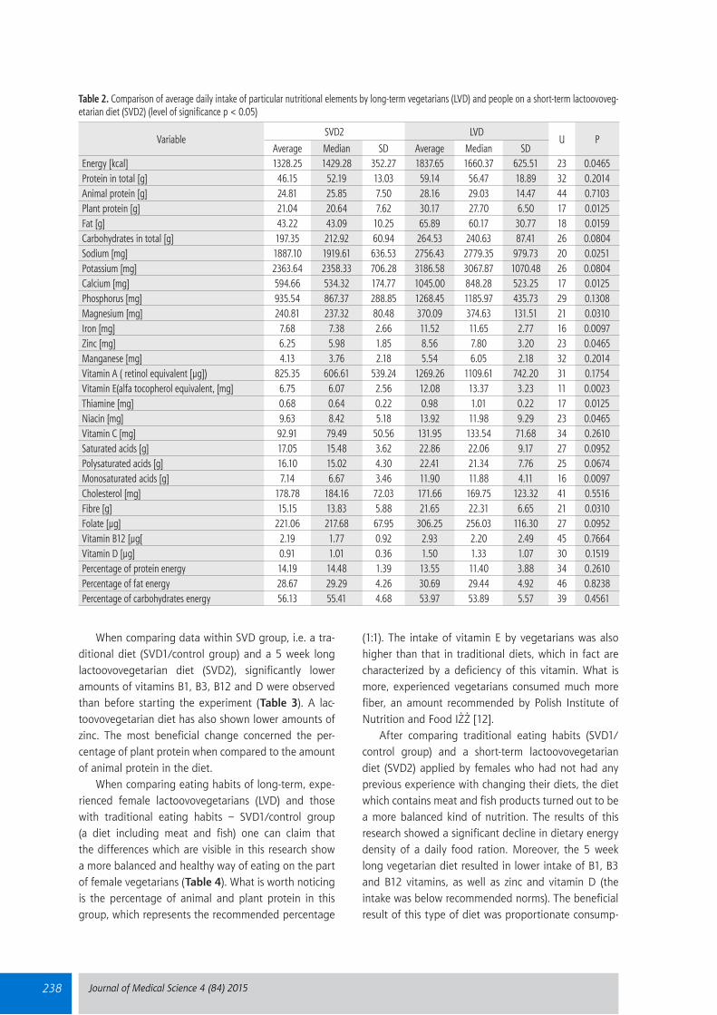
238 Journal of Medical Science 4 (84) 2015
When comparing data within SVD group, i.e. a tra-ditional diet (SVD1/control group) and a 5 week long lactoovovegetarian diet (SVD2), significantly lower amounts of vitamins B1, B3, B12 and D were observed than before starting the experiment (Table 3). A lac-toovovegetarian diet has also shown lower amounts of zinc. The most beneficial change concerned the per-centage of plant protein when compared to the amount of animal protein in the diet.
When comparing eating habits of long-term, expe-rienced female lactoovovegetarians (LVD) and those with traditional eating habits – SVD1/control group (a diet including meat and fish) one can claim that the differences which are visible in this research show a more balanced and healthy way of eating on the part of female vegetarians (Table 4). What is worth noticing is the percentage of animal and plant protein in this group, which represents the recommended percentage
(1:1). The intake of vitamin E by vegetarians was also higher than that in traditional diets, which in fact are characterized by a deficiency of this vitamin. What is more, experienced vegetarians consumed much more fiber, an amount recommended by Polish Institute of Nutrition and Food IŻŻ [12].
After comparing traditional eating habits (SVD1/control group) and a short-term lactoovovegetarian diet (SVD2) applied by females who had not had any previous experience with changing their diets, the diet which contains meat and fish products turned out to be a more balanced kind of nutrition. The results of this research showed a significant decline in dietary energy density of a daily food ration. Moreover, the 5 week long vegetarian diet resulted in lower intake of B1, B3 and B12 vitamins, as well as zinc and vitamin D (the intake was below recommended norms). The beneficial result of this type of diet was proportionate consump-
Table 2. Comparison of average daily intake of particular nutritional elements by long‑term vegetarians (LVD) and people on a short‑term lactoovoveg‑etarian diet (SVD2) (level of significance p < 0.05)
VariableSVD2 LVD
U PAverage Median SD Average Median SD
Energy [kcal] 1328.25 1429 .28 352.27 1837.65 1660.37 625.51 23 0.0465Protein in total [g] 46.15 52.19 13.03 59.14 56.47 18 .89 32 0.2014Animal protein [g] 24 .81 25.85 7.50 28 .16 29.03 14 .47 44 0.7103Plant protein [g] 21.04 20.64 7 .62 30.17 27.70 6.50 17 0.0125Fat [g] 43 .22 43.09 10.25 65.89 60.17 30.77 18 0.0159Carbohydrates in total [g] 197.35 212 .92 60.94 264.53 240.63 87 .41 26 0.0804Sodium [mg] 1887.10 1919 .61 636.53 2756.43 2779.35 979 .73 20 0.0251Potassium [mg] 2363 .64 2358.33 706.28 3186.58 3067.87 1070.48 26 0.0804Calcium [mg] 594.66 534.32 174 .77 1045.00 848 .28 523.25 17 0.0125Phosphorus [mg] 935.54 867 .37 288.85 1268.45 1185.97 435.73 29 0.1308Magnesium [mg] 240.81 237 .32 80.48 370.09 374 .63 131.51 21 0.0310Iron [mg] 7 .68 7 .38 2 .66 11.52 11.65 2 .77 16 0.0097Zinc [mg] 6.25 5.98 1.85 8.56 7.80 3.20 23 0.0465Manganese [mg] 4 .13 3 .76 2 .18 5.54 6.05 2 .18 32 0.2014Vitamin A ( retinol equivalent [μg]) 825.35 606.61 539.24 1269 .26 1109.61 742.20 31 0.1754Vitamin E(alfa tocopherol equivalent, [mg] 6.75 6.07 2.56 12.08 13 .37 3 .23 11 0.0023Thiamine [mg] 0.68 0.64 0.22 0.98 1.01 0.22 17 0.0125Niacin [mg] 9 .63 8 .42 5.18 13 .92 11 .98 9 .29 23 0.0465Vitamin C [mg] 92 .91 79 .49 50.56 131.95 133.54 71 .68 34 0.2610Saturated acids [g] 17.05 15.48 3 .62 22 .86 22.06 9 .17 27 0.0952Polysaturated acids [g] 16.10 15.02 4.30 22 .41 21 .34 7 .76 25 0.0674Monosaturated acids [g] 7 .14 6 .67 3 .46 11.90 11 .88 4 .11 16 0.0097Cholesterol [mg] 178 .78 184 .16 72.03 171 .66 169.75 123 .32 41 0.5516Fibre [g] 15.15 13 .83 5.88 21.65 22 .31 6.65 21 0.0310Folate [μg] 221.06 217 .68 67.95 306.25 256.03 116.30 27 0.0952Vitamin B12 [μg[ 2 .19 1 .77 0.92 2 .93 2.20 2 .49 45 0.7664Vitamin D [μg] 0.91 1.01 0.36 1.50 1 .33 1.07 30 0.1519Percentage of protein energy 14 .19 14 .48 1 .39 13.55 11.40 3 .88 34 0.2610Percentage of fat energy 28 .67 29 .29 4 .26 30.69 29 .44 4 .92 46 0.8238Percentage of carbohydrates energy 56.13 55.41 4 .68 53.97 53.89 5.57 39 0.4561

239The benefits and risks of short-term diet changes on the example of the use a 5-week long lactoovovegetarian diet. Analysis...
tion of animal and plant protein. On analyzing two vari-ants of lactoovovegetarian diets: short-term (LVD) and 5 week long (SVD2), it has been noticed that females who do not have any previous experience with prepar-ing and composing vegetarian meals consumed insuf-ficient amounts of basic microelements and vitamins, specifically calcium, magnesium, zinc, iron, B vitamins of group B, D vitamins and polysaturated fatty acids (Tables 3, 4).
Each tested group made some nutrition mistakes which resulted in insufficient intake of vitamin D and iron, as well as too low intake of vitamin E and cal-cium and magnesium (in the case of traditional and short-term vegetarian diets) (Table 5) and excess of phosphorus in each variant of the diet in question. The ratio of sodium and potassium content in the diet should also be taken into serious consideration.
Discussion
A properly balanced lactoovovegetarian diet does not cause a greater risk of nutrient deficiencies than in the case of a diet rich in meat and fish products [2]. This fact can be proven by this research based on an analy-sis of nutritional surveys of long-term experienced veg-etarians. The analysis has shown that the level of bal-ance and health benefits of their diet were higher than in the two other groups when it comes to daily con-sumption of nutritional antioxidants, such as vitamin E and fiber. Some deficiencies that have been observed concerned fewer nutrients than in the case of the other two groups. As noted in the introduction to this paper, this type of diet does not rule out products of animal origin which are rich source vitamins of group B and calcium. Minerals such as magnesium, iron, phospho-rus, manganese, zinc and copper can be found in many
Table 3. Comparison of average daily intake of particular nutritional elements in the duration of traditional diet (SVD1/control group) and lactoovoveg‑etarian diet (SVD2) (level of significance p < 0.05)
VariableSVD1 SVD2
T PAverage Median SD Average Median SD
Energy [kcal] 1732 .42 1661 .92 621.80 1328.25 1429 .28 352.27 7 0.0663Protein in total [g] 65.84 60.12 20.50 46.15 52.19 13.03 3 0.0209Animal protein [g] 42 .49 38.09 12.70 24 .81 25.85 7.50 1 0.0109Plant protein [g] 22 .89 19.08 9 .92 21.04 20.64 7 .62 20 0.7671FAT [g] 61 .12 53.60 25.06 43 .22 43.09 10.25 7 0.0663Carbohydrates in total [g] 233 .72 213 .76 81 .61 197.35 212 .92 60.94 12 0.2135Sodium [mg] 2565.32 2590.00 831 .76 1887.10 1919 .61 636.53 8 0.0858Potassium [mg] 2902.09 2756.65 670.53 2363 .64 2358.33 706.28 7 0.0663Calcium [mg] 738.10 724 .37 295.06 594.66 534.32 174 .77 11 0.1731Phosphorus [mg] 1167 .86 1090.13 272 .14 935.54 867 .37 288.85 6 0.0506Magnesium [mg] 294.54 278 .33 69.70 240.81 237 .32 80.48 8 0.0858Iron [mg] 9.59 8 .73 2 .69 7 .68 7 .38 2 .66 7 0.0663Zinc [mg] 8 .27 8 .38 1.80 6.25 5.98 1.85 3 0.0209Manganese [mg] 4.15 4 .37 1.75 4 .13 3 .76 2 .18 21 0.8590Vitamin A(retinol equivalent/ [μg]) 815.49 668.08 420.81 825.35 606.61 539.24 21 0.8590Vitamin E (alfa‑tocopherol equivalent [mg]) 7.60 7.02 3.03 6.75 6.07 2.56 16 0.4413Thiamine [mg] 0.95 0.87 0.32 0.68 0.64 0.22 3 0.0209Niacin [mg] 16 .76 14.04 5.81 9 .63 8 .42 5.18 3 0.0209Vitamin C [mg] 109.34 75.39 89 .34 92 .91 79 .49 50.56 20 0.7671Saturated acids [g] 23 .18 21.70 9 .91 17.05 15.48 3 .62 12 0.2135Monosaturated acids [g] 24 .49 22.05 10.50 16.10 15.02 4.30 6 0.0506Polysaturated acids [g] 8 .77 8.08 3 .86 7 .14 6 .67 3 .46 14 0.3139Cholesterol [mg] 228.60 201.42 89.90 178 .78 184 .16 72.03 6 0.0506Fibre [g] 14.60 14 .11 3 .66 15.15 13 .83 5.88 22 0.9528Folate [μg] 243 .28 216 .19 92 .86 221.06 217 .68 67.95 15 0.3743Vitamin B12 [μg] 3 .28 2 .64 1 .42 2 .19 1 .77 0.92 0 0.0077Vitamin D [μg] 2 .66 1 .76 2 .11 0.91 1.01 0.36 1 0.0109Percentage of protein energy 15.81 16.85 2.56 14 .19 14 .48 1 .39 10 0.1386Percentage of fat energy 30.71 30.82 3 .69 28 .67 29 .29 4 .26 13 0.2604Percentage of carbohydrates energy 52.03 50.58 6.05 56.13 55.41 4 .68 9 0.1097

240 Journal of Medical Science 4 (84) 2015
food products of both animal and plant origin. This research has shown that a short-term vegetarian diet has a significant influence on testees’ insufficient sup-ply of nutrients as compared to a traditional diet and a long-term vegetarian diet.
One of such nutrients is zinc, the amount of which was not sufficient in the case of volunteers who under-went our dietary intervention. Products rich in this ele-ment include: brown bread, buckwheat, eggs, rennet
cheese, but also meat and liver (Table 6). Eliminating the last two on the list from one’s diet without find-ing a proper substitute may result in lower zinc intake. Moreover, this research has shown that volunteers examined both before and during their diets consumed insufficient amounts of calcium, iron and magnesium, elements which are present in dairy products, eggs, whole grains, groats, fruit and vegetables. As a conse-quence, eliminating meat and fish (rich in zinc) and eat-
Table 4. Mean value of daily intake of particular minerals and vitamins among females on a traditional diet and a vegetarian diet, recommended norms (level of significance p < 0.05)
Nutrient (average daily intake)Traditional diet
(SVD1/control group)Long‑term vegetarian
diet (LDV)5‑week‑long
lactovegetarian diet (SVD2)Recommended dietary
allowances for women [12]Vitamin B (thiamine) 0.94 0.98 0.68 1 .1Vitamin B3 (niacin) 16 .76 13 .92 9 .62 14Vitamin B12 [μg] 3 .27 2 .93 2 .18 2 .4Vitamin D [μg] 2.65 1.5 0.90 5Zinc [mg] 8 .26 8.56 6.25 8Animal protein [g] 42 .48 28 .16 24.80 0.45g/kg/ body weight /dailyProtein in total [g] 65.84 59.14 46 .14 0.9g/kg body weight/dailyMagnesium [mg] 294.53 370 240.80 320Iron [mg] 9.58 11.52 7 .67 18Calcium [mg] 738.09 1045 594.65 1000Sodium [mg] 2565.31 2756 1887 .1 1500Potassium [mg] 2902.09 3186.5 2363 .64 4700Phosphorus [mg] 1167 .8 1268 .4 935 700Manganese [mg] 4 .14 5.54 4 .12Vitamin A [μg] 815.48 1269 .2 825.35 700Vitamin C [mg] 109.34 131.95 92 .91 75Vitamin E [mg] 7.60 12.08 6.75 8
% of caloric requirement covered by carbohydrates, protein and fats
Protein:15.81Fats:30.71
Carbohydrates:52.03
11.4029 .4453.89
14 .1928 .6756.13
<15%<35%
50–70%Dietary energy density [kcal] 1732 1838 1328 ‑
Table 5. Types of nutritional deficiencies during nutritional intervention (groups SVD1/control group and SDV2) and in log‑term lactoovovegetarian diet (LVD)
Analysis of nutrition interviews in each group Nutrients consumed in insufficient amounts Health benefits
LVD (long‑term vegetarian diet) Potassium, iron, vitamin D, B1High fibre and vitamin E content, beneficial ratio of plant and animal protein consumed
SVD1 (traditional diet)Potassium, iron, vitamin D, vitamin B1, vitamin
E, calcium, magnesiumLower risk of vitamin B3 and B12 deficiencies
SVD2 (5 week long vegetarian diet)B1, B3, B12, D, zinc, magnesium, iron, calcium,
potassium, vitamin EBeneficial ratio of plant and animal protein
consumed
Table 6. Zinc, vitamin B1 [mg], B3 [mg], B12 [μg], and vitamin D [IU] content in particular food products [per 100 g]
Zinc [16]Veal liver (8.40), Ementaler cheese (4.05), beans (3.77), buckwheat (3.50), pork shoulder (3.11), oatmeal (3.10), rye whole‑wheat
bread (2.54), hen’s eggs (1.76)
B1 vitamin [21]Sunflower seeds (1.318), red lentils (1.072), pork (0.98), pistachio nuts (0.82), pea seeds (0.77), millet (0.73), white beans (0.67),
peanuts (0.66), buck‑wheat (0.54) B3 vitamin [21] Peanuts (17.2), liver (13.6), pork (0.8–5.6), trout (8.4), plaice (8.3), cheese (1.2), parsley (1.2)B12 vitamin [22] Liver (25–110), fish (1–15), milk and milk products (0.4–2.2), eggs (1.6) and meat (0.6–1.2)Vitamin D [22, 23] Fresh eel (1200), fresh cod (480), pickles salmon (540), egg yolk (54), cheese (7.6–28)

241The benefits and risks of short-term diet changes on the example of the use a 5-week long lactoovovegetarian diet. Analysis...
ing habits which do not include sufficient amounts of meat-free and rich in zinc products mentioned before led to problems with a balanced amount of zinc in a vegetarian diet. The research also shows a signifi-cantly lower supply of vitamins of group B i.e. B1, B3, B12 (during the 5 week long vegetarian diet), which in turn shows too small consumption of meat-free prod-ucts such as groat, whole grains, seeds and legumes, nuts (vitamin B1, B3) and dairy products (vitamin B12) (Table 6). Lower supply of both zinc and B1, B3, B12 vitamins confirms the fact that a properly balanced veg-etarian diet should include a higher consumption of the food products mentioned before [13, 14]. It is worth noticing that vitamin B3 can be synthesized endoge-nously. Tryptophan is a substrate in this reaction [13]. It has been suggested that the biosynthesis of niacin from this amino acid is sufficient to supply the organism with a proper amount of vitamin B3. However, it is neces-sary to consume 100 g of protein daily. Volunteers did not include such an amount before or after the experi-ment. B12 vitamin is present in products of animal ori-gin exclusively. When choosing a vegetarian diet, one should pay particular attention to the consumption of dairy and eggs which substitute meat, which in turn is the primary source of cobalamin (Table 6) [15].
As far as other minerals are concerned, this research has not shown any significant differences in their amounts for those on a vegetarian diet as com-pared to their diets preceding the experiment. Numer-ous sources point out, nevertheless, that a properly bal-anced vegetarian diet supplies more antioxidant A, C, E vitamins in comparison to a traditional diet, which confirms our results for long-term vegetarians [7]. This is due to the fact that a vegetarian diet is richer in fruit and vegetables than a traditional one. Having no prop-er eating habits before undergoing the vegetarian diet a low intake of fruit and vegetables can influence nutri-tional behavior while diet is in progress. A relatively small amount of these nutrients in both traditional and 5 week long vegetarian diets correlates with a relative-ly small amount of fibers.
Another aspect worth noticing is the excessive amount of phosphorus, both before and during the experiment, as well as in the case of the long-term vegetarian diet (Table 4). Phosphorus is present in many food products [16] and its excess in one’s organ-ism is quite common. It might be suggested, thus, that a diet which eliminates meat and fish products does not influence total phosphorus content, as this element is present to a high extent in seeds and whole grain products.
The research has also shown higher amounts of potassium as compared to those supplied in a tradi-tional diet. Consumption of this element was insuffi-cient in all three types of diets discussed in this paper. Compared to a high concentration of sodium, deficien-cy of potassium was quite significant. However, studies of long-term vegetarians showed that vegetarian diets should include relatively higher amounts of potassium than diets rich in meat and fish. This may be achieved by including dry and fresh fruit and vegetables in one’s diet.
Calcium deficiencies in Poles’ diets are quite com-mon [17]. It has been calculated that average daily intake of this element equals 50% RDA [18]. The Table 4 confirms that traditional eating habits in most cases do not supply one’s organism with sufficient amounts of calcium required for proper metabolism. Here, the amount of calcium consumed in a traditional diet and in a 5 week long vegetarian diet did not differ and in both cases was below the minimum. However, nutritional surveys with long-term vegetarians con-firmed the fact that a lactoovovegetarian diet which included a higher supply of dairy products, as com-pared to a diet rich in meat and fish, does cover the daily requirements of this nutritional element.
This research has also shown that all tested group did not receive enough vitamin D with their diets. Despite the fact that 90% [12] of this compound is syn-thesized endogenously, it is claimed that proper sup-ply of this vitamin is indispensable to maintain proper calcium phosphorus ratio of an organism. Vitamin D is present, among others, in fish, eggs but also in prod-ucts enriched by this element, as well as in mushrooms [19] (Table 6). When concentration of vitamin D and calcium in blood is not sufficient, one might suspect that metabolic processes of these two elements may be disturbed, which might result in calcium absorp-tion disorders and inadequate bone mineralization and some cell changes [20].
In both cases of the lactoovovegetarian diet, i.e. a long-term and a short-term example, the analysis of nutritional surveys showed a beneficial change in the amount of consumed animal protein in relation to plant protein. Adequate supply of animal protein is connect-ed with providing the organism with bigger amounts of unsaturated fatty acids and eliminating saturated fats at the same time.
This research also shows a relatively low dietary energy density, especially in the case of volunteers undergoing a dietary change. In all cases, nutritional surveys were performed on women who described the

242 Journal of Medical Science 4 (84) 2015
level of their physical activity as low, which explains the data. A significant difference between the dietary energy density before choosing a vegetarian diet as compared to data collected during the dietary change proves that these volunteers’ eating habits had not been properly balanced and as a consequence, accounts for the deficient supply of all aforementioned nutrients.
Conclusion
A short-term change of eating habits from a traditional diet into a lactoovovegetarian one by those who do not have previous experience in planning vegetarian meals may result in insufficient supply of numerous nutrients, especially calcium, iron, magnesium and group B vita-mins deficiencies. This is mainly due to the fact that those people do not know how to compile a balanced vegetarian diet. In order to prevent them from all the deficiencies mentioned earlier, it is necessary to pro-vide them with regular assistance and consultations of a dietician.
A properly balanced lactoovovegetarian diet, even one applied over many years, may turn out to be a very beneficial eating habit, especially for those at risk of diabetes or heart disease. Consumption of prod-ucts which comprise this diet, i.e. whole grains, groats, rice, seeds and vegetables and fruit may significantly lower the risk of those diseases. This diet may also have a positive influence on the supply of antioxidants, especially vitamin E. Big dietary awareness and expe-rience in preparing vegetarian meals (which results in adequate supply of dairy and eggs) lowers the risk of many nutritional deficiencies, such as those of calcium.
This research has also proven that each of the diets discussed here may result in deficiencies of some nutri-ents. Two of such nutrients include vitamin D and iron.
Limitations of the study include small research group as well as inclusion of only female volunteers. Therefore results of this study cannot be attributed to the general population. Moreover the studies did not include any biochemical analysis that could assess the actual nutritional status of respondents.
Acknowledgements
Conflict of interest statementThe authors declare that there is no conflict of interest in the authorship or publication of contribution.
Funding sourcesThis study was founded by Poznan University of Medical Sci-ences, grant number 502-14-01125184-10182.
ReferencesBurke LE, Hudson AG, Warziski MT, Styn MA, Music E, 1. Elci OU, Sereika SM. Effects of a vegetarian diet and tre-atment preference on biochemical and dietary variables in overweight and obese adults: a randomized clinical trial. Am J Clin Nutr. 2007 Sep;86(3):588–596.Craig WJ, Mangels AR, American Dietetic Association. 2. Position of the American Dietetic Association: vegetarian diets. J Am Diet Assoc. 2009 Jul;109(7):1266–1282.Fraser G.E. Vegetarian diets: what do we know of their 3. effects on common chronic diseases? Am J Clin Nutr. 2009 May;89(5):1607S-1612S.Medkova IL, Mosiakina LI, Biriukova LS. Estimation of 4. action of lactoovovegetarian and vegan diets on blood level of atherogenic lipoproteins in healthy people. Vopr Pitan. 2002;71(4):17–9.Newby PK, Tucker KL, Wolk A. Risk of overweight and 5. obesity among semivegetarian, lactovegetarian, and vegan women. Am J Clin Nutr. 2005 June;81(6):1267–1274.Lisowska A, Chabasińska M, Przysławski J, Schlegel-6. -Zawadzka M, Mądry E, Walkowiak J. The influen-ce of 24-months of lactoovovegetarian and vegan diet on nutritional status, energy and macronutrient intake. Pediatr Współcz. 2010;12(2):121–125.Leitzmann C. Vegetarian diets: what are the advantages? 7. Forum Nutr. 2005;57:147–156.Sobate J. The contribution of vegetarian diets to human 8. health. Forum Nutr. 2003;56:218–220.Larsson CL, Johansson GK. Dietary intake and nutritional 9. status of young vegans and omnivores in Sweden. Am J Clin Nutr. 2002 July;76(1):100–106.Ball MJ, Bartlett MA. Dietary intake and iron status of 10. Australian vegetarian women. Am J Clin Nutr. 1999 Sep;70(3):353–358.Kim MK, Cho SW, Park YK. Long-term vegetarians have 11. low oxidative stress, body fat, and cholesterol levels. Nutr Res Pract. 2012 Apr;6(2):155–161.Jarosz M (editor). Normy żywienia dla populacji polskiej 12. – nowelizacja. Warszawa: Instytut Żywności i Żywienia; 2012.Nagalski A, Bryła J. Zastosowanie niacyny w terapii. 13. Postępy Hig Med Dośw. 2007 May;61:288–302.Tylicki A, Siemieniuk M. Tiamina i jej pochodne w regula-14. cji metabolizmu komórek. Postępy Hig Med Dośw. 2011 July;65:447–469.Chabasińska M, Przysławski J, Lisowska A, Schlegel-Za-15. wadzka M, Grzymisławski M, Walkowiak J. Typ i czas sto-sowania diety wegetariańskiej a surowicze stężenie wita-miny B12. Prz Gastroenterol. 2008;3(2):63–67.Kunachowicz H, Nadolna I, Iwanow K, Przygoda B. War-16. tość odżywcza wybranych produktów spożywczych. War-szawa: Wydawnictwo Lekarskie PZWL; 1997.Sobaś K, Wądołowska L, Słowińska MA, Człapka-Matya-17. sik M, Niedźwiedzka E, Szczepańska J. Analiza charakte-rystycznych modeli spożycia wapnia przez pary rodzin-ne matka-córka. Probl Hig Epidemiol. 2011 Jan;92(1): 54–62.Paradowska-Stankiewicz I, Trafalska E, Grzybowski 18. A. Realizacja zapotrzebowania na wybrane witaminy i składniki mineralne w diecie młodzieży. Nowa Medy-cyna. 2000;12.

243The benefits and risks of short-term diet changes on the example of the use a 5-week long lactoovovegetarian diet. Analysis...
Siwulski M, Sobieralski K, Sas-Golak I. Wartość odżywcza 19. i prozdrowotna grzybów. Żywn Nauka Technol Jakość. 2014;1(92):16–28.Tukaj C. Właściwy poziom witaminy D warunkiem zacho-20. wania zdrowia. Postępy Hig Med Dośw. 2008;62:502–510.Ziemlański Ś (editor). Normy żywienia człowieka. Fizjo-21. logiczne podstawy. Warszawa: Wydawnictwo Lekarskie PZWL; 2001; 211–280. Kunachowicz H, Nadolna I, Przygoda B, Iwanow K. Tabe-22. le składu i wartości odżywczej żywności. Warszawa: Wydawnictwo Lekarskie PZWL; 2005.Charzewska J, Chlebna-Sokół D, Chybicka A, Czech-Ko-23. walska J, Dobrzańska A, Helwich E, Imiela JR, Karczma-rewicz E, Książyk JB, Lewiński A, Lorenc RS, Lukas W, Łukaszkiewicz J, Marcinowska-Suchowierska E, Milanow-ski A, Milewicz A, Płudowski P, Pronicka E, Radowicki S, Ryżko J, Socha J, Szczapa J, Weker H. Zalecenia dotyczą-
Correspondence address:Marta Pelczyńska (Kramkowska)
Division of Biology of Civilization-Linked DiseasesDepartment of Chemistry and Clinical Biochemistry
Poznan University of Medical Sciences6 Świecickiego Str., 60-781 Poznan, Poland
phone: +48 618 546476fax: +48 618 546477
email: [email protected]
ce profilaktyki niedoborów witaminy D w Polsce. Stan-dardy Medyczne/Pediatia. 2009;6:875–879.
Acceptance for editing: 2015-11-10 Acceptance for publication: 2015-12-31

244 Journal of Medical Science 4 (84) 2015
© 2015 by the author(s). This is an open access article distributed under the terms and conditions of the Creative Commons Attribution (CC BY-NC) licencse. Published by Poznan University of Medical Sciences
DOI: https://doi.org/10.20883/medical.e6
Legal aspects of a healthy diet for children.Comments on the grounds of the directive on foodstuffs in schoolsMonika Urbaniak
Faculty of Health Sciences, Department of Medical Law, Poznan University of Medical Sciences, Poland
Introduction
The aim of this article is to analyze legal solutions for the availability of foodstuffs in schools, binding from Sep 1, 2015. These new solutions are intended to intro-duce into trade in school shops and canteens so called healthy food. Like any revolutionary changes, they generate a series of questions regarding their confor-mity with the Polish Constitution [1] and also contrib-ute to the discussion on overall solutions intended to fight against obesity in children and young people in a broader perspective. The new regulations are pro-tested against by entrepreneurs who run businesses in schools as they state that the list of products approved for sale is too restrictive, due to the fact that it elimi-nates from students’ diets products containing too much sugar. They also point out that students will con-tinue to provide themselves with so called junk food due to the proximity of regular groceries to schools or due to a lack of changes in nutritional habits at home which results in bringing unhealthy food to school.
The Directive issued by the Minister of Health on Aug 26, 2015 on groups of foodstuffs intended to be sold to children and young people in education sys-tem units and requirements for foodstuffs used with-in collective feeding of children and young people in those units [2], which entered into force on Sep 1, 2015, eliminated from school shops food which is con-sidered unhealthy. It needs to be pointed out that the Directive was published in the Journal of Laws of Aug 28, 2015 i.e. three days before it entered into force. The entrepreneurs point out that such a short period of vacatio legis makes it difficult for the addressees of those provisions to adapt to the new legal conditions. The Constitutional Tribunal in its judgment of March 2, 1993 [3] emphasized that “the principle of the rule of law requires that a modification of law binding so far which implies unfavorable effects for the legal situation of entities be in principle introduced under the regime of interim provisions or at least with a proper vacatio legis as they let the entities involved adapt to the new
ABSTRACT
The aim of this article is to analyze legal solutions for the availability of foodstuffs in schools, binding from Sep 1, 2015, targeted to introduce into school shops and canteens so-called healthy food. The Directive issued by the Minister of Health on Aug 26, 2015 on groups of foodstuffs intended to be sold to children and young people in education system units and requirements for foodstuffs used within collective feeding of children and young people in those units eliminated from school shops provide a list of food which is considered unhealthy. The adopted solutions are an introduction to the legal statutory fight against obesity in children and young people and enhance the protection of the health of children at pre-school and school age by limiting access within kindergartens, schools and tutelary-educational institutions to foodstuffs containing significant quantities of ingredients not recommended for their development.
Keywords: food, obesity, liberty of economic activity, children and young people.
O R I G I N A L PA PE R

245Legal aspects of a healthy diet for children.Comments on the grounds of the directive on foodstuffs in schools
legal conditions. The legislator may abandon them – and decide to introduce directly (immediately) a new law – if to do so is justified by an important public interest which cannot be outweighed by the interest of an individual”. In this case, although the modification of binding legal provisions had unfavorable effects on businesses such as school shops, the legislator decided not to extend the period which would allow them to adapt to the new legal situation. Pursuant to the pro-visions of Article 2 of the Act of Nov 28, 2014 on the modification of the Act on food and feeding safety [4], the date of entering into force of Article 52c, which is the basis to issue the Directive in discussion was deter-mined to be Sep 1, 2015. Considering the necessity to adapt by businesses to the new legal situation, the leg-islator passed a relatively long period of vacatio legis. However, there is still a question about the lawfulness of the basis of the Directive issued. If the Act of Nov 28, 2014 on the modification of the Act on food and feeding safety determined the date of its entrance into force to be Sep 1, 2015 and the Directive of the Min-ister of Health was issued on Aug 26, 2015 pursuant to Article 52c of the Act on food and feeding safety, this means that it was issued on the basis of an act which had not yet entered into force. Under §127 of legal technicality [5] a directive should enter into force on the date of entrance into force of the act being the basis for the issuance of the directive. Also, under §128, section 1 of the legal technicality a directive may be issued after the act containing a provision which authorizes its issuance is published and before the act enters into force. In such a ase, the date of entrance into force of a directive is determined as a date no sooner than the date of entrance into force of the act that authorizes the issuance of such a directive. Consid-ering the above, it needs to be stated that the direc-tive in discussion conforms with legal technicality. The rules of legislation proceeding provided for in the act are directly binding for the government legislator [6]. Moreover, Article 7 of the Act of July 20, 2000 on the publication of normative deeds and some other legal deeds [7] clearly states that normative deeds issued on the basis of acts may be published within the period between the date of the publication of the act and the date of its entrance into force; such a deed may not enter into force before the act.
Considering the fact that directives are issued on the basis of acts and in order to execute them and their most important function is to enable the execution of the provisions of the act, it is obvious that the execu-tory provisions should enter into force together with
the new basic provisions [8]. Furthermore, it needs to be remembered that under §13 of the legal technical-ity, together with a draft version of an act, the draft directives which are fundamental for its execution are drawn up. The Directive under discussion undoubtedly is a deed on which the execution of the provisions of the act depends. The Constitution in Article 92 sec-tion 2 provides that directives are issued by authorities specified in the Constitution on the basis of a detailed authorization contained in the act for the purpose of its execution. The authorization should specify the author-ity competent to issue the directive and the scope of matters to be regulated and guidelines regarding the content of the deed. This provision results in the fact that the lawfulness of a directive depends on the accomplishment of the constitutional grounds for its issuance. In the case of the Directive of the Minister of Health of Aug 26, 2015 on groups of foodstuffs intend-ed to be sold to children and young people in educa-tion system units and requirements for foodstuffs used in the collective feeding of children and young people in those units, it needs to be said that the statutory authorization was complete as it specified the author-ity competent to issue it and specified in detail the scope of the matters to be regulated.
Pursuant to the content of §1 of the aforementioned directive, it specifies groups of foodstuffs intended to be sold to children and young people in education sys-tem units. In practice this means that the foodstuffs enlisted in Schedule 1 of the directive cannot be sold in school shops, canteens or vending machines. Moreover, the directive sets forth the requirements for foodstuffs used in the collective feeding of children and young people in education system units. These requirements result from the principles of a rational diet in collective feeding and are based on norms for feeding children and young people and the nutritional and health val-ues of the different foodstuffs.
The adopted solutions are intended, according to the grounds of the draft directive, to enhance the pro-tection of health in children at pre-school and school age by limiting access within kindergartens, schools and tutelary-educational institutions to foodstuffs con-taining significant quantities of ingredients not rec-ommended for their development [9]. It needs to be emphasized that proper diets in children and young people are very important considering the fact that excessive consumption in these groups leads to being overweight or obese caused, among others, by incor-rect diets, well established in the family which means in particular an overly high calorific value for their every-

246 Journal of Medical Science 4 (84) 2015
day diet, too much animal fat and simple sugars accom-panied by the limited physical activity of children and young people [10]. Typical irregularities in the diets of school children and young people are monotony, too much consumption of sugar and sweet things as well as meat and meat products, sweet carbonated drinks, fats and fast foods, combined with too little consump-tion of fruit and vegetables, milk and wholemeal cere-al products, as well as fish [11]. In addition, it needs to be pointed out that disorders of energy balance in the body appear when certain physiological stimuli are removed from the person’s everyday life e.g. when physical effort is limited [12].
The directive was issued on the basis of a statutory authorization referred to in Article 52c section 6 of the Act of Aug 25, 2006 on food and feeding safety [13], modified with the Act of Nov 28, 2014 on the modifi-cation of the Act on food and feeding safety. This Act, in Article 1 point 2, added Part IIA entitled: Foodstuffs and feeding children and young people in education system units. Under Article 52c, section 6 of the Act referred to hereinabove the minister competent for health matters will specify by means of a directive:
the groups of foodstuffs approved for sale to chil- –dren and young people in education system units;the requirements for foodstuffs used in the collec- –tive feeding of children and young people in edu-cation system unitsin consideration of the feeding standards for chil- –dren and young people and the nutritional and health values of foodstuffs.A violation of those provisions will be penalized
with a fine or a civil sanction such as termination of contract. Under Article 52c, section 5 of the Act, if the provisions are violated, the head of a kindergarten, a school head or a head of a unit referred to in Article 2, points 3, 5, 7 of the Act of Sep 7, 1991 on the Edu-cation System (i.e. schools and other educational facili-ties including school youth hostels, centers for reha-bilitation, centers of sociotherapy, special school-edu-cational centers, special educational centers as well as facilities that provide care and education to students during education away from their fixed residence) are authorized to terminate, without notice, the contract with the entity responsible for the sale of foodstuffs or the provision of collective feeding to children and young people, without compensation.
Paternal responsibility for a child is the natural right of each parent. Under Article 48 of the Constitution of the Republic of Poland parents shall have the right to rear their children in accordance with their own convic-
tions. This right, guaranteed in Article 48, section 1, refers to freedom of conscience and belief. Moreover, in Article 72, section 1 the Constitution ensures the protection of the rights of the child. The Constitution recognizes the supreme role of parents in the process of child education and it also covers looking after the child which involves among other things the right to make decisions about the child’s diet. It needs to be said that the democratic legislator should respect the parents’ right to bring up their children in accordance with their convictions i.e. also those which concern the child’s diet. With the introduction of a total ban on the sales of foodstuffs containing excessive quantities of ingredients not recommended for their development, the legislator is interfering too much with the constitu-tional freedom to educate children in accordance with the parents’ own convictions.
Another issue to be considered with respect to the introduction of the new provisions is a question about the lawfulness - in the light of Article 22 of the Con-stitution - of the limitation of the freedom of econom-ic activity by banning the sales of some foodstuffs in education system units. Undoubtedly, the modification of the Act of Aug 25, 2006 on food and feeding safe-ty introduced a limitation into the discussion upon the freedom of economic activity, which was specified in detail in the directive issued by the Minister of Health on the basis of the provisions of the Act. The legislator has the right to limit economic activity for important public reasons, which in its view includes health pre-vention in children and young people.
The principle of the freedom of economic activity is among the fundamental legal principles of the Polish legal order. According to the doctrine of business and administrative law it is one of the fundamental pillars of the economy [14]. However, this principle referred to in Article 20 of the Constitution is not absolute and may be limited on the basis of Article 22 of the Con-stitution pursuant to which a limitation of the freedom of economic activity may be imposed only by means of statute and only for important public reasons. It needs to be emphasized that the provision of Article 22 of the Constitution determines limits to state interference in economic activity. The limits are of two kinds: formal – it says that the limitation upon the freedom of econom-ic activity may be imposed “only by means of statute” and material – saying that they may be imposed “only for important public reasons” [15]. The Constitutional Tribunal in its judgment of Apr 8, 1998 [16] stated that freedom of economic activity may be subject to differ-ent limitations to a larger degree than personal rights

247Legal aspects of a healthy diet for children.Comments on the grounds of the directive on foodstuffs in schools
and freedoms. Undoubtedly, such a value is the pro-tection of health referred to in Article 68 of the Con-stitution and this is an important public reason, which justifies the limitation upon the freedom of economic activity [17]. The conditions of the introduction of limi-tations are set forth in Article 31, section 3 of the Con-stitution under which any limitation upon the exercise of constitutional freedoms and rights may be imposed only by statute, and only when necessary in a demo-cratic state for the protection of its security or public order, or to protect the natural environment, health or public morals, or the freedoms and rights of other per-sons. Such limitations shall not violate the essence of freedoms and rights. The principle of proportionality referred to in Article 31, section 3 of the Constitution is an important barrier which prevents rights and free-dom from being limited in an unjustified or excessive manner by the legislative authorities [18]. In conclusion, the limitation upon the freedom of economic activity in order to protect the value of health should be consid-ered acceptable in terms of the principle of proportion-ality. It is also important to point out that the adopted solution is proportional to its desired purpose because it is impossible to achieve with other, less burdensome methods. Also, the opinion of the government about a draft act - filed by MPs - about the modification of the act on food and feeding safety (files no. 1127 and 1127A) is worth considering. This opinion states that in the statement of reasons for the MPs’ draft act it has not been proven that the requirements of propor-tionality have been met and it is doubtful whether the suggested solutions are capable of guaranteeing the achievement of the desired purpose, considering the fact that children and young people willing to provide themselves with those foodstuffs subject to limitations will be able to acquire them out of units covered by bans, also during breaks between lessons [19].
The legislator assessed in legal terms two values: the freedom of economic activity and the protection of children and young people’s health by introducing a ban on the sales of determined foodstuffs in a situ-ation where, in the legislator’s view, the full exercise of rights was not possible. The limitation upon the freedom of economic activity should be imposed by means of statute, which took place in the Act on food and feeding safety which in Article 52c indicated an authority competent to issue the directive.
On the basisi of the outlined legal status arises the question of whether the limitation upon the free-dom of economic activity for businesses selling food in schools is adequate for the protected values and if
it is possible to achieve the purpose set by the legisla-tor by introducing other means aimed to limit the con-sumption of unhealthy foodstuffs than only by banning their sales in school shops. A possible limitation upon the freedom of economic activity of businesses con-ducting economic activity in schools and other educa-tional centers needs to be considered. After the modi-fications enter into force, they will be the only group not to benefit from the ban but on whom the legislator imposes special duties within the scope of the distribu-tion of so called healthy food in schools. It needs to be agreed with the legislator that the introduced bans are targeted to accomplish vital social purposes such as health protection, although the introduction of a statu-tory ban on the sales of certain foods is not a means necessary to achieve the purpose set by the legislator. Maybe to achieve the desired purpose, it would be suf-ficient to increase children and young people’s aware-ness through education on healthy dieting or also to increase the volume of physical activity in schools. It is easy to imagine children and young people having access to foodstuffs containing significant quantities of ingredients not recommended for their development at home, after school or by going during school breaks to grocery stores located near their school-educational facilities. It seems that the achievement of the purpose, aside from the adopted legal solutions, could be guar-anteed with obesity prevention programs involving, as well as proper dieting, also increased volumes of physi-cal activity for children and young people. Some Pol-ish cities (e.g. in Gdańsk) have introduced programs to fight against obesity in children involving examining children in determined age groups for early detection of civilization diseases.
In conclusion, it needs to be said that the limita-tion upon the freedom of economic activity imposed in the Act on food and feeding safety and the directive issued on the basis thereof is legally acceptable as the introduction of the solutions discussed herein above by the legislator was motivated by the need to protect children and young people’s health, which is a value referred to in Article 31, section 3 of the Constitution. In the margin of this discussion, however, it needs to be added that no later than a month after the directive entered into force, buns reappeared in the list of prod-ucts admissible in school shops [20] which prompts the question of whether this is only a liberalization of the adopted provisions or the begining of a return to the situation before the modifications. Let us hope that the imposed limitations will be a successful tool in the fight against obesity in children and young people and will

248 Journal of Medical Science 4 (84) 2015
lead them to learn and preserve good eating habits. However, it needs to be remembered that the solutions adopted by the legislator are not a complete remedy in the fight against obesity in children and young people.
Acknowledgements
Conflict of interest statementThe authors declare that there is no conflict of interest in the authorship or publication of contribution.
Funding sourcesThere are no sources of funding to declare.
ReferencesKonstytucja Rzeczpospolitej Polskiej z dnia 2 kwietnia 1. 199 7 r., Dz. U. 1997 nr 78 poz. 483 ze zm.Rozporządzenie z dnia 26 sierpnia 2015 roku w sprawie 2. grup środków spożywczych przeznaczonych do sprzeda-ży dzieciom i młodzieży w jednostkach systemu oświa-ty oraz wymagań, jakie muszą spełniać środki spożywcze stosowane w ramach żywienia zbiorowego dzieci i mło-dzieży w tych jednostkach, Dz. U. 2015 poz. 1256Orzeczenie TK z dnia 2 marca 1993 roku, sygn.. akt 9/923. Ustawa z dnia 28 listopada 2014 r. o zmianie ustawy 4. o bezpieczeństwie żywności i żywienia, Dz. U. 2015 poz.Rozporządzenie Prezesa Rady Ministrów z dn. 20 czerw-5. ca 2002 r. w sprawie "Zasad techniki prawodawczej", Dz. U. Nr 100, poz. 908Giderewicz S. Pozorne wytyczne w przepisach upoważ-6. niających do wydania rozporządzenia. Przegląd Legisla-cyjny. 2013;4:51.Ustawa z dnia 20 lipca 2000 r. o ogłaszaniu aktów nor-7. matywnych i niektórych innych aktów prawnych, Dz. U. 2015 poz. 1484 t.j.Kaszubowski K. Komentarz do § 127 rozporządzenia 8. w sprawie „Zasad techniki prawodawczej”. In: Bąkowski T, Bielski P, Kaszubowski K, Kokoszczyński M, Stelina J, Warylewski JK, Wierczyński G. Zasady techniki prawo-dawczej. Komentarz do rozporządzenia, Warszawa 2003, www.lex.pl. Accessed: 1.10.2015.https://legislacja.rcl.gov.pl/docs/516/12273657/122956 9. 73/dokument180495.pdf. Accesed: 29.09.2015.Jarosz M (ed.). Zasady prawidłowego żywienia dzieci 10. i młodzieży oraz wskazówki dotyczące zdrowego stylu życia. Instytut Żywności i Żywienia, Warszawa 2008; 29.
Wanat G, Grochowska-Niedworok E, Kardas M, Całyniuk 11. B. Nieprawidłowe nawyki żywieniowe i związane z nimi zagrożenie dla zdrowia wśród młodzieży gimnazjalnej. Hygeia Public Health. 2011;46(3):381.Oblacińska A. Podstawy teoretyczne nadwagi i otyłości 12. u dzieci i młodzieży. In: Oblacińska A. Wspieranie dzie-cka z nadwagą i otyłością w społeczności szkolnej. War-szawa 2013; 7.Ustawa z dnia 25 sierpnia 2006 r. o bezpieczeństwie 13. żywności i żywienia, Dz. U. z 2015 poz. 594.Ciechanowicz-McLean J. Konstytucyjna zasada wolno-14. ści gospodarczej a ochrona środowiska. Gdańskie studia prawnicze. T. XXXI. 2014; 99.Byrski J, Traple E. Konstytucyjność ustawowego uregulo-15. wania maksymalnego poziomu opłaty interchange. Pań-stwo i Prawo. 2013;6:64.Wyrok Trybunału Konstytucyjnego z dnia 8 kwietnia 16. 1998 r., sygn. K 10/97.Banaszak B. Konstytucja Rzeczypospolitej Polskiej. 17. Komentarz. Warszawa 2009; 137.Ogonowski A. Konstytucyjna wolność działalności gospo-18. darczej w orzecznictwie Trybunału Konstytucyjnego. Przegląd Prawa Konstytucyjnego. 2012;1:229–230.Stanowisko Rządu wobec poselskiego projektu ustawy 19. o zmianie ustawy o bezpieczeństwie żywności i żywie-nia (druki nr 1127 i 1127-A), www.senat.gov.pl. Acces-ses: 1.10.2015.http://www.gazetaprawna.pl/artykuly/897124,szefowa 20. -men-wynegocjowalam-powrot-drozdzowek-do-szkol -bede-negocjowac-kawe-i-wiecej-soli.html. Accessed: 3.10.2015
Acceptance for editing: 2015-11-10 Acceptance for publication: 2015-12-31
Correspondence address:Monika Urbaniak
Department of Medical Law4 Rokietnicka Str., 61-806 Poznan, Poland
phone: +48 61 6584201email: [email protected]

249Journal of Medical Science 4 (84) 2015
© 2015 by the author(s). This is an open access article distributed under the terms and conditions of the Creative Commons Attribution (CC BY-NC) licencse. Published by Poznan University of Medical Sciences
DOI: https://doi.org/10.20883/medical.e7
Liposuction-induced metabolic alterations – the effect on insulin sensitivity, adiponectin, leptin and resistinMagdalena Gibas-Dorna1, Piotr Turkowski2, Małgorzata Bernatek3, Kinga Mikrut1, Justyna Kupsz1, Jacek Piąt ek1
1 Department of Physiology, Poznan University of Medical Sciences, 6 Swiecicki Str., 60-781 Poznan, Poland2 Samir Ibrahim Mandala Beauty Clinic, Chwiałkowskiego Str. 28/7, 61-553 Poznan, Poland3 Chair of Social Medicine, Poznan University of Medical Sciences, 5 Rokietnicka Str., 60-806 Poznan, Poland
Introduction
According to International Society of Aesthetic Plastic Surgery liposuction was the second most frequently performed aesthetic operation in 2013. As with any surgery liposuction carries risks, however, in recent years, improved techniques have made liposuction safer, easier, and less painful. Thus, any intervention that immediately decreases adiposity and is relatively safe could be a viable method not only for aesthetic purpose but also for increasing the efficiency of insulin and improving metabolic profile, especially when com-bined with regular exercise and proper diet. Beneficial change in adipose tissue metabolism may be achieved directly or indirectly by modified release of metaboli-cally active hormones/adipokines, modulated response to these hormones or by increased activity of sym-pathetic afferents to the fat cell. This paper presents
the current state of knowledge on liposuction-induced metabolic changes with regard to release of major adi-pokines.
Types, distribution and function of adipose tissue
Adipose tissue is a metabolically active organ involved in multiple functions. It serves as a buffer against influx of dietary fat and energy depot, plays role in the pro-cesses of satiety control, thermoregulation, reproduc-tion, and exhibits immunomodulatory and endocrine activities. As an endocrine organ adipose tissue secretes hormonally active factors including leptin, adiponectin, resistin, visfatin, interleukin 6 (IL6), tumor necrosis fac-tor (TNF), and many others, and plays role in hormonal metabolism of sex hormones and glucocorticoids [1].
ABSTRACT
Liposuction surgically removes subcutaneous abdominal tissue (SAT) and has almost no effect on visceral abdominal tissue (VAT) depot. However, some authors suggest that deep layers of SAT are functionally similar to VAT and the amount of deep subcutaneous abdominal adipose tissue is strongly related to insulin resistance in a manner nearly identical to that of visceral adiposity. Moreover, SAT determines leptin secretion which indirectly reflects the level of insulin sensitivity in the body. Thus, the immediate removal of SAT could potentially affect metabolic profile of a patient. The current data are conflicting and cannot bring a clear evidence suggesting that liposuction itself results in important metabolic outcomes and, on the other hand, cannot exclude such a possibility. This review summarizes the liposuction-induced metabolic changes with regard to release of major adipokines and insulin sensitivity.
Keywords: liposuction, insulin sensitivity, adiponectin, leptin, resistin, metabolic outcomes.
R E V IE W PA PE R

250 Journal of Medical Science 4 (84) 2015
The division of adipose tissue into two main types, white and brown, reflects functional and anatomical differences. White adipose tissue (WAT) is localized under skin and is designed for energy storage whereas brown adipose tissue (BAT) is present around kidneys, adrenals, aorta, and within mediastinum and neck. BAT plays role in secretion of inflammatory mediators, dissi-pates energy and generates heat. Energetic utilization of fats is regulated by beta-3 adrenergic receptors acti-vation and/or in the mechanism of postprandial ther-mogenesis, which is due to leptin activity and signals from ventromedial hypothalamus in response to post-prandial levels of glucose and insulin. Depending on body’s demands, white adipose tissue acts as a potent buffer maintaining constant level of fatty acids in the circulation by storing lipids in the form of triglycerides and esterified form of cholesterol, and also releasing free fatty acids. WAT is also the major site of leptin and adiponectin production. [2]. The most important local-izations of WAT are visceral abdominal tissue (VAT), subcutaneous abdominal tissue (SAT) and femoral and gluteal subcutaneous regions. Different anatomic dis-tribution of WAT is related with different function, met-abolic and endocrine activity. 2/3 of WAT is present in SAT, while the rest is found around viscera, in the retroperitoneal and gonadal depots, in the mammary glands, liver and skeletal muscles.
Abdominal obesity, also known as a central obesity, results from accumulation of fat in both SAT and VAT and serves as a risk factor for insulin resistance, which in turn favors development of diabetes type II, dyslipi-demia, arteriosclerosis and chronic inflammation with inflammatory cytokines production (eg. TNF alpha, IL6, IL1) and inhibition of anti-inflammatory cytokines release (eg. adiponectin, IL10) [3]. The expression and secretion of adipokines in adipose tissue vary accord-ing to the adipocyte size and number and to the adi-pose tissue depot. For example, adiponectin and leptin expression and secretion are higher in SAT contain-ing larger adipocytes as compared with VAT [4]. Until recently the ideas presenting relationship of regional adiposity with insulin senisitivity/resistance were not consistent and studies differed in the assessment of the importance of WAT and BAT. Some studies have sug-gested that the majority of metabolic activity in the adipose tissue belongs to WAT which is the most criti-cal determinant of insulin sensitivity [5], whereas oth-ers have indicated the dominant role of BAT [6, 7].
The lack of consistent findings is probably attrib-uted to the use of different methodologies (eg. differ-ent type of liposuction procedure), small number of
subjects, not homogenous study groups, differences in patient’s lifestyle (exercise, diet), difficulties in main-taining stable body mass, and low sensitivity of an examination. At the moment, most authors agree that there is a positive correlation between accumulation of VAT and development of insulin resistance, while SAT determines leptin secretion which indirectly reflects the level of insulin sensitivity in the body [8].
Liposuction procedure, metabolic change and maintenance of body weight after surgery
Classic liposuction also known as suction-assisted lipo-suction (SAL) uses aspiration techniques to break down and draw the fat cells out of the body. In this method small cannula is inserted through a small incision and attached to a vacuum device. There are many different types of liposuction according to the volume of infil-tration or wetting solution injected before the surgery: dry, wet, superwet and tumescent technique. Moreover, the surgery could be modified by the new technologies such as power assisted liposuction (PAL), ultrasound assisted liposuction (UAL) and laser assisted liposuction (LAL). According to the volume of solution aspirated liposuction could be divided into two categories: large volume liposuction (> 5 liters aspirated) and small vol-ume liposuction (< 5 liters aspirated). The most popu-lar liposuction types are tumescent and superwet tech-niques with minimal risk of bleeding complications and small volume fluid infusions during surgery. Tumes-cent liposuction is performed under local anesthesia after subcutaneous infusion of fluid (most commonly containing saline, lidocaine, and epinephrine, with or without sodium bicarbonate) in a ratio of 2–3 mL of infiltrate to 1 ml of aspirate; the endpoint of infiltra-tion is tissue turgor [9]. For the best results liposuction candidates should be healthy and physically fit and not more than 20 pounds overweight, their skin should be firm and elastic. The major contraindications include and are not limited to: severe cardiovascular disease, severe coagulation disorders, pregnancy or less than 6 months post-partum, eating disorders, psychiatric problems, morbid obesity, serious life stress within last 6 months [10]. The indications and contraindications for liposuction summarizes Table 1.
Liposuction surgically removes only SAT and has almost no effect on VAT depots. Up to 50% of SAT is localized in the deep layer and, during abdominal lipo-suction, this is the predominant removed tissue. Some

251Liposuction-induced metabolic alterations – the effect on insulin sensitivity, adiponectin, leptin and resistin
authors suggest that deep layers of SAT are function-ally similar to VAT and the amount of deep subcuta-neous abdominal adipose tissue is strongly related to insulin resistance in a manner nearly identical to that of visceral adiposity [11, 12]. The assessment whether liposuction is successful in a longitudinal weight man-agement is the first step toward potential metabolic benefit analysis. In 2001 Commons et al. reviewed 631 cases of liposuction that was performed in the same hospital by the same surgeon over 12 years, with an average follow up 1 year. More than 80% of patients maintained stable body weight within 12 months from surgical procedure [13]. Contrary to this report, there are studies describing rebuilding of removed fat and its redistribution to the abdominal and visceral regions with negative impact on metabolic param-eters and risk factors for coronary artery disease. In prospective analysis of fat distribution Hernandez et al. observed an initial decrease in abdominal SAT (6 weeks after liposuction) with subsequent significant increase in SAT and WAT after 1 year from surgery. Tested metabolic variables (serum adiponectin, free fatty acids, glucose, insulin, triglycerides, sensitiv-ity to insulin) were not significantly changed except an initial drop in leptin level, which after 12 months returned to its basic concentrations. This study, how-ever, was conducted on small study group (n = 14) and the type of surgery was small-volume liposuction (less than 5L of aspirate) [14].
It has been documented that large volume lipo-suction enhances insulin sensitivity markedly, lowers insulinemia and additionally reduces circulating mark-ers of vascular inflammation [15]. The beneficial effect
on insulin sensitivity persists over months from surgery [16, 17].
The number of study participants is one of the major determinants for statistical power and sensitivity of analysis. In a large clinical study (123 obese women) D’Andrea et al. observed positive metabolic changes after 21 and 90 days from large volume liposuction. They noted significantly improved insulin sensitivity, resting metabolic rate, serum adipocytokines, and level of inflammatory markers. The changes were correlated with the decrease in fat mass and waist-hip ratio and remained relatively stable [18]. Rizzo et al. evaluated the effects of dermolipectomy after 40 days from the surgery. They found significant decline in plasma resis-tin, and inflammatory markers (IL6, IL10, TNF) and an increase in plasma adiponectin. Moreover, observed metabolic changes were accompanied by an improve-ment in insulin-mediated glucose uptake, substrate oxi-dation and degree of inflammation [19].
Contrary to these findings, a number of studies reported no, or even diverse effects of liposuction on metabolic profile. They underlined that only negative energy balance induced by diet and/or exercise, not simply a decrease in the mass of adipose tissue, is criti-cal for achieving the metabolic benefits of weight loss. Weight loss in response to energy deficit decreases VAT mass, production of proinflammatory cytokines, size of adipocytes, helps to reduce intrahepatic and myocellu-lar fat, and undoubtedly, these changes bring signifi-cant improvement in most of the metabolic parameters [20]. In addition, some authors report post-liposuction adipose tissue regain and redistribution, preferentially in the abdominal regions of VAT, after months from the
Table 1. General indications and contraindications for liposuction
Indications Absolute contraindications Relative contraindicationsAesthetic indications; reduced body mass Psychiatric diseases; eating disorders Previous liposuction complications
Multiple systemic lipomatosis Morbid obesitySome medications which cannot
or will not be stoppedDercum’s disease Unrealistic expectations Previous liposuction in areas "of interest"Lipomas Ucontrolled drug or alcohol addiction Lifestyles which cannot or will not be changed
Chronic LymphedemaUse of some specific medications, which cannot be
stoppedMore than 50 pounds weight
loss during last yearAxillary hyperhidrosis Pregnancy or less than 6 months post‑partum period Excessive smoking or alcohol usePost‑ablative surgery Immunosuppressive therapy (steroids) Some psychiatric and/or social problemsGynecomasty Anticoagulant therapy
Major surgery and/or general anesthesia within last 6 months
Stroke or heart attack within last 6 monthsAllergy to lidocaine
Serious life stress within last 6 monthsUncontrolled diabetes

252 Journal of Medical Science 4 (84) 2015
surgery. This, in turn, may possibly result in consequenc-es of metabolic dysregulation after surgery [14, 21].
For better understanding of the metabolic altera-tions after liposuction a number of studies determined its effect on major adipokines: adiponectin, leptin and resistin. These adipose tissue derived hormones are involved in maintenance of metabolic homeostasis (Table 2).
Adiponectin
Adiponectin is a hormone/cytokine primarily produced in the WAT. Changes in its serum concentration are associated with metabolic profile and risk factors for cardiovascular disorders. Serum level of adiponectin is reversibly correlated to BMI, insulinemia and trig-lycerides. The number of studies have shown that adi-ponectin increases insulin sensitivity via enhancement of fatty acids oxidation and inhibition of hepatic glu-coneogenesis, and exhibits anti-inflammatory and anti-atherogenic activity in blood vessels [22, 23]. There are three forms of this hormone: LMW (low molecular weight), MMW (middle molecular weight), and HMW (high molecular weight). It has been shown that antidi-abetic and antiatherogenic properties of adiponectin are related to HMW activity [24, 25].
A number of authors demonstrated that surgical removal of fat improves metabolic parameters like insu-lin sensitivity, and increases adiponectin secretion [18, 26]. As reported by Giugliano et al., super wet tech-nique of liposuction was associated with improvement in insulin sensitivity measured by HOMA-IR (Homeosta-sis Model Assessment - Insulin Resistance), reduction in circulating markers of vascular inflammation (IL6, IL18, TNF alfa, CRP), and elevated serum adiponectin with-in six months of stable body weight after liposuction. Moreover, there was a positive correlation between the
amount of fat aspirate and adiponectin and changes in HOMA [27]. Similar findings were described by Maher et al. after 12 weeks of observation. Using tumescent technique and aspirating more than 5L of fat they not-ed that insulin sensitivity has greatly improved in obese women with and without coexisting diabetes. The level of adiponectin was increased in both groups, but not significantly in patients without diabetes [28]. It has been shown that weight reduction increases adiponec-tin plasma level. Thus, after liposuction an inhibitory effect of obesity on its production is possibly “turned off” [26]. On the other hand, there are negative stud-ies that deny the existence of metabolic benefits and elevated adiponectin levels in response to liposuction. They underline that regulation of adipokine production is multifactorial in response to negative energetic bal-ance that could be induced by exercise and/or dietary restriction only. Physical effort as a nonpharmacologi-cal and nonsurgical intervention evidently reduces VAT level, even if body mass has not been decreased so markedly [20, 29]. Decreased size of adipocytes decreases leptin release, which is a well known factor inhibiting adiponectin production, the other mecha-nisms involve improvement in BAT blood flow, anti-oxidant effects, and high catecholamine level [30]. Most recent data indicate that irrespectively of exercise training small-volume liposuction down regulates the expression of adiponectin genes in SAT and its serum level [31]. To clarify these discrepancies, however, fur-ther studies performed on large population cohorts and including mechanistic insights are required.
Leptin
Leptin is a hormone secreted primarily by WAT. It acts as a satiety factor in signaling whole body energy bal-ance. High levels of circulating leptin signal adequate
Table 2. General metabolic activities of adiponectin, leptin and resistin in humans
Hormone Site of production Metabolic activity
Adiponectin white adipose tissue
increases fatty acids oxidation –inhibits hepatic gluconeogenesis –increases insulin sensitivity and glucose uptake –has anti‑inflammatory and anti‑atherogenic activity –
Leptinwhite adipose tissue, brown adipose tissue,
stomach, placenta, skeletal muscle
inhibits appetite and enhances thermogenesis –stimulates sympathetic activity increasing rate of metabolism –inhibits insulin secretion –increases insulin sensitivity –increases lipolysis –decreases hepatocyte lipogenesis preventing from lipotoxicity –
Resistinmononuclear blood cells, white adipose tissue,
skeletal muscle, pancreas
pro‑inflammatory molecule –mediates insulin resistance –other metabolic functions are still unclear –

253Liposuction-induced metabolic alterations – the effect on insulin sensitivity, adiponectin, leptin and resistin
energy stores whereas low levels are consistent with an energy deficit. At the hypothalamic level leptin inhib-its appetite and enhances thermogenesis by decreas-ing the activity of orexigenic neurons (Npy/AgRP/GABA containing neurons) and increasing the activity of the anorexigenic neurons (POMC/CART containing neurons). Leptin, through its central activity, favors catabolism in BAT which is partially a consequence of its stimulatory effect on sympathetic nervous system [32]. Barzilai et al. showed that peripheral leptin stim-ulates its receptors in VAT and selectively decreases visceral adiposity preventing from development of an insulin resistance [33]. The current data clearly indicate counterregulation of insulin by leptin through inhibi-tion of insulin secretion, increase in hepatic insulin extraction, suppression of insulin lipogenesis by lep-tin lipolysis, and modulation of peripheral tissue and brain sensitivity to insulin action [34, 35]. Decreasing hepatocyte lipogenesis leptin prevents development of lipotoxicity, the condition which often contributes to the insulin resistance. Leptin-mediated inhibitory feedback on insulin secretion is related with decrease in adipogenesis and parallel increase in overall insu-lin sensitivity. The direct stimulatory effect of leptin on glucose uptake and increased insulin sensitivity is limited to muscle, because prolonged exposure of adi-pocytes to leptin results in a loss of insulin sensitivi-ty and an inhibition of insulin stimulated lipogenesis. [36, 37]. The reciprocal effects on synthesis of leptin and insulin are regulated by complex mechanisms that occur within adiopoinsular axis. Both insulin and glu-cose increase the synthesis of leptin, while leptin alone acts as a potent inhibitor for insulin secretion prevent-ing occurrence of hyperinsulinemia and lipotoxicity. On the other hand, hyperinsulinemia interferes with leptin signaling at the peripheral receptor level and facilitates leptin resistance [38].
Normally leptin concentrations tend to increase with increasing adiposity, however, in obese individuals this interaction is somehow inhibited and leptin resistance develops [39]. As suggested by authors, this resistance abolishes only selected actions of leptin. Enriori et al. demonstrated that although hyperleptinemia in obese subjects was linked with resistance to anorexigenic effects of leptin, the ability to activate dorsomedial nucleus of hypothalamus and sympathetic drive to BAT was sustained [40]. Additionally, peripheral resistance to leptin in WAT was also widely described [41]. Periph-eral leptin resistance may contribute to decreased lep-tin ability to modify the adipocyte insulin responsive-ness and to maintain normal basal rate of lipolysis.
Almost all cases of obesity contribute to increased lep-tin resistance and deregulation of its central anorexi-genic activities. Thus, it has been postulated that lipo-suction by decreasing adiposity could possibly improve both leptin and insulin sensitivity. Most authors agree that one of the early effects of liposuction is lowered leptin level but the relationship between hypoleptine-mia and insulin sensitivity is not so evident. In animal model, Schrebier et al. reported an increasing trend of leptin level at 42 day after liposuction [42]. In humans, SAL liposuction was followed by an early drop in serum leptin concentration (after 1 week), which additional-ly correlated with voluntary changes in energy intake. The effect was not significant after 6 weeks from sur-gery [43]. Another study demonstrated the effect of low level laser therapy (LLLT) on serum leptin and lipid profile in overweight and obese women. Authors observed increase in serum triglycerides and decrease in leptin level. However, the effect was limited to over-weight patients only (BMI 25–29.9) [44]. In an open parallel-group clinical trial Robles-Cervantes et al. test-ed metabolic profile in two groups of obese women: “liposuction plus diet” and “diet only”. They found a marked decrease in leptin level 1 month after lipo-suction, however this change was not correlated with insulin sensitivity Described by many authors delayed return of leptin concentrations to the levels before sur-gery with simultaneous maintenance of body weight (when yo-yo effect is absent) may indicate potential metabolic benefits resulting from improved peripheral and central leptin sensitivity [36]. In meta-analysis of 15 studies on cardiovascular metabolic markers after suction assisted lipectomy only leptin and fasting insu-lin were the variables that were significantly associated with the amount of aspirated fat, whereas the other factors such as inflammatory markers were not [45].
Resistin
Resistin is yet another cytokine that may contribute to obesity and impaired metabolic profile. It was original-ly named for its resistance to insulin, and was proposed to impair glucose tolerance and insulin action, but the exact role of this peptide in humans still remains unclear. In animal model, resistin suppresses insulin stimulated glucose uptake and induces insulin resis-tance [46]. Excessive adiposity increases expression of resistin genes in adipose tissue and resistin works as a feedback regulator of adipogenesis in rats [47]. How-ever, unlike the rodents, human resistin is predominant-ly expressed in human mononuclear cells in response to

254 Journal of Medical Science 4 (84) 2015
inflammatory stimuli and, because of that, cannot be
considered as a fat-derived cytokine [48]. On the other
hand, in vitro studies on isolated human adipocytes
have shown that antigenic stimuli increase secretion of
resistin from adipose tissue. Moreover, culturing adipo-
cytes with recombinant human resistin evokes proin-
flammatory cytokine release and up-regulates media-
tors of insulin signaling pathway [49]. Most studies on
genetic programming confirm the correlation between
increased gene expression, inflammatory response and
risk for diabetes [50]. In 2001 Stepan et al. described
the link between diabetes and level of serum resistin.
They have found that administration of anti-resistin
antibody improves blood sugar and insulin action in
mice with diet-induced obesity. Moreover, treatment
of normal mice with recombinant resistin impaired
glucose tolerance and insulin action [51]. According to
recent large case-controlled clinical studies on humans,
elevated levels of resistin are associated with the devel-
opment of diabetes type 2, possibly through inflam-
matory processes, but the mechanistic insights require
further evaluation [52]. Resistin levels appear to cor-
relate positively with SAT and majority of studies con-
firm this relationship [53]. A longitudinal analysis of
patients on a weight reduction program including diet-
ing and exercise, has brought an evidence that serum
resistin change is positively correlated with changes in
BMI, body fat, fat mass, visceral fat area, and mean
glucose and insulin in patients [54]. Little is known
about the effect of abdominal lipoplasty on resistin
level. D’Andrea et al. reported decrease in serum resis-
tin after large volume liposuction. Their report was
based on observation of a large study group, so they
were able to detect even discrete effect of liposuction
on levels of studied variables [18]. Ma et al. confirmed
beneficial metabolic effects of liposuction describing
lowered resistin level with no change in inflammatory
markers (CRP and IL6), and improved insulin sensitiv-
ity after 3 months from surgery. However, their study
group was very small and consisted of 16 individu-
als only [55]. Similar effect but after dermolipectomy
was presented by Rizzo et al. who found that after 40
days following surgery decrease in serum resistin was
accompanied by a significant improvement in insulin-
mediated glucose uptake [19]. Ramos-Gallardo et al.
suggested that in patients with impaired lipid pro-
file, liposuction can reduce the metabolism of choles-
terol by lowering the level of resistin, which is known
to increase the production of LDL and degrades LDL
receptors in the liver [56].
Summary
Numerous clinical and experimental studies have
brought conflicting findings about abdominal lipo-
suction surgery and its metabolic effects including
adipokines secretion and insulin sensitivity. The pos-
sible explanation includes differences in a lifestyle of
examined subjects (diet, physical activity), difficulties
in maintaining stable body mass, experimental meth-
odology (eg. differences in the follow-up time after
surgery or the methods used to test for insulin sensitiv-
ity), and the type of liposuction procedure used (eg.
large vs. small volume liposuction). Moreover, most
of published observations was based on examination
of the small number of study participants. Thus, the
current data cannot bring a clear evidence suggesting
that liposuction itself results in important metabolic
outcomes, on the other hand, the possibility that lipo-
suction may serve as a new strategy for rapid restora-
tion of impaired metabolic profile cannot be complete-
ly excluded. Some of the scientists suggest its addi-
tive/facilitating effect when combined with reduced
calorie intake and increased energy expenditure [18].
Indeed, more trials performed on large population,
that address the long-term effects of SAL on metabol-
ic markers and are similar in experimental design and
study homogeneity, are required to evaluate this con-
cept in details.
Acknowledgements
Conflict of interest statementThe authors declare that there is no conflict of interest in the authorship or publication of contribution.
Funding sourcesThere are no sources of funding to declare.
ReferencesKershaw EE, Flier JS. Adipose Tissue as an Endocrine 1. Organ. J Clin Endocrinol & Metab. 2004;89(6):2548–2556.Siemińska L. Adipose tissue. Pathophysiology, distribu-2. tion, sex differences and the role in inflammation and cancerogenesis. Pol J Endocrinol. 2007;58(4):330–342.Alberti KG, Zimmet PZ. Definition, diagnosis and classi-3. fication of diabetes mellitus and its complications. Part 1: diagnosis and classification of diabetes mellitus pro-visional report of a WHO consultation. Diabet Med. 1998;15:539–553.Wajchenberg BL. Subcutaneous and visceral adipose tis-4. sue: their relation to the metabolic syndrome. Endocr Rev. 2000;21(6):697–738.Abate N, Garg A, Peshock RM, Stray-Gundersen J, Grun-5. dy SM. Relationships of generalized and regional adiposi-

255Liposuction-induced metabolic alterations – the effect on insulin sensitivity, adiponectin, leptin and resistin
ty to insulin sensitivity in men. J Clin Invest. 1995;96:88–98.Park KS, Rhee BD, Lee KU, Kim SY, Lee HK, Koh CS, Min 6. HK. Intraabdominal fat is associated with decreased insulin sensitivity in healthy young men. Metabolism. 1991;40:600–603.Rendell M, Hulthen UL, Tornquist C, Groop L, Mattiasson 7. I. Relationship between abdominal fat compartments and glucose and lipid metabolism in early postmenopau-sal women. J Clin Endocrinol Metab. 2001;86:744 –749.Cnop M, Landchild MJ, Vidal J, Havel PJ, Knowles NG, 8. Carr DR, Wang F, Hull RL, Bovko EJ, Retzlaff BM, Walden CE, Knopp RH, Kahn SE. The concurrent accumulation of intra-abdominal and subcutaneous fat explains the asso-ciation between insulin resistance and plasma leptin con-centrations: distinct metabolic effects of two fat com-partments. Diabetes. 2002;51:1005–1015.Rohrich RJ, Kenkel JM, Janis JE, Beran SJ, Fodor PB. An 9. update on the role of subcutaneous infiltration in suction assisted lipoplasty. Plast Reconstr Surg. 2003;111:926–7. American Society for Aesthetic Plastic Surgery. ASAPS 10. Statistics on cosmetic surgery. New York: American Socie-ty for Aesthetic Plastic Surgery, 2003.Kelley DE, Thaete FL, Troost F, Huwe T, Goodpaster BH. 11. Subdivisions of subcutaneous abdominal adipose tissue and insulin resistance. Am J Physiol Endocrinol Metab. 2000;278:E941–E948.Perez RA. Liposuction and diabetes type 2 development 12. risk reduction in the obese patient. Med Hypotheses. 2007;68(2):393–396.Commons GW, Halperin B, Chang CC. Large-volume lipo-13. suction: a review of 631 consecutive cases over 12 years. Plast Reconstr Surg. 2001;108:1753–1763.Hernandez TL, Kittelson JM, Law CK, Ketch LL, Stob 14. NR, Lindstrom RC, Scherzinger A, Stamm ER, Eckel RH. Fat Redistribution Following Suction Lipectomy: Defen-se of Body FAT and Patterns of Restoration. Obesity. 2011;19:1388–1395. Giugliano G, Nicoletti G, Grella E, Giugliano F, Esposito 15. K, Scuderi N, D Andrea F Effect of liposuction on insulin resistance and vascular inflammatory markers in obese women. Br J Plast Surg. 2004;57(3):190–194.Giese SY, Bulan EJ, Commons GW, Spear SL, Yanovski JA. 16. Improvements in cardiovascular risk profile with large-volume liposuction: a pilot study. Plast Reconstr Surg. 2001;108:510–520.González-Ortiz M, Robles-Cervantes JA, Cárdenas-Cama-17. rena L, Bustos-Saldańa R, Martínez-Abundis E. The effe-cts of surgically removing subcutaneous fat on the meta-bolic profile and insulin sensitivity in obese women after large-volume liposuction treatment. Horm Metab Res. 2002;34:446–449.D’Andrea F, Grella R, Pizzo MR, Grella E, Nicoletti G, Bar-18. bieri M, Paolisso G. Changing the metabolic profile by large-volume liposuction: a clinical study conducted with 123 obese women. Aest Plast Surg. 2005;29(6):472–8; discussion 479–80, 481.Rizzo MR, Paolisso G, Grella R, Barbieri M, Grella E, Rag-19. no E, Grella R, Nicoletti G, D'Andrea F. Is dermolipecto-my effective in improving insulin action and lowering inflammatory markers in obese women? Clin Endocrinol. 2005;63(3):253–258.
Klein S, Fontana L, Young VL, Coggan AR, Kilo C, Pat-20. terson PW, Mohammed BS. Absence of an effect of lipo-suction on insulin action and risk factors for coronary heart disease. N Engl J Med. 2004;350(25):2549–57.Weber RV, Buckley MC, Fried SK, Kral JG. Subcutaneous 21. lipectomy causes a metabolic syndrome in hamsters. Am J Physiol Regul Integr Comp Physiol. 2000; 279: R936–R943.Diez JJ, Iglesias P. The role of the novel adipocyte-derived 22. hormone adiponectin in human disease. Eur J Endocri-nol. 2003;148:293–300.Lihn AS, Pedersen SB, Richelsen B. Adiponectin: action, 23. regulation and association to insulin sensitivity. Obes Rev. 2005;6(1):13–21.Moroi M, Akter S, Nakazato R, Kunigasa T, Masai H, 24. Furuhashi T, Fukusa H, Koda E, Sugi K, Jesmin S. Lower ratio of high-molecular-weight adiponectin level to total may be associated with coronary high-risk plaque. BMC Research Notes. 2013;6:83. http://www.biomedcentral.com/1756–0500/6/83.Heidemann C, Sun Q, van Dam RM, Meigs JB, Hang C, 25. Tworoger SS, Mantzoros CS, Hu FB. Total and high-mo-lecular-weight adiponectin and resistin in relation to the risk for type 2 diabetes in women. Ann Intern Med. 2008;149(5):307–16.Yang WS, Lee WJ, Funahashi T, Tanaka S, Matsuzawa Y, 26. Chao CL, Chi-Ling Chen CL, Tai TY, Lee-Ming Chuang LM. Weight reduction increases plasma levels of an adi-pose-derived anti-inflammatory protein, Adiponectin. J Clin Endocrinol Metab. 2001;86(8):3815- 3819 (Erratum. 2002, J. Clin Endocrinol Metab. 2001;87:1626.Giugliano G, Nicoletti G, Grella E, Giugliano F, Esposito 27. K, Scuderi N, D Andrea F. Effect of liposuction on insulin resistance and vascular inflammatory markers in obese women. Br J Plast Surg. 2004;57(3):190–194.Maher A, Kamel I. Effect of large-volume abdominal 28. liposuction on serum adiponectin level and its metabo-lic consequences in obese women. Egypt J Plast Reconstr Surg. 2009;33(2):201–208.Mohammed BS, Cohen S, Reeds D, Young VL, Klein S. 29. Long-term effects of large-volume liposuction on meta-bolic risk factors for coronary heart disease. Obesity. 2008;16(12):2648–2651.Berggren JR, Hulver MW, Houmard JA. Fat as an 30. endocrine organ: influence of exercise. J App Physiol. 2005;99(2):757–764.Solis MY, Artioli GG, Montag E, Painelli V, Saito FL, Lima 31. FR, Roschel H, Gualano B, Lancha AH, Benatti FB. The liposuction-induced effects on adiponectin and selected cytokines are not affected by exercise training in women. Int J Endocrinol. 2014;2014:315382.Correira MLG, Morgan DA, Mitchell JL, Sivitz WI, Mark 32. AL., Haynes WG. Role of corticotrophin-releasing fac-tor in effects of leptin on sympathetic nerve activity and arterial pressure. Hypertension. 2001;38:384–388.Barzilai N, Wang J, Massilon D, Vuguin P, Hawkins M, Ros-33. setti L. Leptin selectively decreases visceral adiposity and enhances insulin action. J Clin Invest. 1997;100:3105–3110Borer KT. Counterregulation of insulin by leptin as key 34. component of autonomic regulation of body weight. World J Diabetes. 2014;5:606–629.

256 Journal of Medical Science 4 (84) 2015
Harris RB. Direct and indirect effects of leptin on adipocy-35. te metabolism. Biochim Biophys Acta. 2014;1842(3):414–23.Muller G, Ertl J, Gerl M, Preibisch G. Leptin impairs meta-36. bolic actions of insulin in isolated rat adipocytes. J Biol Chem. 1997; 272:10585–10593.Yildiz BO, Haznedaroglu IC. Rethinking leptin and insulin 37. action: therapeutic opportunities for diabetes. Int J Bio-chem Cell Biol. 2006; 38(5–6):820–830.Kellerer M, Lammers R, Fritsche A, Strack V, Machicao 38. F, Borboni P, Ullrich A, Haring HU. Insulin inhibits leptin receptor signalling in HEK293 cells at the level of janus kinase-2: a potential mechanism for hyperinsulinaemia- associated leptin resistance. Diabetologia. 2001;44:1125–1132.Jequier E. Leptin signaling, adiposity, and energy balan-39. ce. Ann N Y Acad Sci. 2002;967:379–88.Enriori PJ, Sinnayah P, Simonds SE, Garcia Rudaz C, 40. Cowley MA. Leptin action in the dorsomedial hypotha-lamus increases sympathetic tone to brown adipose tis-sue in spite of systemic leptin resistance. J Neurosci. 2011;31:12189–12197.Wang Z, Zhou YT, Kakuma T, Lee Y, Kalra SP, Kalra PS, 41. Pan W, Unger RH. Leptin resistance of adipocytes in obesity: role of suppressors of cytokine signaling. Bio-chem Biophys Res Commun. 2000;277:20–26.Schreiber JE, Singh NK, Shermak MA. The effect of 42. liposuction and diet on ghrelin, adiponectin, and lep-tin levels in obese Zucker rats. Plast Reconstr Surg. 2006;117(6):1829–1835.Talisman R, Belinson N, Modan-Moses D, Canti H, Oren-43. stein A, Barzilai Z, Parret G. The Effect of Reduction of the Peripheral Fat Content by Liposuction-Assisted Lipectomy (SAL) on Serum Leptin Levels in the Posto-perative Period: A Prospective Study. Aest Plast Surg. 2001;25(4):262–265.Salem ES, Serry ZM, Tawfik MS, HF Aboel Magd, Youssef 44. SS. The photo biological effect of low level laser thera-py on serum level of leptin, cholesterol and triglycerides in overweight and obese females. Arab J Nuc Sci Apel. 2013;46(3):307–312.Danilla S, Longton C, Valenzuela K, Cavada G, Norambu-45. ena H, Tabilo C, Erazo C, Benitez S, Sepulveda S, Schulz R, Andrades P. Suction-assisted lipectomy fails to impro-ve cardiovascular metabolic markers of disease: A meta-analysis. Journal of Plastic, Reconstructive & Aesthetic Surgery. 2013;66:1557–1563.Shuldiner AR, Yang R, Gong DW. Resistin, obesity, and 46. insulin resistance—the emerging role of the adipocyte as an endocrine organ. N Engl J Med. 2001;345:1345–1346.Li J, Yu X, Pan W, Unger RH. Gene expression profile of 47. rat adipose tissue at the onset of high-fat-diet obesity. Am J Phys Endocrinol Metab. 2002;282:E1334–E1341.Patel L, Buckels AC, Kinghorn IJ, Murdock PR, Holbro-48. ok JD, Plumpton C, Macphee CH, Smith SA. Resistin is expressed in human macrophages and directly regulated by PPAR gamma activators. Biochem Biophys Res Com-mun. 2003;300:472–476.
Correspondence address:Magdalena Gibas-Dorna
Department of PhysiologyPoznan University of Medical Sciences
5 Swiecickiego Str., 60-781 Poznan, Polandphone: +48 61 8546540email: [email protected]
Kusminski CM, da Silva NF, Creely SJ, Fisher FM, Harte 49. AL, Baker AR, Kumar S, McTernan PG. The in vitro effe-cts of resistin on the innate immune signaling pathway in isolated human subcutaneous adipocytes. J Clin Endocri-nol Metab. 2007;92:270–276.Osawa H, Yamada K, Onuma H, Murakami A, Ochi M, 50. Kawata H, Nishimiya T, Niiya T, Shimizu I, Nishida W, Hashiramoto M, Kanatsuka A, Fujii Y, Ohashi J, Maki-no H. The G/G genotype of a resistin single-nucleotide polymorphism at –420 increases type 2 diabetes mellitus susceptibility by inducing promoter activity through spe-cific binding of Sp1/3. Am J Hum Genet. 2004;75:678–686.Steppan CM, Bailey ST, Bhat S, Brown EJ, Banerjee RR, 51. Wright CM, Patel HR, Ahima RS, Lazar MA. The hormone resistin links obesity to diabetes. Nature. 2001;409:307–312.Chen BH, Song Y, Ding EL, Roberts CK, Manson JE, 52. Rifai N,. Buring JE, Gaziano JM, Liu S. Circulating levels of resistin and risk of type 2 diabetes in men and women: results from two prospective cohorts. Diabetes. 2009;32:329–334.Won JC, Park CY, Lee WY, Lee ES, Oh SW, Park SW. Asso-53. ciation of plasma levels of resistin with subcutaneous fat mass and markers of inflammation but not with metabo-lic determinants or insulin resistance. J Korean Med Sci. 2009;24:695–700.Azuma K Katsukawa F, Oguchi S, Murata M, Yamazaki H, 54. Shimada A, Saruta T. Correlation between serum resist-in level and adiposity in obese individuals. Obes Res. 2003;11:997–1001.Ma GE, Liu P, Lei H, Chen J, Liu ZJ. Effect of liposuction on 55. adipokines, inflammatory markers and insulin resistance. Zhonghua Zheng Xing Wai Ke Za Zhi. 2010;26(1):26–8.Ramos-Gallardo G, Pérez Verdin A, Fuentes M, Godi-56. nez Gutierrez S, Ambriz-Plascencia AR, Gonzalez-Garcia I, Gomez-Fonseca SM, Madrigal R, Gonzalez-Reynoso LI, Figueroa S, Toscano Igaruta X, Jimenez Gutierrez DF. Effect of abdominoplasty in the lipid profile of patients with dyslipidemia. Plast Surg Int. 2013;2013:861348.
Acceptance for editing: 2015-11-10 Acceptance for publication: 2015-12-31
257Journal of Medical Science 4 (84) 2015
© 2015 by the author(s). This is an open access article distributed under the terms and conditions of the Creative Commons Attribution (CC BY-NC) licencse. Published by Poznan University of Medical Sciences
DOI: https://doi.org/10.20883/medical.e8
Institutions of health’s care. Aspects European and judicialPiotr Stępniak
Poznan University of Medical Sciences, Poland
Introduction
Health protection for people facing jurisdiction in con-sequence of different types of crime starts to be an issue of great importance, especially in connection with “social” opening of polish penitentiary system after 1989. Although the problem is vital, polish penitentia-ry thought seems to pass it over, what can be noticed especially in penitentiary sciences, which focuses main-ly on the crisis in penitentiary reeducation [1]. From this point of view the problem is a bit “one-tracked”. There are many voices pointing the essence, causes and con-sequences of the mentioned problem for working with people, who came into collision with the law – in the wide perspective for state’s penal policy, but solution proposals are still few. Usually they limit to an analy-sis of single action programs, description of therapeu-tic activities in a particular prison etc. This perspective lacks of the possibility to generalize mentioned descrip-tions of successful experiences to the status of scientific theory. This is the main reason why in Polish peniten-tiary thought standstill prevails. It causes that instead of extrapolating mentioned programs to a higher level than analytic sentences most of theorists tend to reach to penitentiary classics like Michel Foucault [2] or Erv-ing Goffman [3].
The problem of crisis may be defined as the “prob-lem of crisis in scientific thought” about reeducation,
it’s aims, elements and methods. The solution, how-ever, requires a new theory of influence adequate to nowadays challenges and standards.
Present paper is then a part of a trend to find the premises for the theory’s construction. I will try to com-bine the point of view presented by social sciences, criminology and medical sciences, what will become a starting point for analysis and in further perspective – for research on some theoretical models.
The issue is not as simple as it may seem – it begins on the level of definition of the reeducation itself. It is a fact that to this day it remains impossible to work out one, universal concept of reeducation. In the face of controversies classics always give the best solu-tion – in their understanding reeducation is the pro-cess of change done in persons’ personality, which aims to eliminate or reduce social disadaptation. For S. Jedlewski, and especially for Cz. Czapów [4], reeduca-tion is a system of caring, educational and therapeutic actions (influences). It seems that mentioned triad is broad in meaning, so it may refer to different age cat-egories in people affected by these actions. Unfortu-nately confrontation with “real reeducation ” especially penitentiary one falls out not really well.
Therefore, despite the fact that possibility of moral revival in people, who are in collision with the law, is nowadays often remonstrated, what may be argued with opinion that the prison didn’t improve anybody
ABSTRACT
Present paper is then a part of a trend to find the premises for the theory’s construction. I will try to combine the point of view presented by social sciences, criminology and medical sciences, what will become a starting point for analysis and in further perspective – for research on some theoretical models.
Keywords: European Union, justice, medico-social institutions, convicts’ health care.
R E V IE W PA PE R

258 Journal of Medical Science 4 (84) 2015
yet – there appears a question concerning specification of current priorities.
Taking into consideration classical reeducation thought and my former papers on social work [5] I would like to propose here a system of factors influ-encing people, who are in collision with the penal law:
Therapy. –Social reinforcement. –Education. –It is easy to notice that this system is alike to one
presented in my former papers [6], but here appears a legible turn in priorities towards treatment and social support. It goes with trends present in so called “old EU members” especially in United Kingdom and France [7].
More precise characteristics of mentioned actions should be preceded by a comment concerning social reinforcement. So – it can be defined as a supreme aim, which can be realized by i.e. protection of con-victs’ health. After B. Dubois and K.K. Miley I will pres-ent it here as „the way, in which people, institutions and communities obtain control over their lives” [8]. According to J. Rappaport “the idea of reinforcement (authorization) suggests both – person deciding about his/her live and his/her democratic participation in live of community, often realized via institutions like schools, neighborhood, churches and other voluntary organizations. Reinforcement brings psychological sense of control and influence on things that happen with and to a person, it also refers to the possibility of having a real influence on society, politics and law. Therefore it is a multi-level construct, which applies to single citizens and institutions or local communities, it suggests studies on people in a certain context” [9].
Reinforcement is then both – the aim and the pro-cess. As an aim it signifies an ultimate state, e.g. when person under charge obtains power to complete inte-gration with surrounding community. As a process it is expressed by facilitating, making possible and favor-ing or promoting the ability to competent, adaptive functioning. It is obvious that in this process actions aiming to maintain good state of health play leading role. Above mentioned conclusion lays on a belief that: “people, as long as they have proper support from milieu, are fighting, active organisms, able to organize their lives and develop their hidden potentials” [10].
From supporting convicts actions model perspec-tive, direction of efforts to obtain change is deter-mined by basic problem, which is client’s “departure” to dregs of society in consequence of committing a crime. In case of juveniles it brings a threat to their physical, mental and social development, so the threat
of demoralization, which is often connected with fam-ily pathology and upbringing in environment socially downgraded, where health care is usually on a very low level. On latter issue the process of causing change in convict, judged in consequence of getting into colli-sion with penal law, should be oriented.
Initial condition of success is the maintenance relatively good state of convict’s health. In already described perspective process of “health repair” should be proceeded on three basic levels:
Single person work level –Group work level –Social institutions level, making no difference for –convicts placed in prison, so – in isolation, and those, who are released (especially in the condi-tions of probation supervision). The issue of levels needs a few words of comment.
It seems that the fact of convict’s isolation from society by imprisonment, is not an obstacle for health support-ing actions for a convict and family. These actions are complex and may be proceeded simultaneously inside and outsider the prison. The fact that prison system in nowadays more open to society is not meaningless here – this is why the problem of convicts health can not remain hidden from society, as it happened before. Prisons’ openness, what is worth emphasizing, enables to include in prisoners supportive actions services and institutions of health care and social assistance, which operate in open environment. Therefore convicts dur-ing imprisonment may be interested in cooperation.
Mentioned institutions may also focus on convicts’ families. So – casework in health protection is typical for cases commissioned by court. It is taken on a base of legal mandate, like judgment made by court (e.g. absolute imprisonment or conditional stay of the car-rying out of a sentence connected with probation). It dominated is old EU member countries and in Poland in 1960s. It pressures direct work with individual. Five basis orientations can be pointed here:
Traditional, i.e. medical –Psychosocial –Functional –Problem-oriented –Socio-behavioural. –Focusing on social reeducation of convicts (but –
formulated individually) is common for all orientations in the work context.
In the middle 1950s in Western Europe some atten-tion focused on taking care of families of justice admin-istration clients. On the beginning working with family was a part of casework. Still quite soon ward’s (prisoner

259Institutions of health’s care. Aspects European and judicial
or person under probation in open environment) behav-iour started to be perceived not as a personality product, but as an effect of family interactions. It all started famil-ial approach, which is a basis for framework program of taking care of dynamic system individual (patient) – surrounding. British system of helping prisoners based on supporting bonds with family during imprisonment (what is often a difficult situation for relatives, also affecting health [11]) can be an example here.
General familial approach relies on acknowledging the influence of familial processes, roles and the way that state of health in family members affects health of an individual included in executive penal proceed-ings. On the beginning focus was on individual pathol-ogy, but quickly family pathology was centered, espe-cially health negligence – it all caused farming four approaches to work on heath issues with families.
Thus in 1950s psychodynamic approach was used. It involved taking into consideration the influ-ence of family members’ personalities on their health and convict’s health. In early 1960s theorists initiated approach involving denying the possibility to com-municate about health in dysfunctional families. Fol-lowing was a structure approach, which dominated in 1970s. Its aim was to work with disorganized families and served as a way to study environmental influenc-es, family development stages, and organizational fac-tors like interaction patterns and rules. It served health interventions in cases of family crises using the method of planned, short-term problem solving. In the 1970s eclectic approach to ward’s families’ health occurred – it involved using techniques of evaluation and interven-tion strategies from different theoretical models, e.g. psychodynamic model, communication theory, struc-tural model or crisis intervention model. Together they presume existence of many factors, which should be taken into consideration by medical and social staff to understand family’s functioning, intervention aims and potential possibilities and forms of pro-health actions.
Group-work methods were applied to professional social work in 1930s, and group-work theories were created on 1940s. Group-work is defined as a planned effort made for change, based on a conviction that people experience through interactions and group pro-cesses, because group is an organism in which men-tioned processes occur on many levels. In other words, people responsible for convicts’ health care should use group structure and group processes to evoke change in single group members. Helping practice concern-ing convicts’ health care should then use both – medi-co-social context of the group itself, and means which
are used by group members to sustain or change atti-tudes, interpersonal relations and develop abilities of effective coping and preserving good state of health in their surroundings. It is necessary to notice that group therapy may be preceded only in little groups.
English author G. Konopka describes in this con-text group-work as a method of medico-social work. This should help single person to improve functioning in a society through intentional experiencing within a group and lead to more effective coping with one’s problems concerning group or community, especially those related with health care [12].
As a method of acting in legal cases in an open environment group-work didn’t became popular as much as mentioned above familial approach. One of the main reasons is a peculiar character of criminal cir-cles. They create difficult to modify hermetic systems of values, they are usually closed structures, rarely sub-mitting to interventions. Specific solidarity of its mem-bers and following high level of inner integration cause the existence of informal groups, which are an alterna-tive for those created by medico-social staff – directed by administration task groups. Despite all it seems to be a promising method of medico-social work, which meaning will grow with the process of opening pris-ons’ to the society. Prisons were the place where group therapy proved to be effective (e.g. addiction therapy programs like duet for convicted alcoholics) [13]. The character of institutions favours creating by penitentia-ry service special purpose groups for prisoners.
A version of group-work is combining individual actions taken by every social worker with the work done in interdisciplinary teams. The level of complication in convicts’ problems is often high, so social workers face the necessity of cooperating with different specialists (e.g. psychologists, psychiatrists and doctors of many other specialties).
The last level of convicts’ health care refers to its institutional dimension. Some comment on organiz-ing local communities’ health care seems necessary here. Medico-social staff actions in community involve arrangements, but also assistance organizations devel-opment and conducting reforms in health service. It is than acting on macro systems, focusing on communi-ty organization models and following conclusions for social policy and the process of its administration.
General philosophy of arranging local communities is based on following assumptions:
Human communities often require help to satisfy –their needs in terms of health care. Like individuals needing help to manage these problems.

260 Journal of Medical Science 4 (84) 2015
Human communities may develop a capacity of –solving their problems, especially those concerning health. People wish for change and are able to alter. –Democracy requires participation in health protec- –tion, taking actions concerning community prob-lems and for people to acquire abilities, which enable this participation. People should participate in making, adjusting or –controlling crucial changes in health protection, in the community premises. Changes in communities live, made or prepared by –its members, have the meaning and permanence, impossible for imposed ones. “Holistic” approach enables dealing with those –problems, which are insoluble using “fragmentary” approach. It is crucial for solving health problems. Holistic model of convicts’ health care seems to
be the most effective for its protection, especially in the process of social reeducation. The standard of its realization depends on society’s wealth i.e. possessed funds, and public opinion support in addition. However European public opinion is not always well oriented in the topic. Furthermore, in the beginning of XXI century it becomes a bit populist towards methods of treating criminals, with an attitude rather towards punishing than supporting [14]. This is why the model probably will come to life [15]. However it may not speak against comparison of engaged in convicts’ health protection institutions system in “old EU member-countries” and in Poland. This comparison will be the last part of the present paper. It was based on following documents:
Rapport from conference „L’insertion des jeunes –en difficulte et le fonds social europeen. Approche comparee en Europe”, chich took place in Vau-creeson near Paris at 15–17.10.2007 (organization: Centre National de Formation et d’Etudes de la Protecion Judiciaire de la Jeunesse).Rapport from „Practice into Policy conference”, –which took place in London at 20–21.11.2007 (organization: Centre for Economic & Social Inclu-sion –London).I took part in both mentioned.Furthermore:Studies: Prise en charge Medici – psycho – sociale. –Ed. Le Comede. Comite Medical pour les exiles. Hopital de Bicetre, Le Cremlin (Paris 2005). Administration Penitentiaire. Rapport annual –d’activite 2006. Ed. Ministre de la Justice. Paris.Le guide de sortant de prison. Observatoire Interna- –tional des prisons. Ed. La Decouverte, Paris 2006.
Analysis presented below is a first stage of compar-ative analysis mentioned system and Polish institutions, it includes confrontation of information from cited doc-uments and concerning both compared areas.
To characterize present system of health care insti-tutions for convicts in Western Europe it is necessary to notice, that it formed under the influence of tradi-tion developmental social services. Evolution of these services in XXth century in EU countries took place in following 5 basic stages:
The stage of gradual passage from charity and vol- –untary work to professional actions (1900–1920).The stage of forming working methods based main- –ly on North-American experiences (1920–1940).The stage of inner differentiation of social services, –caused by variety of realized tasks (1940–1955).The stage of “casework as a basic method of act- –ing” re-discovery (1955–1960).The stage of “comprehensive approach” to working –methods, based on acting in reliance on group and local communities (after 1960). French author C. de Robertis notices that equally
important moment for social services development, alike to passage from voluntary to professional work, was combining occupation of social assistant and social nurse in one called a social worker. It happened before II World War (in France in 1938) [16]. After II World War to a group of social workers also other specialists and professions working and helping people in difficult situations, which impede integration with society, were included. Although had rather informal character. They were employed in a variety of institutions like social assistance houses and prisons, what caused a fact, that they were not a homogenous group any more, because institutions created new places of actions, aims and tasks.
Currently medico-social staff works with such a het-erogeneous group of people needing help and support like: elderly, disabled, homeless, unemployed, mentally distorted, socially unadapted people or criminals etc. The face the problems like children abuse or neglect, lack of care or incapability of elderly people, lack of accommodation, poverty, addiction from drugs or chemical substances and crime. They prepare rapports for courts concerning topics like: health care, treatment possibilities – original and consequent, health support for families, gerontology, possibilities of creating pre-vention systems, unemployment counter acting etc. In following presentations I’ll focus on this group of socio-medical staff, whose aim is convicts’ health pro-tection.

261Institutions of health’s care. Aspects European and judicial
The second important developmental factor for modern convicts’ health care institutions system was gradual cessation of private funds for medico-social services. In consequence currently the core of health care institutions for this group is located in a public sector. Still, the sector of societies and foundations is an important “supplement”. It seems necessary to emphasize that although means for the institutions are transferred from public funds, both institutions and money are administrated by societies themselves or private persons.
To sum up, current division of health care institutions for convicts contains following categories (Table 1).
Source: author’s own study after: Prise en charge Medici – psycho – sociale. Ed. Le Comede. Comite Medical pour les exiles. Hopital de Bicetre, Le Crem-lin (Paris 2005) oraz : Le guide de sortant de prison. Observatoire International des prisons. Ed. La Decou-verte, Paris 2006.
From above presented table appears that:When we compare institutions of health care for –convicts in public and societal sector in “old EU member countries” and in Poland it turns out that in first case both sectors developed proportional-ly. In Poland there are no departmental services in societal sector, what causes that the public sector
is a monopolist in the scope of health services for convicts. In the same time public sector in Poland is inefficient in providing health care for all demand-ing convicts, especially in situation of prisons over-population, so it needs a kind of institutional sup-port. One of the possible ways contains table 1. Situation looks much better after analysis of both –sectors concerning medical services and institu-tions supporting departmental medical services. Except medical services in schools the rest of ser-vices developed in both – old EU member coun-tries and in Poland, however some differences to the detriment of Poland occur [17]. In our country this sector is less extended, what may be caused by a fact that it develops for a quite short time, fur-thermore old EU member countries are wealthier, so they have greater funds, which may be allocated in health care.Situation referring to sanitary services and social –assistance is quite alike. In Poland societal sector, although represented on the level of all services and institutions, except those created by religious congregations, is poor. The reasons are resemble to these presented in point 2.It seems that further evolution of health care insti-
tutions for convicts judged by common courts will be
Table 1. Institutions of health protection for jurisdiction clients in old EU member countries and in Poland (state from 2006)
Types of social services
Sector’s characteristicsPublic Societal
Old EU members
PolandOld EU
membersPoland
Basic medical and services and institutionsDepartmentalPrisons’ medical services + + + –Juridical medical services + + + –Supporting (universal health service – medical services for convicts)Health care, prevention, and treatment institutions + + + +Specialist health institutions (specialist hospitals, clinics, mental hospitals, rehab institutions) + + + +Schools’ medical services + + + –Sanitary and social assistance institutionsRegional services of social hygiene + – + –Social actions of Armed Forces + – – –Municipal services and social assistance offices + + + +Medical and social services for emigrants and and profit‑emigrants + + + +Charity institutions – – + +Religous Congregations – + – –Red Cross + + – –Others – – + +
+ exists – does not exist Source: author’s own study after: Prise en charge Medici-psycho-sociale. Ed. Le Comede. Comite Medical pour les exiles. Hopital de Bicetre, Le Cremlin (Paris 2005) oraz : Le guide de sortant de prison. Observatoire International des prisons. Ed. La Decouverte, Paris 2006

262 Journal of Medical Science 4 (84) 2015
connected with both – consolidation of public sector in old EU member countries and more definite state and social support for societal sector in Poland. Prac-tice and hitherto experiences of old EU member coun-tries prove that this sector has large developmental possibilities. It also enables flexibility in administrating health protection institutions and possessed funds, so it is a vital support for health care institutions system traditionally located in old EU countries and in Poland in the public sector.
Table analysis would be incomplete without an indication of the fact that European health care institu-tions working for jurisdiction may be divided according to the type of environment they act in or according to subordination to the Ministry of Justice. Concerning the latter organizations located formally inside and outside jurisdiction. First group consists of:
Medico-social services in probation services. –Medico-social services in educational institutions –and reformatories.Medico-social services in prisons organized as auto- –nomic services or as a part of probation services. In Poland there are no medico-social services in –juridical probation service [18]. In second group following European institutions and medico-social services are located:Childcare centers, –Centers of medical and social service for family, –Institutions and organizations of common health –service,Mental health clinics, –Medical and social services for schools. –All kinds of these services are present in Poland. To sum up, institutions and services included in first
group may be named as proper medico – social ser-vices in Justice (services medico – sociaux aupres de la Justice), those from the second – supporting (sub-ordinate to Ministries, e.g. Ministry of Public Health; services authorized by Justice – services habilites par la Justice).
In practice these services cooperate closely and their tasks often overlap. It is worth emphasizing that courts may order to both, however the scope of compe-tencies is defined by the law and authorizations done by Minister of Justice (habilitation). It is worth to notice that in the first group public sector services prevail, in case of prisons it has monopoly on medical services for people during imprisonment.
In the majority of Western European countries insti-tutions and organizations dealing with health care of convicts are a part of public sector, while those from
societal sector are taking care of cases recognized by
courts in a guardianship procedure.
To the complete comparison of present medico-so-
cial service systems in old EU member countries and
in Poland should be added that in both exists sepa-
rate network of medico-social services for juveniles
and adults subordinate to the Ministries of Justice. In
Poland this network is less developed. In France it is
a organizational part of great system of legal youth
protection, which have in the Ministry of Justice own
autonomic General Directory (Protection Judiciaire de
la Jeunesse) [19].
To summarize all what has been said in pres-
ent paper concerning the comparison of institu-
tions of health protection for people, who appear
before the court in old EU member countries and in
Poland I would like to formulate some more general
remarks.
The issue of health care for jurisdiction “clients”
obtained great meaning in Western Europe after II
World War. It remains an integral part of social and
penal policy of single EU member countries. In con-
sequence whole system if medico-social institutions
located in public and societal sector are harmonizing.
Poland seems to be a bit behind, however in the last
few years clear progress is observed. Its determinants
are e.g. constant extension of public sector and creat-
ing and development of societal sector. This direction
should hold, because consequences for jurisdiction are
consequences for a whole society.
Acknowledgements
Conflict of interest statementThe authors declare that there is no conflict of interest in the authorship or publication of contribution.
Funding sourcesThere are no sources of funding to declare.
ReferencesMachel H. Sens i bezsens resocjalizacji penitencjarnej. 1. Casus polski. Kraków 2006.Foucault M. Surveiller et punir. Editio Gallimard 2005.2. Goffman E. Sigma. Notes on the Management of Spo-3. iled Identity, Harmondsworth. Pelikan Books 1973 (1st edition). Czapów C. Wychowanie resocjalizujące. Warszawa 1980. 4. Czapów C, Jedlewski S. Pedagogika resocjalizacyjna. Warszawa 1971.Stępniak P. Kryzys resocjalizacji penitencjarnej a praca 5. socjalna. Przegląd Więziennictwa Polskiego. 44–45/200. Stępniak P. Praca i pracownicy socjalni w przeciwdziała-6. niu przestępczości. In: Ambrozik W, Stępniak P (Eds.).

263Institutions of health’s care. Aspects European and judicial
Służba więzienna wobec problemów resocjalizacji peni-tencjarnej. Poznań – Warszawa – Kalisz 2004. Preis en charge Medici-psycho-socjale, Le Comede. Comi-7. te Medical pour les Exiles. Hopital de Bicetre, Le Crem-lin-Bicetre. Ministere des Solidarites, de la Sante et de la Famille.Dubois B, Miley K. Praca socjalna, Warszawa 1996; 156.8. Rappaport J. Terms of empowering/exemplars of preven-9. tion. Toward a theory of gerontological servical services. New York, Columbia University Pres, without publishing date; p. 122. Rappaport J. op. cit. p. 131.10. Stępniak P. Angielski model pomocy dla więźniów i ich 11. rodzin. Opieka – Wychowanie – Terapia. 1991;1–2.Konopka G. Social group work: A helping process. Engle-12. wood Cliffs 1999; 89. Machel H. See: op. cit., pp. 234–241.13. Salas D. La volonte de punir. Essai sur le populisme penal. 14. Hachette Litteratures 2005 (Paris).Edgy penal populism was seen in Poland during ruling of 15. Jarosław Kaczyński (2005–2007). de Robertis C. Metodyka dzialania w pracy socjalnej. 16. Warszawa 1996; p. 119.For recommendations for mentioned services check alre-17. ady cited in footnote no. 6: Prise en charge Medici – psy-cho – socjale... See also a guide for released convicts:
Correspondence address:Piotr Stępniak
Poznan University of Medical Sciencesemail: [email protected]
Le guide du sortant de prison…, pp. 254 0 275 „Le suivi medical. Le suivi psychiatrique”.The bill from 27 July 2001 on probation officers, Dz. U. 18. Nr 98, poz. 107 with further changes do not stipulate its creating.For details see my paper: Środowisko otwarte jako alter-19. natywa dla więzienia. Z doświadczeń francuskich. Poznań 1997.
Acceptance for editing: 2015-11-10 Acceptance for publication: 2015-12-31

264 Journal of Medical Science 4 (84) 2015
© 2015 by the author(s). This is an open access article distributed under the terms and conditions of the Creative Commons Attribution (CC BY-NC) licencse. Published by Poznan University of Medical Sciences
DOI: https://doi.org/10.20883/medical.e9
Presacral schwannoma. Case descriptionRoman Jankowski1, Jeremi Kościński1, Bartosz Sokół1, Stanisław Malinger2, Janusz Szymaś3
1 Chair and Department of Neurosurgery and Neurotraumatology, Poznan University of Medical Sciences, Poland2 Chair and Department of General Surgery, Surgery of Gastroenterological Oncology and of Plastic Surgery,
Poznan University of Medical Sciences, Poland3 Chair and Department of Clinical Pathomorphology, Poznan University of Medical Sciences, Poland
Introduction
Tumours of the presacral regions occur at the frequen-cy of one case per 40,000 persons admitted to hospi-tal [1]. Schwannoma accounts for 3% to 3.2% tumours in this anatomical region. Abernanthey et al. (1986), working in Mayo Clinic in the period of 33 years col-lected a series of only 13 giant schwannomas of the presacral region [2].
The presacral region, also referred to as retrorectal region, is frontally confined by the rectum, dorsally by the sacral and coccygeal bones and on the inferior side by muscles of pelvic floor. Lateral limits are marked by ureters and iliac blood vessels [3].
Tumours of the spinal presacral region pose a com-plex problem, requiring collaboration of specialists in surgery, gynecology, urology and neurosurgery.
The objective of the study is the presentation of the case of a 23-year-old female with a giant schwannoma in the retrorectal region and the analysis of the prob-
lems resulting from diagnosis and surgical treatment of tumours located in this anatomic region.
Case description
23-year-old female patient during routine gyneco-logical examination was diagnosed with a tumour of small pelvis. The tumour was confirmed by per rectum examination and transrectal ultrasonography (T-USG). Magnetic resonance imaging (MR) of abdominal cavity (Figure 1a, b) disclosed pathological solid mass (in size of 130 mm x 74 mm x 63 mm) with sharply outlined margins in the presacral area in small pelvis. In sagit-tal plane the tumour was located beginning at lower edge of L5 vertebral body down to interface of S2–S3 sacral segments, inducing an osteoblastic reaction in the sacral bone on its left side and bulging through widened sacral foramina at the level of S1 and S2 segments on the left side. In T2-weighted images the
ABSTRACT
Schwannomas in the presacral region of vertebral column occur sporadically and are usually diagnosed incidentally during diagnostic procedures applied as a response to nonspecific complaints associated with vertebral column or abdominal cavity. This study focuses not only on the presentation of the case of the patient with giant schwannoma in the retrorectal area, but on the highlighting of the problems associated with diagnosis and treatment of tumours located in this anatomic region as well. The presented case involves a 23-year old woman. The diagnosis of the disease was made during gynecological examination accompanied by ultrasonography of pelvic organs. Neurological examination disclosed no deviations from the normal condition. MR imaging allowed to determine precise location of the tumour and its anatomic relations to pelvic visceral and vascular structures. The patient underwent a successful surgery using laparotomy. Histological examination revealed structures of schwannoma. Surgical radicality and the lack of relapse were confirmed by MR imaging taken five years after the surgery.
Keywords: schwannoma, presacral space, surgery.
C A SE S T U DY

265Presacral schwannoma. Case description
tumour manifested a non-uniform signal intensity and the examination disclosed fine foci of destruction with-in the tumour. The tumour, out of necrotic areas, dem-onstrated evident amplification following intravenous administration of paramagnetic contrast agent (gado-linium). The tumour compressed and modelled uterine corpus. The left ovary manifested normal structure,
was compressed and frontally and medially displaced. Left parametrium with urinary bladder were also com-pressed. The radiologist put forward the diagnosis of schwannoma.
The patient was admitted to the Department of Neurosurgery in a good general condition, with no complaints reported. Neurological examination
Figure 1. MRI in (a) sagittal and (b) axial projections presents location of the tumour in the presacral region
a
b
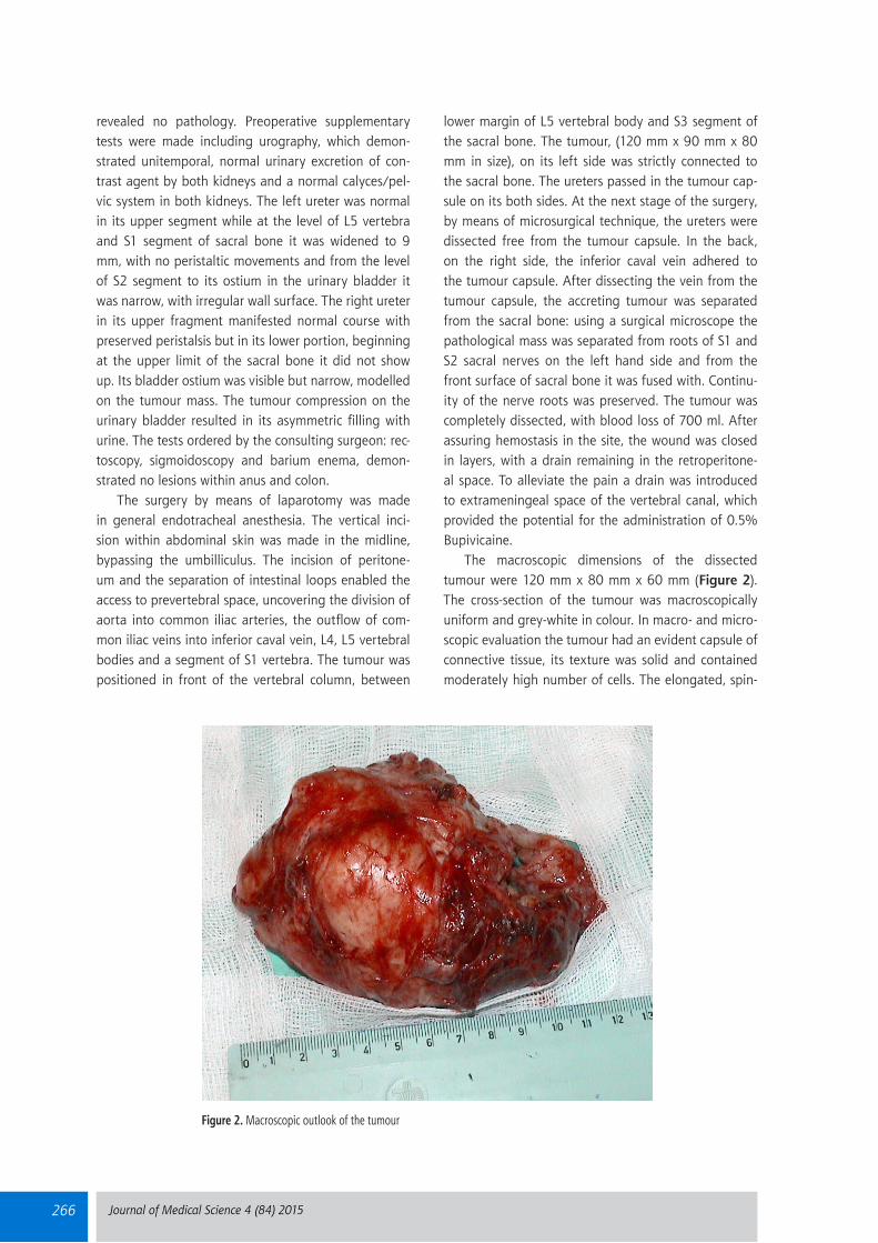
266 Journal of Medical Science 4 (84) 2015
revealed no pathology. Preoperative supplementary tests were made including urography, which demon-strated unitemporal, normal urinary excretion of con-trast agent by both kidneys and a normal calyces/pel-vic system in both kidneys. The left ureter was normal in its upper segment while at the level of L5 vertebra and S1 segment of sacral bone it was widened to 9 mm, with no peristaltic movements and from the level of S2 segment to its ostium in the urinary bladder it was narrow, with irregular wall surface. The right ureter in its upper fragment manifested normal course with preserved peristalsis but in its lower portion, beginning at the upper limit of the sacral bone it did not show up. Its bladder ostium was visible but narrow, modelled on the tumour mass. The tumour compression on the urinary bladder resulted in its asymmetric filling with urine. The tests ordered by the consulting surgeon: rec-toscopy, sigmoidoscopy and barium enema, demon-strated no lesions within anus and colon.
The surgery by means of laparotomy was made in general endotracheal anesthesia. The vertical inci-sion within abdominal skin was made in the midline, bypassing the umbilliculus. The incision of peritone-um and the separation of intestinal loops enabled the access to prevertebral space, uncovering the division of aorta into common iliac arteries, the outflow of com-mon iliac veins into inferior caval vein, L4, L5 vertebral bodies and a segment of S1 vertebra. The tumour was positioned in front of the vertebral column, between
lower margin of L5 vertebral body and S3 segment of the sacral bone. The tumour, (120 mm x 90 mm x 80 mm in size), on its left side was strictly connected to the sacral bone. The ureters passed in the tumour cap-sule on its both sides. At the next stage of the surgery, by means of microsurgical technique, the ureters were dissected free from the tumour capsule. In the back, on the right side, the inferior caval vein adhered to the tumour capsule. After dissecting the vein from the tumour capsule, the accreting tumour was separated from the sacral bone: using a surgical microscope the pathological mass was separated from roots of S1 and S2 sacral nerves on the left hand side and from the front surface of sacral bone it was fused with. Continu-ity of the nerve roots was preserved. The tumour was completely dissected, with blood loss of 700 ml. After assuring hemostasis in the site, the wound was closed in layers, with a drain remaining in the retroperitone-al space. To alleviate the pain a drain was introduced to extrameningeal space of the vertebral canal, which provided the potential for the administration of 0.5% Bupivicaine.
The macroscopic dimensions of the dissected tumour were 120 mm x 80 mm x 60 mm (Figure 2). The cross-section of the tumour was macroscopically uniform and grey-white in colour. In macro- and micro-scopic evaluation the tumour had an evident capsule of connective tissue, its texture was solid and contained moderately high number of cells. The elongated, spin-
Figure 2. Macroscopic outlook of the tumour

267Presacral schwannoma. Case description
dle-shaped cells manifested a slightly blurred margins of cytoplasm. The cells formed parallel or interwoven bands. The nuclei of cells were elongated, rod-shaped and positioned in the long axis of a cell. Nuclear chro-matin had uniform character, with nucleolus noted only occasionally. Palisade set-ups of cell nuclei were not encountered in the examined case. Occasionally, the tumour texture contained fibrous acellular regions and foci of xanthomatous cells. The vascular supply was relatively rich. The blood vessels frequently mani-fested thickened walls, with the presence of homoge-nous hyaline masses. Routine histological examination of the surgical material allowed for the diagnosis of schwannoma, manifesting, according to WHO, I degree of biological malignancy (Figure 3).
The post-operational course was free of compli-cations. After healing of the post-operational wound, on the ninth day following the operation, the patient showed a normal condition in neurological examina-tion, complained of no pain and was released home.
The patient continued her studies, then took up pro-fessional work, staying under control in the outpatient clinic. Currently the patient reports no complaints and consecutive neurological examinations demonstrate no deviations from a normal condition. MR control exami-nation of lumbal/sacral vertebral column made 5 years after the operation revealed a condition following a complete removal of the tumour (Figure 4).
Discussion
Schwannomas in the retrorectal region are slowly grow-ing tumours leading to transplacement of surrounding anatomic structures. They are observed more frequent-ly among females [4, 5].
The tumour evokes ailments when it reaches a large size. The clinical symptoms result from compression from the tumour on the neighbouring anatomic struc-tures: nervous, vascular and visceral. Most frequently the patient complains of a discomfort in abdominal
Figure 3. Histological preparation of the tumour. H&E staining, magnification 150 x

268 Journal of Medical Science 4 (84) 2015
cavity, lumbosacral pain, pain in the lumbar/sacral region, hypogastric pain radiating to groins, frequent urination or difficulties in passing urine. Some patients complain of unpleasant sensation of a filled anus and constipation. Sporadically, neurological defects are also observed [6, 7].
The symptoms are usually nonspecific and in over 26% the tumours are completely asymptomatic [3]. In such cases presacral schwannomas are recognised during physical examination of the abdominal cav-ity, gynecological examination “per rectum” or dur-ing radiological examination made due to nonspecific complaints [6]. This confirms our case, in which suspi-cion of a tumour in the presacral region was put for-ward by a gynecologist during a routine gynecological examination. The presence of a tumour in the retrorec-tal space may hamper a delivery of a newborn [6, 7].
Tumours located in this anatomic region may mani-fest an inborn character (meningeal hernias, dysontoge-netic tumours), be of primary origin (chordoma, osseous giant cell tumour, chondrosarcoma, immature neuro-ma), of metastatic or inflammatory character [3, 6].
Apart from the above mentioned tumours, schwan-noma has to be distinguished from ovarian tumours, ureteral tumours, retroperitoneal sarcoma or abscess in iliopsoas muscle [8–11].
Nevertheless, despite such broad range of differen-tial diagnosis, similarly to the discussed case, the classi-cal variant of schwannoma presents no diagnostic dif-ficulties in the postoperative material already on the level of routine morphological techniques. In doubtful cases, immunohistochemical techniques and occasion-ally electron-microscopic techniques turned out to be useful.
Figure 4. Control MRI in sagittal projection (a) and axial projection (b) presents the condition following complete excision of the tumour
a
b

269Presacral schwannoma. Case description
Schwannoma stems from ventral roots of sacral nerves. In plain radiography and computer-assisted tomography (CT) lesions of erosion type and irregular destruction of frontal pelvic foramens of sacral bone may be evident [12]. In CT examination and in magnet-ic resonance imaging (MRI) schwannoma is very well confined, manifests smooth edges and transplaces the neighbouring anatomic structures. The tumour is fre-quently featured by a heterogenous pattern, with cen-trally located cysts and it manifests marginal augmen-tation following administration of a contrast agent [4]. Lesions of cystic type are present much more frequent-ly (in 60%) in schwannomas than in other tumours [4, 13]. MRI patterns of schannomas present hemorrhagic, necrotic and calcified foci. The lesion is hypointense on T1 weighted images and hyperintense on T2 weighted images [14].
Needle biopsy of tumours in the retrorectal region is a safe approach to make the diagnosis strict. Histo-logical evaluation of the obtained material in 95% cas-es allows to conclude whether the lesion is of a benign or a malignant character. The tumour type can be pre-cisely defined in 81% cases [15]. Indications for needle biopsy in cases of schwannomas are controversial due to their sufficiently distinct image in MRI examination. Proponents of needle biopsy are of the opinion that such a biopsy should be made in patients with the tumour evoking no clinical symptoms and the patient does not decide to undergo surgery [7].
The size and location of the tumour have to be very precisely determined prior to surgery. A particu-lar attention should be paid to position of arterial and venous blood vessels in the anatomic region, to the determination of tumour relationships to ureters, uri-nary bladder, posterior wall of rectum and other close-ly positioned anatomic structures. The risk of intraop-erative injuries has to be evaluated before taking the decision about the surgery. In our case, this led to the extension of diagnostic procedures (apart from MRI) by rectoscopy, sigmoidoscopy, barium enema to the large intestine.
Depending of tumour location, size, relationships to other anatomic structures and type of tumour in the presacral region various surgical approaches are applied, including frontal access (through the peritone-al cavity), posterior access (through the sacral region), frontal and posterior in parallel, transrectal and trans-vaginal one [3, 6, 16]. Schwannomas are approached through the frontal access. In the case of small dimen-sion tumours the endoscopic technique is used with transrectal or transvaginal operative approach [17].
Surgical treatment of schwannoma aims at its com-plete removal. Problems with reaching the aim stem from the size of the tumour, its rich blood supply and vicinity to important anatomic structures. The tumours in this area are usually supplied with blood through sacral arteries, medial and lateral arteries, lumbar arteries and internal iliac arteries.
During the surgery, the surgeon should focus on preserving continuity of sacral nerve roots (S1, S2, S3) which exit from sacral bone through pelvic foramina, fused with the capsule of the tumour. Damage to the nerves results in dysfunction of vesical and anal sphinc-ter muscles, disturbs sexual functions and leads to pare-sis of greatest gluteal muscle [1]. In order to preserve the continuity of nerve roots S1 to S3 a microsurgical technique has been applied during our operation.
Complete excision of a tumour in the presacral region is possible in cases of benign tumours and in three quarters of cases of malignant tumours. A partial tumour resection is made when there are no chances for a complete removal of the pathological lesion [7]. In such cases relapses if schwannoma are described, usually within a time distance of a few years [18].
Complications occur quite frequently, in 1/3 of the cases, but they are usually of reversible character [3]. They most frequently include the injury to venous or arterial blood vessels, a disturbed function of vesical and anal sphincters and in men disturbances in erec-tion. Deficits in sensation in perineal region and frontal surface of thighs were also described [2, 3, 18].
In patients with asymptomatic schwannoma the possibility of conservative treatment with periodic con-trol of MR examination should be taken into account. Strauss et al. treated 28 patients with schwannoma in retroperitoneal space, out of which 8 had the tumour accidentally detected due to their nonspecific com-plaints and signs in MR imaging [7]. The diagnosis of schwannoma was established by needle biopsy and the patients were subject to further observation. After 32 months no progression of the tumour was revealed by MRI control, the patients reported no pains and neurological examination detected no abnormalities. However, other authors reported very good results of surgical treatment applied to asymptomatic presacral schwannomas [19, 20].
Application of radiotherapy in cases of incomplete excision of schwannoma is controversial and rather not recommended as it may induce the transforma-tion of schwannoma cells and the development of malignant neuroma [21, 22]. Stereotactic radiosur-gery is recommended in cases of small schwannomas,

270 Journal of Medical Science 4 (84) 2015
with the diameter of less than 30 mm, positioned in the sacral bone [23].
Conclusions
Giant schwannomas of sacral bone occur rarely and pose diagnostic and therapeutic difficulties. They manifest a slow local growth and induce nonspecific symptoms.
Their preoperative procedures require magnetic resonance imaging of lumbosacral vertebral column and multispecialistic evaluation of pelvic visceral struc-tures.
Complete tumour excision is equal to cure.
Acknowledgements
Conflict of interest statementThe authors declare that there is no conflict of interest in the authorship or publication of contribution.
Funding sourcesThere are no sources of funding to declare.
ReferencesFeldenzer JA, Mc Gauley JL, Mc Gillicuddy JE. Sacral and 1. presacral tumors: problems in diagnosis and manage-ment. Neurosurg. 1989;25:884–891.Abernathey CD, Onofrio BM, Scheithauer B. Pairolero PC, 2. Shives TC. Surgical management of giant sacral schwan-nomas. J Neurosurg. 1986;65:286–295.Lev-Chelouche D, Gutman M, Goldman G.Even-Sapir E, 3. Meller I, Issakov J,Presacral tumors: A practical classifica-tion and treatment of unique and heterogenous group of diseases. Surgery 2003;133:473–478.Hughes MJ, Thomas JM, Fisher C, Moskovic EC, Imaging 4. features of retroperitoneal and pelvic schwannomas Clin Radiol. 2005 60, 886–93.Li Q, Gao C, Juzi JT,HAO X, Analysis of 82 cases of retro-5. peritoneal schwannoma. ANZ J Surg. 2007;77:237– 240.Klimo P, Rao G, Schmidt R, Schmidt MH, Nerve sheath 6. tumours involving the sacrum. Case report and clasifica-tion scheme. Neurosurg Focus 2003;15:1–6.Strauss DC, Qureshi YA, Hayes AJ, Thomas M. Manage-7. ment of benign retroperitoneal schwannomas: a single--center experience. Am J Surg. 2011;202:194–198.. Van Roggen JF, Hogendoorn PC. Soft tissue tumours of 8. the retroperitoneum. Sarcoma 2000;4:17–26.Aran T, Guven S, Gocer S, Ersoz S, Bozkaya H, Large retro-9. peritoneal schwannoma mimicking ovarian carcinoma: case report and literature review. Eur J Gynaecol Oncol. 2009;30:446–448.Sharma SK, Koleski FC, Husain AN, Albala DM, Turkt 10. TM, Retroperitoneal schwannoma mimicking an adrenal lesion. World J Urol. 2002;20:232–233.
Liu YW, Chiu HH, Huang CC,TU CA, Retroperitoneal 11. schwannoma mimicking a psoas abscess. Clin Gastroen-terol Hepatol. 2007;5:A32.Chiang ER, Chang MC, Chen TH. Giant retroperitoneal 12. schwannoma from the fifth lumbar nerve root with verte-bral body osteolysis: a case report and literature review. Arch Orthop Trauma Surg. 2009;129:495–499.Takatera H, Takiuchi H, Namiki M,mTakaha M, Ohnis-13. hi S, Sonoda T. Retroperitoneal schwannoma. Urology 1986;28:529 –231.Hoaraua N, Slimb K, Da Inesa D. CT and MR imaging of 14. retroperitoneal schwannoma. Diagnostic and Interventio-nal Imaging 2013;94:1133–1139.Hoeber I, Spillane AJ, Fisher C, Thomas JM, Accuracy of 15. biopsy techniques for limb and limb girdle soft tissue tumours. Ann Surg Oncol. 2001;8:80–87.Asakage N. Laparoscopic resection of a retroperitoneal 16. schwannoma. Asian J Endosc Surg 2012;5:25–30.Konstantinidis K, Theodoropoulos GE, Sambalis G, Geo-17. rgiou M, Vorias M, Laparoscopic resection of presac-ral schwannomas. Surg Laparosc Endosc Percutan Tech. 2005;15:302–304.Pongsthorn C, Ozawa H, Aizawa T,Kuskabe T, Nakamura 18. T, Itoi E,Giant sacral schwannoma: a report of six cases. Ups J Med Sci. 2010;115:146–152.Rao W, Wang G, Xiu D. Laparoscopic resection of a retro-19. peritoneal schwannoma adherent to vital vessels. Surg Laparosc Endosc Percutan Tech. 2009;19, E 21–23.Yoshino T, Yoneda K. Laparoscopic resection of a retrope-20. ritoneal ancient schwannoma: a case report and review of the literature. Anticancer Res. 2008;28:2889 –2891.Isler MH, Fogaca MF, Mankin HJ. Radiation induced 21. malignant schwannoma arising in a neurofibroma. Clin Orthop. 1996;325:251–256. Kotoura Y, Shikata J, Yammamuro T, Kasahara K, Ivasaki 22. R, Nakashima Y, et al. Radiation therapy for giant intra-sacral schwannoma. Spine 1991;16:239–242.Gertszten PC, Ozhasoglu C, Burton SA, Kalnicki S, Welch 23. WC, Feasibility of frameless single fraction stereotactic radiosurgery for spinal lesions. Neurosurg Focus 2002;13, Article 2.
Acceptance for editing: 2015-11-10 Acceptance for publication: 2015-12-31
Correspondence address:Jeremi Kościński
Chair and Department of Neurosurgery and Neurotraumatology
Poznan University of Medical Sciences49 Przybyszewskiego Str., 60-355 Poznan, Poland
phone: +48 607715837fax: +48 61 8691430
email: [email protected]

271Journal of Medical Science 4 (84) 2015
Journal of Medical Science Instructions for Authors
AbbreviationsAbbreviations should be defined at first mention, by putting abbreviation between brackets after the full text . Ensure consistency of abbreviations throughout the article . Avoid using them in the title and abstract . Abbreviations may be used in tables and figures if they are defined in the table footnotes and figure legends.
Trade namesFor products used in experiments or methods (particularly those referred to by a trade name), give the manufacturer's full name and location (in parentheses) . When possible, use generic names of drugs .
Title page The first page of the manuscript should contain the title of the article, authors’ full names without degrees or titles, authors’ institutional affiliations including city and country and a running title, not exceeding 40 letters and spaces. The first page should also include the full postal address, e‑mail address, and telephone and fax numbers of the corresponding author .
AbstractThe abstract should not exceed 250 words and should be structured into separate sections: Background, Methods, Results and Conclusions . It should concisely state the significant findings without reference to the rest of the paper. The abstract should be followed by a list of 3 to 6 Key words. They should reflect the central topic of the article (avoid words already used in the title) .
The following categories of articles can be proposed to the Journal of Medical Science:
ORIGINAL RESEARCH
Original articles: Manuscripts in this category describe the results of original research conducted in the broad area of life science and medicine . The manuscript should be presented in the format of Abstract (250‑word limit), Keywords, Introduction, Material and Methods, Results, Discussion, Perspectives, Acknowledgments and References . In the Discussion section, statements regarding the importance and novelty of the study should be presented . In addition, the limitations of the study should be articulated . The abstract must be structured and include: Objectives, Material and Methods, Results and Conclusions. Manuscripts cannot exceed 3500 words in length (excluding title page, abstract and references) and contain no more than a combination of 8 tables and/or figures. The number of references should not exceed 45.
Brief Reports: Manuscripts in this category may present results of studies involving small sample sizes, introduce new methodologies, describe preliminary findings or replication studies . The manuscript must follow the same format requirements as full length manuscripts. Brief reports should be up to 2000 words (excluding title page, abstract and references) and can include up to 3 tables and/or figures. The number of references should not exceed 25.
REVIEW ARTICLES
Review articles: These articles should describe recent advances in areas within the Journal’s scope. Review articles cannot exceed 5000 words length (excluding title page, abstract and references) and contain no more than a combination of 10 tables and/or figures. Authors are encouraged to restrict figures and tables to essential data that cannot be described in the text. The number of references should not exceed 80.
A THOUSAND WORDS ABOUT… is a form of Mini‑Reviews. Manuscripts in this category should focus on latest achievements of life science and medicine. Manuscripts should be up to 1000 words in length (excluding title page, abstract and references) and contain up to 5 tables and/or figures and up to 25 most relevant references. The number of authors is limited to no more than 3 .
Journal of Medical Science (JMS) is a PEER‑REVIEWED, OPEN ACCESS journal that publishes original research articles and reviews which cover all aspects of clinical and basic science research . The journal particularly encourages submissions on the latest achievements of world medicine and related disciplines . JMS is published quarterly by Poznan University of Medical Sciences .
ONLINE SUBMISSION:Manuscripts should be submitted to the Editorial Office by an e‑mail attachment: nowinylekarskie@ump .edu .pl . You do not need to mail any paper copies of your manuscript .
All submissions should be prepared with the following files:– Cover Letter– Manuscript– Tables– Figures – Supplementary Online Material
COVER LETTER: Manuscripts must be accompanied by a cover letter from the author who will be responsible for correspondence regarding the manuscript as well as for communications among authors regarding revisions and approval of proofs . The cover letter should contain the following elements: (1) the full title of the manuscript, (2) the category of the manuscript being submitted (e .g . Original Article, Brief Report), (3) the statement that the manuscript has not been published and is not under consideration for publication in any other journal, (4) the statement that all authors approved the manuscript and its submission to the journal, and (5) a list of at least two referees.
MANUSCRIPT: Journal of Medical Science publishes Original Articles, Brief Reports, Review articles, Mini‑Reviews, Images in Clinical Medicine and The Rationale and Design and Methods of New Studies . From 2014, only articles in English will be considered for publication . They should be organized as follows: Title page, Abstract, Introduction, Materials and Methods, Results, Discussion, Acknowledgments, Conflict of Interest, References and Figure Legends . All manuscripts should be typed in Arial or Times New Roman font and double spaced with a 2,5 cm (1 inch) margin on all sides . They should be saved in DOC, DOCX, ODT, RTF or TXT format . Pages should be numbered consecutively, beginning with the title page .
Ethical GuidelinesAuthors should follow the principles outlined in the Declaration of Helsinki of the World Medical Association (www .wma .net) . The manuscript should contain a statement that the work has been approved by the relevant institutional review boards or ethics committees and that all human participants gave informed consent to the work . This statement should appear in the Material and Methods section . Identifying information, including patients' names, initials, or hospital numbers, should not be published in written descriptions, illustrations, and pedigrees . Studies involving experiments with animals must be conducted with approval by the local animal care committee and state that their care was in accordance with institution and international guidelines . Authorship:According to the International Committee on Medical Journal Ethics (ICMJE), an author is defined as one who has made substantial contributions to the conception and development of a manuscript . Authorship should be based on all of the following: 1) substantial contributions to conception and design, data analysis and interpretation; 2) article drafting or critical advice for important intellectual content; and 3) final approval of the version to be published . All other contributors should be listed as acknowledgments. All submissions are expected to comply with the above definition.
Conflict of InterestThe manuscript should contain a conflict of interest statement from each author. Authors should disclose all financial and personal relationships that could influence their work or declare the absence of any conflict of interest. Author’s conflict of interest should be included under Acknowledgements section .

272 Journal of Medical Science 4 (84) 2015
OTHER SUBMISSIONS
Invited Editorials: Editorials are authoritative commentaries on topics of current interest or that relate to articles published in the same issue . Manuscripts should be up to 1500 words in length. The number of references should not exceed 10. The number of authors is limited to no more than 2 .
Images in Clinical Medicine: Manuscripts in this category should contain one distinct image from life science or medicine. Only original and high‑quality images are considered for publication. The description of the image (up to 250 words) should present relevant information like short description of the patient's history, clinical findings and course, imaging techniques or molecular biology techniques (e.g. blotting techniques or immunostaining) . All labeled structures in the image should be described and explained in the legend. The number of references should not exceed 5. The number of authors is limited to no more than 5.
The Rationale, Design and Methods of New Studies: Manuscripts in this category should provide information regarding the grants awarded by different founding agencies, e .g . National Health Institute, European Union, National Science Center or National Center for Research and Development . The manuscript should be presented in the format of Research Project Objectives, Research Plan and Basic Concept, Research Methodology, Measurable Effects and Expected Results . The article should also contain general information about the grant: grant title, keywords (up to five), name of the principal investigator and co‑investigators, founding source with the grant number, Ethical Committee permission number, code in clinical trials (if applicable). Only grant projects in the amount over 100,000 Euro can be presented. Manuscripts should be up to 2000 words in length (excluding references) and can include up to 5 tables and/or figures. The abstract should not exceed 150 words. The number of authors is limited to the Principal Investigator and Co‑investigators.
Acknowledgements Under acknowledgements please specify contributors to the article other than the authors accredited . List here those individuals who provided help during the research (e .g ., providing language help, writing assistance or proof reading the article, etc .) . Also acknowledge all sources of support (grants from government agencies, private foundations, etc .) . The names of funding organizations should be written in full .
ReferencesAll manuscripts should use the 'Vancouver' style for references. References should be numbered consecutively in the order in which they appear in the text and listed at the end of the paper. References cited only in Figures/Tables should be listed in the end. Reference citations in the text should be identified by Arabic numbers in square brackets . Some examples: This result was later contradicted by Smith and Murray [3] . Smith [8] has argued that . . . Multiple clinical trials [4–6, 9] show . . .
List all authors if there are six or fewer; if there are seven or more, list first six follower by “et al .” . Journal names should be abbreviated according to Index Medicus .
Some examplesStandard journal articles1. Fassone E, Rahman S. Complex I deficiency: clinical features, biochemistry and
molecular genetics. J Med Genet. 2012 Sep;49(9):578–590.
2 . Pugh TJ, Morozova O, Attiyeh EF, Asgharzadeh S, Wei JS, Auclair D et al . The genetic landscape of high‑risk neuroblastoma. Nat Genet. 2013 Mar;45(3):279–284.
BooksPersonal author(s)1. Rang HP, Dale MM, Ritter JM, Moore PK. Pharmacology. 5th ed. Edinburgh: Churchill
Livingstone; 2003.
Editor(s) or compiler(s) as authors2. Beers MH, Porter RS, Jones TV, Kaplan JL, Berkwits M (editors). The Merck manual of
diagnosis and therapy . 18th ed . Whitehouse Station (NJ): Merck Research Laboratories; 2006.
Chapter in the book1 . Phillips SJ, Whisnant JP . Hypertension and stroke . In: Laragh JH, Brenner BM, editors .
Hypertension: pathophysiology, diagnosis, and management . 2nd ed . New York: Raven Press; 1995. p. 465–478.
TABLES: Tables should be typed on sheets separate from the text (each table on a separate sheet) . They should be numbered consecutively with Arabic numerals . Tables should always be cited in text (e .g . table 2) in consecutive numerical order . Each table should include a compulsory, concise explanatory title and an explanatory legend . Footnotes to tables should be typed below the table body and referred to by superscript lowercase letters . No vertical rules should be used . Tables should not duplicate results presented elsewhere in the manuscript (e.g. in figures).
FIGURES: All illustrations, graphs, drawings, or photographs are referred to as figures and must be uploaded as separate files when submitting a manuscript. Figures should be numbered in sequence with Arabic numerals . They should always be cited in text (e.g. figure 3) in consecutive numerical order. Figures for publication must only be submitted in high‑resolution TIFF or EPS format (minimum 300 dpi resolution). Each figure should be self‑explanatory without reference to the text and have a concise but descriptive legend. All symbols and abbreviations used in the figure must be defined, unless they are common abbreviations or have already been defined in the text. Figure Legends must be included after the reference section of the Main Text .
Color figures: Figures and photographs will be reproduced in full colour in the online edition of the journal. In the paper edition, all figures and photographs will be reproduced as black‑and‑white.
SUPPLEMENTARY ONLINE MATERIAL: Authors may submit supplementary material for their articles to be posted in the electronic version of the journal . To be accepted for posting, supplementary materials must be essential to the scientific integrity and excellence of the paper . The supplementary material is subject to the same editorial standards and peer‑review procedures as the print publication.
Review ProcessAll manuscripts are reviewed by the Editor‑in‑Chief or one of the members of the Editorial Board, who may decide to reject the paper or send it for external peer review . Manuscripts accepted for peer review will be blind reviewed by at least two experts in the field. After peer review, the Editor‑in‑Chief will study the paper together with reviewer comments to make one of the following decisions: accept, accept pending minor revision, accept pending major revision, or reject . Authors will receive comments on the manuscript regardless of the decision . In the event that a manuscript is accepted pending revision, the author will be responsible for completing the revision within 60 days.
CopyrightThe copyright to the submitted manuscript is held by the Author, who grants the Journal of Medical Science (JMS) a nonexclusive licence to use, reproduce, and distribute the work, including for commercial purposes .