Embed Size (px)
Citation preview
PRACTICE GUIDELINES FOR MANAGEMENT OF THE DIFFICULT AIRWAY 1 (Approved by House of Delegates on October 21, 1992,
and last amended October 16, 2002)
Practice guidelines are systematically developed recommendations that assist the practitioner
and patient in making decisions about health care. These recommendations may be adopted,
modified, or rejected according to clinical needs and constraints.
Practice guidelines are not intended as standards or absolute requirements. The use of practice
guidelines cannot guarantee any specific outcome. Practice guidelines are subject to revision as
warranted by the evolution of medical knowledge, technology, and practice. They provide basic
recommendations that are supported by analysis of the current literature and by a synthesis of expert
opinion, open forum commentary, and clinical feasibility data.
This revision includes data published since the "Practice Guidelines for Management of the
Difficult Airway" were adopted by the American Society of Anesthesiologists in 1992; it also
includes data and recommendations for a wider range of management techniques than was
previously addressed.
Purpose of the Guidelines for Difficult Airway Management
The purpose of these Guidelines are to facilitate the management of the difficult airway and
to reduce the likelihood of adverse outcomes. The principal adverse outcomes associated with
the difficult airway include (but are not limited to): death, brain injury, cardiopulmonary arrest,
unnecessary tracheostomy, airway trauma and damage to teeth.
1Developed by the American Society of Anesthesiologists Task Force on Difficult Airway Management: Robert A. Caplan, M.D. (Chair); Jonathan L. Benumof, M.D.; Frederic A. Berry, M.D.; Casey D. Blitt, M.D.; Robert H. Bode, M.D.; Frederick W. Cheney, M.D.; Richard T. Connis, Ph.D.; Orin F. Guidry, M.D.; David G. Nickinovich, Ph.D.; and Andranik Ovassapian, M.D. Supported by the American Society of Anesthesiologists under the direction of James F. Arens, M.D., Chair, Committee on Practice Parameters. A list of the references used to develop these Guidelines is available by writing to the American Society of Anesthesiologists. Address reprint requests to the American Society of Anesthesiologists: 520 N. Northwest Highway, Park Ridge, Illinois 60068-2573
Practice Guidelines for Management of the Difficult Airway
Focus
The primary focus of these Guidelines is the management of the difficult airway encountered
during administration of anesthesia and tracheal intubation (Appendix A). Some aspects of the
Guidelines may be relevant in other clinical contexts. The Guidelines do not represent an
exhaustive consideration of all manifestations of the difficult airway or all possible approaches to
management.
Application
The Guidelines are intended for use by anesthesiologists and by individuals who deliver
anesthetic care and airway management under the direct supervision of an anesthesiologist. The
Guidelines apply to all types of anesthetic care and airway management delivered in anesthetizing
locations and is intended for all patients of all ages.
Task Force Members and Consultants
The ASA appointed a Task Force of ten members to (1) review the published evidence; (2)
obtain the opinions of anesthesiologists selected by the Task Force as consultants; and (3) build
consensus within the community of practitioners likely to be affected by the Guidelines. The Task
Force included anesthesiologists in both private and academic practices from various geographic
areas of the United States, and consulting methodologists from the ASA Committee on Practice
Parameters.
These Practice Guidelines update and revise the 1993 publication of ASA's Guidelines for
Management of the Difficult Airway.1 The Task Force revised and updated the Guidelines by
means of a five-step process. First, original published research studies relevant to the revision and
update were reviewed and analyzed. Second, the panel of expert consultants was asked to (1)
participate in a survey related to the effectiveness and safety of various methods and interventions
2
Practice Guidelines for Management of the Difficult Airway
that might be used during management of the difficult airway, and (2) review and comment on draft
reports. Third, the Task Force held an open forum at a major national anesthesia meeting to solicit
input from attendees on a draft of the Guidelines. Fourth, the consultants were surveyed to assess
their opinions on the feasibility and financial implications of implementing the Guideline. Finally,
all of the available information was used by the Task Force to finalize the Guidelines.
Availability and Strength of Evidence
Evidence-based guidelines are developed by a rigorous analytic process. To assist the reader,
these Guidelines make use of several descriptive terms that are easier to understand than the
technical terms and data that are used in the actual analyses. These descriptive terms are defined
below:
The following terms describe the strength of scientific data obtained from the scientific
literature.
Supportive: There is sufficient quantitative information from adequately designed studies to
describe a statistically significant relationship (p < 0.01) between a clinical intervention and
a clinical outcome, using meta-analysis.
Suggestive: There is enough information from case reports and descriptive studies to provide a
directional assessment of the relationship between a clinical intervention and a clinical
outcome. This type of qualitative information does not permit a statistical assessment of
significance.
Equivocal: Qualitative data have not provided a clear direction for clinical outcomes related to
a clinical intervention and (1) there is insufficient quantitative information or (2) aggregated
comparative studies have found no quantitatively significant differences among groups or
conditions.
3
Practice Guidelines for Management of the Difficult Airway
The following terms describe the lack of available scientific evidence in the literature.
Inconclusive: Published studies are available, but they cannot be used to assess the relationship
between a clinical intervention and a clinical outcome because the studies either do not meet
predefined criteria for content as defined in the “Focus” of these Guidelines, or do not
provide a clear causal interpretation of findings because of research design or analytic
concerns.
Insufficient: There are too few published studies to investigate a relationship between a clinical
intervention and clinical outcome.
Silent: No studies that address a relationship of interest were found in the available published
literature.
The following terms describe survey responses from the consultants for any specified issue.
Responses are assigned a numeric value of agree = +1, undecided = 0 or disagree = -1. The average
weighted response represents the mean value for each survey item.
Agree: The average weighted response must be equal to or greater than +0.30 (on a scale of –1
to 1) to indicate agreement.
Equivocal: The average weighted response must be between -0.30 and +0.30 (on a scale of –1
to 1) to indicate an equivocal response.
Disagree: The average weighted response must be equal to or less than -0.30 (on a scale of –1
to 1) to indicate disagreement.
Guidelines Evaluation of the Airway
History. There is insufficient published evidence to evaluate the effect of a bedside medical
history on predicting the presence of a difficult airway. Similarly, there is insufficient
4
Practice Guidelines for Management of the Difficult Airway
evidence to evaluate the effect of reviewing prior medical records on predicting the presence
of a difficult airway. There is suggestive evidence that some features of a patient's medical
history or prior medical records may be related to the likelihood of encountering a difficult
airway. This support is based upon recognized associations between a difficult airway and a
variety of congenital, acquired, or traumatic disease states. In addition, the Task Force
believes that the description of a difficult airway on a previous anesthesia record or
anesthesia document offers clinically suggestive evidence that difficulty may recur. The
consultants and Task Force agree that a focused bedside medical history and a focused
review of medical records may improve the detection of a difficult airway.
Recommendations.
An airway history should be conducted, whenever feasible, prior to the initiation of
anesthetic care and airway management in all patients. The intent of the airway history is to
detect medical, surgical, and anesthetic factors that may indicate the presence of a difficult
airway. Examination of previous anesthetic records, if available in a timely manner, may
yield useful information about airway management.
Physical Examination. In patients with no gross upper airway pathology or anatomical
anomaly, there is insufficient published evidence to evaluate the effect of a physical
examination on predicting the presence of a difficult airway. However, there are suggestive
data that findings obtained from an airway physical examination may be related to the
presence of a difficult airway. This support is based upon recognized associations between
the difficult airway and a variety of airway characteristics. The consultants and Task Force
agree that an airway physical examination may improve the detection of a difficult airway.
5
Practice Guidelines for Management of the Difficult Airway
Specific features of the airway physical examination have been incorporated into rating
systems intended to predict the likelihood of a difficult airway. Existing rating systems have
been shown to exhibit modest sensitivity and specificity. The specific effect of the airway
physical examination on outcome has not been clearly defined in the literature.
There is insufficient published evidence to evaluate the predictive value of single features
of the airway physical examination versus multiple features on predicting the presence of a
difficult airway. The consultants and Task Force agree that prediction of a difficult airway
may be improved by the assessment of multiple features. The Task Force does not regard
any rating system as fail-safe.
Recommendations.
An airway physical examination should be conducted, whenever feasible, prior to the
initiation of anesthetic care and airway management in all patients. The intent of this
examination is to detect physical characteristics that may indicate the presence of a difficult
airway. Multiple airway features should be assessed. (Table 1)
Additional Evaluation. The airway history or physical examination may provide indications
for additional diagnostic testing in some patients. The literature suggests that certain
diagnostic tests may identify features associated with a difficult airway. The literature does
not provide a basis for using specific diagnostic tests as routine screening tools in the
evaluation of the difficult airway.
Recommendations:
Additional evaluation may be indicated in some patients to characterize the likelihood or
nature of the anticipated airway difficulty. The findings of the airway history and physical
6
Practice Guidelines for Management of the Difficult Airway
examination may be useful in guiding the selection of specific diagnostic tests and
consultation.
Basic Preparation for Difficult Airway Management
The literature is silent regarding the benefits of informing the patient of a known or
suspected difficult airway, the availability of equipment for difficult airway management, or
the availability of an individual to provide assistance when a difficult airway is encountered.
However, there is strong agreement among consultants that preparatory efforts enhance
success and minimize risk.
The literature suggests that either traditional preoxygenation (3 or more minutes of tidal
volume ventilation) or fastrack preoxygenation (i.e., four maximal breathes in 30 seconds) is
effective in delaying arterial desaturation during subsequent apnea. The literature supports
the greater efficacy of traditional preoxygenation when compared to fastrack preoxygenation
in delaying arterial desaturation during apnea.
The literature supports the efficacy of supplemental oxygen in reducing hypoxemia after
extubation of the trachea.
Recommendations.
At least one portable storage unit that contains specialized equipment for difficult airway
management should be readily available. Specialized equipment suggested by the Task
Force is listed in Table 2.
If a difficult airway is known or suspected, the anesthesiologist should:
1. Inform the patient (or responsible person) of the special risks and procedures pertaining to management of the difficult airway.
2. Ascertain that there is at least one additional individual who is immediately available
to serve as an assistant in difficult airway management.
7
Practice Guidelines for Management of the Difficult Airway
3. Administer face mask preoxygenation before initiating management of the difficult airway. The uncooperative or pediatric patient may impede opportunities for preoxygenation.
4. Actively pursue opportunities to deliver supplemental oxygen throughout the process
of difficult airway management. Opportunities for supplemental oxygen administration include (but are not limited to) oxygen delivery by nasal cannulae, facemask, LMA, insufflation, or jet ventilation during intubation attempts; and oxygen delivery by facemask, blow-by, or nasal cannulae after extubation of the trachea.
Strategy for Intubation of the Difficult Airway
The literature suggests that the use of specific strategies facilitate the intubation of the
difficult airway. Although the degree of benefit for any specific strategy cannot be
determined from the literature, there is strong agreement among consultants that a
preplanned strategy may lead to improved outcome.
Preplanned strategies can be linked together to form airway management algorithms.
The Task Force considers the technical and physiologic complexity of life-threatening airway
events to be sufficiently similar to life-threatening cardiac events to encourage the use of
algorithms in difficult airway management.
Recommendations.
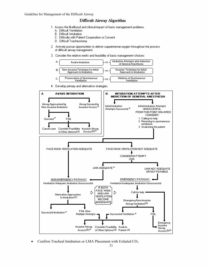
The anesthesiologist should have a preformulated strategy for intubation of the difficult
airway. The algorithm represented in Figure 1 is a strategy recommended by the Task Force.
This strategy will depend in part upon the anticipated surgery, the condition of the patient,
and the skills and preferences of the anesthesiologist.
The strategy for intubation of the difficult airway should include: 1. An assessment of the likelihood and anticipated clinical impact of four basic
problems that may occur alone or in combination: a. Difficult ventilation. b. Difficult intubation. c. Difficulty with patient cooperation or consent. d. Difficult tracheostomy
8
Practice Guidelines for Management of the Difficult Airway
2. A consideration of the relative clinical merits and feasibility of three basic management choices:
a. Awake intubation versus intubation after induction of general anesthesia b. Use of non-invasive techniques for the initial approach to intubation versus
the use of invasive techniques (i.e., surgical or percutaneous tracheostomy or cricothyrotomy).
c. Preservation of spontaneous ventilation during intubation attempts versus ablation of spontaneous ventilation during intubation attempts.
3. The identification of a primary or preferred approach to:
a. Awake intubation. b. The patient who can be adequately ventilated but is difficult to intubate. c. The life-threatening situation in which the patient cannot be ventilated or
intubated. 4. The identification of alternative approaches that can be employed if the primary
approach fails or is not feasible: a. Table 3 displays options for difficult airway management. b. The uncooperative or pediatric patient may restrict the options for difficult
airway management, particularly options that involve awake intubation. Airway management in the uncooperative or pediatric patient may require an approach (e.g., intubation attempts after induction of general anesthesia) that might not be regarded as a primary approach in a cooperative patient.
c. The conduct of surgery using local anesthetic infiltration or regional nerve blockade may provide an alternative to the direct management of the difficult airway, but this approach does not represent a definitive solution to the presence of a difficult airway, nor does it obviate the need for a preformulated strategy for intubation of the difficult airway.
5. The use of exhaled CO2 to confirm tracheal intubation.
Strategy for Extubation of the Difficult Airway
The literature does not provide a sufficient basis for evaluating the benefits of an
extubation strategy for the difficult airway. The Task Force regards the concept of an
extubation strategy as a logical extension of the intubation strategy. Consultant opinion
strongly supports the use of an extubation strategy.
Recommendations. The anesthesiologist should have a preformulated strategy for extubation of the difficult
airway. This strategy will depend in part upon the surgery, the condition of the patient, and
the skills and preferences of the anesthesiologist.
9
Practice Guidelines for Management of the Difficult Airway
The preformulated extubation strategy should include:
1. A consideration of the relative merits of awake extubation versus extubation before the return of consciousness.
2. An evaluation for general clinical factors that may produce an adverse impact on
ventilation after the patient has been extubated. 3. The formulation of an airway management plan that can be implemented if the
patient is not able to maintain adequate ventilation after extubation. 4. A consideration of the short-term use of a device that can serve as a guide for
expedited reintubation. This type of device is usually inserted through the lumen of the tracheal tube and into the trachea before the tracheal tube is removed. The device may be rigid to facilitate intubation and/or hollow to facilitate ventilation.
Follow-Up Care
Although the literature is insufficient to evaluate the benefits of follow-up care, this
activity is strongly supported by consultant opinion. The Task Force has identified several
fundamental concepts that merit consideration.
Recommendations.
The anesthesiologist should document the presence and nature of the airway difficulty in
the medical record. The intent of this documentation is to guide and facilitate the delivery of
future care. Aspects of documentation that may prove helpful include (but are not limited to):
1. A description of the airway difficulties that were encountered. The description should distinguish between difficulties encountered in face mask or LMA ventilation and difficulties encountered in tracheal intubation.
2. A description of the various airway management techniques that were employed.
The description should indicate the extent to which each of the techniques served as beneficial or detrimental role in management of the difficult airway.
The anesthesiologist should inform the patient (or responsible person) of the airway
difficulty that was encountered. The intent of this communication is to provide the patient
(or responsible person) with a role in guiding and facilitating the delivery of future care. The
information conveyed may include (but is not limited to): the presence of a difficult airway,
10
Practice Guidelines for Management of the Difficult Airway
the apparent reasons for difficulty, how the intubation was accomplished, and the
implications for future care. Notification systems such as a written report or letter to the
patient, a written report in the medical chart, communication with the patient's surgeon or
primary care giver, a Medic-Alert bracelet or equivalent identification system, or chart
flags may be considered.
The anesthesiologist should evaluate and follow the patient for potential complications of
difficult airway management. These complications include (but are not limited to): edema,
bleeding, tracheal and esophageal perforation, pneumothorax, and aspiration. The patient
should be advised of the potential clinical signs and symptoms associated with life-
threatening complications of difficult airway management. These signs and symptoms
include, but are not limited to sore throat, pain or swelling of the face and neck, chest pain,
subcutaneous emphysema, and difficulty swallowing.
11
Practice Guidelines for Management of the Difficult Airway
Appendix A:
Definition of the Difficult Airway
A standard definition of the difficult airway cannot be identified in the available literature. For these Guidelines, a difficult airway is defined as the clinical situation in which a conventionally trained anesthesiologist experiences difficulty with face mask ventilation of the upper airway, difficulty with tracheal intubation, or both.
The difficult airway represents a complex interaction between patient factors, the clinical
setting, and the skills of the practitioner. Analysis of this interaction requires precise collection and communication of data. The Task Force urges clinicians and investigators to use explicit descriptions of the difficult airway. Descriptions that can be categorized or expressed as numerical values are particularly desirable, as this type of information lends itself to aggregate analysis and cross-study comparisons. Suggested descriptions include (but are not limited to):
1. Difficult Face Mask Ventilation.
It is not possible for the anesthesiologist to provide adequate face mask ventilation
due to one or more of the following problems: inadequate mask seal, excessive gas leak, r excessive resistance to the ingress or egress of gas. o
Signs of inadequate face mask ventilation include (but are not limited to): absent or
inadequate chest movement, absent or inadequate breath sounds, auscultatory signs of severe obstruction, cyanosis, gastric air entry or dilatation, decreasing or inadequate SpO2 , absent or inadequate exhaled CO2 , absent or inadequate spirometric measures of exhaled gas flow, and hemodynamic changes associated with hypoxemia or hypercarbia (e.g., hypertension, tachycardia, arrhythmia).
2. Difficult Laryngoscopy.
It is not possible to visualize any portion of the vocal cords after multiple attempts at conventional laryngoscopy.
3. Difficult Tracheal Intubation.
Tracheal intubation requires multiple attempts, in the presence or absence of tracheal pathology.
4. Failed Intubation.
Placement of the tracheal tube fails after multiple intubation attempts.
12
Practice Guidelines for Management of the Difficult Airway
Appendix B:
Methods and Analyses 2
The scientific assessment of these Guidelines was based on the following statements or evidence linkages. These linkages represent directional statements about relationships between clinical care and clinical outcome in difficult airway management.
1. Evaluation of the Airway
a. A directed history detects a difficult airway and reduces airway-related adverse outcomes.
b. A directed airway physical examination detects a difficult airway and reduces airway-related adverse outcomes.
c. Diagnostic tests (e.g. radiography) detect a difficult airway and reduce airway-related adverse outcomes.
2. Basic Preparation for Difficult Airway Management
a. Informing the patient with a known or suspected difficult airway reduces airway-related adverse outcomes.
b. Availability of equipment for management of a difficult airway (i.e., a portable storage unit) reduces airway-related adverse outcomes.
c. Availability of an assigned individual to provide assistance when a difficult airway is encountered reduces airway-related adverse outcomes.
d. Preanesthetic preoxygenation by face mask before induction of anesthesia delays arterial desaturation and prevents hypoxemia during subsequent apnea.
3. Strategies for Intubation/Ventilation
a. Awake intubation improves intubation success and reduces airway-related adverse outcomes.
b. Adequate face mask ventilation after induction: 1. Rigid laryngoscopic blades of alternative design or size improve intubation success
and reduce airway-related adverse outcomes. 2. Fiberoptic guided intubation improves intubation success and reduces airway-
related adverse outcomes. 3. An intubating stylet, tube-changer or gum elastic bougie improves intubation
success and reduces airway-related adverse outcomes. 4. A lighted stylet or lightwand improves intubation success and reduces airway-
related adverse outcomes. 5. Retrograde intubation improves intubation success and reduces airway-related
adverse outcomes. c. The laryngeal mask airway:
1. The laryngeal mask airway improves ventilation and reduces airway-related adverse outcomes.
2. The laryngeal mask airway versus face mask improves ventilation and reduces airway-related adverse outcomes.
13
2 Readers with special interest in the statistical analysis used in establishing these Guidelines can receive further information by writing to the American Society of Anesthesiologists: 520 N. Northwest Highway, Park Ridge, Illinois 60068-2573
Practice Guidelines for Management of the Difficult Airway
3. The laryngeal mask airway versus tracheal intubation results in equivalent ventilation and reduces perioperative airway-related outcomes.
4. The laryngeal mask airway versus oropharyngeal airway results in equivalent ventilation and reduces perioperative airway-related outcomes.
5. The laryngeal mask airway as an intubation conduit reduces airway-related adverse outcomes.
d. Inadequate face mask ventilation after induction - cannot intubate: 1. The laryngeal mask airway for emergency ventilation reduces airway-related
adverse outcomes. 2. A rigid bronchoscope for difficult airway management reduces airway-related
adverse outcomes. 3. The esophageal tracheal combitube for difficult airway management reduces
airway-related adverse outcomes. 4. Transtracheal jet ventilation reduces airway-related adverse outcomes.
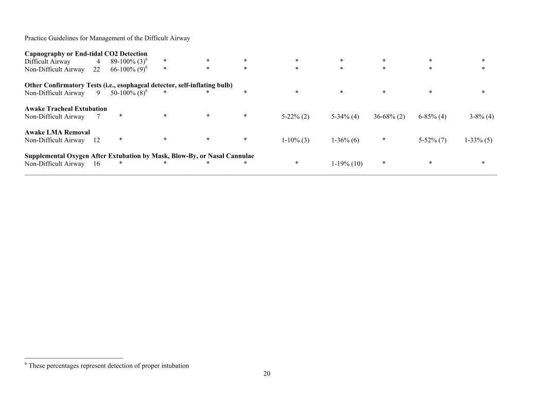
4. Confirmatory Tests of Tracheal Intubation a. Capnography or end-tidal CO2 detection verifies tracheal intubation and leads to fewer
adverse outcomes. b. Other confirmatory tests (i.e., esophageal detectors or self-inflating bulbs) verify
tracheal intubation and lead to fewer adverse outcomes. c. Fiberoptic confirmation of tracheal intubation
5. Awake Extubation
Awake extubation reduces airway-related adverse outcomes.
6. Supplemental Oxygen a. Supplemental oxygen delivery before induction by face mask or insufflation reduces
airway-related adverse outcomes. b. Supplemental oxygen delivery after extubation by face mask, blow-by, or nasal
cannulae of the trachea reduces airway-related adverse outcomes.
7. Follow-Up Care a. Post-extubation care and counseling reduces adverse airway-related outcomes. b. Documentation of a difficult airway and management reduces subsequent adverse
airway-related outcomes. c. Registration with an emergency notification service reduces subsequent adverse
airway-related outcomes. Scientific evidence was derived from aggregated research literature and from surveys, open
presentations and other consensus-oriented activities. For purposes of literature aggregation, potentially relevant clinical studies were identified via electronic and manual searches of the literature. The electronic search covered a 37-year period from 1966 through 2002. The manual search covered a 60-year period from 1943 through 2002. More than 3000 citations were initially identified, yielding a total of 1106 non-overlapping articles that addressed topics related to the 30 evidence linkages. After review of the articles, 538 studies did not provide direct evidence, and were subsequently eliminated. A total of 569 articles contained direct linkage-related evidence. Of these, 255 articles either used or included subjects with difficult airways.
14
Practice Guidelines for Management of the Difficult Airway
A directional result for each study was initially determined by a literature count, classifying each outcome as either supporting a linkage, refuting a linkage, or neutral. The results were then summarized to obtain a directional assessment of support for each linkage, with the intent of conducting meta-analyses where appropriate. Summary statistics for selected outcomes commonly reported in the literature are shown in Table 4. These descriptive statistics separate the reported outcome data for difficult and non-difficult airway subjects.
There was an insufficient number of acceptable studies to conduct meta-analysis for the
difficult airway.3 However, two evidence linkages contained studies pertinent to the Guidelines, with sufficient statistical information to conduct formal meta-analyses. These two linkages were: linkage 2d (preanesthetic preoxygenation for 3 minutes versus 4 maximal breaths) and linkage 6b (supplemental oxygen delivery by mask, blow-by, or nasal cannulae after extubation of the trachea).
Weighted mean effect sizes were determined for continuous outcome measures, and Mantel-
Haenszel odds-ratios were determined for dichotomous outcome measures. An acceptable significance level was set at p < 0.01 (one-tailed). Tests for heterogeneity of the independent studies were conducted to assure consistency among the study results. DerSimonian-Laird random-effects odds ratios were considered when significant heterogeneity was found. To control for potential publishing bias, a "fail-safe N" value was calculated. No search for unpublished studies was conducted, and no reliability tests for locating research results were done. For time to desaturation, the weighted mean effect size was d=1.57 (CI=0.98-2.14) for linkage 2d (preanesthetic preoxygenation for 3 minutes versus 4 maximal breaths). For reduced frequency of hypoxemia, the fixed-effects odds ratio was OR=5.98 (CI=3.16-11.31) for linkage 6b (suppl-emental oxygen delivery by mask, blow-by, or nasal cannulae after extubation of the trachea).
Interobserver agreement among Task Force members and two methodologists was
established by interrater reliability testing. Agreement levels using a Kappa (k) statistic for two-rater agreement pairs were as follows: (1) type of study design, k = 0.64 - 0.78; (2) type of analysis, k = 0.78 - 0.85; (3) evidence linkage assignment, k = 0.89 - 0.95; and (4) literature inclusion for database, k = 0.62 - 1.00. Three-rater chance-corrected agreement values were as follows: (1) study design, Sav = 0.73, Var (Sav) = 0.008; (2) type of analysis, Sav = 0.80, Var (Sav) = 0.008; (3) linkage assignment, Sav = 0.93 Var (Sav) = 0.003; (4) literature database inclusion, Sav = 0.80 Var (Sav) = 0.032. These values represent moderate to high levels of agreement.
The findings from the literature were supplemented by the opinions of Task Force
members as well as by surveys of the opinions of 50 anesthesiologists selected as consultants on the basis or their recognized interest in airway management. The kappa statistic (k) was used to obtain a quantitative measure of agreement among consultants. Consultants exhibited strong agreement (k > 0.75) on the potential beneficial effects of the following activities: conduct of the airway history and physical examination, advance preparation of the patient and equipment, formulation of strategies for intubation and extubation of the difficult airway, and provision of follow-up care.
15
3 Meta-analytic data for non-difficult airway patients can be obtained by writing to the American Society of Anesthesiologists.
Practice Guidelines for Management of the Difficult Airway
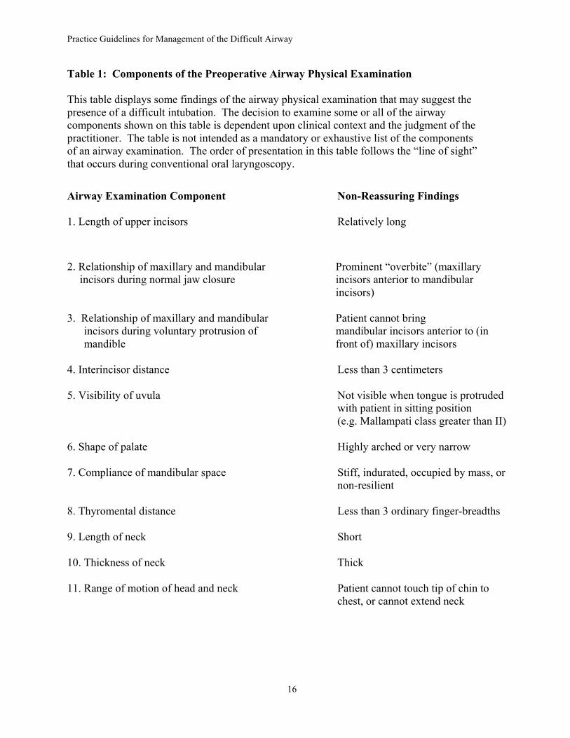
Table 1: Components of the Preoperative Airway Physical Examination This table displays some findings of the airway physical examination that may suggest the presence of a difficult intubation. The decision to examine some or all of the airway components shown on this table is dependent upon clinical context and the judgment of the practitioner. The table is not intended as a mandatory or exhaustive list of the components of an airway examination. The order of presentation in this table follows the “line of sight” that occurs during conventional oral laryngoscopy. Airway Examination Component Non-Reassuring Findings 1. Length of upper incisors Relatively long 2. Relationship of maxillary and mandibular Prominent “overbite” (maxillary
incisors during normal jaw closure incisors anterior to mandibular incisors) 3. Relationship of maxillary and mandibular Patient cannot bring
incisors during voluntary protrusion of mandibular incisors anterior to (in mandible front of) maxillary incisors
4. Interincisor distance Less than 3 centimeters 5. Visibility of uvula Not visible when tongue is protruded with patient in sitting position (e.g. Mallampati class greater than II) 6. Shape of palate Highly arched or very narrow 7. Compliance of mandibular space Stiff, indurated, occupied by mass, or non-resilient 8. Thyromental distance Less than 3 ordinary finger-breadths 9. Length of neck Short 10. Thickness of neck Thick 11. Range of motion of head and neck Patient cannot touch tip of chin to chest, or cannot extend neck
16
Practice Guidelines for Management of the Difficult Airway
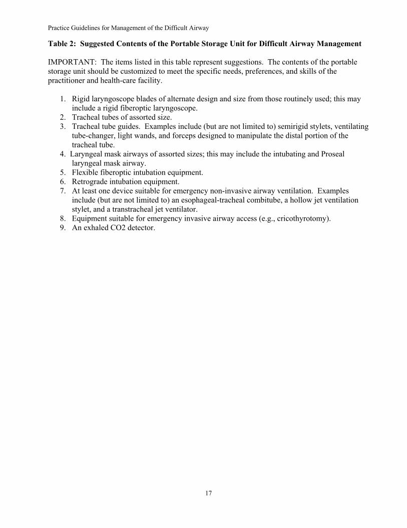
Table 2: Suggested Contents of the Portable Storage Unit for Difficult Airway Management IMPORTANT: The items listed in this table represent suggestions. The contents of the portable storage unit should be customized to meet the specific needs, preferences, and skills of the practitioner and health-care facility.
1. Rigid laryngoscope blades of alternate design and size from those routinely used; this may
include a rigid fiberoptic laryngoscope. 2. Tracheal tubes of assorted size. 3. Tracheal tube guides. Examples include (but are not limited to) semirigid stylets, ventilating
tube-changer, light wands, and forceps designed to manipulate the distal portion of the tracheal tube.
4. Laryngeal mask airways of assorted sizes; this may include the intubating and Proseal laryngeal mask airway.
5. Flexible fiberoptic intubation equipment. 6. Retrograde intubation equipment. 7. At least one device suitable for emergency non-invasive airway ventilation. Examples
include (but are not limited to) an esophageal-tracheal combitube, a hollow jet ventilation stylet, and a transtracheal jet ventilator.
8. Equipment suitable for emergency invasive airway access (e.g., cricothyrotomy). 9. An exhaled CO2 detector.
17
Practice Guidelines for Management of the Difficult Airway
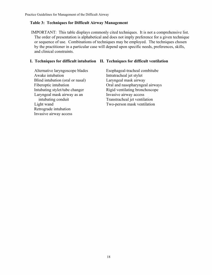
Table 3: Techniques for Difficult Airway Management IMPORTANT: This table displays commonly cited techniques. It is not a comprehensive list.
The order of presentation is alphabetical and does not imply preference for a given technique or sequence of use. Combinations of techniques may be employed. The techniques chosen by the practitioner in a particular case will depend upon specific needs, preferences, skills, and clinical constraints.
I. Techniques for difficult intubation II. Techniques for difficult ventilation Alternative laryngoscope blades Esophageal-tracheal combitube Awake intubation Intratracheal jet stylet Blind intubation (oral or nasal) Laryngeal mask airway Fiberoptic intubation Oral and nasopharyngeal airways Intubating stylet/tube changer Rigid ventilating bronchoscope Laryngeal mask airway as an Invasive airway access intubating conduit Transtracheal jet ventilation Light wand Two-person mask ventilation Retrograde intubation Invasive airway access
18
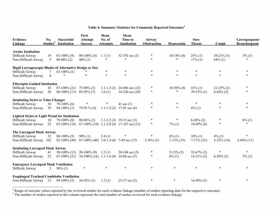
Table 4: Summary Statistics for Commonly Reported Outcomes4 First Mean Mean Evidence No. Successful Attempt No. of Time to Airway Sore Laryngospasm/ Linkage Studies5 Intubation Success Attempts Intubation Obstruction Hypoxemia Throat Cough Bronchospasm Awake Intubation Difficult Airway 19 81-100% (9) 80-100% (4) 1.1 (1) 52-192 sec (3) * 10-14% (4) 25% (1) 10-21% (3) 3% (1) Non-Difficult Airway 5 80-88% (2) 40% (1) * * * * 17% (1) 64% (1) * Rigid Laryngoscopic Blades of Alternative Design or Size Difficult Airway 7 63-100% (2) * * * * * * * * Non-Difficult Airway 8 * * * * * * * * * Fiberoptic-Guided Intubation Difficult Airway 43 87-100% (21) 75-80% (3) 1.1-1.3 (2) 24-406 sec (12) * 10-30% (4) 41% (1) 21-25% (2) * Non-Difficult Airway 26 88-100% (13) 85-95% (7) 1.0 (1) 16-220 sec (20) * * 39-53% (2) 6-64% (2) * Intubating Stylet or Tube-Changer Difficult Airway 10 78-100% (6) * * 41 sec (1) * * * * * Non-Difficult Airway 10 94-100% (7) 75-93 % (4) 1.1-1.2 (2) 17-91 sec (6) * * 6% (1) * * Lighted Stylet or Light Wand for Intubation Difficult Airway 10 79-100% (8) 80-86% (2) 1.1-2.2 (2) 19-33 sec (3) * * 6-20% (2) * 6% (1) Non-Difficult Airway 22 63-100% (16) 67-100% (10) 1.1-2.0 (4) 17-107 sec (12) * 7% (1) 10-49% (8) * * The Laryngeal Mask Airway Difficult Airway 32 80-100% (5) 90% (1) 2.4 (1) * * 6% (1) 10% (1) 4% (1) * Non-Difficult Airway 162 82-100% (49) 67-100% (40) 1.0-1.4 (6) 7-49 sec (15) 2-36% (5) 1-13% (15) 7-71% (33) 2-32% (14) 2-60% (11) Intubating Laryngeal Mask Airway Difficult Airway 41 30-100% (13) 20-100% (9) 1.3 (1) 20-168 sec (5) * 5-13% (2) 33-67% (2) * * Non-Difficult Airway 25 67-100% (21) 34-100% (16) 1.1-1.6 (4) 10-86 sec (5) * 6% (1) 14-21% (2) 6-28% (2) 3% (1) Emergency Laryngeal Mask Ventilation Difficult Airway 3 98% (1) * * * * * * * * Esophageal Tracheal Combitube Ventilation Non-Difficult Airway 13 94-100% (5) 38-92% (3) 1.2 (1) 25-27 sec (2) * * 16-48% (3) * *
4 Range of outcome values reported by the reviewed studies for each evidence linkage (number of studies reporting data for the respective outcome) 5 The number of studies reported in this column represent the total number of studies reviewed for each evidence linkage
Practice Guidelines for Management of the Difficult Airway
Capnography or End-tidal CO2 Detection Difficult Airway 4 89-100% (3)6 * * * * * * * * Non-Difficult Airway 22 66-100% (9)6 * * * * * * * * Other Confirmatory Tests (i.e., esophageal detector, self-inflating bulb) Non-Difficult Airway 9 50-100% (8)6 * * * * * * * * Awake Tracheal Extubation Non-Difficult Airway 7 * * * * 5-22% (2) 5-34% (4) 36-68% (2) 6-85% (4) 3-8% (4) Awake LMA Removal Non-Difficult Airway 12 * * * * 1-10% (3) 1-36% (6) * 5-52% (7) 1-33% (5) Supplemental Oxygen After Extubation by Mask, Blow-By, or Nasal Cannulae Non-Difficult Airway 16 * * * * * 1-19% (10) * * * __________________________________________________________________________________________________________________________________________
206 These percentages represent detection of proper intubation
Guideline for Management of the Difficult Airway
• Confirm Tracheal Intubation or LMA Placement with Exhaled CO2 21
Guideline for Management of the Difficult Airway
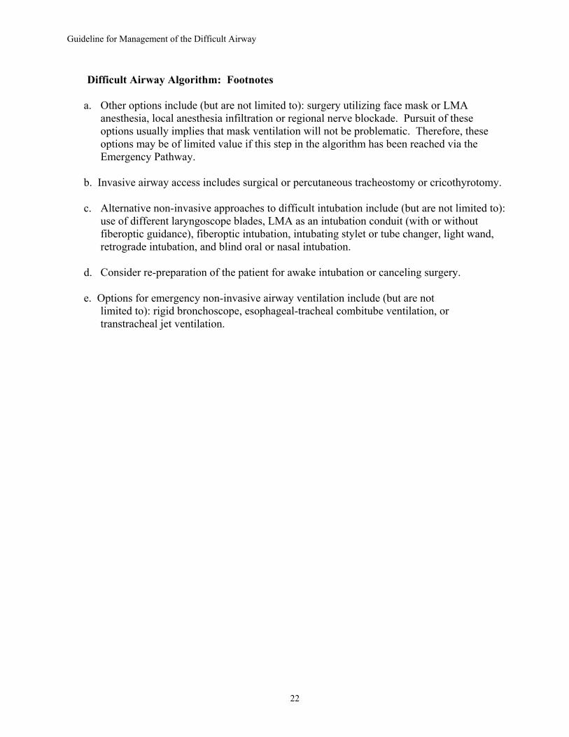
Difficult Airway Algorithm: Footnotes
a. Other options include (but are not limited to): surgery utilizing face mask or LMA anesthesia, local anesthesia infiltration or regional nerve blockade. Pursuit of these options usually implies that mask ventilation will not be problematic. Therefore, these options may be of limited value if this step in the algorithm has been reached via the Emergency Pathway.
b. Invasive airway access includes surgical or percutaneous tracheostomy or cricothyrotomy. c. Alternative non-invasive approaches to difficult intubation include (but are not limited to):
use of different laryngoscope blades, LMA as an intubation conduit (with or without fiberoptic guidance), fiberoptic intubation, intubating stylet or tube changer, light wand, retrograde intubation, and blind oral or nasal intubation.
d. Consider re-preparation of the patient for awake intubation or canceling surgery. e. Options for emergency non-invasive airway ventilation include (but are not limited to): rigid bronchoscope, esophageal-tracheal combitube ventilation, or
transtracheal jet ventilation.
22