Embed Size (px)
Citation preview

1
PRE-CLINICAL
PERIOD From. To
Name :
Permanent Address:
Present Address :
Phone :
Name & Address of Parent / Guardian
Blood Group :
Allergic to :

2
3
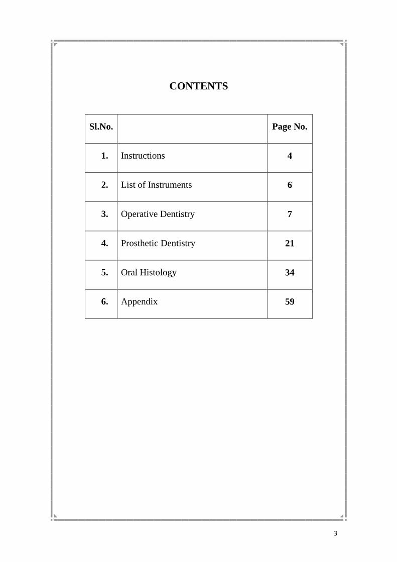
CONTENTS
Sl.No. Page No.
1. Instructions 4
2. List of Instruments 6
3. Operative Dentistry 7
4. Prosthetic Dentistry 21
5. Oral Histology 34
6. Appendix 59

4
Instructions
Before commencing the work, the student should get his/her
instruments checked and obtain the signature of the staff member.
On satisfactory completion of each case, the work should be
assessed as good, fair, poor and the signature of the staff member
obtained
The record must be carefully preserved and submitted to the Head
of the Department at the end of every year.

5
PRE-CLINICAL RECORD
DEPARTMENT OF
CONSERVATIVE DENTISTRY & ENDODONTICS
From............................ To..............................

6
Armamentarium
Conservative Dentistry :
1. Spirit Lamp
2. Mouth Mirror
3. Probe
4. Tweezer
5. Cotton Holder
6. Cement Spatula
7. Chip syringe
8. Contra angle hand piece
9. Straight hand piece
10. Airotor hand piece
11. Burs for airotor hand piece
(Round, Straight fissure, Tapering fissure, inverted cone)
12. Dappen dish
13. Amalgam carrier
14. Mortar & pestle
15. Amalgam plugger
16. Straight chisel
17. Excavator
18. Hoe
19. Hatchet
20. Gingival marginal trimmers
21. Plastic instruments
22. Matrix retainer & bands (Ivory No. 8)Universal retainer & bands
(Ivory No. 1)Universal retainer & bands
23. Mandrils
24. Stones
25. Amalgam polishing Kit-1
26. Inlay polishing rubber stone
27. A set of reamers and files (45-80 size)
28. Ball Burnisher
29. Diamond Carver for Amalgam
7
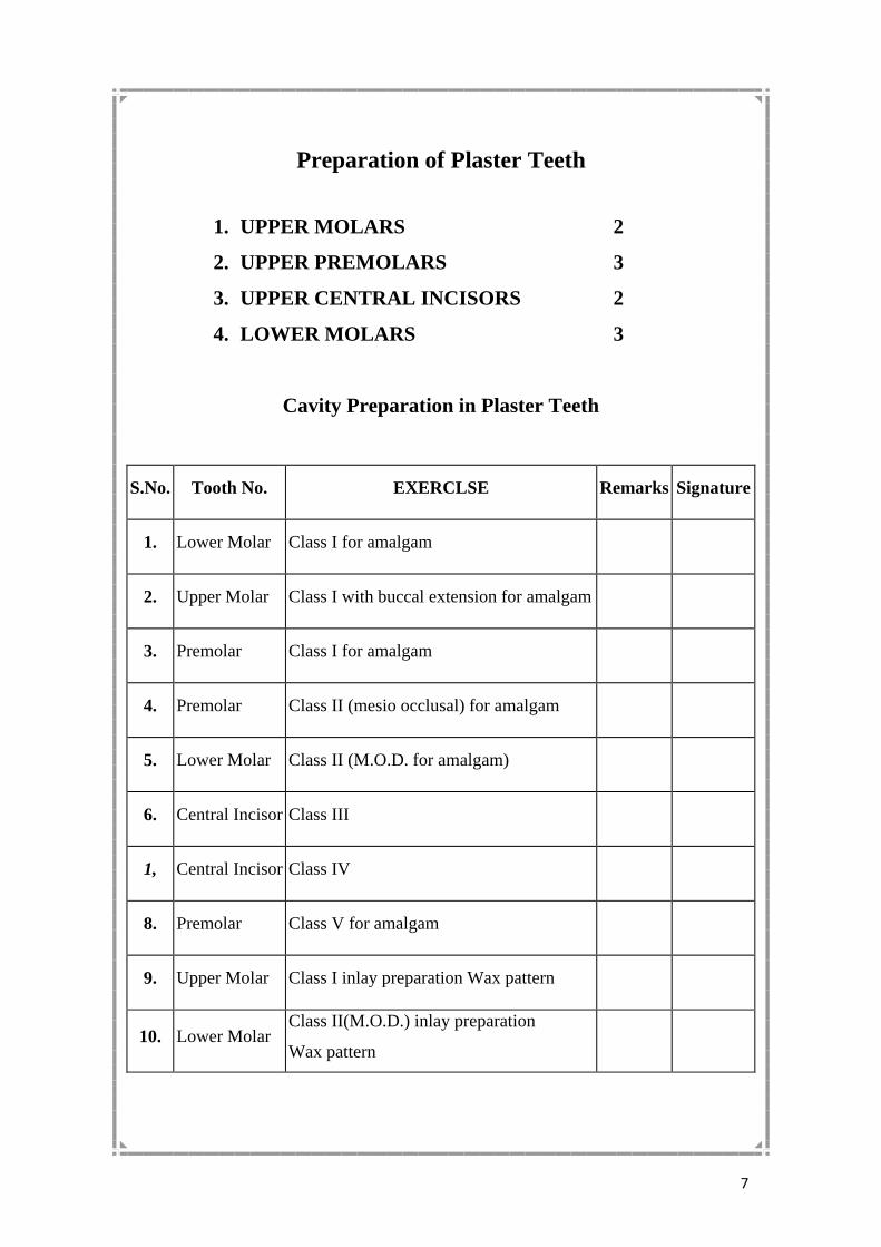
Preparation of Plaster Teeth
1. UPPER MOLARS 2
2. UPPER PREMOLARS 3
3. UPPER CENTRAL INCISORS 2
4. LOWER MOLARS 3
Cavity Preparation in Plaster Teeth
S.No. Tooth No. EXERCLSE Remarks Signature
1. Lower Molar Class I for amalgam
2. Upper Molar Class I with buccal extension for amalgam
3. Premolar Class I for amalgam
4. Premolar Class II (mesio occlusal) for amalgam
5. Lower Molar Class II (M.O.D. for amalgam)
6. Central Incisor Class III
1, Central Incisor Class IV
8. Premolar Class V for amalgam
9. Upper Molar Class I inlay preparation Wax pattern
10. Lower Molar Class II(M.O.D.) inlay preparation
Wax pattern

8
Setting of Natural Sound Teeth
(Excluding third molars) - Upper & Lower Models
Instructions to the Students
1. Before commencing work the instruments must be arranged on a
clean towel on the table.
2. Take special care in handling the equipment.
3. All upper teeth should be treated under indirect vision.
4. Before handing over the exercise, the restorations should be
polished.
5. All procedures should be handled using gloves and mask.
9
CAVITY PREPARATION IN NATURAL TEETH
CAVITY PREPARATION FOR AMALGAM
S.No Tooth No EXERCISE Remarks Signature
1. 34 Class I Preparation
Cement Lining
Amalgam Filling
2.
36
Class I Preparation
Cement Lining
Amalgam Filling
3.
15
Class I Preparation
Cement Lining
Amalgam Filling
4.
16
Class I with buccal extension
Cement Lining
Amalgam Filling
5.
37
Class I with buccal extension
Cement Lining
Amalgam Filling
6.
25
Class II Mesio-Occlusal
Cement Lining
Amalgam Filling
7.
26
Class II Mesio-Occluso-distal
Cement Lining
Amalgam Filling
8.
27
Class II Mesio-Occlusal
Cement Lining
Amalgam Filling

10

11
9.
45
Class II Mesio-Occlusal
Cement Lining
Amalgam Filling
10.
46
Class II Mesio-Occlusal-distal
Cement Lining
Amalgam Filling
11.
36
Class V Preparation
Cement Lining
Amalgam Filling
Cavity Preparation for Inlay
S.No Tooth No. EXERCISE Remarks Signature
12.
35
Class I Inlay Preparation
Wax Pattern, Sprue & Reservoir
Attachment Investing & Casting,
Trimming, Polishing & Cementing
13.
47
Class II Mesio Occlusal Inlay
Preparation
Wax Pattern, Sprue & Reservoir
Attachment Investing & Casting,
Trimming, Polishing & Cementing
Natural Tooth Cavity Preparation
S. No Tooth No. EXERCISE Remarks Signature
1.
Class I Cavity Preparation
2.
Class I Cavity Preparation
3.
Pulp Capping Cavity Preparation
Calcium hydroxide Sub-base
filling

12
13
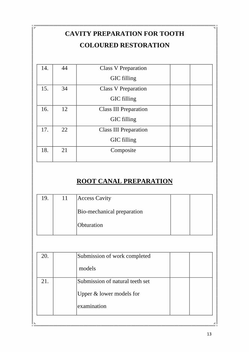
CAVITY PREPARATION FOR TOOTH
COLOURED RESTORATION
14. 44 Class V Preparation
GIC filling
15. 34 Class V Preparation
GIC filling
16. 12 Class III Preparation
GIC filling
17. 22 Class III Preparation
GIC filling
18. 21 Composite
ROOT CANAL PREPARATION
19.
11
Access Cavity
Bio-mechanical preparation
Obturation
20. Submission of work completed
models
21. Submission of natural teeth set
Upper & lower models for
examination

14

15
RECORD OF WORK DONE
Department of Conservative Dentistry & Endodontics
Name of the Student:
From..............................................................To...........................................
Attendance and Progress:
Remarks:
Verified and Found Correct:
Signature of Assistant Professor:
Signature of Professor&
Head of the Department
with Seal

16

17
Attendance and Progress
Lecture clinical Progress Signature
Date
Jan.
Feb.
March
April
May
June
July
Aug
Sept.
Oct.
Nov.
Dec.

18

19
Record of Work Done
By Mr./Mrs.
From....................................................To.....................................................
Remarks:
Signature of Head of the Department

20

21
PROSTHETICS
I year B.D.S Assessment Signature
EXERCISE No. 1
Preparation of Plaster Blocks 4
2 cm x 2 cm x 2cm
EXERCISE No. 2
(A) Preparation of Edentulous Models - 2 sets
1. Impression
2. Boxing of the Impression
3. Cast
4. Finishing
(B) Preparation of Dentulous Models - 2 Sets
1. Impression
2. Boxing of the Impression
3. Cast
4. Finishing
5. Signature

22
EXERCISE No. 3
EXERCISE No. 4
Partial Denture CI I & CI IV (Kennedy design)
1. Impression
2. Cast
3. Base plate adaptation
4. Bite rim
5. Articulation
6. Teeth Setting
7. Flasking
8. Packing and curing
9. Finishing
EXERCISE No. 5
Preparation of Complete Dentures
1. Impression
2. Cast
3. Adaptation of Base Plate
4. Occlusal Rim Preparation
5. Mounting on the Articulator
6. Setting of Teeth
7. Wax up
8. Flasking
9. Packing and Curing
10. Polishing and Finishing
Preparation of special Tray Resin
Tray
Shellac
Tray
1. Impression
2. Cast
3. Wax Pattern
4. Flasking
5. Packing
6. Finishing and Polishing
Assessment
Signature
23
II Year
PROSTHETICS:
EXERCISE No.1
Kennedys Class II
(Cast Partial Denture in Cobalt
Chromium Base Metal Alloy)
1. Impression
2. Cast
3. Surveying and Designing
4. Duplication and Preparation of Refractory cast
5. Designing and Preparation of Wax Pattern
6. Attachment of Sprues
7. Investing
8. Preheating and Wax elimination
9. Casting
10. Trimming, Finishing and Polishing
EXERCISENo.2
Rebasing Maxillary Denture
1. Impression
2. Cast Preparation
3. Articulation
4. Removal of the palatal, labial and buccal
portion of the Denture
5. Waxup
6. Flasking
7. Packing
8. Curing
Assessment
Signature

24
EXERCISE No. 3
Repair of Complete Denture
1. Assemble fractured parts
2. Fixation with model cement
3. Preparation of cast
4. Creation of space at the fractured site
and placement of retention grooves
5. Wax up
6. Processing the Denture
7. Trimming and Polishing
EXERCISE No. 4
Acrylic Cap Splint
1. Impression
2. Cast Preparation
3. Fracture and assemble to normal
Occlusion
4. Articulation
5. Preparation of Wax pattern
6. Polishing and Finishing
EXERCISE No. 5
One Clinical Impression of the Mouth
1. Impression
2. Cast Preparation
3. Finishing
EXERCISE No. 6
COMPLETE DENTURE WAX PATTERN
I
Setting
II
Setting
III
Setting
IV
Setting
V
Setting
1 . Preparation of Casts
2. Preparation of Occlusal Rims
3. Articulation
4. Arrangement of Teeth
5. Waxing, Carving & Polishing
Assessment & signature
Assessment
Signature

25
Attendance and Progress
Lecture Clinical Progress Signature
Date
Jan.
Feb.
March
April
May
June
July
Aug
Sept.
Oct.
Nov.
Dec.

26

27
Record of Work Done
By Mr./Ms
From.............................................. To.................................................
Exercises: I Year B.D.S.
II Year B.D.S
Complete Denture
Arrangement of teeth
Waxing, Carving & Polishing
Attendance:
Remarks :
Signature of External Examiner Signature of Head of the Department.

28

29
Carve Teeth in Wax Natural Size
I. B.D.S
1. Tooth No. 11 & 21
2. Tooth No. 12 & 22
3. Tooth No. 13 & 23
4. Tooth No. 14 & 24
5. Tooth No. 16 & 26
6. Tooth No. 41 & 31
7. Tooth No. 32 & 42
8. Tooth No. 33 & 43
9. Tooth No. 45 & 35
10. Tooth No. 46 & 36
11. Tooth No. 47 & 37
Assessment
Signature
1 1
2 2
3 3
4 4
6 6
1 1
2 2
3 3
5 5
6 6
7 7

30

31
Attendance and Progress
Lecture Clinical Progress Signature
Date
Jan.
Feb.
March
April
May
June
July
Aug
Sept.
Oct.
Nov.
Dec.

32

33
Record of Work Done
By Mr./ Mrs.
From........................................... To..............................................
Remarks:
Signature of Head of the Department

34

35

36

37

38

39

40

41

42

43

44

45

46

47

48

49

50

51

52

53

54

55

56

57

58

59

60

61

62

63

64

65

66

67

68

69

70

71

72
73

74

75

76

77

78

79

80

81

82

83

84

85

86

87

88
Attendance and Progress
Lecture Clinical Progress Signature
Date
Jan.
Feb.
March
April
May
June
July
Aug
Sept.
Oct.
Nov.
Dec.

89

90
Record of Work Done
By Mr./ Mrs.
From........................................... To..............................................
Remarks:
Signature of Head of the Department

91

92
Tooth Designation for Dental purposes Two Digit System
Introduction:
Over the Years since dentistry first required a method of tooth designation
various systems have been used. These have included multiple digit systems giving
quadrants and an indication of the teeth of the type set out in this international
Standard and those using an angular/grid, representation, let the quadrant with a 1 to 8
single digit system for the teeth.
The wide use of computers for information storage and the increasing need for
simple communication on dental matters by word of mouth, in print and / or write has
resulted in fresh thought being given to the requirements of a tooth designation
system. The two digit system detailed in this International Standard has been
compiled and endorsed by the Federation Dentair international (FDI)to Satisfy the
following requirements.
a. Simple to understand and teach
b. Easy to pronounce in conversation and dictation
c. Readily communicable in print and or write
d. Easy to translate into computer 'input'
e. Easily adaptable to standard charts used in general practice
Details of the factors relating to the decision of the FDI to adopt the two digit
system are recorded in appendix I of the FDI Chronicle of the 58th Annual Session
Scope and Field of Application:
This International standard establishes tooth designation system for dental
purposes using two digits to designate each tooth.
Principle:
The first digit indicates the quadrant and the second digit the tooth in the
quadrant. Quadrants are allotted the digits 1 to 4 for the permanent and 5 to 8 for the
deciduous teeth in a clockwise sequence starting at the upper right side, teeth within
the same quadrant allotted the digits 1 to 8(deciduous teeth 1 to 5) from the midline
backwards.. The digits shall be pronounced separately, thus the permanent canines are
teeth one three and four-three on the right side.
Designation System:
The designation system is illustrated below:
Permanent Teeth
(upper right) (upper left)
18 17 16 15 14 13 12 11 21 22 23 24 25 26 27 28
48 47 46 45 44 43 42 41 31 32 33 34 35 36 37 38
(lower right) (lower left)
Deciduous Teeth
(upper right) (upper left)
55 54 53 52 51 61 62 63 64 65
85 84 83 82 81 71 72 73 74 75
(lower right) (lower left)

93
Angles Classification of Malocclusion
Class I : The Lower dental arch is in normal relation to the upper dental
arch as evinced by the occlusion of the mesio buccal cups of the
upper first permanent molar in the buccal groove of the lower
first permanent molar.
Class II : The lower dental arch is in distal relation to the upper dental arch
as evinced by the disto buccal cusp of the upper first permanent
molar occluding in the buccal groove of the lower first
permanent molar.
Div. 1. The upper incisors are proclined.
Div. 2. Upper central incisors show lingual inclination and
overlapped by the upper lateral incisors.
Sub Div. Class II Occlusion on one side only
Class III : The lower is in mesial relation to the upper dental arch Mesio
buccal of the upper first permanent molar occludes distal to
the buccal groove of the lower first permanent Molar.

94
ORTHODONTICS
Angles Classification on Malocclusion
Class I : when the Mesiobuccal cusp, of the upper 1st permanent molar e
Mesiobuccal groove of the lower 1st permanent molar
Variations are: (i) Irregularity of individual teeth
(ii) Irregularity of a group of teeth
I. The Individual variations:
1. Labioversion
2. Linguoversion
3. Mesioversion
4. Distoversion
5. Torsiversion
6. Axioversion
7. Buccoversion
8. Palatoversion
II. The group variations are:
1. Bimaxillary Protrusion
2. Bimaxillary Spacing
3. Bimaxillary Crowding
4. Anterior Open Bite
Class II: When the distobuccal cusp of the upper 1st permanent molar
lies in the mesiobuccal groove of the lower 1 st permanent
molar There are two divisions of II Malocclusion namely.
(a) Class II Division I : When the molar relationship is of Class II nature
on both sides and the upper incisor are proclined.

95
CLASS II : Division 1-subdivision : when the molar relations is of Class
II nature on one side and of Class I nature on the opposite
side and is accompanied with proclination of upper incisors.
(B)
CLASS II : Division 2 : when the molar relations is of Class II nature on
both sides and upper incisors show lingual inclination and
lateral incisors are proclined
CLASS II :Division 2 - subdivision : when the molar relations is of
Class II on one side and of Class I nature on the other side
CLASS III : When the lower 1st permanent molar lies mesial to the
upper 1st permanent molar by a premolar width or a cuspal
width
CLASS III : Subdivision : when the rnolar relations is of Class III nature
on one side and of Class I nature on the other side
Black's Classification of Cavities
CLASS I : cavities occuririg in the pit and fissure defects in the occlusal
surfaces of the bicuspids and molars, the lingual surfaces of
the upper incisors and the buccal and lingual grooves that are
found occasionally on the occlusal surface of molars
CLASS II : Cavities in the proximal surfaces of posteriors.
CLASS III :Cavities in the proximal surfaces of anteriors without
involving incisal angle.
CLASS IV : Cavities in the proximal surfaces of anteriors involving the
incisal angle.
CLASS V : Cavities in the gingival one third of any tooth.

96
Kennedy's Classification of Partially Edentulous Arches
C1 I Bilateral edentulous areas located posterior to the remaining
natural teeth
C1 II Unilateral edentulous areas located posterior to the
remaining natural teeth
C1 III Unilateral edentulous area with natural teeth remaining both
anterior and posterior to it
C1 IV A single, anterior (crossing the midline) edentulous area
located anterior to the remaining natural teeth

97
Impaction of teeth:
Definition:
A tooth which is completely or partially unerupted and positioned
against another tooth ,bone ,soft tissue so that its further eruption is
unlikely described according to its anatomic position(Archer)
Classification of impaction:
Winter's classification:
Based on the position of the long axis of the impacted tooth in
relation to the long axis of the 2nd molar
1. vertical: the long axis of the third molar is parallel to that of the
2nd molar.
2. horizontal:the long axis of the third molar is at right angle to that
of the 2nd molar .
3. mesioangular impaction.
4. distoangular impaction:
all the previous four classes can come in:
a - lingual deflection.
b - buccal deflection.
5. inverted impaction

98
A. Relation of the tooth to the ascending ramus of the mandible and
to the distal surface of the 2nd molar: (Pell &Gregory)
Class1: The space between the anterior part of the ascending ramus
and the distal surface of the 2nd molar is sufficient to
accommodate the mesiodistal diameter of the crown of the third
molar.
Class2: The space between the anterior part of the ascending ramus
and distal surface of the 2nd molar is less than the mesiodistal
diameter of the crown of the third molar (part of the tooth located
within the ramus.
Class3: All the third molar is located within the ascending ramus of
the mandible.
B.Relative depth of the third molar in bone:
This show the superior inferior relationship of the tooth in relation
to the occlusal plane. (Pell & Gregory)
Position A: the highest portion of the tooth is on level with or
above the occlusal plane.
Position B: the highest portion is below the occlusal plane but
above the cervical margin of the 2nd molar
Position C:the highest point of the tooth is below the cervical
margins of the 2nd molar (deep impaction)

99
Classification of impacted maxillary third molar:
1. 2nd molar (as before)
2. The relationship of tooth to maxillary sinus :
a-sinus approximation : (s.a) where no bone or very thin bone exist
between the impacted teeth and floor of sinus.
b-no sinus approximation : (n.s.a) where 2 mm or more of bone exist
between the floor of sinus and impacted teeth.
Classification of impacted maxillary cuspids:
• Class1: palatally impacted cuspids ,these could be in vertical,
horizontal, semivertical position.
• Class2:labialy impacted cuspide which could be in vertical,
horizontal, semivertical.
• Class3:impacted cuspid located both in the palatal and labial
surfaces.
• Class4: impacted cuspid that are present in an edentulous maxilla
and may assume any of the previous three classes.