Embed Size (px)
Citation preview

PRECLINICAL EVIDENCE FOR THE EFFICACY OF ANGIOTENSIN RECEPTOR
ANTAGONISM IN A RODENT MODEL OF VULNERABILITY
TO COMORBID DEPRESSION AND CARDIOVASCULAR DISEASE
by
Kristen A. Stedenfeld
B.A. in Psychology, New York University, 2004
Submitted to the Graduate Faculty of
Arts and Sciences in partial fulfillment
of the requirements for the degree of
Doctor of Philosophy
University of Pittsburgh
2011

ii
UNIVERSITY OF PITTSBURGH
COLLEGE OF ARTS AND SCIENCES
This dissertation was presented
by
Kristen A. Stedenfeld
It was defended on
June 7, 2011
and approved by
Peter J. Gianaros, PhD, Associate Professor, Department of Psychiatry
Linda Rinaman, PhD, Professor, Department of Neuroscience
Susan R. Sesack, PhD, Professor, Department of Neuroscience
Etienne L. Sibille, Associate Professor, Department of Psychiatry
Dissertation Advisor: Alan F. Sved, Professor and Chairman, Department of Neuroscience

iii
Copyright © by Kristen A. Stedenfeld
2011

iv
Major depressive disorder and cardiovascular disease are highly comorbid, and the
presence of one disorder greatly increases the likelihood of the other. Remarkably, depressed but
otherwise healthy patients with no history of cardiovascular disease are as likely to have a heart
attack as patients with established cardiovascular disease.
Experimental studies have used chronic mild stress (CMS), a rodent model of depression
that uses a series of unpredictable, intermittent, and variable mild stressors to induce anhedonia,
one of the core diagnostic criteria for major depression. CMS also induces a constellation of
behavioral, physiological, and neuroendocrine responses that closely resemble those observed in
depressed patients, including alterations in autonomic control of the heart marked by decreased
heart rate variability (HRV). Commonly prescribed antidepressants might not improve
cardiovascular alterations associated with depression, even when depressive signs are
ameliorated. There is evidence, however, that candesartan, an angiotensin type 1 receptor
(AT1R) antagonist (ARB) often prescribed for cardiovascular disorders, has anxiolytic and
possibly antidepressant effects in animal models.
To study the possible antidepressant effects of candesartan, we first established a robust
rodent model of vulnerability to depression, since severity of depression is correlated with
PRECLINICAL EVIDENCE FOR THE EFFICACY OF ANGIOTENSIN RECEPTOR
ANTAGONISM IN A RODENT MODEL OF VULNERABILITY
TO COMORBID DEPRESSION AND CARDIOVASCULAR DISEASE
Kristen A. Stedenfeld, PhD
University of Pittsburgh, 2011

v
severity of cardiovascular changes in humans. We found that rats selectively-bred for low
locomotor responses to a novel environment (bLR) were especially vulnerable to CMS-induced
anhedonia and cardiovascular changes. Conversely, selectively-bred high-responder rats (bHR)
were resilient to the behavioral and cardiovascular changes induced by CMS.
Finally, we compared the effects of candesartan and the SSRI fluoxetine on CMS-
induced anhedonia and cardiovascular changes. We found that candesartan has profound
antidepressant effects, including rapid reversal of anhedonia, and attenuated anxiety-like
behavior. Furthermore, candesartan reversed cardiovascular changes, including clinically
relevant markers of risk for cardiac mortality. Thus the major findings of these studies are
twofold: (1) bHR/bLR rats exposed to CMS offer a robust model of the interactions of
predisposition and environmental stress that may contribute to depression and comorbid
cardiovascular disease and (2) candesartan and other ARBs may be novel therapies for the
treatment of comorbid depression and cardiovascular disease, and may be more effective than
traditionally-prescribed antidepressants such as SSRIs.
vi
TABLE OF CONTENTS
ACKNOWLEDGEMENTS .................................................................................................. XII
LIST OF ABBREVIATIONS ............................................................................................. XVI
1.0 INTRODUCTION.....................................................................................................1
1.1 DEPRESSION AND CARDIOVASCULAR DISEASE ARE HIGHLY
COMORBID .....................................................................................................................1
1.2 MAJOR DEPRESSION: A DISORDER OF MOOD AND PHYSIOLOGY .2
1.2.1 Diagnostic criteria for depression ................................................................2
1.2.2 Physiological changes that co-occur with depression ..................................4
1.3 AUTONOMIC CONTROL OF THE HEART IS ALTERED IN
DEPRESSION AND CARDIOVASCULAR DISEASE ..................................................4
1.3.1 Normal autonomic control of the heart ........................................................4
1.3.2 Reduced heart rate variability is a marker of cardiac mortality risk .........7
1.4 ANIMAL MODELS OF DEPRESSION: UNPREDICTABLE CHRONIC
MILD STRESS ................................................................................................................ 10
1.4.1 Modeling vulnerability to depression: High- and low-responder rats ...... 11
1.4.2 Novelty-seeking predicts emotionality in screened supplier-bred rats ..... 11
1.4.3 Selectively-bred rats represent the extreme ends of a spectrum of
behavior and mood ................................................................................................. 12

vii
1.5 ANTIDEPRESSANT TREATMENT: HOW CAN YOU MEND A BROKEN
HEART? ………………………………………………………………………………….13
1.6 THE RENIN-ANGIOTENSIN SYSTEM IN STRESS AND DEPRESSION
…………………………………………………………………………………..13
1.7 COULD AN ANTAGONIST OF THE RENIN-ANGIOTENSIN SYSTEM
BE USED TO TREAT COMORBID DEPRESSION AND CARDIOVASCULAR
DISEASE? ....................................................................................................................... 14
2.0 NOVELTY-SEEKING BEHAVIOR PREDICTS VULNERABILITY IN A
RODENT MODEL OF DEPRESSION .................................................................................. 16
2.1 ABSTRACT .................................................................................................... 16
2.2 INTRODUCTION .......................................................................................... 17
2.3 EXPERIMENTAL PROCEDURES .............................................................. 19
2.4 RESULTS ........................................................................................................ 24
2.5 DISCUSSION .................................................................................................. 31
3.0 VULNERABILITY TO ANHEDONIC BEHAVIOR IS ASSOCIATED WITH
CARDIOVASCULAR ALTERATIONS IN A RODENT MODEL OF DEPRESSION ...... 36
3.1 ABSTRACT .................................................................................................... 36
3.2 INTRODUCTION .......................................................................................... 37
3.3 EXPERIMENTAL PROCEDURES .............................................................. 39
3.4 RESULTS ........................................................................................................ 43
3.5 DISCUSSION .................................................................................................. 59
4.0 CANDESARTAN REVERSES ANHEDONIA AND CARDIOASCULAR
ALTERATIONS IN A RODENT MODEL OF DEPRESSION ............................................ 66
viii
4.1 ABSTRACT .................................................................................................... 66
4.2 INTRODUCTION .......................................................................................... 67
4.3 EXPERIMENTAL PROCEDURES .............................................................. 70
4.4 RESULTS ........................................................................................................ 76
4.5 DISCUSSION .................................................................................................. 91
5.0 GENERAL DISCUSSION ...................................................................................... 99
5.1 SUMMARY AND INTERPRETATIONS OF CURRENT FINDINGS ....... 99
5.2 POSSIBLE MECHANISMS BY WHICH CANDESARTAN REVERSES
DEPRESSION-LIKE BEHAVIOR AND CARDIOVASCULAR DYSFUNCTION .. 103
5.3 FUTURE DIRECTIONS .............................................................................. 106
5.4 RELEVANCE AND CONCLUSIONS ......................................................... 108
BIBLIOGRAPHY ................................................................................................................. 110

ix
LIST OF TABLES
Table 1: Comparison of body weight at baseline and at each of four weeks of CMS .................. 24
Table 2: Peak and duration of heart rate and blood pressure responses to three CMS stressors ... 50
Table 3: Frequency domain components of HRV, third quarter of the dark period ..................... 55

x
LIST OF FIGURES
Figure 1: Diagnostic criteria for major depressive disorder, from the Diagnostic and Statistical
Manual of Mental Disorders (DSM-IV). .....................................................................................3
Figure 2: Schematic of sympathetic and parasympathetic control of the heart ..............................6
Figure 3: Diagram demonstrating the concept of frequency domain analysis. ..............................9
Figure 4: An example of a typical chronic mild stress (CMS) schedule...................................... 21
Figure 5: bHR and bLR pre-CMS locomotor activity in response to a novel environment.......... 26
Figure 6: Percent preference for sucrose across four weeks of CMS .......................................... 28
Figure 7: Latency to approach and consume a food pellet in the novelty-suppressed feeding test
................................................................................................................................................. 30
Figure 8: 24-hour resting heart rate and blood pressure.............................................................. 46
Figure 9: Heart rate responses to three CMS stressors during CMS week 4 ............................... 49
Figure 10: Time-domain HRV in bHR/bLR rats across four weeks of CMS .............................. 53
Figure 11: Frequency domain components of HRV, final quarter of the dark period .................. 54
Figure 12: Increases in HR and BP in response to a novel cage stress in bHR/bLR rats ............. 57
Figure 13: Duration of the increase in HR and BP in response to a novel cage stress ................. 58
Figure 14: Diagram of experimental procedures and typical CMS schedule ............................... 71
Figure 15: Percent sucrose preference across 9 weeks of CMS .................................................. 79
xi
Figure 16: Increase in body weight across 9 weeks of CMS ...................................................... 80
Figure 17: Results from novelty-suppressed feeding test (NSF) test during CMS week 9 .......... 82
Figure 18: Results from the forced swim test (FST) during CMS week 8 ................................... 84
Figure 19: Line graphs of 24-hour resting HR and BP across 9 weeks of CMS .......................... 86
Figure 20: Changes in time-domain HRV across 9 weeks of CMS, with and without drug
treatment. .................................................................................................................................. 88
Figure 21: Frequency domain components of HRV during CMS week 9 ................................... 90

xii
ACKNOWLEDGEMENTS
The completion of these experiments was only possible due to the efforts of a large and diverse
group of supportive and hard-working people. I hope that anyone reading this section will
forgive me for its length and occasional sentimentality. When it comes to the expression of
gratitude, I feel that it’s best not to be stingy.
I am so grateful for the opportunities given to me by the Department of Neuroscience, the
Center for Neuroscience at the University of Pittsburgh, and the Center for the Neural Basis of
Cognition. It is inspiring to be surrounded by so many wonderful, smart, passionate people.
Boundless thanks go to my advisor and mentor, Dr. Alan Sved. Alan is a thoughtful
mentor, an inspiring teacher and an awesome scientist. I would like to thank Alan for his
generosity in encouraging my projects, his commonsense approach to experiments, his patience
with my often inconvenient schedule, for always pushing me to be a better, more careful
scientist, for imparting both his knowledge and his passion for science, and most of all for his
kindness.
Many thanks go to my wonderful committee members for their support, encouragement
warmth, and enthusiasm. Thank you to Dr. Etienne Sibille, the chair of my committee, for his
practical and straightforward outlook. Thank you to Dr. Susan Sesack for her humor and candor.
Thank you to Dr. Peter Gianaros for his advice and guidance during my adventure with heart rate
variability. And special thanks go to Dr. Linda Rinaman, who was the first person to show me

xiii
how to pick up a rat during my rotation in her lab, and who has been supportive ever since. I
would also like to thank Dr. Randall Sakai, my outside examiner, for adding another unique
voice to my diverse committee. Thanks also to Dr. Ed Stricker and Dr. Pat Card, former
committee members who have each been powerful sources of support and guidance at critical
points in my graduate career.
I am so thankful for the friendly encouragement of Joan Blaney and Marlene Nieri, and
for the staff of the Department of Neuroscience fiscal office who keep the rest of us going: Mary
Spanoudakis, Debbie Glumac, Kathy Short, and Joni Mainier. They are an impressive group of
women who do so much and yet always find the time to say hello.
I would like to thank my collaborators, Dr. Ilan Kerman and Dr. Sarah Clinton from the
University of Alabama Birmingham, and Dr. Huda Akil and Dr. Stan Watson from the
University of Michigan, who supplied me with the selectively-bred strains of rats used in these
studies. I absolutely could not have done these experiments without them! I am especially
thankful to Ilan and Sarah who have been such great role models for me as I figure out how to
balance work and science and parenthood. I would also like to thank Dr. Angela Grippo, who
pioneered this line of research. Angela’s publications were the inspiration for these projects, and
she was always willing to share her knowledge and encourage me along the way. The research of
Dr. Juan Saavedra is the other half of the inspiration behind these experiments, and I am grateful
and humbled at the opportunity to collaborate with him.
Thank you to Dr. Israel Christie for his tutelage in the fine art of frequency domain
analysis, and the use of his fantastic software, Physioscripts. Without his help, I would still be
sorting through an unending pile of numbers. Thank you to Dr. Debbie Chapman and Dr. Joe
Newsome who went out of their way to find a way for me to do these experiments in our animal

xiv
facility. Thank you to Barb Cohen and Michelle Reddy for caring for my rats and making
accommodations for these sometimes-strange experiments.
Many thanks to the members of the Sved lab including Xia Li who taught me to be
careful with brains, and Dr. Judy Sved who taught me excellent surgical technique. Thanks also
to Dr. Gina Cano and Vivian Allahyari, for their camaraderie and encouragement. I was also
fortunate to have the help of two outstanding undergraduate students, Nichole Johnson and Jenna
Parrish, who always kept me laughing and reminded me how fun and strange science can be.
I am especially grateful for the help, support, and friendship of Myriam Stricker, who
entered my life as a coworker and quickly became one of my most treasured friends. There is not
a single area of my life that Myriam’s generosity and kindness has left untouched, and I am a
better person because of her friendship. I could not have completed these experiments or
finished my dissertation without her considerable help, nor would the process have been nearly
as enjoyable. Myriam, from the bottom of my heart: Thank you.
A number of dear friends have made my graduate experience a truly wonderful one,
including (but not limited to) Dr. Kate Cosgrove, Dr. Ericka Holmstrand, Jesse Wood, and Joy
Balcita-Pedicino. An extra special bit of gratitude is owed to Dr. Layla Banihashemi, who has
been such a fabulous friend and supporter from my very first day of graduate school. Thank you,
Layla, for always listening.
I am so blessed to be buoyed by the love of an extraordinary family, including my
parents, Karen and Randy Maynard and Dennis Stedenfeld. My mother, Karen, deserves much
of the credit for nurturing my curiosity, my fascination with taking things apart to figure out how
they work, and my admiration of the resilience of the human spirit. Warm thanks go to my
incredible sister, Laura Stedenfeld, for her steadfast encouragement and support. I would also

xv
like to acknowledge the profound influence of my late grandfather, Arthur O’Brien, who was the
first person to show me how science could explain the things in nature I loved so much. Many
thanks to my family-in-law, who have adopted me as one of their own from the beginning
(whether I liked it or not): Joseph Schmich, Tina Schmich and Lori Nelson, Meredith Schmich,
and Dustin Schmich. You are all a bunch of lunatics, and I love you.
In closing, I would like to express my tremendous love and gratitude for my husband
Ben, my partner in all things, closest friend, and all-around favorite person. I honestly don’t
know what I would do without him. And last, but far from least, thank you to my amazing
daughter, Adelaide. Every day that I spend with her I’m reminded of what a great big beautiful
world we live in. I am so very lucky to have been able to spend my time studying one small
corner of it.

xvi
LIST OF ABBREVIATIONS
5-HTT – Serotonin transporter
ACE – Angiotensin converting enzyme
ANS – Autonomic nervous system
ARB – Angiotensin II type 1 receptor blocker
AT1Rs – Angiotensin type 1 receptors
bHR – Selectively-bred high-responder rat
bLR – Selectively-bred low-responder
BP – Blood pressure
BrdU - 5-bromo-2'-deoxyuridine
CMS – Chronic mild stress
CRH – Corticotrophin-releasing hormone
DSM-IV – Diagnostic and statistical manual of mental disorders, 4th edition
EPMZ – Elevated plus maze
FRL – Flinders resistant line
FSL – Flinders sensitive line
FST – Forced swim test
HAB – High anxiety-like behavior rat
HR – Heart Rate
xvii
HiR – High-responder rat (supplier-bred)
HPA – Hypothalamic pituitary adrenal
HF – High frequency
HRV – Heart rate variability
IBI – Inter-beat interval
IL-1β – Interleukin 1β
IL-6 – Interleukin-6
LAB – Low anxiety-like behavior
LF – Low frequency
LoR – Low-responder rat (supplier-bred)
NSF – Novelty-suppressed feeding
PVN – paraventricular nucleus of the hypothalamus
RAS – Renin angiotensin system
SA – Sinoatrial
SEM – Standard error of the mean
SPT – Sucrose preference test
SSRI – Selective serotonin reuptake inhibitor
TNF- - Tumor necrosis factor -
TP – Total power
VLF – Very low frequency

1
1.0 INTRODUCTION
1.1 DEPRESSION AND CARDIOVASCULAR DISEASE ARE HIGHLY COMORBID
Emotions, personality, and mood can have a tremendous impact on human physiology, and
prolonged periods of emotional distress can have serious repercussions on long-term health. A
profound example of this interaction of psychology and physiology is the highly comorbid
relationship between major depressive disorder and cardiovascular disease. Incredibly,
depressed but otherwise healthy patients with no history of cardiovascular disease are as likely to
have a heart attack as patients with established cardiovascular disease [1, 2]. This relationship
exists independent of common risk factors, such as increased body mass index, smoking, or
preexisting cardiac pathophysiology [3]. Furthermore, depression is considered a significant risk
factor for coronary heart disease and cardiac mortality [2-4]. In the six months following a
myocardial infarction, mortality was 3.5 times higher in patients who were also depressed than
those who were not [2]. The association between depression and cardiovascular disease is also
bidirectional, and patients who have recently had a major cardiac event, such as a myocardial
infarction are more likely to be diagnosed with depression. Indeed, while approximately 2-9% of
American adults suffer from depression, the prevalence of this disorder in patients recovering
from myocardial infarction is between 20-40% [5-7]. Cardiovascular disease and major
depression are currently two of the most detrimental disorders in developed countries and

2
prevalence of both disorders continues to rise [8, 9]. Although the comorbid, bidirectional
relationship between depression and cardiovascular disease is gaining attention [10], many
important questions regarding the relationship between the two disorders, and the mechanisms
that might connect them, remain unanswered.
1.2 MAJOR DEPRESSION: A DISORDER OF MOOD AND PHYSIOLOGY
1.2.1 Diagnostic criteria for depression
Major depressive disorder is a complex and devastating disorder with significant behavioral,
physiological, and neuroendocrine alterations. It is estimated that somewhere between 20 - 25%
of the population will suffer from some form of depression in their lifetime [11]. The Diagnostic
and Statistical Manual of Mental Disorders (DSM-IV) lists two main criteria of the disorder:
persistent depressed mood, and anhedonia, a lack of or reduction in pleasure for things that used
to be pleasurable. A clinical diagnosis of major depression requires either both of these criteria
or one of these criteria accompanied by a certain number of the following changes: changes in
body weight or appetite, changes in sleep pattern, low energy or fatigue, diminished
concentration, feelings of worthlessness, guilt, or despair, or recurrent thoughts of death or
suicide [12] (Figure 1).

3
Figure 1: Diagnostic criteria for major depressive disorder, from the Diagnostic and Statistical
Manual of Mental Disorders (DSM-IV).

4
1.2.2 Physiological changes that co-occur with depression
In addition to these diagnostic criteria, a number of neuroendocrine and physiological changes
have been observed in depressed patients: increased levels of sympathetic activity, marked by
increased norepinephrine excretion [13], hyperactivation of the hypothalamic pituitary adrenal
(HPA) axis [14-17], increased levels of immune factors, such as pro-inflammatory cytokines
[18], and cardiovascular changes, such as increased resting heart rate (HR) and decreased heart
rate variability (HRV) [19-21] . Decreased HRV is an especially critical symptom of depression,
as it is also used as a clinical marker for increased risk of cardiac mortality [22], and will be
discussed in detail below.
1.3 AUTONOMIC CONTROL OF THE HEART IS ALTERED IN DEPRESSION
AND CARDIOVASCULAR DISEASE
1.3.1 Normal autonomic control of the heart
Heart rate is controlled by the sinoatrial (SA) node, a cluster of autorhythmic cells located in the
right atrium of the heart, which generates electrical impulses that cause the heart to contract. The
SA node is often called the pacemaker of the heart, and receives neural input from both
complementary branches of the autonomic nervous system (ANS). The sympathetic branch of
the ANS, also called the ―fight or flight‖ branch has excitatory effects on the periphery, working
to mobilize the body to deal with the source of stress: blood flow to muscles increases, digestion

5
is inhibited, and HR is increased. The parasympathetic branch, sometimes called the ―rest and
digest‖ branch, is generally inhibitory, helping to return the body to rest after a stress,
decreasing HR, and aiding in activities such as digestion, sexual arousal, and growth. These
dual branches of the ANS work together to modulate heart rate based on the state of the
organism. At rest, both branches are active, though the parasympathetic branch predominates to
keeps HR at a rate that is lower than intrinsic SA node activity. This is sometimes referred to as
a parasympathetic ―brake‖ on HR, and allows an extra level of control over HR. In times of
stress, HR can be increased by inhibiting parasympathetic activity and increasing sympathetic
activity; conversely, HR is decreased by increasing parasympathetic and inhibiting sympathetic
activity. The sympathetic and parasympathetic branches of the ANS are each controlled by
brainstem areas of the central nervous system (Figure 2). Pre-sympathetic neurons in the
brainstem (e.g. rostral ventrolateral medulla and raphe pallidus) project to the intermediolateral
cell column in the spinal cord, which project in turn to the sympathetic ganglia that innervate the
heart. The parasympathetic (i.e. vagal) branch originates in the nucleus ambiguus and dorsal
motor nucleus.

6
Figure 2: Schematic of sympathetic and parasympathetic control of the heart

7
1.3.2 Reduced heart rate variability is a marker of cardiac mortality risk
In a healthy cardiovascular system, the sympathetic and parasympathetic branches of the ANS
work to ensure proper cardiovascular function under the varying conditions experienced by the
organism. However, prolonged periods of chronic stress can have lasting effects on cardiac
autonomic tone, characterized by an increase in sympathetic activity, a decrease in
parasympathetic activity, or both. Time-domain HRV is a measurement of the variability in time
between heart beats [i.e., the standard deviation of the inter-beat interval (IBI)], and reflects the
interaction of sympathetic and parasympathetic influences on HR [22]. A reduction in HRV
indicates a shift in sympathovagal balance toward higher sympathetic tone, and is a significant
predictor of mortality following myocardial infarction [23, 24].
Depression is highly comorbid with a number of cardiovascular disturbances that fall
under the general term of cardiovascular disease. Specifically, these include atherosclerosis,
ventricular arrhythmia, myocardial infarction, and sudden cardiac death [7, 25-27]. Depression is
also associated with increased risk of progression of heart failure [28]. Each of these
cardiovascular pathologies, in turn, has been associated with prolonged sympathovagal
imbalance, as measured by reduced HRV. Reduced HRV is consistently reported after
myocardial infarction, predicts future cardiac pathology such as ventricular arrhythmia and
sudden death [29], and is associated with higher incidence of cardiovascular mortality [30, 31].
Indeed, reduced HRV predicts the progression of atherosclerosis even more reliably than
common risk factors such as smoking status or lipid measurements [32]. Additionally, reduced
HRV is frequently observed in medically well depressed patients and reductions in HRV are
significantly worse in severely depressed patients [21, 33]. Indeed, major to severe depression

8
doubles the cardiac mortality risk, as predicted by reduced HRV, in patients who are otherwise
medically healthy [1].
While HRV in the time domain (discussed above) is useful as an index of overall
autonomic influence of HR, frequency domain analysis of HRV can be used as an index of
relative sympathetic and parasympathetic components that contribute to HRV. Frequency
domain analysis identifies and measures rhythmic fluctuations in HR, and examines power (e.g.
variability) as a function of frequency. The frequency distributions produced by this method are
separated into frequency bands that have been shown to correspond to specific components of
the ANS (Figure 3). Total power across the frequency spectrum is related to overall autonomic
balance [22]. The high frequency (HF) band corresponds to the respiratory frequency, and is
used as an index of vagal or parasympathetic influence on HRV [22, 34]. Low frequency (LF)
HRV is influenced by sympathetic and parasympathetic elements [22, 35] and very low
frequency (VLF) power likely reflects modulation by the renin-angiotensin system [29, 35, 36]
or its effects on parasympathetic influence on the heart [36]. Reduced VLF is an especially
useful measure, as it strongly predicts cardiac mortality, and accounts for up to 30% of the
mortality risk of depression after myocardial infarction [31].

9
Figure 3: Diagram demonstrating the concept of frequency domain analysis.
Frequency domain analysis first separates waveform data (Fig 3A) into its component frequencies (Fig
3B) and then identifies and quantifies the amount of fluctuation (i.e. variability) in HR that is accounted
for by a given frequency band. Pharmacological studies have shown that these frequency bands can be
used as an index of autonomic function (Fig 3C).

10
1.4 ANIMAL MODELS OF DEPRESSION: UNPREDICTABLE CHRONIC MILD
STRESS
The comorbid relationship between depression and cardiovascular disease has been identified
and firmly established because of important work in clinical and epidemiological research [2-4,
6, 10, 20, 37-45]. However, work in experimental animal models has significantly contributed to
a more complete understanding of the mechanisms that are common to both disorders. Of the
available animal models of depression, one of the currently best-validated is the chronic mild
stress (CMS) model of depression used in rodents [46, 47]. In this model, an animal is exposed
to a variety of mild, yet unpredictable, stressors over the course of many weeks. CMS induces a
constellation of altered behavioral, physiological, and neuroendocrine responses that closely
resembles aspects of human depression [46]. The unpredictable nature of CMS is critical, and
predictable stress has been shown to have an opposite effect, improving measures of mood and
cognitive function in rats [48]. Depression-like behaviors have been evaluated by measuring
anhedonia, one of the core signs of depression [12]. Anhedonia has been operationally defined
using a variety of measures, including decreases in sucrose preference [49-53], approach to
palatable food [54], brain stimulation reward [47, 55], and even palatable food-induced
dopamine release in nucleus accumbens and prefrontal cortex [56, 57]. CMS-induced anhedonia
is reversed by chronic, but not acute, administration of common antidepressants [46, 47, 53, 58-
63], demonstrating that CMS is an appropriate model for studying the effects of chronic
antidepressant treatment. CMS has proven to be a very useful tool for studying physiological and
neural alterations that may take place during the development of depression [58, 64-66]. For
instance, a series of studies that used CMS to induce anhedonic behavior in rats also reported
changes in cardiovascular function that parallel those observed in depressed patients, including a

11
small increase in HR, decreased HRV, and an increased susceptibility to arrhythmia [49, 53, 67,
68]. However, a common criticism of the CMS model is that results can vary across
experiments, even within labs. Indeed, in preliminary experiments performed in our lab, CMS-
exposed rats had a modest but significant decrease in sucrose preference, which was variable
across individual rats. Furthermore, preference for sucrose was positively correlated with pre-
CMS locomotor activity in an open field. These results suggested that individual differences in
behavioral traits might predispose certain rats to be vulnerable or resilient to the anhedonic
effects of CMS.
1.4.1 Modeling vulnerability to depression: High- and low-responder rats
Although factors such as major life stress certainly contribute to the onset of depression, some
individuals are more likely than others to become depressed. Indeed, depression is most likely
aused by a complex interaction of genetic predisposition and environmental factors, and
epidemiological studies estimate that genetic factors account for approximately 40-50% of risk
for depression [11, 69]. Most individuals experience stressful life events, yet only a portion of
individuals will experience serious anxiety or depressive disorders as a result of these events
[70].
1.4.2 Novelty-seeking predicts emotionality in screened supplier-bred rats
One way to approach the issue of vulnerability and resilience has been to correlate baseline
individual differences in behavior with either vulnerability or resistance to markers of mood
disorders (e.g. behavior, genes) [71-75]. For instance, based on locomotor activity in a novel

12
environment, Sprague-Dawley rats can be classified as either high-responders (HiR), which have
high levels of exploration, or low-responders (LoR) which have low levels of exploration [71,
74, 76-78]. Since low exploratory activity is interpreted as a sign of anxiety-like behavior due to
rodents’ innate aversion to novel, open spaces, it is not surprising that this classification predicts
emotional reactivity in other behavioral tests. Rats classified as LoR have higher anxiety-like
behavior than HiR rats in tests such as the elevated plus maze (EPMZ) and the light-dark box
[71, 76, 79, 80].
1.4.3 Selectively-bred rats represent the extreme ends of a spectrum of behavior and
mood
One approach that has been used to study trait differences in rats is to selectively breed animals
at the opposite end of the normal distribution, and this strategy has recently been applied to
locomotor activity in a novel environment. These selectively-bred strains (bred HiR [bHR] and
bred LoR [bLR], respectively) are thought to represent the extremes of the normal distribution of
the Sprague-Dawley population. Individual differences in anxiety-like behavior persisted and
intensified as rats were selectively-bred on the trait of LoR or HiR [71, 81], making them a
convincing model of inherited susceptibility or resilience to mood disorders. Indeed, as the
results in Chapter 2 demonstrate, bHR and bLR rats appear to be resilient and susceptible,
respectively, to CMS-induced depression-like symptoms, and CMS-exposed bLR are an
especially robust model of anhedonic behavior.

13
1.5 ANTIDEPRESSANT TREATMENT: HOW CAN YOU MEND A BROKEN
HEART?
If depression and cardiovascular disease are connected, does treating depression also improve
decreased HRV? In the case of tricyclic antidepressants, the answer is decidedly no, as those
drugs are known to have cardiotoxic properties [82-84], leading doctors to discourage their use
in patients that are especially vulnerable to cardiovascular dysfunction [85, 86]. In the case of
selective serotonin reuptake inhibitors (SSRIs), reports are much less clear. Some studies have
reported that SSRIs improve cardiovascular outcomes and reduce mortality [19, 87], while others
have found that SSRIs do not improve mortality risk, even when depression is in remission [88].
Finally, some studies have reported that SSRIs may actually exacerbate cardiac mortality,
especially when administered in conjunction with beta-blockers, which are commonly prescribed
for various cardiovascular disorders [89]. Cognitive behavioral therapy also improves signs of
depression but does not alter cardiac mortality [90]. These varied results present the troubling
possibility that in some individuals the cardiac mortality risk associated with depression may
persist even when depression is treated.
1.6 THE RENIN-ANGIOTENSIN SYSTEM IN STRESS AND DEPRESSION
Many hormonal and neurohumoral systems are activated in both depression and heart disease.
One of these systems is the renin angiotensin system (RAS), which has an established role in
cardiovascular regulation [91, 92]. Heightened levels of angiotensin II, the peptide that mediates
the effects of the RAS, are considered a risk factor for heart failure [93], and can directly damage

14
cardiac myocytes [94]. Furthermore, drugs that inhibit the RAS such as angiotensin II type 1
receptor (AT1R) blockers (ARBs) and angiotensin converting enzyme (ACE) inhibitors have
been shown to reduce mortality from heart failure [95], possibly by reversing the reduction in
VLF mentioned above [29, 35, 36].
In rats, the RAS is also known to modulate both the HPA axis and the sympathetic
nervous system responses to stress. Angiotensin II increases sympathetic nerve activity,
stimulates norepinephrine release, and directly activates sympathetic ganglionic cells [91, 96,
97]. AT1Rs are located at every level of the HPA axis [98, 99], and blockade with a centrally-
acting ARB attenuates the HPA response to acute stress [100]. Chronic pretreatment with the
ARB candesartan also greatly reduces anxiety-like behavior in rats [101], and prevents gastric
ulceration associated with a chronic stress [102, 103].
There is also some evidence that drugs that inhibit the RAS may have antidepressant
properties. There is some evidence indicating that ACE inhibitors improve mood and cognitive
function in hypertensive patients [104, 105], and antidepressant use is lower in hypertensive
patients taking ARBs or ACE inhibitors [106]. Finally, drugs that inhibit the RAS have positive
antidepressant-like effects in the forced swim test in mice [107, 108], a behavioral test with high
predictive validity for antidepressant drug efficacy.
1.7 COULD AN ANTAGONIST OF THE RENIN-ANGIOTENSIN SYSTEM BE
USED TO TREAT COMORBID DEPRESSION AND CARDIOVASCULAR DISEASE?
Major depression and cardiovascular disease are highly comorbid, and depression is considered a
significant risk factor for cardiac mortality. In fact, severity of depression is correlated with a

15
reduction in HRV, a clinical marker of risk for cardiac mortality [33]. Thus individuals who are
more vulnerable to depression may be also be at risk for increased vulnerability to cardiovascular
dysfunction. We hypothesized that bHR/bLR rats would (1) provide a model of increased
resilience or susceptibility to depression-like behavior brought on by the CMS model of
depression (Chapter 2) and (2) susceptibility to depression-like behavior would be associated
with significant changes in cardiovascular function, such as decreased HRV, particularly in the
VLF range (Chapter 3). Furthermore, typically prescribed antidepressants might not improve
cardiovascular risk, though they reverse depressive behavior. We tested whether an antagonist of
the RAS, a system known to be involved in both cardiovascular dysfunction and mood, would
reverse both anhedonic behavior and changes in cardiovascular function induced by the CMS
model of depression (Chapter 4). These studies demonstrate that bLR rats exposed to CMS
provide a robust model for studying the relationship between depression and cardiovascular
disease and present evidence that antagonists of the RAS alleviate both anhedonic behavior and
cardiovascular changes induced by CMS.

16
2.0 NOVELTY-SEEKING BEHAVIOR PREDICTS VULNERABILITY IN A
RODENT MODEL OF DEPRESSION
This chapter was published as: Stedenfeld, K. A., Clinton, S. M., Kerman, I. A., Akil, H., Watson,
S. J., Sved, A. F. Novelty-seeking behavior predicts vulnerability in a rodent model of
depression. Physiol Behav. 2011,103:210-6.
2.1 ABSTRACT
The onset of major depressive disorder is likely precipitated by a combination of heredity and
life stress. The present study tested the hypothesis that rats selectivity bred on a trait related to
emotional reactivity would show differential susceptibility or resilience to the development of
depression-like signs in response to chronic mild variable intermittent stress (CMS). Male
Sprague-Dawley rats that were bred based on the trait of either high or low locomotor activity in
response to a novel environment were exposed to four weeks of CMS or control conditions.
Changes in hedonic behavior were assessed using weekly sucrose preference tests and anxiety-
like behavior was evaluated using the novelty-suppressed feeding test. During four weeks of
CMS, bred low responder (bLR) rats became anhedonic at a faster rate and to a larger degree
than bred high responder (bHR) rats, based on weekly sucrose preference tests. Measures of
anxiety-like behavior in the novelty-suppressed feeding test were also significantly increased in

17
the CMS-exposed bLR rats, though no differences were observed between CMS-exposed bHR
rats and their unstressed controls. These findings present further evidence that increased
emotional reactivity is an important factor in stress susceptibility and the etiology of mood
disorders, and that bHR and bLR rats provide a model of resistance or vulnerability to stress-
induced depression. Furthermore, exposing bHR and bLR rats to CMS provides an excellent way
to study the interaction of genetic and environmental factors in the development of depression-
like behavior.
2.2 INTRODUCTION
Reliable animal models of depression are crucial to the success of preclinical studies of mood
disorders such as major depressive disorder. Chronic mild stress (CMS), an animal model of
depression first developed in the late 1980’s, has gained favor in recent years for several reasons.
First, it uses variable and unpredictable chronic intermittent stress, an established risk factor for
depression [11, 109]. Second, it provokes changes in behavior and physiology that closely
resemble the human disorder [46, 47]. Finally, CMS-induced depression-like signs can be
reversed with antidepressant treatment [46, 60-62]. Additionally, the extended timecourse of the
CMS paradigm, which typically lasts 4-6 weeks, is more appropriate for studying the effects of
chronic drug treatment than acute measurements of so-called ―despair behavior‖ such as the
forced swim test [47, 110-113]. The depression-like behavior produced by CMS has typically
been evaluated by measuring anhedonia, a key component of DSM-IV criteria for major
depressive disorder. Reduced sucrose preference, for instance, can be reversed by chronic but not
acute administration of anti-depressant drugs and is not changed by drugs that are ineffective as

18
antidepressants [58, 64, 65, 114]. The CMS model of depression in rodents strongly mimics both
the symptoms of major depressive disorder and the time course of typical responsiveness to
pharmacological treatment.
However, environmental factors are not solely responsible for the onset of depression,
and a key question in the study of mood disorders is that of vulnerability and resilience among
individuals [70]. Researchers have begun to address this issue by examining baseline individual
differences in behavior that correlate with either vulnerability or resistance to behavioral,
genetic, hormonal, and molecular markers of mood disorders [71-75]. One behavioral trait that
has been examined in this regard in rats is locomotor behavior in novel environments [71, 74, 76-
78]. Outbred Sprague-Dawley rats can be classified based on their activity level in a novel
environment. Rats classified as high-responders (HiR) have high levels of exploratory activity,
whereas rats with low activity in a novel environment are classified as low responders (LoR).
Low exploratory activity is interpreted as a sign of anxiety-like behavior due to rodents’ innate
aversion to novel, open spaces. Likewise, increased exploration is considered indicative of less
anxiety-like behavior. Since the HiR/LoR designation is based on exploration of a novel
environment, it is not surprising that this classification predicts emotional reactivity in other
behavioral tests. Specifically, outbred rats classified as LoR have higher anxiety-like behavior
than HiR rats in the elevated plus maze, light-dark box, and the open field test [71, 76, 79]. LoR
rats’ tendency toward higher emotional reactivity is also apparent in the forced swim test, in
which they display more depression-like behavior than HiR rats [80].
In order to study the possible interaction of genetics and behavioral phenotype in HiR
versus LoR animals, rats have been selectively bred based on that trait [71, 81]. These
selectively-bred strains of each type (bred HiR [bHR] and bred LoR [bLR], respectively)

19
represent the extremes of the normal distribution of the Sprague-Dawley population. The
patterns of anxiety- and depression-like behavior discussed above have been preserved and
enriched across generations of bHR and bLR rats [71, 81], raising the possibility that the
bHR/bLR rat strains may represent a model of inherited susceptibility or resilience to mood
disorders. We therefore hypothesized that studying bHR/bLR rats in the CMS model of
depression would reveal an interaction of genetic predisposition and environmental stress,
providing a model of vulnerability or resilience to stress-induced depression.
2.3 EXPERIMENTAL PROCEDURES
Animals and Housing
Rats (male, Sprague-Dawley) used in this study were selectively-bred for locomotor traits
according to procedures outlined in Stead et al., 2006 [71] and Clinton et al., 2007 [81]. These
rats were from the F20 generation bred at the Molecular and Behavioral Neuroscience Institute at
the University of Michigan; 16 Low-Responder (bLR) rats and 15 High Responder (bHR) rats
were shipped to the University of Pittsburgh when they were approximately 3 months old (400-
450g). Prior to shipping, locomotor activity in a novel environment similar to the housing cage,
was tested in all rats. All subsequent testing was performed at the University of Pittsburgh. Rats
were housed individually in plastic cages (length x width x height: 40 x 22 x 19 cm) with 3-5 cm
of bedding (course cut Aspen chips; P.J. Murphy) and wire lids. Standard rat chow (Purina) and
tap water were available ad libitum. Rooms were kept at a constant temperature of 23C under
12 h light: 12 h dark lighting conditions (lights on at 0700). CMS-exposed and Control groups

20
(see descriptions below) were housed in separate rooms under similar conditions. The University
of Pittsburgh Animal Care and Use Committee approved all animal protocols that were used.
Chronic Mild Stress Protocol
After a two-week period to collect baseline measurements, rats were either exposed to CMS for a
total of 5 weeks, or were handled according to standard animal care practices (Control group).
CMS and Control groups were single-housed in separate rooms. The following individual
stressors were used in the CMS protocol in varying order across different weeks (Figure 4):
continuous overnight lighting; overnight water deprivation (18h) followed by 1 hour of empty
water bottle replacement; 40-degree cage tilt; stroboscopic lighting (2-6 h; Chauvet mini-strobe
CH-730; 8-12 flashes per second, 35 watts); overnight paired housing with another CMS-
exposed rat (18h); damp bedding (300-500mL lukewarm water added to cage bedding); white
noise (radio static, 85dB, 1-4 h, continuous or intermittent); and predator odor exposure (30-60
minutes exposure to 20uL undiluted 2,4,5-trimethylthiazoline (TMT; Pherotech Intl.) placed on a
piece of filter paper and hung in each rat’s cage). These mild environmental stressors were
chosen to minimize the amount of interaction between experimenter and animal and have been
shown in previous studies to be effective in inducing depression-like behaviors in rats [49-53, 58,
68, 111]. All CMS rats were exposed to the same schedule of stressors, although the schedule of
these stressors was altered on a weekly basis and multiple stressors occasionally overlapped.
However, overnight food- and water-deprivation was only combined prior to the sucrose
preference test (below).

21
Figure 4: An example of a typical chronic mild stress (CMS) schedule.
CMS-exposed rats were exposed to a variety of intermittent stressors, which were applied each week, on
different days and in different combinations. Control rats were housed in a separate room and were
handled according to usual animal care protocols with the addition of a weekly sucrose preference test
(SPT). SPT was always administered on Friday.

22
Sucrose Preference Test
The CMS protocol has been shown to decrease rodents’ preference for sweet solutions (see [46]
for a review), which is thought to represent anhedonia, a core symptom of major depression [12].
This anhedonic behavior is commonly assessed in rats via the sucrose preference test (SPT) [49-
53, 64, 68, 114] and can be reversed by chronic antidepressant treatment [60-62]. To accustom
the rats to the taste of sucrose, ad libitum water was replaced with 1% sucrose solution for one
week. Tap water was returned one day before the first baseline SPT was administered. SPT was
administered once per week to both CMS and Control groups. Rats were food- and water-
deprived for 18 hours prior to the test. Two graduated burets were placed on the cage filled with
either 1% sucrose solution or tap water. Rats were allowed to drink freely for one hour and total
volume consumed was recorded. Preference for sucrose was calculated as [(mL sucrose / total
mL consumed) *100].
Novelty-suppressed Feeding
The novelty-suppressed feeding test (NSF) is often used as a measure of depression-like
behaviors. Like the open field test, the NSF test is based on rodents’ innate fear of novel spaces.
However, the NSF test introduces an additional component of motivation, as the food-deprived
animal’s drive to eat conflicts with its fear of novel open spaces. Chronic, but not acute,
administration of antidepressants reduces these latencies, giving the NSF test excellent predictive
validity for the timecourse of antidepressant efficacy [64, 115, 116].
During CMS week 5, rats were food-deprived 24 hours prior to the test, with free access
to water and were moved to the dimly lit testing room one to two hours before the test. Rats were
placed into one corner of an open field apparatus (17 in. x 17 in. x 12 in.) with clear acrylic walls

23
and an opaque white acrylic floor. Light level in the open field was maintained at 16-20 Lux and
the walls and floor were wiped with Novalsan (chlorhexidine diacetate) between trials. A food
pellet was placed in the center of the open field and rats were placed in one corner. Latencies to
approach and to begin eating were recorded with a limit of 15 minutes. As soon as the rat was
observed to eat, or the 15-minute time limit was reached, the rat was removed from the open
field and placed in the home cage and observed until it began to eat in the home cage. Previous
studies have demonstrated that home cage consumption is the same across treatment and control
groups [64, 115, 116].
Data Analysis
Statistics were performed using Microsoft Excel and SPSS 16.0. Values are expressed as mean
± SEM. For comparisons of four groups, 2-way ANOVA (strain*treatment) was used, with
Tukey HSD post-hoc tests where appropriate. Repeated measures ANOVA was used to identify
significant differences in SPT measures across time, also with Tukey HSD post-hoc test. To
compare proportion of animals in each group with SPT scores less than 50%, a 2-analysis was
performed. Pearson correlation was used to examine the relationship between body weight and
sucrose preference or fluid intake. For all tests, a two-tailed p-value of 0.05 or less was
considered significant.

24
2.4 RESULTS
Body Weight
Body weights were measured once per week, at the same time that food and water were removed
prior to the weekly sucrose preference test. At baseline, before exposure to CMS, there were no
significant differences within strains (Table 1). However, body weights were significantly
higher in bHR rats than bLR rats (p<0.01), a difference that persisted throughout the experiment
and that we have observed in previous populations of bHR/bLR rats (unpublished observation,
Clinton, Kerman, Akil & Watson). There was also a significant difference between treatment
groups (p<0.01) in the total amount of weight gained during the experiment. While both strains
of control rats gained similar amounts of weight, CMS-exposed rats did not gain weight during
the experiment (Table 1).
Table 1: Comparison of body weight at baseline and at each of four weeks of CMS
*: p<0.01 compared to opposite strain
**: p<0.01 compared to opposite treatment

25
Characterization of bHR/bLR rats
To confirm behavioral phenotype, bHR (n=15) and bLR (n=16) rats that were used in this study
were exposed to a novel environment before being shipped to the University of Pittsburgh. As
anticipated, bHR rats were markedly more active in a novel environment (Figure 5). The two
groups were further assigned to CMS-exposed or control groups (n=8 per group, except bHR-
Control n=7) and there were no baseline difference (p > 0.05) in locomotor scores between pre-
assigned treatment groups (Figure 5). CMS-exposed rats were exposed to a variety of intermittent
stressors, which were applied each week, on different days and in different combinations. Control rats
were housed in a separate room and were handled according to usual animal care protocols with the
addition of a weekly sucrose preference test (SPT). SPT was always administered on Friday.

26
Figure 5: bHR and bLR pre-CMS locomotor activity in response to a novel environment
Prior to other experimental procedures, locomotor activity in response to a novel environment
was tested. There was a significant difference between bHR (n=15) and bLR rats (n=16) in
baseline locomotor activity (p<0.01) but no difference within strains in the subgroups of rats that
were subsequently exposed to CMS or not.

27
Exposure to chronic mild stress
Rats of each rat strain were exposed to 4 weeks of intermittent CMS (Figure 4) and sucrose
preference, used as a measure of anhedonia, was tested at weekly intervals. At baseline, all
groups had similar robust preference for 1% sucrose versus water. In control rats of both strains,
sucrose preference remained stable during the experimental period. In contrast, after two weeks
of CMS, bLR-CMS rats showed significantly reduced sucrose preference (p=0.01; Figure 6)
compared to both control groups and bHR-CMS rats (p=0.02), as well as their own baseline
(p<0.001). bLR-CMS rats had SPT scores that were significantly less than 50% (p<0.05), which
persisted throughout the CMS period. bHR-CMS rats did not have significantly reduced SPT
scores until Week 4. Body weight was not correlated with either sucrose preference (r =.023,
n.s.) or sucrose intake during the SPT (r = 0.37, n.s.).

28
Figure 6: Percent preference for sucrose across four weeks of CMS
Sucrose preference was assessed at weekly intervals. At baseline and Week 1, groups had no significant
difference in sucrose preference. bLR-CMS rats (n=8) had reduced sucrose preference compared to the
other three groups starting in Week 2. In Weeks 3 and 4 there was a significant Treatment x Strain
interaction (p=0.02). Values are means SEM. *:p<0.05 compared to other groups.
29
Performance in novelty-suppressed feeding test
The NSF test was used as an additional measurement of depressive behavior [64, 115-117].
Latency to approach the food and latency to consume the food were recorded, with longer
latencies in each parameter considered indicative of anxiety-like behavior [64, 115-117]. There
was a significant interaction of strain and treatment; the bLR-CMS group scored higher on both
measures, taking significantly longer to approach (Figure 7A) and begin to eat the food pellet
(Figure 7B). In fact, only one of the eight bLR-CMS rats ate the food pellet within the 15-
minute time limit of the test. Scores were not significantly different between bHR-CMS rats and
either control group (Figure 7A & 7B). All rats were observed eating chow within two minutes
of being returned to the home cage, with no significant differences among groups. Finally, only
bLR rats (both CMS-exposed and control) defecated during the NSF test, a factor that is often
interpreted to indicate increased anxiety during behavioral tests [118-121].

30
Figure 7: Latency to approach and consume a food pellet in the novelty-suppressed feeding test
Depression-related anxiety was further assessed in the Novelty-Suppressed Feeding test. Following 24 h
food deprivation, bLR-CMS rats had increased latency to approach (4A) and eat (4B) a food pellet placed
in the center of an open field. Values are means SEM. *: Significant difference from opposite
treatment. Significant difference from opposite strain. 7A: Latency to approach food pellet.
Significant effects of treatment (p = 0.03) and strain (p = 0.05) as well as an interaction of the two (p =
0.03) were found. 7B: Latency to eat food pellet. Treatment, Strain, and Treatment*Strain: p<0.001

31
2.5 DISCUSSION
The key observation from these studies is that rats selectively bred for low and high locomotor
responses to a novel environment show markedly different susceptibility to develop signs of
depression in response to chronic mild intermittent stress. bLR rats showed signs of anhedonia
sooner and to a greater degree than bHR rats, and this was confirmed with behavior in the NSF
test, a test which is sensitive to antidepressant drugs [64, 117].
Supplier-bred Sprague-Dawley rats exhibit a range of behavior in novel
environments [74, 122]. Based solely on this locomotor behavior, Sprague-Dawley rats can be
separated into two groups: high-responders and low-responders (HiR & LoR), and these
designations appear to predict emotional reactivity [71, 81]. The rats used in this study were
selectively bred for novelty-seeking behavior in a novel environment over multiple generations.
Differences in behavior and neurobiology have been well-characterized in bHR and bLR rats; in
stressful or novel environments bHR rats have exaggerated locomotor activity, higher HPA
activity and less anxiety [71, 81]. Additionally, bHR rats have increased drive for reward, are
more inclined to self-administer drugs of abuse, and have higher dopaminergic tone in nucleus
accumbens, compared to bLR rats [123, 124]. Based on these studies it has been postulated that
differences in emotional reactivity between bHR and bLR rats represent a model of vulnerability
to stress and mood disorders and may be a valuable tool for exploring genetic and environmental
interactions of such disorders.
The current study used intermittent CMS as a model of stress-induced depression.
CMS reproduces many of the complex symptoms typically observed in depressed human
patients, including the core symptom of anhedonia, making it a highly useful animal model of
depression [11, 46, 47, 60-62, 109-111, 113]. In rodents, anhedonia is most commonly measured

32
by a reduction in sucrose preference, which can be reversed by antidepressant drugs with a time
course consistent with antidepressant efficacy and is not changed by drugs that are ineffective as
antidepressants [58, 64, 65]. These results demonstrate the validity of the CMS time course in
predicting antidepressant drug actions. After four weeks of CMS exposure, bLR rats showed
significantly greater anhedonia, as compared to either bHR rats exposed to CMS or control (non-
CMS-exposed) groups of both strains. It is notable that this reduction in sucrose preference in
bLR rats occurred at a faster rate and to a greater degree compared not only to the bHR rats in
this study, but also to supplier-bred Sprague-Dawley rats in studies from other labs [49-53, 68]
including ours (Stedenfeld & Sved, unpublished observation). Furthermore, these scores were
significantly less than 50%, the expected score if rats exhibited no preference for either sucrose
or water. It is possible, then, that this markedly reduced preference may reflect not only a lack of
interest, but an actual aversion to the previously-rewarding sucrose solution. Such extreme
anhedonia is noted in severe cases of major depression in human patients, including the
melancholic subtype [12, 125, 126], which may be more likely to occur following stressful life
events and in patients with low sensation-seeking personalities [127-132]. The melancholic
subtype also may be related to a reduction in reward processing [133, 134], paralleling similar
observations in bLR rats such as a reduction in dopaminergic tone in nucleus accumbens [124]
and decreased propensity to self-administer drugs of abuse [123]. Melancholic patients also
display increased anxiety, increased HPA axis responses to stress [135, 136] and increased levels
of stress-related neuropeptides in the paraventricular nucleus of the hypothalamus (PVN) [137].
Differences between bHR and bLR behavior were also seen in the NSF test, which is
used to measure anxiety-like behavior as well as antidepressant efficacy [115, 116]. Although
these may seem like two very different endpoints for a single test, there is, in fact a high rate of

33
comorbidity between anxiety disorders and depression [138, 139], and the two disorders share
some overlapping characteristics such as irritability, sleep disturbances, and difficulty
concentrating [12]. However, in addition to being a central symptom of anxiety disorders,
anxiety itself is often a component of the negative affect associated with depression [140, 141].
Although anxiety is not explicitly stated in the diagnostic criteria for major depression [12], up to
90% of depressed patients report anxiety as a symptom [142]. Indeed, anxiety without
depression is much more common than depression without anxiety [140, 141]. Depressed
patients with anxiety symptoms at the time of remission are more likely to experience a relapse
than patients without anxiety [143]. This is especially true in the melancholic subtype of
depression discussed above [129, 130].
We found that bLR-CMS rats took a substantially longer time than bHR-CMS rats to
approach and then to consume the offered food pellet. In fact, only one of the eight bLR-CMS
rats ate within the fifteen-minute time limit of the test, though there were no significant
differences in home cage consumption among groups. Home cage food consumption is an
important control for this particular test and indicates that NSF behavior in the open field is due
to increased anxiety in bLR-CMS rats, rather than a difference in appetite or food-related reward.
While feelings of anxiety may be a component of both depression and anxiety disorders,
the core symptom of anhedonia is unique to depression, [12]. bLR rats show heightened
anhedonia (reduced sucrose preference) as well as anxiety-like behavior in the NSF compared to
bHR rats, suggesting that the bLR/bHR model is a model of susceptibility or resilience to
depression, rather than anxiety disorder per se. These substantial contrasts in behavior are most
likely due to the interaction of inborn changes in neurobiology and the stress of the CMS model.

34
There are a number of rat strains available that have been selectively bred for behavioral
characteristics that may correspond to changes in mood or emotionality. One such strain is the
Flinders Sensitive Line (FSL), which was originally derived by breeding Sprague Dawley rats
for altered sensitivity to the acetylcholinesterase inhibitor diisopropyl fluorophosphate. This
strain was later described as a putative model of depression because the rats exhibit behavioral
and physiological similarities to depressed patients such as abnormalities in sleep and reduced
appetite and psychomotor function [144, 145]. However, although the FSL rats share several
characteristics in common with depression, they lack what many consider a key characteristic:
anhedonia [144-147]. Interestingly, although both FSL and FRL rats develop signs of anhedonia
in response to CMS [145, 147] they do not differ from each other in this regard. Thus, the FSL
model lacks evidence of a key criterion of depression and the FSL/FRL rats cannot be considered
a dichotomous model of vulnerability versus resistance to depression.
Another selectively-bred rat strain was developed by breeding Wistar rats based on either
high (HAB) or low anxiety-like behavior (LAB) in the elevated plus maze (EPMZ) [148]. The
heightened anxiety-like behavior in HAB rats extends to other similar behavioral measures of
anxiety (e.g. light-dark box, [149]) and neurobiological correlates of increased anxiety, such as
increases in corticotrophin releasing hormone (CRH) in the paraventricular hypothalamus of
HAB rats, as compared to LAB rats [150]). It has been suggested that HAB rats may be a model
of depression because of their increased immobility in the forced swim test (FST), a model of
learned helplessness frequently used to screen for antidepressant drug efficacy[151]. However,
the FST has proven to be more useful as a tool for drug discovery than as a standalone model of
depression-like behavior, given that acute SSRI treatment increases active behaviors and
decreases immobility in unstressed rats [151]. However, measures of anhedonia (e.g., SPT) do

35
not appear to have been reported for HAB rats at baseline and during CMS. Additionally, LAB
rats show a marked increase in so-called antisocial behavior [149] and aggression [152, 153].
These findings suggest that the HAB/LAB model may be useful in the study of anxiety and
social behavior, but, at present, it is difficult to compare HAB/LAB rats to bLR/bHR rats as a
model of selective vulnerability to depression.
A different approach to developing animal models of neuropsychiatric disorders has been
to target specific neurotransmitter systems. For example, a transgenic rat has recently been
introduced that lacks the serotonin transporter (5-HTT) [154]. These rats, as well as mice lacking
the 5-HTT, display evidence of increased behavioral anxiety and increased immobility in the
FST [155-157]. The 5-HTT knockout rat also shows reduced 24-hour sucrose consumption
[155]. These and other transgenic rodent lines with specific neurochemical alterations may be
beneficial in determining the role of individual neurotransmitters in depression and have been
suggested to be useful animal models for research on the mechanisms underlying depression and
its treatment.
In summary, the present study demonstrates that bLR and bHR rats provide a model of
vulnerability and resistance to depression-like behavior following a period of chronic
intermittent stress. This model provides excellent opportunities to further study the interaction of
genetic predisposition to mood disorders and environmental stress. Future studies will further
elucidate the underlying neurobiology associated with these differences in behavior and will
explore the relationship between depressive behavior and altered autonomic and cardiovascular
regulation in bLR rats.

36
3.0 VULNERABILITY TO ANHEDONIC BEHAVIOR IS ASSOCIATED WITH
CARDIOVASCULAR ALTERATIONS IN A RODENT MODEL OF DEPRESSION
3.1 ABSTRACT
Major depressive disorder and cardiovascular disease are highly comorbid. We hypothesized that
rats with vulnerability to depression would also show susceptibility to depression-related
changes in cardiovascular function. Male Sprague-Dawley rats that were selectively-bred based
on either high or low locomotor response to a novel environment were exposed to four weeks of
Chronic Mild Stress (CMS), a model of depression employing a series of unpredictable,
intermittent, and variable mild stressors. We previously showed that bred low-responder (bLR)
rats are more vulnerable to CMS-induced anhedonia compared to bred high-responder (bHR)
rats, as measured by preference for a dilute sucrose solution. In the present study, radiotelemetry
transmitters were implanted to chronically record heart rate (HR) and blood pressure (BP). Heart
rate variability (HRV) and frequency domain analysis of HRV were calculated as clinically
relevant measures of autonomic function. Before CMS began, BP and HR were similar in bLR
and bHR rats except during the light period (7am-7pm) when BP was higher in bHR rats.
During the fourth week of CMS, CMS-exposed bLR rats showed increased resting HR,
decreased HRV, and decreased frequency domain measures related to parasympathetic function,
in addition to increased cardiovascular reactivity to an acute mild stress. bLR rats have an

37
increased inherent sensitivity to CMS-induced depression-like behavior that is associated with
vulnerability to significant cardiovascular alterations similar to those seen in patients with
depression. This study further demonstrates the usefulness of the bLR/bHR model as a robust
model of susceptibility and resistance to CMS-induced depression and associated cardiovascular
and autonomic changes.
3.2 INTRODUCTION
Major depressive disorder is considered a significant risk factor for coronary heart disease and
cardiac mortality [2, 3]. This comorbid relationship is independent of common risk factors such
as increased body mass index, smoking, or preexisting cardiac pathophysiology [3]. Indeed,
depressed but otherwise healthy patients with no history of cardiovascular disease are as likely to
suffer an adverse cardiac event as patients with established cardiovascular disease [1, 2].
Furthermore, this association between depression and cardiovascular disease is bidirectional.
For example, while approximately 5% of American adults suffer from depression, the prevalence
of this disorder in patients who have survived a myocardial infarction is several-fold higher [5,
6]. This bidirectional relationship between depression and cardiovascular disease has been
firmly established in the clinical and epidemiological literature [6, 10, 37].
The relationship between depression and cardiovascular disease has also received support
from animal research. Much of this work has utilized one of the best-validated rodent models of
depression, the depression-like state induced in rats by intermittent unpredictable chronic mild
stress (CMS) [46, 47, 49-53, 67, 110]. CMS uses a series of mild, unpredictable changes in the
rodents’ environment to induce a depression-like syndrome. Perhaps the most crucial depression-

38
like sign produced by CMS is anhedonia, typically defined in rodents as a reduction in
preference for sweet solutions [46, 47, 110, 158]. In addition to physiological changes such as
increases in hypothalamic-pituitary-adrenal (HPA) axis activity and circulating pro-inflammatory
factors [51, 52], CMS-induced anhedonia is associated with potentially detrimental alterations in
cardiovascular function that mirror those observed in depressed patients, including increased
resting heart rate (HR) and reduced HR variability (HRV) [50, 52, 53]. Reduced HRV is used
clinically as an indicator of risk for cardiovascular disease and heart attack [3, 23, 31], and can
be attributed to a shift in autonomic drive to the heart, typically reflecting an increase in
sympathetic activation, a decrease in parasympathetic activation, or both, as reflected by spectral
analysis of HR rhythm [34, 159].
In human patients, the severity of depression is related to the degree of cardiovascular
disturbance. Reduced HRV and increased resting HR are significantly worse in severely
depressed patients [21, 33]. Patients with major to severe depression who are otherwise
medically healthy have twice the mortality risk as those with minor depression [1].
Furthermore, the risk of cardiac death in patients with coronary artery disease is higher in
patients with moderate to severe depression and has been shown to persist and even increase in
the years following hospitalization for coronary artery disease [38]. These findings suggest that
individuals with increased vulnerability to depression or more severe forms of depression are at
high risk of cardiovascular disease and cardiac death.
Recent data from our lab has shown that rats selectively-bred for low locomotor activity
in a novel environment (bred Low Responder (bLR) rats) are especially vulnerable to
depression-like behavior [160]. In rodents, low exploratory activity in a novel environment is
interpreted as a sign of anxiety-like behavior, because rats have an innate aversion to novel, open

39
spaces. Conversely, increased exploration is considered indicative of less anxiety-like behavior.
bLR rats and their bred-high-responder (bHR) counterparts have been selectively-bred based on
this behavior, a distinction that has also been observed in behavior across a variety of tests of
mood and emotionality [71, 76, 161]). For example, in behavioral tests such as the elevated plus
maze and light-dark box, bHR rats display significantly less anxiety-like behavior than bLR rats
[71]. Following exposure to CMS bLR rats show signs of anhedonia, demonstrated by a
decreased preference for a dilute sucrose solution, more rapidly and to a much greater degree
than bHR rats. This apparent vulnerability to CMS in bLR rats and resistance in bHR rats was
also seen in anxiety-like responses in a novelty-suppressed feeding test [160].
The current study expands upon these behavioral findings, using cardiovascular data
recorded from the same group of bHR and bLR rats to examine the comorbid relationship
between depression and cardiovascular disturbances. Specifically, we hypothesized that bLR
rats exposed to CMS would be especially susceptible to the cardiovascular changes that co-occur
with depression-like behaviors, and that these changes would be more severe in bLR rats than
bHR rats. This experiment also permitted an examination of potential differences in baseline
cardiovascular function and responses to mild stress in bLR and bHR rats.
3.3 EXPERIMENTAL PROCEDURES
Animals and Housing
The present study represents an additional analysis of the bLR and bHR rats for which the
behavioral consequences of CMS have already been reported [160]. Details of the behavioral
studies and methods can be found in that report. As noted in that previous report, the bLR

40
(n=16) and bHR (n=15) rats used in these studies were from the F20 generation bred from
Sprague-Dawley rats at the Molecular and Behavioral Neuroscience Institute at the University of
Michigan and shipped to the University of Pittsburgh when they were approximately 3 months
old (400-450g). At the University of Pittsburgh the rats were housed singly in plastic tubs under
controlled environmental conditions with a 12/12 light dark cycle (lights on at 7 am). Food
(Purina Chow) and tap water were available ad libitum except as noted as a part of CMS and
testing procedures. All animal protocols conform with the NIH Guide for the Care and Use of
Laboratory Animals and were approved by the University of Pittsburgh Animal Care and Use
Committee.
Telemetry transmitter implantation
After one week to acclimate to the housing facility at the University of Pittsburgh, telemetry
transmitters (PA-C40, Data Sciences International) were implanted in all rats to record BP, HR,
and HRV. Under isofluorane anesthesia (2% in 100% oxygen, delivered at 1-1.5 L/min), the
body of the transmitter was anchored to the abdominal muscle in the intraperitoneal space and
the catheter was implanted into the femoral artery. Wounds were closed with silk suture and
Ketoprofen (2mg/kg, s.c.) was administered as an analgesic. Rats were allowed 7-10 days to
recover before baseline recordings began.
Chronic Mild Stress Protocol
After a two-week period to collect baseline measurements, rats were either exposed to CMS for a
total of 5 weeks, or were handled according to standard animal care practices (Control group).

41
An example of the CMS schedule is shown in Figure 4 (p.19) and details of the CMS protocol
are described in our previous publication [160].
Sucrose Preference Test
Anhedonic behavior was assessed in rats at weekly intervals using a sucrose preference test
(SPT). The testing methods and results have been reported in detail in Stedenfeld et al. [160]
(Chapter 2).
Radiotelemetry Data Collection & Analysis
BP and HR measurements were collected using Dataquest ART 4.0 software (Data Sciences
International) as a 10-second average every 2 minutes for the majority of the experiment.
Continuous, beat-by-beat recordings of pulsatile BP were also made at selected points in the
study in order to calculate HRV and components of its power spectrum. Due to hardware
limitations, continuous recordings for HRV were taken from only 8 rats at a time, of the same
treatment group (ie, CMS or Control), with 4 rats from each strain. HRV values are from the
same time of day, but different days during the same week of the experiment. CMS was
continued during recordings; therefore, recordings from CMS-exposed rats were sometimes
taken during exposure to various ongoing stressors, including exposure to strobe light or white
noise, water deprivation, and paired housing.
The telemetry transmitter also recorded activity level on the same schedule as BP and
HR. The PA-C40 telemetry transmitter model records activity based on changes in transmitter
signal strength, providing a crude index of overall activity.

42
HRV analysis was focused on the dark period, when rats are typically more active.
Values from the first quarter of the dark period (time: 7pm – 10pm), when rats had a large
increase in activity (data not shown) were evaluated, as were values from the third quarter of the
dark period (1am – 3am). Time-domain HRV was calculated from continuous recordings of
pulsatile BP, as the standard deviation of inter-beat-intervals (IBIs) within each of these two
three-hour segments.
To assess autonomic contributions to HRV, frequency components of HRV were
analyzed using the custom open source HRV analysis program, Physioscripts, which employs a
band limited variance technique [34]. The sequence of IBIs was resampled at 10 Hz to create a
regularly spaced time series. In addition to applying thresholds and limits to eliminate artifacts,
sequences were also visually inspected for artifact, and missing or excluded points were replaced
by linear interpolation. A bandpass filter was applied across the frequency band of interest
before calculating the variance of the filtered time series. Boundaries for the frequency bands
were selected based on previous studies in rats [162, 163]: total power (TP) 0-5 Hz, high
frequency (HF) 1-3 Hz, low frequency (LF) 0.04-1.0 Hz, and very low frequency (VLF) 0-0.39
Hz. Frequency distribution analysis using this technique produces results analogous to spectral
analysis using Fourier transform [22], but provides more robust results due to the filtering
technique used, which omits frequencies outside of the specified bands [159].
Cage switch stress
Cage switch stress was used to assess cardiovascular reactivity to an acute mild stressor. During
week 5 of CMS, HR and BP were continuously recorded for 50 minutes during the weekly cage
change, in which the rat was transferred from a cage with week-old soiled bedding to a clean

43
cage with fresh bedding. This occurred during the light cycle, between 10:00am and 12:00pm; a
similar cage transfer occurred once per week to all animals during the 8 weeks they were housed
at the University of Pittsburgh.
Data Analysis
Statistical analyses were performed using Microsoft Excel and PASW Statistics 18.0 (SPSS).
Values are expressed as mean ± SEM. For comparisons of four groups, 2-way ANOVA (Strain
x Treatment) was used, with Tukey HSD post-hoc tests where appropriate. Repeated measures
ANOVA was used to identify significant differences across time, with Tukey HSD test as
appropriate. For all tests, a two-tailed p-value of 0.05 or less was considered significant.
3.4 RESULTS
Sucrose Preference Test
As we reported previously [160], CMS-exposed and control groups of both strains showed a
robust preference for sucrose during the baseline period before CMS was started. During week 2
of CMS, CMS-exposed bLR rats showed a significantly reduced sucrose preference (compared
to their baseline control, as well as the bLR-Control group and both bHR groups). This persisted
throughout the remainder of the experiment. In contrast, the bHR showed a mildly reduced
sucrose preference only after 4 weeks of CMS.

44
Baseline cardiovascular measurements in bHR and bLR rats
At baseline, before CMS began, values for HR and BP during the light and dark period were
within the typical range for adult Sprague-Dawley rats (Figure 8A-D) and showed a clear
circadian pattern. Baseline HR values did not significantly differ between bLR and bHR rats
during the light or dark period, though they tended to be higher in bHR rats. bHR rats had
significantly higher resting BP than bLR rats during the dark period (p<0.05), but not the light
period. Baseline HRV values were not significantly different. Activity, assessed by changes in
telemetry signal strength as the rat moves about its cage, showed a strong circadian pattern, with
increased activity during the dark phase. As expected and reported previously [71, 81, 160],
bHR rats were more active across the 24-hour period than bLR rats (p=0.003, data not shown).
Heart rate and blood pressure during CMS exposure
HR was significantly elevated in bLR-CMS rats compared to bLR-Control and bHR-CMS rats
during the dark phase of weeks 3 and 4 of CMS (Figure 8A, p < 0.05). There were no significant
differences in HR during the light period during any week of CMS (Figure 8B). There were also
no significant differences in BP between CMS-exposed and control groups at any time point
(Figure 8C & 8D). However, bHR rats had significantly higher BP than bLR rats during the
light period of CMS week 4 (Figure 8D, p=0.003); this effect was mainly driven by the
significant difference between the bHR-Control group, and the bLR-Control group (p=0.05).
Additionally, the difference between light-period and dark-period HR and BP values did not
change significantly over the four weeks of CMS, suggesting that cardiovascular circadian
rhythms remained intact. Circadian rhythms in activity were also intact for all groups and there

45
were no significant differences in 24-hour activity between CMS-exposed and Control rats of
either strain (data not shown).

46
Figure 8: 24-hour resting heart rate and blood pressure
HR and BP were recorded chronically throughout the experiment, across the dark period (7pm-7am) and
the light period (7am-7pm). HR during the dark phase was elevated in bLR-CMS rats during weeks 3 and
4 of CMS (Fig 8A). There were no significant differences in HR during the light phase (Fig 8B), BP
during the dark phase (Fig 8C), or BP during the light phase (Fig 8D). Values are means SEM;
n=8/group except bHR-Ctrl, n=7. *:p<0.05 compared to Controls.

47
Cardiovascular response to CMS protocol stressors
BP and HR were recorded throughout the CMS protocol, allowing for analysis of the impact of
the various stressors on these parameters. However, during continuous overnight recordings of
beat-by-beat data for HRV analysis only 8 rats could be recorded at one time. Therefore, this
data set was not entirely comprehensive, but includes cardiovascular data from smaller groups of
rats during a subset of stressors. We have chosen to present data from a few of these stressors as
representative examples of cardiovascular responses to a range of CMS stressors. The following
stressors were chosen because recordings were available for both bLR-CMS and bHR-CMS rats
for at least two of the four weeks of CMS: overnight wet bedding plus paired housing, strobe
light, and loud white noise. The combination of overnight wet bedding and paired housing
produced the largest and most sustained HR response (Table 2, Figure 9A). During this weekly
stressor, CMS-exposed bHR (n=4) and bLR rats (n=4) were paired with another CMS-exposed
rat of either the same or opposite strain. The same eight rats remained in their home cage and
were recorded from each week and were always paired with an unfamiliar rat of similar body
weight whose transmitter was turned off. Once the intruder rat was placed in the cage, 300-
500mL of lukewarm water was poured on the bedding, often wetting the rats as well. Aggressive
behavior was sometimes observed in the first 30-60 minutes of paired housing, but was never
sustained or severe enough to require separation. Following onset of the stressor, all rats had a
significant increase in HR and BP (Figure 9A). There were no significant differences in peak
HR response between bHR and bLR rats in any of the 4 weeks of CMS (Table 2). Peak
response was defined as the difference between the highest recorded value in the 15 minutes
following stressor onset and at t = -10 minutes. During each of the four weeks of CMS, bLR-
CMS rats had a more sustained HR response than bHR-CMS rats, as measured by a larger area

48
under the curve (AUC), for the first 240 minutes (4 hours) of stress (p<0.01, Table 2). There
were no significant differences between bLR-CMS and bHR-CMS rats in peak BP response or
AUC during any of the 4 weeks of CMS.
Most stressors used in the CMS protocol, however, did not produce large cardiovascular
responses. For example, a strobe light turned on during the dark period elicited a very small
increase in HR and BP (Figure 9B). Peak HR response and AUC were not significantly different
between bLR-CMS and bHR-CMS rats. Similarly, loud (85dB) white noise caused a small and
transient increase in HR and BP (Figure 9C). The peak HR response to white noise was
significantly larger and more sustained in bLR-CMS rats than bHR-CMS rats during CMS week
2 (p<0.01, Table 2). For both of these stressors, there were no significant differences in peak BP
response or AUC for BP response between bLR-CMS and bHR-CMS rats. These data
demonstrate that the CMS protocol produced small to moderate increases in HR and BP, and that
these changes were sometimes larger or more sustained in bLR rats than bHR rats.

49
Figure 9: Heart rate responses
to three CMS stressors during
CMS week 4
9A: HR response to Paired
housing plus wet bedding. CMS
rats were paired overnight in a
cage with dampened bedding.
There was no significant
difference in peak HR response,
but bLR-CMS rats had a longer
duration of HR response,
measured by AUC (p<0.01).
9B: HR response to Strobe light
during the dark period. There
were no significant differences in
peak response or duration.
9C: HR response to white noise.
There were no significant
differences in peak response or
duration.

50
Table 2: Peak and duration of heart rate and blood pressure responses to three CMS stressors
Peak increase and area under the curve (AUC) were calculated for HR and BP responses to each of three CMS stressors: paired
housing plus wet bedding, strobe light, and white noise. Repeated measures ANOVA yielded no significant effects for any of these
measures. Values are means SEM. #p<0.01 compared to opposite strain (one-way ANOVA).

51
Heart Rate Variability
Time-domain HRV (s.d. of the IBI) was measured each week throughout the CMS period.
Analysis of HRV focused on the dark period, when rats are more active, and for which we have
lengthy continuous cardiovascular recordings. During the first quarter of the dark period, there
was a significant overall effect of strain (p=0.03) and stress (p=0.006), as well as a significant
interaction of time and stress (p=0.05) (Figure 10). Baseline (pre-CMS) values are not included
in this analysis because they were not recorded across the dark period; however, as noted above,
baseline HRV values were not significantly different among groups (data not shown). There
were also no significant differences among groups during weeks 1-3 of CMS. During CMS
week 4, bLR-CMS rats had significantly reduced HRV compared to bLR-Control (p<0.01) and
bHR-CMS (p<0.05) groups (Figure 10). bHR-CMS rats also had reduced HRV compared to
their controls (p<0.01).
Frequency domain analysis of HRV was performed to evaluate possible sources of the
reduction in HRV. During the fourth week of CMS, both bLR-CMS and bHR-CMS rats had
reduced total power compared to unstressed controls (p<0.05, Figure 11A). bLR-CMS rats had
a significant reduction in HF power (Figure 11B) compared to controls (p<0.05), and bLR rats
had lower LF power than bHR rats (p<0.05), regardless of whether they were exposed to CMS or
not (Figure 11B). Both bLR-CMS and bHR-CMS rats had reduced VLF power, compared to
unstressed controls (p<0.05, Figure 11D).
Interestingly, this significant reduction in HRV in bLR-CMS and bHR-CMS rats was not
evident across the entire dark period. During the third quarter of the dark period (0100 – 0400),
for instance, there was not a significant difference in HRV during the fourth week of CMS

52
(Table 3). Furthermore, there were no significant differences in frequency domain components
of HRV, with the exception of an increase in LF power in bHR-CMS rats (p<0.01, Table 3).
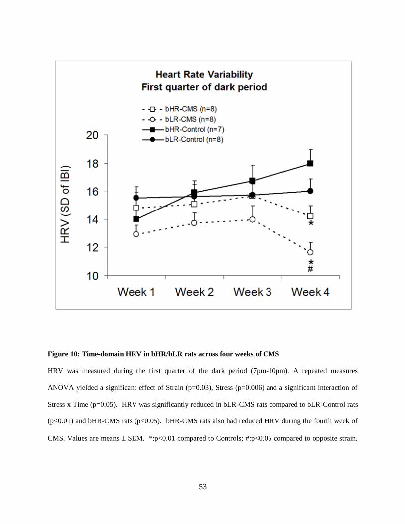
53
Figure 10: Time-domain HRV in bHR/bLR rats across four weeks of CMS
HRV was measured during the first quarter of the dark period (7pm-10pm). A repeated measures
ANOVA yielded a significant effect of Strain (p=0.03), Stress (p=0.006) and a significant interaction of
Stress x Time (p=0.05). HRV was significantly reduced in bLR-CMS rats compared to bLR-Control rats
(p<0.01) and bHR-CMS rats (p<0.05). bHR-CMS rats also had reduced HRV during the fourth week of
CMS. Values are means SEM. *:p<0.01 compared to Controls; #:p<0.05 compared to opposite strain.
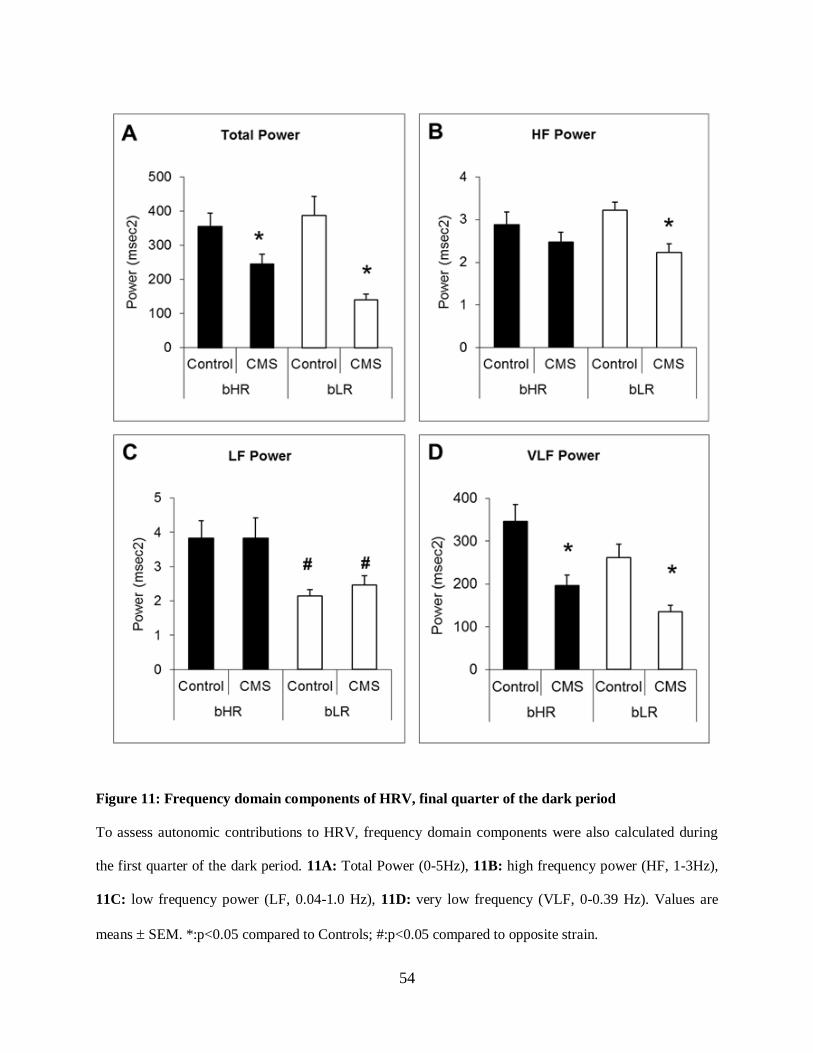
54
Figure 11: Frequency domain components of HRV, final quarter of the dark period
To assess autonomic contributions to HRV, frequency domain components were also calculated during
the first quarter of the dark period. 11A: Total Power (0-5Hz), 11B: high frequency power (HF, 1-3Hz),
11C: low frequency power (LF, 0.04-1.0 Hz), 11D: very low frequency (VLF, 0-0.39 Hz). Values are
means SEM. *:p<0.05 compared to Controls; #:p<0.05 compared to opposite strain.

55
Table 3: Frequency domain components of HRV, third quarter of the dark period
Values are means SEM. *:p<0.01 compared to Controls; #:p<0.01 compared to opposite strain.

56
Cardiovascular reactivity to an acute novel stress
During Week 5 of CMS, HR and BP were monitored during an acute stress to evaluate
cardiovascular reactivity. Baseline BP and HR were recorded 10 minutes before being placed in
a cage with clean bedding. All groups were within the normal range for Sprague-Dawley rats at
rest. Just before the cage change there was a significant baseline effect of CMS for both HR and
BP, which was mainly due to a significant difference in HR (p=0.03) and BP (p=0.04) between
bHR-Control rats and bLR-CMS rats; there were no other significant differences among groups
at baseline.
Immediately following placement in a cage with clean bedding, all groups showed a
significant increase from baseline in both HR and BP (Figures 12A & 12C). Both strains of
CMS-exposed rats had significantly higher peak increases in BP compared to controls (p < 0.05;
Figure 12D). bLR-CMS rats had a significantly larger peak increase in HR compared to bHR-
CMS rats (p = 0.02) and both control groups (p = 0.004; Figure 12B). There was also a
significant main neffect of stress on the area under the curve (AUC) of cardiovascular responses
during the first 40 minutes following stressor onset (i.e., t=0-40). AUC for HR (Figure 13A)
and BP (Figure 13B) responses to novel cage placement was higher in bLR-CMS rats compared
to bLR-Control rats (one-tailed ANOVA, p = 0.02 and p = 0.03, respectively). Additionally,
there was a significant main effect of stress on the duration of the HR and BP responses (one-
tailed ANOVA, p = 0.05 and p = 0.04, respectively). bLR-CMS rats had HR and BP responses
that remained elevated above baseline for significantly longer than bLR-Control rats (BP: bLR-
CMS: 41±7min vs. bLR-Control: 22±3min, p=0.03; HR: bLR-CMS: 49±7min vs. bLR-Control:
27±5min, p=0.02). This relationship was not observed in bHR rats (BP: bHR-CMS: 30±4 vs.
bHR-Control 28±8, p=0.40; HR: bHR-CMS: 42±9 vs. bHR-Control 43±15, p=0.48).

57
Figure 12: Increases in HR and BP in response to a novel cage stress in bHR/bLR rats
HR and BP increases from baseline after rats were transferred to a new cage with clean bedding. Values
are means ± SEM. n=8/group, except bHR-Control, which had n=7 and two recordings from this group
were not usable, thus n=5. *: p<0.01 compared to Controls; #p<0.01 compared to opposite strain. 12A:
bLR-CMS rats had the largest peak increase in heart rate. Baseline heart rate (bpm); t = -10 minutes:
bHR-Ctrl: 329±17, bHR-CMS: 291±12, bLR-Ctrl: 317±20, bLR-CMS: 268±7. 12B: Bar graph of peak
increase in heart rate from baseline 5 minutes after cage switch was performed. 12C: Both strains of
CMS-exposed rats had significantly higher peak blood pressure responses compared to controls. Baseline
blood pressure (mmHg), t = -10 minutes: bHR-Ctrl: 104±4, bHR-CMS: 95±3, bLR-Ctrl: 99±4, bLR-
CMS: 92±1 (significant effect of treatment, p<0.05). 12D: Bar graph of peak increase in blood pressure
from baseline 10 minutes after cage switch was performed.

58
Figure 13: Duration of the increase in HR and BP in response to a novel cage stress
13A: Bar graph of area under the curve (AUC) for heart rate response to cage switch.
13B: Bar graph of area under the curve (AUC) for blood pressure response to cage switch
Values are means ± SEM. n=8/group, except bHR-Control, which had n=7 and two recordings from this
group were not usable, thus n=5. *:p<0.05 vs. Controls of same strain.

59
3.5 DISCUSSION
This chapter presents evidence that vulnerability to chronic stress-induced depressive behavior is
associated with alterations in cardiovascular function. These changes include increased resting
HR, reduced HRV, reduced frequency domain power, and increased cardiovascular reactivity to
acute stress. Behavioral data from the rats used in the current study were featured in a previously
published report [160] that demonstrated increased vulnerability of bLR rats to CMS-induced
depressive behavior compared to bHR rats. These two selectively-bred strains of Sprague
Dawley rats were bred based on their low and high locomotor responses to a novel environment,
a behavioral phenotype that appears to correspond with similarly divergent behavior in tests of
mood and anxiety-like behavior [71, 81]. When bHR and bLR rats were exposed to CMS, bLR-
CMS rats showed decreased preference for sucrose compared to bHR-CMS rats, bLR-Control
(unexposed) rats, and their own pre-stress baselines beginning in the second week of CMS. This
anhedonic behavior persisted throughout the experiment in bLR-CMS rats, but was not observed
in bHR-CMS rats until the final week, and even then was less robust, suggesting that bLR and
bHR rats provide an excellent model in which to study the interaction of environmental stress
and heritable predisposition to depression.
Baseline differences between bLR and bHR rats
The current study expands on previous behavioral findings in bLR and bHR rats by incorporating
cardiovascular parameters that were chronically recorded throughout the CMS period. We first

60
examined whether inherent behavioral differences in locomotion and emotionality in bLR and
bHR rats extended to differences in cardiovascular function during the baseline period before
CMS began. Though bHR rats tended to have higher HR and BP than bLR rats during both the
dark and the light periods, this relationship only reached significance when comparing BP during
the dark period. In addition, there were no significant differences in HRV at baseline.
By definition, bHR rats have a higher locomotor response to a novel environment ([71,
81, 123, 124, 160, 161]. This increase in locomotion was also apparent in 24-hour home cage
activity, as assessed by telemetry transmitter signal strength. During the baseline period, bHR
rats were significantly more active than bLR rats. Interestingly, when cardiovascular reactivity
to cage change was measured during Week 5 of CMS, bHR-Control rats tended to have longer
BP and HR responses than bHR-CMS rats, as quantified by AUC. Though this difference did not
reach statistical significance, bHR rats’ extended cardiovascular response may be due in part to
their inherent heightened locomotor response to a novel cage, and reduced cardiovascular
response in bHR-CMS rats may suggest that CMS blunts the acute locomotor response to
novelty, even though 24-hour activity level is not different between strains.
CMS alters cardiovascular function
Previous studies using CMS have found significant changes in cardiovascular function that
mirror those observed in human patients with cardiovascular disease [49, 50, 52, 53]. We
hypothesized that bLR-CMS rats, which are especially sensitive to depression-like behavior
brought on by CMS exposure, would also have an increased sensitivity to CMS-associated
cardiovascular changes. Indeed, there was a significant increase in resting HR during the dark
period in the bLR-CMS group compared to bHR-CMS and control groups during CMS weeks 3

61
and 4. No such increase in HR was observed during the light period (0700-1900), and no
significant differences were seen in BP during any week, either during the dark or light period.
Previous studies have reported a similar elevation in HR recorded during the light period at the
end of a four week period of CMS [49, 50, 52, 53] and elevated resting HR is frequently
observed in depressed patients throughout the light-dark cycle [164-166].
The CMS model was developed to mimic the types of daily stressors that, over time,
result in depression-like signs [46]. The stressors used include changes to the animals’
environment, such as overnight lighting and water deprivation, and are assumed to be relatively
mild. Despite the frequent use of the CMS model, to our knowledge there has not been a report
of the physiological or autonomic impact of individual intermittent mild stressors. Because HR
and BP were monitored chronically throughout this experiment, we were able to record
cardiovascular responses to many of these stressors, and have analyzed data from strobe light,
white noise, and the combination of wet bedding and paired housing. Overall, stressors
produced small to moderate increases in BP and HR, with the largest increases being in response
to paired housing combined with wet bedding. Though there were no significant differences in
peak HR or BP response between bLR and bHR rats, the HR response was prolonged in bLR-
CMS rat, as measured by area under the curve once depression-like behaviors have emerged.
More typical were the small increases in HR and BP in response to strobe light and loud white
noise. bHR and bLR rats did not differ in their response to strobe light turned on during the dark
period. bLR rats had higher peak and longer HR responses to white noise during week 2 of CMS,
but not during week 4, suggesting that this group may have adapted to the stressor. It is possible
that the longer duration of HR response to some stressors in bLR-CMS rats may be indicative of
an underlying difference in sensitivity to stress in bLR rats. As cardiovascular reactivity to these

62
stressors is only one component of autonomic responses to stress, further studies employing
measurements of HPA reactivity such as plasma corticosterone or other circulating factors such
as cytokines across the 4 weeks of CMS would further clarify this issue.
CMS enhances cardiovascular reactivity to acute stress
The depression-like changes in bLR rats’ behavior were also associated with an increase in
cardiovascular reactivity to acute stress, as measured by an increase in HR and BP in response to
a novel cage. Notably, the stressor used, switching to a clean cage, is a procedure that occurs
each week to all rats as their cages are cleaned. Despite the assumed familiarity of this
procedure, bLR-CMS rats had a larger increase in HR than either bHR-CMS or control groups.
This difference between bLR-CMS and bHR-CMS reactivity appears to be specific to the
autonomic regulation of the heart, as both bHR-CMS and bLR-CMS rats had similar BP
responses to the cage change, compared to control groups. Additionally, AUC of the HR and BP
response to stress was significantly higher in bLR-CMS than bLR-Control rats, and HR and BP
remained elevated longer in bLR-CMS rats; however, this relationship was not observed in bHR
rats. This increased magnitude of cardiovascular response may be further evidence of the
considerable effect of CMS on bLR rats. Baseline BP and HR values just prior to the cage
switch test were slightly lower in the rats exposed to CMS, though such differences were not
observed when BP or HR were monitored over longer periods of time.
CMS reduces heart rate variability
In addition to the CMS-induced increase in resting HR and heightened cardiovascular reactivity,
HRV was significantly reduced in both bHR-CMS and bLR-CMS animals during the fourth

63
week of CMS. Additionally, HRV was significantly lower in bLR-CMS rats than bHR-CMS
rats. Thus, the group that had the greatest degree of anhedonia [160], also had the most reduced
HRV. This association of a large reduction in sucrose preference and substantially reduced HRV
in bLR rats closely mirrors the correlative relationship between severity of depression and
reductions in HRV apparent in depressed patients [21, 33].
Reduced HRV is a critical risk factor for cardiac mortality and likely reflects a disruption
in sympathovagal balance to the heart. Reduced HRV in the time domain (s.d. of IBI), can
represent an increase in sympathetic activity, a decrease in parasympathetic activity, or both.
Frequency domain or spectral analysis of HRV is often used in both experimental and clinical
studies as a noninvasive method of assessing autonomic contributions to a global reduction in
HRV. Total power across the frequency spectrum is related to overall autonomic balance
whereas the HF band corresponds to the respiratory frequency, and is used as an index of vagal
or parasympathetic influence on HRV. LF HRV appears to be influenced by both sympathetic
and parasympathetic elements [22, 35] and VLF power may reflect cardiovascular modulation by
the renin-angiotensin system [29, 35, 36] or its effects on parasympathetic influence on the heart.
Frequency domain analysis of HRV during the fourth week of CMS revealed that bLR-CMS rats
had reduced total power, HF, and VLF compared to bLR-Control rats (p<0.05). Reduced HF
power has been observed in both clinical [43] and experimental studies [167] of cardiovascular
dysfunction associated with depressive symptoms, and likely reflect a decrease in vagal control
of the SA node [168]. Reductions in total power and VLF are most strongly associated with
mortality after myocardial infarction, even more so than HF or LF [24], and account for nearly
30% of the risk conferred on cardiac mortality by depression [31], making it one of the most
reliable measurements of mortality risk. Taken together, these data suggest that the large

64
reduction in frequency-domain components of HRV seen in bLR-CMS rats may be due to a
decrease in parasympathetic control of the heart mirroring the clinical findings of HRV
alterations associated with depression.
Interestingly, this reduction in HRV was not apparent across the entire dark period. For
example, when the same HRV analysis was performed during the third quarter of the dark period
(0100 – 0400), a period less influenced by changes in the light cycle, there were no significant
differences in HRV or its frequency domain components, with the exception of bHR-CMS LF
power (Table 3). Experimental studies that measure HRV in animals typically do so acutely,
upon termination of the experiment, and only for 5- 20 minutes. This is an appropriate analysis
method, and, indeed, 20 minutes is the recording length recommended by the Task Force of The
European Society of Cardiology and the North American Society of Pacing and
Electrophysiology in the prominent article that discusses this method [22]. However, our results
suggest that longer recording periods, which may be more comparable to human studies using
Holter recordings, likely yield more complex results than short recording periods. Even in the
case of animals with extreme anhedonia and greatly reduced HRV, as in the current study,
reduced HRV is not apparent at every time period.
Decreased HRV has been reported in various animal models of depression. A series of
studies by Grippo et al used CMS to induce anhedonia in rats and demonstrated reduced HRV
following 4 weeks of CMS [49, 50, 52, 53, 68]. Similar findings have been reported in studies
using social isolation to induce anhedonia in the monogamous prairie vole (for a review, see
[158]). After a 4 week period of isolation from their housing partner, both time-domain HRV
and HF power (in this case measured by respiratory sinus arrhythmia) were reduced in both male
and female prairie voles [167]). HRV was also reduced in the Flinders Sensitive Line (FSL) rat

65
[169], a strain of rat originally bred for its thermoregulatory sensitivity to cholinergic drugs that
has also been shown to be susceptible to depression-like behavior [144]. However, this
alteration may be due to physiological changes that are unrelated to the depression-like behavior
of the FSL rat, as HRV was also reduced in the Flinders Resistant Line (FRL) rat, and there was
no significant difference between FSL and FRL rats. Furthermore, HRV measurements were
taken acutely, under urethane anesthesia, which makes them difficult to compare to chronic
measures in conscious rats.
In conclusion, vulnerability to CMS-induced depression-like behavior also confers a
vulnerability to significant cardiovascular alterations associated with human depression. These
include increased resting HR, reduced HRV, changes in frequency domain components of HRV,
and increased cardiovascular reactivity. Frequency-domain analysis of HRV suggests that these
alterations may be due in part to a decrease in parasympathetic function. This robust model of
susceptibility and resistance to CMS-induced depression and associated cardiovascular changes
should provide a powerful approach to examining the mechanisms underlying this association
and potential therapeutic interventions for treating these serious comorbid conditions.

66
4.0 CANDESARTAN REVERSES ANHEDONIA AND CARDIOASCULAR
ALTERATIONS IN A RODENT MODEL OF DEPRESSION
4.1 ABSTRACT
Major depressive disorder and cardiovascular disease share a comorbid bidirectional
relationship, wherein the presence of one increases the likelihood of the other. Antidepressants
such as selective serotonin reuptake inhibitors (SSRIs), might not improve cardiovascular
mortality, even when depressive signs are ameliorated. Candesartan, an AT1R antagonist often
prescribed for hypertension and other cardiovascular disorders, has been shown to be anxiolytic
in animal models, and may have antidepressant properties. This study used adult male rats that
were selectively-bred for low locomotor response to a novel environment, which are especially
vulnerable to chronic mild stress (CMS) -induced signs of depression and associated
cardiovascular changes. Rats were exposed to CMS for five weeks, which induced anhedonia
and decreased heart rate variability (HRV) and frequency-domain components of HRV,
clinically relevant markers of risk for cardiac death; control rats were housed under standard
conditions. During the sixth week of the experiment, rats were implanted with subcutaneous
osmotic minipumps containing candesartan, the SSRI fluoxetine, or vehicle, and CMS or control
housing was continued for an additional four weeks. Candesartan rapidly reversed anhedonic
behavior measured by sucrose preference during the first week of treatment. Candesartan also
67
reduced CMS-induced anxiety-like behavior in the novelty-suppressed feeding test, and had
positive effects in the forced swim test in unstressed rats. Furthermore, candesartan reversed the
CMS-induced reduction in HRV and its frequency domain components. Although fluoxetine
also reversed anhedonic and anxiety-like behavior, and had positive results in the FST, it did not
reverse the reduction in HRV and even decreased HRV in control (unstressed) rats. These
results provide robust evidence that candesartan is a novel, effective treatment for comorbid
depression and cardiovascular disease.
4.2 INTRODUCTION
Major depressive disorder and cardiovascular disease are highly comorbid, and the presence of
one disorder increases the likelihood of the other. Depression is an independent risk factor for
coronary heart disease both in patients with cardiovascular disease as well as medically healthy
individuals [1, 38, 39, 44, 170] and is a significant independent predictor of mortality within 18
months following a heart attack [2, 4]. Frasure-Smith and colleagues [2] found that in the six
months following a myocardial infarction, mortality was 3.5 times higher in patients who were
also depressed than those who were not.
Animal models of depression have been extremely useful in providing support for this
relationship between depression and cardiovascular disease and establishing an experimental
model in which to study possible underlying mechanisms and methods of treatment. For
instance, the chronic mild stress (CMS) model of depression in rodents, which uses a series of
intermittent unpredictable mild stressors to induce an anhedonic state [46, 47], also incorporates
many of the cardiovascular disturbances associated with human depression, including increased

68
resting heart rate (HR) and decreased HR variability (HRV). Decreased HRV is a risk marker
for heart attack and cardiovascular mortality [23, 31, 40], and more severe signs of depression
are associated with larger reductions in HRV [21, 33].
Recent studies from our lab have shown that rats selectively-bred for reduced locomotor
behavior in a novel environment (bred low responder or bLR rats) are especially vulnerable to
the anhedonic effects of CMS [160] and that depression-like behaviors in these animals were
also associated with changes in cardiovascular function, including decreased HRV, and
decreased very low frequency power of HRV, a component of frequency-domain analysis that
corresponds to parasympathetic activity and is a predictor of cardiac mortality [3, 31]. Bred high
responder (bHR) rats, on the other hand, were resistant to anhedonia, and had only modest
reductions in HRV.
There are conflicting reports regarding whether antidepressant therapy improves reduced
HRV or risk for cardiac mortality. Tricyclic antidepressants have known cardiotoxic properties
and may exacerbate existing cardiovascular conditions [82-84], and their use in patients with
cardiovascular disease has been discouraged [85, 86]. While some studies indicate that selective
serotonin reuptake inhibitors (SSRIs) improve cardiovascular outcomes [19, 87], others find that
SSRIs do not improve mortality risk, even when depression is in remission [88]. Still other
studies report that SSRIs may worsen outcomes, especially when administered in conjunction
with beta-blockers, which are commonly prescribed for various cardiovascular disorders [89]. In
rats exposed to 4 weeks of CMS, simultaneous treatment with the SSRI fluoxetine prevented the
anhedonic effects of CMS, but did not fully prevent the reduction in HRV [53].
The renin angiotensin system (RAS) has an established role in cardiovascular regulation,
and drugs that inhibit the RAS such as AT1R blockers (ARBs) and angiotensin converting

69
enzyme (ACE) inhibitors have been shown to reduce mortality from heart failure [95], possibly
by reversing the reduction in VLF mentioned above [29, 35, 36]. The RAS also plays a key role
in the physiological response to acute and chronic stress in studies in rodents. Chronic
pretreatment with an ARB reduces anxious behavior in tests such as the elevated plus maze
[101], attenuates the hypothalamic pituitary adrenal (HPA) axis response to an acute stress [100]
and prevents gastric ulceration associated with a chronic stress [102, 103]. Furthermore, drugs
that inhibit the RAS may have antidepressant properties. The ARB losartan and the ACE
inhibitor captopril both give positive antidepressant-like effects in the forced swim test in mice
[107, 108], a supposed animal model of despair behavior. There is some evidence indicating that
ACE inhibitors improve mood and cognitive function in hypertensive patients [104], and
antidepressant use is lower in hypertensive patients taking ARBs or ACE inhibitors [106].
The current study investigates the potential antidepressant effects of the ARB candesartan
on CMS-induced changes in behavior and cardiovascular function. These experiments were
performed in bLR rats, which are especially vulnerable to depression-like behavior [160], and
were compared with the effects of the SSRI fluoxetine. We hypothesized that candesartan would
reverse CMS-induced anhedonia and the reduction in HRV and its power spectral components,
possibly more effectively than fluoxetine.

70
4.3 EXPERIMENTAL PROCEDURES
Animals and Housing
Adult male bred low responder (bLR) rats were selectively-bred from a Sprague Dawley line at
the Molecular and Behavioral Neuroscience Institute at the University of Michigan and shipped
to the University of Pittsburgh when they were approximately 2 months old (250-300g). This
experiment used 48 bLR rats that were singly housed in plastic bins with wire lids. CMS-
exposed and control rats were housed in separate adjacent rooms under similar conditions. The
housing rooms were kept at a controlled temperature and humidity on a 12:12 light-dark cycle
with lights on at 7:00 am. Food (Purina Chow) and tap water were available ad libitum except
where noted as a part of the CMS protocol or testing procedures. Body weight was measured
weekly for the duration of the experiment. All animal protocols conform to the NIH Guide for
the Care and Use of Laboratory Animals and were approved by the University of Pittsburgh
Animal Care and Use Committee. Figure 14A shows an outline of the experimental procedures.

71
Figure 14: Diagram of experimental procedures and typical CMS schedule
Figure 14A: Rats were implanted with telemetry transmitters and allowed 7-10 days to recover. After a
baseline recording period, rats were exposed to control conditions or CMS for a total of 9 weeks. Drugs
were administered via subcutaneous osmotic minipump beginning in week 6. The novelty-suppressed
feeding test (NSF) was administered in weeks 5 and 9 and the forced swim test (FST) was administered in
week 8. Figure 14B: Rats were exposed to either 9 weeks of intermittent unpredictable stressors or
control conditions. Control rats were housed in separate rooms and handled according to usual animal
care protocols with the addition of a weekly sucrose preference test (SPT).

72
Telemetry transmitter implantation surgery
Rats were left undisturbed for one week after arriving at the animal facility at the University of
Pittsburgh. After this time, telemetry transmitters (PA-C40, Data Sciences International) were
implanted in a subset (n=30) of the rats to record blood pressure (BP), heart rate (HR), and HR
variability (HRV); the remaining rats (n=18) were not implanted with transmitters (due to limited
availability of transmitters for this experiment). Under isoflurane anesthesia (2% in 100%
oxygen, delivered at 1-1.5 L/min), the body of the transmitter was anchored to the abdominal
muscle in the intraperitoneal space and the catheter was implanted into the femoral artery.
Wounds were closed with silk suture and a single dose of Ketoprofen (2mg/kg, s.c.) was
administered as an analgesic. Rats were allowed 7-10 days to recover from surgery before
baseline recordings began.
Chronic Mild Stress protocol
Baseline cardiovascular and sucrose preference data (see below) were collected for two weeks
before CMS began. Rats were either exposed to CMS (n=24) or were handled according to
standard animal care procedures (Control group, n=24) for a total of 9 weeks. The CMS protocol
included the following individual stressors, used in varying order across weeks as previously
described in detail [160]: continuous overnight lighting; overnight water deprivation (18h)
followed by 1 hour of empty water bottle replacement; 40-degree cage tilt; stroboscopic lighting
(2-6 h; Chauvet mini-strobe CH-730; 8-12 flashes per second, 35 watts); overnight paired
housing with another CMS-exposed rat (18h); damp bedding (300-500mL lukewarm water
added to cage bedding); white noise (radio static, 85dB, 1-4 h, continuous or intermittent); and
predator odor exposure (30-60 minutes exposure to a small vial of undiluted 2,4,5-

73
trimethylthiazoline (TMT; Contech Intl.) opened in the housing room). A typical week of
stressors is diagrammed in Figure 14B.
Sucrose Preference Test
Anhedonia was measured using the sucrose preference test (SPT) as previously described in
detail [160]. Rats were accustomed to the taste of sucrose by replacing ad libitum water with 1%
sucrose for one week. Tap water was returned to the cage for one day, and then rats were food-
and water-deprived for 18 hours before the first baseline SPT was administered. During the test,
two graduated burets filled with either 1% sucrose solution or tap water were placed on the cage
and rats were allowed to drink freely for one hour. Total volume consumed of each fluid was
recorded, and preference for sucrose was calculated as [(mL sucrose / total mL consumed) *100].
SPT was administered once per week to both CMS and Control groups (Figure 14). Body weight
was measured weekly for the duration of the experiment.
Radiotelemetry data collection and analysis
Details of our cardiovascular data collection and analysis methods can be found in the previous
chapter. Blood pressure (BP) and heart rate (HR) were recorded as a 10-second average every 2
minutes using Dataquest ART 4.0 software (Data Sciences International). For HRV analysis,
pulsatile BP was also recorded from groups of 8 rats at a time on different days of the same
week, for 16-24 hours at a time. CMS was continued throughout the recordings.
Time-domain HRV was calculated as the standard deviation of the inter-beat interval
(IBI). To evaluate autonomic contributions to HRV, frequency components of HRV were
analyzed using the custom open source HRV analysis program, Physioscripts, which employs a

74
band limited variance technique [34] (see Chapter 3). Briefly, the event sequence of IBIs was
resampled at 10 Hz, inspected for artifact, and a bandpass filter was applied before calculating
the variance within the specified frequency range. Boundaries for the frequency ranges were as
follows [163]: total power (TP) 0-5 Hz, high frequency (HF) 1-3 Hz, low frequency (LF) 0.04-
1.0 Hz, and very low frequency (VLF) 0-0.39 Hz.
Drug administration
At the beginning of week 6, rats were implanted with osmotic minipumps (Alzet, 2ML4)
containing one of the following: candesartan (0.5mg/kg/day; Astra Zeneca), fluoxetine
(4mg/kg/day; BioTrend), or 0.9% sterile saline (2mL). These doses were chosen based on
previous reports that demonstrated their effects on behavior and neurobiology [66, 100, 101,
171]. Minipumps were loaded and primed for several hours in 37ºC saline and were then
surgically implanted subcutaneously in the interscapular region and wounds were closed with
silk suture. A single dose of Ketoprofen (2mg/kg, s.c.) was administered as an analgesic. Rats
received 3-5 days of drug before the first post-drug SPT was administered (Figure 14A: CMS
Week 6).
Forced Swim Test
The Forced Swim Test (FST) is a common test of antidepressant drug efficacy, with
antidepressant drugs decreasing immobility in this test and increasing time spent in active
behavior (i.e., swimming and climbing) [151, 172, 173]. This test was performed during CMS
week 8. During swim sessions, rats were placed in a clear Plexiglas cylinder (20cm wide x
61cm high) filled 45 cm high with 23-25ºC tap water. Two sessions were administered: a 15-

75
minute session to acclimate the rats to the test and apparatus and a 5-minute testing session 24
hours later, which was videotaped for behavioral scoring. Based on the protocol in Cryan et al
[151], three behaviors were scored: climbing, during which the rat’s front paws move in and out
of the water; swimming, in which the rat’s front paws remain underwater, while the rat is mobile;
and floating or immobility, in which the rat’s only movements are to keep its head above the
water. The 5-minute testing period was divided into sixty 5-second bins, and the predominant
behavior during each bin was scored. Scorers were kept blind to the experimental groups.
Novelty-suppressed feeding test
During Week 5 and Week 9 of CMS, anxiety-like behavior was assessed using the novelty-
suppressed feeding test (NSF). Rats were food deprived for 24 hours before being placed in an
open field (43cm x 43cm) with a food pellet in the center. Latencies to approach and to consume
the food pellet were recorded. Once the rat bit the pellet, or a 15-minute time limit was reached,
it was returned to its home cage, and latency to eat in the home cage was measured. Testing took
place between 10:00am and 1:00pm.
Data Analysis
Statistical analyses were performed using Microsoft Excel and PASW Statistics 18.0 (SPSS).
Values are expressed as mean ± SEM. One-way ANOVA was used for comparisons of CMS
and Controls, and for comparisons of drug and treatment groups, a 2x3 ANOVA (Stress x Drug)
was used, with Tukey HSD post-hoc tests where appropriate. Repeated measures ANOVA was
used to identify significant differences across time, with least square difference (LSD) or Tukey

76
HSD test as appropriate. Pearson’s correlation was used to compare HRV and sucrose intake.
For all tests, a two-tailed p-value of 0.05 or less was considered significant.
4.4 RESULTS
Sucrose Preference Test
Weekly sucrose preference tests were used to assess anhedonic behavior (Figure 15A). At
baseline, before CMS began, there was no significant difference between groups (p=0.48). By
the end of the first week of CMS, the CMS-exposed rats reduced their preference for sucrose by
approximately 30% (p=0.003), and this persisted through CMS week 5, consistent with our
previous studies using bLR rats exposed to CMS [160]. In the beginning of week 6, rats were
implanted with osmotic minipumps containing vehicle, candesartan or fluoxetine. When tested at
the end of week 6, CMS-exposed rats receiving candesartan had significantly higher sucrose
preference scores than those receiving vehicle (p=0.04) (Figure 15B), and sucrose preference in
these rats was not different from either baseline or control rats. The CMS group receiving
fluoxetine was not significantly different from CMS-Veh rats during week 6, but was not
significantly different from the CMS-candesartan group either, due to the marked variability in
the fluoxetine-treated group (Figure 15B). After four weeks of drug treatment (CMS week 9),
CMS-Cand and CMS-Flx groups were not significantly different from unstressed Controls,
though the untreated CMS-Veh group remained significantly anhedonic (p<0.001). No
significant differences in SPT were observed among Control groups receiving vehicle, fluoxetine
or candesartan at any time during the experiment. Body weight, measured weekly throughout

77
the experiment, did not differ between groups at any time during the experiment (Figure 16);
across the 9 weeks of the experiment rats typically gained approximately 55 grams.

78

79
Figure 15: Percent sucrose preference across 9 weeks of CMS
15A: Anhedonia was assessed weekly via SPT. There were no significant differences in sucrose
preference at baseline. CMS-exposed rats had significantly lower sucrose preference starting in CMS
week 1 and continuing throughout the experiment. Values are means ± SEM. #: p<0.001, CMS vs.
Control; *: p<0.01, CMS-Veh vs. CMS-Cand; ɸ: p<0.01, CMS-Veh vs. CMS-Flx. n=8 rats/group, except
during week 5, as noted below.
15B: Individual changes in sucrose preference in CMS-exposed rats from CMS week 5 to week 6. Group
means are represented in black. Candesartan rapidly increased sucrose preference scores, rats receiving
fluoxetine had variable responses, and rats receiving saline did not have a significant change in sucrose
preference. Note that one rat from the CMS-Flx group was excluded from this graph because it did not
drink during week 5.

80
Figure 16: Increase in body weight across 9 weeks of CMS
There were no significant differences among groups in increases in body weight or absolute body weights
(not shown) during the experiment. Baseline body weights are listed next to group names in the figure
legend. Values are means ± SEM. n=8 rats/group.

81
Performance in the novelty-suppressed feeding test
The NSF was used to assess anxiety-like behavior. It was performed once in the fifth week of
CMS (see Figure 14A) to confirm anxiety-like behavior before drugs began [64, 115, 116] and
repeated during CMS week 9. At that time, both fluoxetine and candesartan significantly reduced
latencies to approach (Figure 17A) and eat (Figure 17B) the food pellet (p<0.05). There were no
significant differences among unstressed controls in either of these measures. Among all groups,
there were no significant differences in either the number of times rats approached the pellet
(Figure 17C) or latency to begin eating the in the home cage (Fig 17D). These data indicate that
candesartan and fluoxetine have similar anxiolytic effects in this model.

82
Figure 17: Results from novelty-suppressed feeding test (NSF) test during CMS week 9
Anxiety-like behavior was assessed using the NSF test during the final week of CMS. Candesartan and
fluoxetine reduced latencies to approach (17A) and eat (17B) a food pellet placed in the center of an open
field (p<0.05). There were no significant differences in the number of times rats approached the food
pellet (17C) or the latency to eat upon returning to the home cage (17D). Values are means ± SEM. *:
p<0.05 vs. rats receiving vehicle; #:p<0.05 vs. control rats receiving the same drug. n=8 rats/group.

83
Performance in the forced swim test
The FST is frequently used as a predictive test of antidepressant drug efficacy. This test was
performed during CMS week 8, after 3 weeks of drug treatment. In unstressed Controls,
candesartan significantly decreased time spent immobile (p=0.04) and increased swimming
behavior (p=0.05, Figure 18A) but not climbing. Similarly, fluoxetine reduced immobility
(p=0.02) and increased swimming (p=0.05) but not climbing. In the CMS-exposed group, there
were no significant differences among treatment groups in immobility, swimming or climbing
(Fig 18B), but all CMS groups showed decreased immobility and increased swimming compared
to control rats treated with vehicle.

84
Figure 18: Results from the forced swim test (FST) during CMS week 8
The FST was used to evaluate antidepressant actions of candesartan and fluoxetine. 18A: In control rats,
immobility was decreased by candesartan (p=0.04) and fluoxetine (p=0.02) and swimming behavior was
also increased by both drugs (p=0.05 for both). 18B: In CMS-exposed rats, there were no significant
differences among treatment groups. Values are means ± SEM. *: p<0.05 vs. rats receiving vehicle. Note
that two rats from the Control-Flx group were excluded from analysis because they were large enough to
balance on their tails during the FST testing period.

85
Resting blood pressure and heart rate
HR and BP were chronically recorded throughout the experiment in 5 of the 8 rats in each group.
At baseline, there were no significant differences in HR between groups. For the first four weeks
of CMS, CMS-exposed rats tended to have higher HR than Control rats, but this difference was
not statistically significant (Figure 19B). There were also no significant differences in HR once
drug administration began. There were no significant differences in BP from baseline through
Week 6 of CMS (Figure 19A). During week 7 of CMS, one week after drug treatment had
begun, CMS and Control rats receiving candesartan had significantly lower BP compared to rats
from the CMS group receiving vehicle (p<0.005). Hypotension persisted in these groups for the
remainder of the experiment. CMS-Flx rats also had reduced BP during CMS week 7 (p<0.01),
but BP returned to pre-drug levels during weeks 8 and 9. In addition to analysis of 24-hour
averages, light-period and dark-period HR and BP were also analyzed separately, and no
significant differences were detected.

86
Figure 19: Line graphs of 24-hour resting HR and BP across 9 weeks of CMS
HR and BP were chronically recorded throughout the experiment. Values are means ± SEM. *: p<0.01 vs.
rats receiving vehicle; #:p<0.01 vs. control rats receiving the same drug. 19A: Candesartan significantly
decreased 24-hour resting BP in both CMS-exposed and control rats during CMS weeks 7-9 (p<0.005).
Fluoxetine also decreased BP in CMS-exposed rats during CMS week 7 (p<0.01). 19B: There were no
significant differences in 24-hour resting HR. n=8 rats/group.

87
Heart Rate Variability
Time domain HRV was analyzed during the second quarter of the dark period (10p-1a) across all
weeks of the experiment; this period was chosen because it was common to recordings in all rats
during each week. During CMS weeks 1-4, there was a significant effect of CMS (p=0.01,
Figure 20A). CMS reduced HRV during the first week (p<0.01), but was not significantly
different from controls in weeks 2 and 3. HRV was significantly reduced compared to controls
during the fourth and fifth weeks of CMS (p<0.005, Figure 20B), and remained significantly
reduced in CMS-Vehicle rats for the duration of the experiment. HRV was significantly
correlated with sucrose intake during week 5 (R=0.50, p=0.005, n=48), the final week of CMS
before drugs were administered. HRV was also analyzed during the 3rd
quarter of the dark
period, but as in the previous study (see Chapter 3) no differences were noted in this later period.
Candesartan, fluoxetine, or vehicle were administered beginning CMS week 6, and there
was a significant CMS x Drug interaction in HRV (p<0.05) during CMS weeks 7-9.
Candesartan reversed the CMS-induced decrease in HRV by week 9 (p=0.03 vs. CMS-Veh,
Figure 20B), and did not significantly change HRV in Control animals. Fluoxetine did not
reverse the reduction in HRV in CMS, and during week 9, HRV in CMS-Flx rats was not
significantly different CMS-Veh rats (Figure 20B). Fluoxetine also significantly decreased
HRV in the control group, starting in week 7 (p=0.05) and continuing throughout the experiment.
As in the weeks before CMS began, HRV remained reduced in CMS-Veh rats compared to
Control-Veh rats (p=0.003).

88
Figure 20: Changes in time-domain HRV across 9 weeks of CMS, with and without drug treatment.
HRV was measured during the second quarter of the dark period (11p-1a). 20A: Line graph of HRV
across 9 weeks of CMS. 20B: Bar graph highlighting HRV during CMS week 5 (week before drug) and
CMS week 9 (after 4 weeks of drug). Values are means ± SEM. *:p<0.01 vs. CMS-Veh; #:p<0.05 vs.
Control-Veh; ɸ:p<0.05 vs. rats receiving fluoxetine of the same stress group. n=8 rats/group.

89
Frequency domain analysis of HRV
To evaluate autonomic influences on HRV, frequency domain analysis was performed. During
week 5, the final week of CMS before drug treatment began, CMS-exposed rats had a significant
decrease in total power (p=0.003), VLF power (p=0.004), and LF power (p=0.02) compared to
control rats (data not shown). Sucrose intake was significantly correlated with total power
(R=0.48, p=0.008) and VLF power (R=0.48, p=0.009) during week 5. By CMS week 9, total
power, VLF, HF and LF were still reduced in CMS-Veh rats compared to Control-Veh rats
(p<0.05) (Figure 21). Candesartan reversed this decrease in each of these frequency bands, and
total power, VLF, HF, and LF in CMS-Cand rats were significantly higher than CMS-Veh rats
(p<0.05). Fluoxetine did not reverse CMS-induced decreases in total power, VLF and LF, and
these values were not significantly different from the CMS-Veh group. Fluoxetine also reduced
total power, VLF and LF in Control rats, compared to the Control-Veh group (p<0.05) and the
Control-Cand group (p<0.05). HF was not significantly different among Control groups.

90
Figure 21: Frequency domain components of HRV during CMS week 9
To assess autonomic contributions to HRV, frequency domain components were also calculated during
the second quarter of the dark period. 21A: Total Power (0-5Hz), 21B: high frequency power (HF, 1-
3Hz), 21C: low frequency power (LF, 0.04-1.0 Hz), 21D: very low frequency (VLF, 0-0.39 Hz). Values
are means ± SEM. *:p<0.01 vs. CMS-Veh; #:p<0.05 vs. Control-Veh; ɸ:p<0.05 vs. rats receiving
fluoxetine of the same stress group. n=8 rats/group.

91
4.5 DISCUSSION
Cardiovascular disease and depression are highly comorbid. Though the mechanism that
connects them is not known, some common physiological, hormonal, and neurotransmitter
systems are disrupted in both disorders. One such system is the RAS, which is well-known for its
effects on cardiovascular function and may also be involved in altered mood states such as
anxiety and depression [100, 101, 108, 171, 174]. The major finding of this study is that
candesartan, an ARB used clinically in patients with hypertension or cardiac disease, reverses
both the behavioral and the cardiovascular alterations induced by the CMS model of depression.
Additionally, the effects of candesartan in our model occur faster and more completely than the
SSRI fluoxetine.
CMS induces depression-like effects
bLR rats, which are especially vulnerable to the anhedonic effects of CMS [160], were exposed
to 5 weeks of CMS before drug treatment began. CMS induced anhedonia, as measured by
preference for a dilute sucrose solution, increased anxiety-like behavior in the NSF test, and
reduced HRV and its frequency components. These data replicate previous findings from our lab
[160] and others [49-53, 68] regarding the effects of CMS on behavior and cardiovascular
function.
Candesartan reverses the behavioral effects of CMS
Drug treatment was started in the beginning of CMS week 6, and rats were tested for anhedonic
behavior at the end of the week, 3-5 days after the start of drug administration. In this span of
time, candesartan fully reversed anhedonia. Each rat in the CMS-Cand group drank 100%

92
sucrose within days of receiving candesartan, and this group was not significantly different from
control groups on this measure for the remainder of the experiment. Thus, candesartan has
robust antidepressant properties in this model. Previous clinical data indicate that patients taking
ACE inhibitors have improved mood [104] and those taking ARBs are less likely to be taking
antidepressants [106]. This study provides further experimental evidence of the mood-altering
properties of ARBs as well as an appropriate model in which to study them.
AT1Rs are located at every level of the HPA axis [98], and blockade with a centrally-
acting ARB like candesartan attenuates the HPA response to acute stress [100]. Chronic
pretreatment of candesartan greatly reduces anxiety-like behavior in the elevated plus maze in
rats, possibly via HPA axis blockade or prevention of the sympathoadrenal response [101]. In
the current study, candesartan also had anxiolytic effects in the NSF test. The NSF test has been
used frequently in models of depression because chronic, but not acute, antidepressant
administration reduces behavioral latencies, providing excellent predictive validity for the time
course of antidepressants ([64, 115, 116]). In CMS-exposed rats, candesartan attenuated the
latency to approach and to begin to eat the food pellet. Latencies to eat upon return to the home
cage were not different among groups, indicating that reduced latencies in the open field are not
due to differences in hunger or appetitive value of the food pellet. These results corroborate
previous reports of the anxiolytic properties of candesartan in a test with relevance for the effects
of antidepressant drugs.
Candesartan reverses CMS-induced reduction in HRV
bLR rats’ vulnerability to CMS-induced anhedonia is associated with a decrease in HRV,
indicating an imbalance in sympathovagal control of the heart (Chapter 3). Reduced HRV is a

93
significant marker of risk for cardiac mortality that has been observed in both depression and
cardiovascular disease patients [19, 31, 37, 40]. Candesartan reversed the CMS-induced decrease
in HRV, restoring it to control (i.e. unstressed) levels. As mentioned in Chapter 3, frequency
domain analysis of HRV is a technique that indirectly measures autonomic contributions to
HRV. Total power across the frequency spectrum is related to overall autonomic balance, HF
power is used as an index of parasympathetic influence on HRV, and LF power appears to be
influenced by both sympathetic and parasympathetic elements [22, 35]. Especially pertinent to
this study, VLF power may reflect cardiovascular modulation by the renin-angiotensin system
[29, 35, 36] or its effects on parasympathetic influence on the heart. In human patients, VLF is
considered one of the most reliable indices of risk for cardiac mortality [24], and accounts for
nearly 30% of the depression-related risk for heart attack and death [31]. CMS significantly
reduced these frequency components compared to unstressed controls, and this reduction was
completely reversed by candesartan. Thus, in addition to improving anhedonic behavior within
days, candesartan restores sympathovagal balance, measured by HRV and its frequency
components.
Fluoxetine reverses the behavioral, but not the cardiovascular effects of CMS
Fluoxetine also reversed CMS-induced anhedonia, though more slowly than candesartan.
Individual responses to the initial week of fluoxetine treatment varied, which may parallel the
varied responses of depressed patients to the initiation of SSRI treatment [175]. Nonresponse to
initial antidepressant treatment is an especially serious issue. Approximately 30% of depression
patients do not show improvement after the first course of antidepressant pharmacotherapy
[176], and reports suggest that response rates decrease with each subsequent course of treatment

94
[177]. The weeks-long delay in reversal of anhedonia with SSRIs, also observed in our animal
model, is a clinically significant problem; this delay in complete therapeutic efficacy may
contribute to patient non-adherence and discontinuation of antidepressant therapy. Therefore, a
drug that quickly and robustly improves anhedonia, one of the core signs of depression [12],
would be highly beneficial to patient outcomes. Additionally, fluoxetine, like candesartan,
reduced anxiety-like behavior in the NSF, as demonstrated in previous reports [64, 115, 116],
although this was tested only after 4 weeks of drug treatment so differences in time course for
the development of this action were not assessed. The more rapid time course of candesartan
versus fluoxetine in reversing CMS-induced anhedonia suggests that these two drugs may be
exerting their antidepressant effects via very different mechanisms.
Currently, there is no consensus in the clinical literature as to whether SSRIs reduce the
cardiac mortality risk associated with depression, even as they relieve signs of depression [88,
90]. Various studies have shown that SSRIs either improve [19, 87], have no effect on [88], or
worsen [89] cardiovascular disease and mortality. A study using the CMS model of depression
demonstrated that fluoxetine prevented anhedonia, but not reductions in HRV [53]. We found
that fluoxetine did not reverse the CMS-induced decrease in HRV. Though fluoxetine increased
HRV slightly in CMS-exposed rats, this increase was not statistically significant from untreated
CMS-exposed rats. Fluoxetine also did not improve decreases in the frequency components of
HRV. Thus, despite its improvement of the anhedonic and anxiety-like behavior of CMS, we
found that fluoxetine does not change the potentially detrimental cardiovascular effects
associated with CMS, at least during the time course of this experiment.
Interestingly, fluoxetine administration decreased HRV in control rats to the same degree
as CMS exposure. This suggests that fluoxetine may have detrimental cardiovascular effects
95
independent of its effect on mood, possibly due to interference with normal serotonergic
influence of cardiovascular function [178]. Indeed, as mentioned previously, SSRIs can produce
cardiovascular disturbance such as arrhythmia in healthy patients without cardiovascular disease
[84]. Findings such as these may become increasingly relevant as SSRIs are prescribed for a
variety of conditions not directly related to mood [179-181].
Candesartan has antidepressant effects in the forced swim test
The FST is often referred to as a test of learned helplessness [151, 172, 173], operationally
defined as an increase in immobility and a decrease in swimming and climbing behaviors. No
differences were observed among CMS-exposed rats receiving vehicle or drugs. This is likely an
indication of the limited utility of the FST, rather than the effects of CMS on FST behavior. The
FST was initially developed to test antidepressant drug efficacy, and not depression-like
behavior, per se [172, 182]. When examined in the context of other robust measures of
depression-related behaviors, especially the core symptom of anhedonia, these results and past
reports suggest that the FST is most reliably employed as an acute test to predict antidepressant
drug effects [182, 183].
In Control rats not exposed to CMS, candesartan reduced immobility and increased
swimming behavior to the same degree as fluoxetine. Past reports have suggested that
antagonists of the serotonergic system increase swimming behavior in the FST, while
catecholaminergic antagonists increase climbing behavior [151, 173]. Thus our data suggest that
one possible mechanism by which candesartan might be exerting this change is via an interaction
with the central serotonergic system [184]. Similar antidepressant effects of drugs that inhibit
the RAS have been reported in acute tests of antidepressant efficacy. For instance, the AT1R

96
antagonist losartan and the ACE inhibitor captopril both give positive anti-depressant like effects
in the FST in mice [107, 108]. Previous data from our lab confirm that chronic candesartan also
reduces immobility and increases swimming in supplier-bred Sprague Dawley rats [185].
Rapid effects of candesartan suggest a novel antidepressant mechanism
The positive effect of candesartan and fluoxetine in the FST, specifically the increase in
swimming behavior, suggests that the two drugs may exert their effects via a similar mechanism.
Indeed, there is evidence that the RAS does interact with the serotonergic system [186].
However, the rapid reversal of anhedonia by candesartan suggests that it may be working via a
different mechanism. One of the major clinical concerns regarding SSRI treatment is the delay
in onset that most patients experience. Though SSRIs such as fluoxetine acutely increase
synaptic levels of serotonin, downstream effects such as increases in brain derived neurotrophic
factor (BDNF) or increases in hippocampal neurogenesis may be necessary for full
antidepressant efficacy [64, 187, 188]. This paradox remains a controversial issue, and the
question of the acute versus the long-term mechanisms of SSRI efficacy remains largely
unanswered.
Despite candesartan’s rapid reversal of anhedonia, HRV is not improved until three
weeks later. This suggests that though they are highly comorbid, depression and cardiovascular
dysfunction are dissociable. Indeed, in addition to reports of antidepressant effects on HRV,
there is evidence that cognitive behavioral therapy improves signs of depression but does not
alter cardiac mortality [90]. Therefore, improving the psychological aspects of depression—
whether by antidepressant treatment or by psychotherapy—does not necessarily reduce
cardiovascular risk.

97
In addition to interactions with the serotonergic system, there are a number of
mechanisms, both direct and indirect, by which candesartan may be reversing anhedonia and
reduced HRV. For instance, chronic peripheral administration of candesartan reduces the
sympathoadrenal response to stress [189]. The dose and administration of candesartan used in
the current study have been shown to have central effects as well [190], and may also be exerting
direct effects on central AT1Rs in the hypothalamus or brainstem to alter both cardiovascular
and behavioral changes of CMS. Indeed, AT1Rs are co-localized with corticotrophin-releasing
hormone (CRH) positive neurons in the PVN [99], and two weeks of pretreatment with
candesartan reduces the HPA response to isolation stress [100, 189]. Candesartan also has
demonstrated anti-inflammatory effects [191], decreasing, for example, cold-restraint stress-
induced increase in gastric ulcers in rats [102], and peripheral and central release of cytokines in
response to an endotoxin [192, 193]. Plasma levels of pro-inflammatory cytokines, especially
interleukin-6 (IL-6), IL-1β, and tumor necrosis factor-a (TNF-a) are increased in depression and
are thought to contribute to some of the somatic symptoms of depression, such as fatigue and
reduced appetite [18]. Furthermore, there is evidence that inflammatory markers such as
cytokines are correlated with decreased HRV [42, 194, 195], and cytokine levels are increased in
chronic heart failure in humans [196]. Indeed, we found that CMS increased plasma levels of
IL-1β four-fold in plasma samples collected at the termination of the current experiment, and
candesartan attenuated this large increase to control levels (Stedenfeld, Sved & Saavedra,
unpublished observations), as did fluoxetine.
Given the distinct latencies of the reversal of behavioral and cardiovascular effects of
CMS, candesartan may be affecting these changes via one or more of the above mechanisms. Of
course, isolating the precise mechanism is additionally complex because each of the systems

98
mentioned above—the sympathoadrenal system, the HPA axis, and pro-inflammatory
cytokines—also influences the others.
Candesartan is a novel, effective treatment for comorbid depression and cardiovascular
disease
Currently, candesartan is frequently prescribed for hypertension and heart failure, is tolerated
well with few negative side effects, and has been shown to reduce cardiac mortality [197]. This
study presents evidence that candesartan has profound antidepressant effects, including rapid
reversal of anhedonia, and attenuated anxiety-like behavior. Furthermore, candesartan reverses
the reduced HRV associated with anhedonia, thereby markedly decreasing risk for cardiac
mortality. These findings suggest that candesartan and other ARBs may be novel therapies for
the treatment of comorbid depression and cardiovascular disease, and may be more effective than
traditionally-prescribed antidepressant treatments such as SSRIs.

99
5.0 GENERAL DISCUSSION
5.1 SUMMARY AND INTERPRETATIONS OF CURRENT FINDINGS
Epidemiological and clinical studies using human patients have provided convincing evidence
that depression and cardiovascular disease are highly comorbid [1, 6, 10, 20, 31, 37, 40, 41, 44]
and that depression is an independent risk factor for cardiac mortality [2-4, 31], both in patients
with existing cardiovascular disease [38, 43] and in medically well individuals [1].
Experimental animal models have been useful in further elucidating mechanisms that may be
common to both disorders [49-52, 162]. The studies presented in this dissertation used the CMS
model of depression, a well-validated animal model that uses intermittent unpredictable mild
stressors to induce anhedonia, one of the core diagnostic criteria for depression [12, 46, 47, 49-
52, 68, 198]. CMS also induces a constellation of behavioral, physiological, and neuroendocrine
responses that closely resemble those observed in depressed patients [47], including a decrease
in HRV, a sign of altered autonomic control of the heart and a marker of cardiac mortality risk
[49, 53, 68]. CMS-induced anhedonia is reversed by antidepressant treatment [53, 60, 61],
however there is evidence that pretreatment with an SSRI does not improve an anhedonia-
associated decrease in HRV, even when depression is improved [53]. This phenomenon has
been reported in human patients as well [88, 89], suggesting that in some individuals the cardiac
mortality risk associated with depression may persist even when depression is treated.

100
Many hormonal and neurohumoral systems are activated in both depression and heart
disease. One of these systems is the RAS, which has an established role in cardiovascular
regulation [91, 92]. Additionally, antagonists of the RAS such as the ARB candesartan greatly
reduce the behavioral, neuroendocrine, and sympathoadrenal effects of stress [98, 100, 101, 174,
189], and there is some evidence that ARBs may have antidepressant properties as well [104,
106, 108]. Therefore, the ultimate goal of these experiments was to test the hypothesis that
candesartan reverses both the anhedonic and cardiovascular changes induced by CMS, and to
compare these results with those of the SSRI fluoxetine.
Preliminary CMS experiments in our lab showed that supplier-bred Sprague Dawley rats
had variable responses to four weeks of CMS, and final sucrose preference was positively
correlated with pre-CMS locomotor response to an open field (Stedenfeld & Sved, unpublished
observation). Indeed, one approach to developing models of genetic predisposition to mood
disorders is to selectively-breed animals from within two opposite ends of a behavioral spectrum.
We took advantage of one such model, in which rats were selectively-bred based on locomotor
response to novelty, bHR and bLR rats (described in detail in Chapter 2). Previously reported
differences in mood-related behavior in bHR versus bLR rats [71, 81], led us to hypothesize that
the bHR/ bLR distinction might also predict behavioral responses to CMS-exposure. We present
these results in Chapter 2, providing evidence that bLR rats are especially vulnerable to the
anhedonic effects of CMS and also have increased anxiety-like behavior compared to bHR rats.
Therefore, the bHR/bLR model appears to be ideal for studying the interactions between
inherited vulnerability and environmental stress that may result in depression. At present, little is
known about the underlying neurobiology that might contribute to this difference between
bHR/bLR rats, and more research in this area is necessary.

101
We expanded upon these behavioral findings in Chapter 3 to demonstrate that the
respective vulnerability and resistance of bLR and bHR rats to anhedonia is bLR rats had large
decreases in HRV and its frequency components, increased resting HR, and increased
cardiovascular reactivity to an acute stressor. associated with CMS-induced changes in
cardiovascular function that mirror those in depressed patients. These results parallel reports that
depressed patients display large decreases in HRV [31] [19], and severity of depression is
correlated with reduced HRV [21, 33]. Therefore, the bHR/bLR model is a additionally useful
for the study of comorbid depression and cardiovascular disturbances.
It is likely that multiple genes are working together to produce vulnerability to mood disorders
such as depression [70]. Models such as the bHR/bLR model take advantage of inborn trait
variability to integrate knowledge of behavioral endophenotypes and the genetic and molecular
basis of that behavior. As we learn more about how the prenatal environment and early life
experience shape the development of neural circuits [150, 199], these types of models will
become ever more useful.
Having established a robust model of vulnerability to CMS-induced anhedonia and
cardiovascular alterations, in Chapter 4 we tested the effectiveness of candesartan, an ARB
found to reduce cardiovascular mortality in human patients [197], in reversing CMS-induced
anhedonia and reduced HRV. Candesartan reversed anhedonia within the first week of
treatment, and improved reduced HRV by the fourth week of treatment. It also reduced anxiety-
like behavior in CMS-exposed rats and improved scores on the FST in control rats. By contrast,
fluoxetine also improved anhedonia, but only after 4 weeks of treatment. Fluoxetine did not
improve HRV or its frequency components and, in fact, these measures were decreased in
control rats receiving fluoxetine. Thus, candesartan has both robust antidepressant effects and

102
improves CMS-induced changes in autonomic control of cardiovascular function, and may be
more effective than fluoxetine in the treatment of comorbid depression and cardiovascular
disease.
Notably, only one dose of each drug was administered in this experiment, via
subcutaneous minipump. The dose of candesartan (0.5mg/kg/day, s.c.) was chosen based on
reports that this dose and route of administration has effects on central AT1Rs in areas such as
the PVN [98, 100, 101, 189, 190]. It is not clear whether peripherally-administered candesartan
is accessing the brain by crossing the blood-brain barrier directly or by accessing AT1Rs in
circumventricular organs [98]. This dose of candesartan caused a significant hypotension in both
CMS-exposed and control rats. However, in control rats, other measures of cardiovascular
function, such as 24-hour resting HR, HRV, and frequency components of HRV were not
affected by candesartan, suggesting that candesartan is not causing significant changes in overall
sympathetic or parasympathetic control of cardiovascular function. Additionally, rats receiving
candesartan did not have any noticeable behavioral differences from rats receiving fluoxetine in
tests that required physical exertion, such as the FST.
The dose of fluoxetine (4mg/kg/day, s.c.) was chosen based on reports that it attenuated
chronic stress-induced CRH expression in the PVN [66]. Using this chronic dose of fluoxetine,
we observed a delay in onset of full antidepressant actions that parallels the one seen in
depression patients. There were no changes HRV within 4 weeks of fluoxetine treatment,
though it is possible that this change may have become apparent with a longer treatment period.
For instance, four weeks of pretreatment with fluoxetine (10mg/kg/day, i.p.) partially prevented a
CMS-induced decrease in HRV in Sprague Dawley rats [53].

103
5.2 POSSIBLE MECHANISMS BY WHICH CANDESARTAN REVERSES
DEPRESSION-LIKE BEHAVIOR AND CARDIOVASCULAR DYSFUNCTION
Candesartan had temporally distinct effects on behavioral and cardiovascular outcomes. This
suggests that though depression and cardiovascular disease have a comorbid, bidirectional
relationship, they are dissociable, and can be resolved individually. Thus it is likely that the
effects of candesartan are mediated via interactions with one or more of the following
physiological systems, all of which play a role in both depression and cardiovascular disease
[108, 200].
Inhibition of HPA axis activity
Clinical studies have linked extended hyperactivity of the HPA axis not only with anxiety
disorders but also depression [14, 15, 109, 201] . Depressed patients have an increase in HPA
activity marked by increased levels of CRH in the cerebrospinal fluid and increased plasma
cortisol [17, 202]. In rats, AT1Rs have been found at each level of the HPA axis [98]. They are
heavily co-localized with CRH-positive neurons in the parvocellular region of the PVN, and
appear to directly modulate their activity [99]. Angiotension stimulation increases CRH synthesis
[99] and two weeks of pretreatment with candesartan reduces the HPA response to isolation
stress [100, 189]. AT1Rs are also located in the median eminence, the site where CRH is
released into the portal circulation, the pituitary, where ACTH is released to the bloodstream,
and the adrenal glands, which release corticosterone into the circulation [98].

104
Although previous studies have found a modest increase in plasma corticosterone
following 4 weeks of CMS [51], preliminary results from the current studies do not strongly
suggest a central role of altered HPA activity. Indeed, terminal plasma corticosterone levels were
not significantly higher in CMS-exposed versus control rats (from Chapter 4), nor were there
significant drug effects across groups (Stedenfeld, Sved & Saavedra, unpublished observation).
However, CMS is not typically used for longer than 4-6 weeks, and therefore little is known
about adaptations that might take place in longer applications. Furthermore, it is possible that
acute stress-evoked levels of corticosterone, which were not measured, may be different among
groups.
Inhibition of sympathoadrenal responses to CMS
There are significant interactions between the HPA axis and the sympathoadrenal response to
stress and depression [203], and sympathoadrenal activity is increased in depression patients and
in patients with cardiovascular disease, as marked by increase norepinephrine excretion levels
[13]. In rats, AT1Rs are expressed in the adrenal cortex [204], and chronic peripheral
administration of candesartan prevents the cold-restraint stress-induced increase in adrenal
content of tyrosine hydroxylase mRNA, epinephrine and norepinephrine [91, 189].
Sympathoadrenal activity is also regulated by pre-sympathetic brainstem areas such as the rostral
ventrolateral medulla, which is rich in AT1Rs [205, 206].
Effects on hippocampal neurogenesis
There is evidence that antidepressants stimulate adult hippocampal neurogenesis [187, 188] , and
that hippocampal neurogenesis may be required for the full therapeutic effects of antidepressants

105
[64]. Indeed, the latency of therapeutic actions of antidepressants is consistent with the time
course of integration of newly adult-born hippocampal neurons [207, 208]. However, though
there is some evidence that angiotensin II may have apoptotic effects [209], ARBs do not appear
to increase neurogenesis [210-212]. Data from our lab demonstrating that chronic candesartan
does not increase the number of hippocampal progenitor cells labeled with 5-bromo-2'-
deoxyuridine (BrdU) [185] support this observation.
Anti-inflammatory effects and attenuation of circulating cytokines
Pro-inflammatory cytokines such as TNF-a, IL-6, and IL-1B are increased in depression [18].
Furthermore, cytokine levels are increased in chronic heart failure and following myocardial
infarction in humans [196, 213], and there is evidence that inflammatory markers such as
cytokines are correlated with decreased HRV [42, 194, 195]. In addition to its direct antagonism
of AT1Rs, candesartan has demonstrated anti-inflammatory effects. Candesartan decreases the
immune response to an endotoxin [192, 193], is neuroprotective [191, 214], and decreases
stress-induced gastric ulcerations [102, 103]. As mentioned in the discussion section of Chapter
4, we found that CMS increased plasma levels of IL-1β four-fold, and candesartan attenuated this
large increase to control levels (Stedenfeld, Sved & Saavedra, unpublished observation).
Therefore, it is possible that candesartan is having positive effects on anhedonia and
cardiovascular function via an anti-inflammatory mechanism. The rapid reversal of anhedonia
would support this idea, as a single dose of etanercept, a TNF-a antagonist, was able to decrease
chronic heart failure-induced anhedonia to control levels in rats [55].
Antagonists of the RAS also have positive effects on cerebrovascular inflammation, and
ARBs such as candesartan have been shown to reduce mortality after ischemic stroke [215],

106
possibly by increasing cerebral blood flow [216]. These effects have been shown to be
independent of effects on AT1Rs, and may instead involve decreasing oxidative stress [217] or
restoring endothelial function [191, 218]. There is also evidence that global and regional
cerebral blood flow is altered in depression, and antidepressant therapies restore proper blood
flow [219, 220]; it has been suggested that increases in cerebral blood flow could act as a
biomarker of antidepressant drug effects [221]. It is possible, then, that candesartan is reversing
anhedonia via anti-inflammatory effects on the cerebral vasculature that increase cerebral blood
flow.
5.3 FUTURE DIRECTIONS
Findings presented in this dissertation provide evidence of a model of increased vulnerability to
CMS-induced depression-like behavior and comorbid cardiovascular dysfunction, marked by
reduced HRV and frequency domain components of HRV. The bHR/bLR rat model offers an
excellent way of studying the genetic and environmental interactions that result in depression-
like behavior. However, as previously mentioned, the neurobiological mechanisms that
differentiate the bHR and bLR rats are not well-known. Though some information is available
from supplier-bred rats that were classified as high- or low-responder rats (e.g. LR rats have
increased hippocampal glucocorticoid receptors [76]), not all of these measures have been
replicated in bHR/bLR rats. Indeed, these analyses are planned for tissues collected from the
current studies.
Furthermore, there is evidence that not all characteristics of the supplier-bred HR/LR
classified rats parallel those in selectively-bred bHR/bLR rats. For instance, it has been reported

107
that when exposed to a repeated social defeat stress, rats classified as HR had greater anxiety-like
behavior and reduced sucrose preference compared to unstressed controls [222]. However, there
were only very small differences, if any, between HR and LR rats on these measures, further
demonstrating that the bHR/bLR model is truly unique in the marked difference in behavior and
autonomic cardiovascular function between the two rat strains. Yet this raises the question of
how the bHR/bLR rat strains relate to typical Sprague Dawley rats. Do bHR/bLR rats still
represent extreme ends of a continuum of normal Sprague Dawley rat behavior, or do they now
represent an entirely new rat strain? Exposing supplier-bred HR/LR rats to four weeks of CMS
(e.g. replicating Chapters 2 and 3) would be one way to address many of these questions.
This dissertation also presents evidence that the ARB candesartan reverses both
anhedonia and changes in cardiovascular function in bLR rats that are vulnerable to these CMS-
induced changes. The question of exactly how candesartan is effecting these changes was
addressed at length in the previous section and remains an important area of research. Some of
these mechanisms will be investigated in future analyses of brains and plasma collected from this
experiment. These candidate mechanistic systems are located both centrally and in the
periphery, and it is likely that the subcutaneous dose of candesartan we used has effects in both.
Answering the question of where candesartan’s actions are necessary for its antidepressant
effects—in the brain or in the periphery—perhaps by infusing candesartan directly into the
ventricles rather than subcutaneously, would be a further step toward understanding the
mechanism by which candesartan is working. Finally, if it is determined that candesartan is
working via an indirect mechanism, such as by anti-inflammatory actions, it is possible that that
system could be a specific target for further antidepressant drug discovery.

108
5.4 RELEVANCE AND CONCLUSIONS
Cardiovascular disease and major depression are currently two of the most detrimental disorders
in developed countries [8, 9]. The comorbid relationship between depression and cardiovascular
disease has received attention in recent years as being a serious health concern warranting
increased education and awareness in the clinical and health-provider communities [10].
Depression is an independent risk factor for coronary heart disease both in patients with
cardiovascular disease as well as medically healthy individuals [1, 38, 39, 44, 170] and is a
significant independent predictor of mortality within 18 months following a heart attack [2, 4]. It
is alarming, then, that many reports conclude that antidepressant drugs might not improve
cardiac mortality, even when they improve depression [19, 53, 84, 90, 223]. Because current
antidepressant drugs have a delay in reaching full effect and have varying success rates and
tolerability [176, 224], increased research efforts are necessary in antidepressant drug discovery.
Furthermore, those efforts should focus on drugs that also address physiological changes that
accompany depression. Progress is already being made in this area, as drugs that regulate
peptides such as CRH [16], oxytocin [225], vasopressin [226], melatonin [227], and, indeed, the
RAS [108] are gaining attention [228].
Furthermore, research into the physiological corollaries of mental disorders has
implications beyond finding appropriate pharmacological treatment. Hopefully, as depression is
increasingly recognized as a disorder with serious physiological repercussions and not merely a
mental disorder, the stigma of such disorders will be further eradicated. There is growing
evidence, for example, of improved outreach and treatment for depression and anxiety disorders
in vulnerable populations such as postpartum mothers [229, 230] and veterans returning from
combat [231, 232]. The experiments described in this dissertation, and future experiments that

109
examine the relationship between depression and cardiovascular disease, will increase our
understanding of the possible mechanisms connecting these two disorders and will encourage
more effective treatments in order to improve patients’ quality of life.
110
BIBLIOGRAPHY
[1] Penninx, B. W., Beekman, A. T., Honig, A., Deeg, D. J., Schoevers, R. A., van Eijk, J. T., et
al. Depression and cardiac mortality: results from a community-based longitudinal study. Arch
Gen Psychiatry. 2001,58:221-7.
[2] Frasure-Smith, N., Lesperance, F., Talajic, M. Depression following myocardial infarction.
Impact on 6-month survival. JAMA. 1993,270:1819-25.
[3] Carney, R. M., Blumenthal, J. A., Catellier, D., Freedland, K. E., Berkman, L. F., Watkins, L.
L., et al. Depression as a risk factor for mortality after acute myocardial infarction. Am J Cardiol.
2003,92:1277-81.
[4] Frasure-Smith, N., Lesperance, F., Talajic, M. Depression and 18-month prognosis after
myocardial infarction. Circulation. 1995,91:999-1005.
[5] Schleifer, S. J., Macari-Hinson, M. M., Coyle, D. A., Slater, W. R., Kahn, M., Gorlin, R., et
al. The nature and course of depression following myocardial infarction. Arch Intern Med.
1989,149:1785-9.
[6] Lesperance, F., Frasure-Smith, N., Talajic, M. Major depression before and after myocardial
infarction: its nature and consequences. Psychosom Med. 1996,58:99-110.
[7] Glassman, A. H., Shapiro, P. A. Depression and the course of coronary artery disease. Am J
Psychiatry. 1998,155:4-11.
[8] Murray, C. J., Lopez, A. D. Evidence-based health policy--lessons from the Global Burden of
Disease Study. Science. 1996,274:740-3.
[9] Mathers, C. D., Loncar, D. Projections of global mortality and burden of disease from 2002
to 2030. PLoS Med. 2006,3:e442.
[10] Lichtman, J. H., Bigger, J. T., Jr., Blumenthal, J. A., Frasure-Smith, N., Kaufmann, P. G.,
Lesperance, F., et al. Depression and coronary heart disease: recommendations for screening,
referral, and treatment: a science advisory from the American Heart Association Prevent ion
Committee of the Council on Cardiovascular Nursing, Council on Clinical Cardiology, Council
on Epidemiology and Prevention, and Interdisciplinary Council on Quality of Care and
Outcomes Research: endorsed by the American Psychiatric Association. Circulation.
2008,118:1768-75.
[11] Nestler, E. J., Barrot, M., DiLeone, R. J., Eisch, A. J., Gold, S. J., Monteggia, L. M.
Neurobiology of depression. Neuron. 2002,34:13-25.
[12] Association, A. P. Diagnostic and Statistical Manual of Mental Disorders, 4th Edition.
Washingtion, DC: American Psychiatric Association; 1994.
[13] Otte, C., Neylan, T. C., Pipkin, S. S., Browner, W. S., Whooley, M. A. Depressive
symptoms and 24-hour urinary norepinephrine excretion levels in patients with coronary disease:
findings from the Heart and Soul Study. Am J Psychiatry. 2005,162:2139-45.

111
[14] Holsboer, F. The rationale for corticotropin-releasing hormone receptor (CRH-R)
antagonists to treat depression and anxiety. J Psychiatr Res. 1999,33:181-214.
[15] Holsboer, F. The corticosteroid receptor hypothesis of depression.
Neuropsychopharmacology. 2000,23:477-501.
[16] Holsboer, F., Ising, M. Central CRH system in depression and anxiety--evidence from
clinical studies with CRH1 receptor antagonists. Eur J Pharmacol. 2008,583:350-7.
[17] Nemeroff, C. B., Widerlov, E., Bissette, G., Walleus, H., Karlsson, I., Eklund, K., et al.
Elevated concentrations of CSF corticotropin-releasing factor-like immunoreactivity in
depressed patients. Science. 1984,226:1342-4.
[18] Dantzer, R., O'Connor, J. C., Freund, G. G., Johnson, R. W., Kelley, K. W. From
inflammation to sickness and depression: when the immune system subjugates the brain. Nat Rev
Neurosci. 2008,9:46-56.
[19] Carney, R. M., Freedland, K. E., Stein, P. K., Skala, J. A., Hoffman, P., Jaffe, A. S. Change
in heart rate and heart rate variability during treatment for depression in patients with coronary
heart disease. Psychosom Med. 2000,62:639-47.
[20] Carney, R. M., Freedland, K. E., Stein, P. K. Anxiety, depression, and heart rate variability.
Psychosom Med. 2000,62:84-7.
[21] Krittayaphong, R., Cascio, W. E., Light, K. C., Sheffield, D., Golden, R. N., Finkel, J. B., et
al. Heart rate variability in patients with coronary artery disease: differences in patients with
higher and lower depression scores. Psychosom Med. 1997,59:231-5.
[22] Heart rate variability. Standards of measurement, physiological interpretation, and clinical
use. Task Force of the European Society of Cardiology and the North American Society of
Pacing and Electrophysiology. Eur Heart J. 1996,17:354-81.
[23] Kleiger, R. E., Miller, J. P., Bigger, J. T., Jr., Moss, A. J. Decreased heart rate variability
and its association with increased mortality after acute myocardial infarction. Am J Cardiol.
1987,59:256-62.
[24] Bigger, J. T., Jr., Fleiss, J. L., Steinman, R. C., Rolnitzky, L. M., Kleiger, R. E., Rottman, J.
N. Frequency domain measures of heart period variability and mortality after myocardial
infarction. Circulation. 1992,85:164-71.
[25] Glassman, A. H. Depression, cardiac death, and the central nervous system.
Neuropsychobiology. 1998,37:80-3.
[26] Grippo, A. J., Johnson, A. K. Stress, depression and cardiovascular dysregulation: a review
of neurobiological mechanisms and the integration of research from preclinical disease models.
Stress. 2009,12:1-21.
[27] Gehi, A., Mangano, D., Pipkin, S., Browner, W. S., Whooley, M. A. Depression and heart
rate variability in patients with stable coronary heart disease: findings from the Heart and Soul
Study. Arch Gen Psychiatry. 2005,62:661-6.
[28] Kop, W. J., Synowski, S. J., Gottlieb, S. S. Depression in heart failure: biobehavioral
mechanisms. Heart Fail Clin. 2011,7:23-38.
[29] Bonaduce, D., Marciano, F., Petretta, M., Migaux, M. L., Morgano, G., Bianchi, V., et al.
Effects of converting enzyme inhibition on heart period variability in patients with acute
myocardial infarction. Circulation. 1994,90:108-13.
[30] Curtis, B. M., O'Keefe, J. H., Jr. Autonomic tone as a cardiovascular risk factor: the dangers
of chronic fight or flight. Mayo Clin Proc. 2002,77:45-54.

112
[31] Carney, R. M., Blumenthal, J. A., Freedland, K. E., Stein, P. K., Howells, W. B., Berkman,
L. F., et al. Low heart rate variability and the effect of depression on post-myocardial infarction
mortality. Arch Intern Med. 2005,165:1486-91.
[32] Huikuri, H. V., Jokinen, V., Syvanne, M., Nieminen, M. S., Airaksinen, K. E., Ikaheimo, M.
J., et al. Heart rate variability and progression of coronary atherosclerosis. Arterioscler Thromb
Vasc Biol. 1999,19:1979-85.
[33] Stein, P. K., Domitrovich, P. P., Kleiger, R. E., Schechtman, K. B., Rottman, J. N. Clinical
and demographic determinants of heart rate variability in patients post myocardial infarction:
insights from the cardiac arrhythmia suppression trial (CAST). Clin Cardiol. 2000,23:187-94.
[34] Allen, J. J., Chambers, A. S., Towers, D. N. The many metrics of cardiac chronotropy: a
pragmatic primer and a brief comparison of metrics. Biol Psychol. 2007,74:243-62.
[35] Akselrod, S., Gordon, D., Ubel, F. A., Shannon, D. C., Berger, A. C., Cohen, R. J. Power
spectrum analysis of heart rate fluctuation: a quantitative probe of beat-to-beat cardiovascular
control. Science. 1981,213:220-2.
[36] Taylor, J. A., Carr, D. L., Myers, C. W., Eckberg, D. L. Mechanisms underlying very-low-
frequency RR-interval oscillations in humans. Circulation. 1998,98:547-55.
[37] Carney, R. M., Freedland, K. E., Miller, G. E., Jaffe, A. S. Depression as a risk factor for
cardiac mortality and morbidity: a review of potential mechanisms. J Psychosom Res.
2002,53:897-902.
[38] Barefoot, J. C., Helms, M. J., Mark, D. B., Blumenthal, J. A., Califf, R. M., Haney, T. L., et
al. Depression and long-term mortality risk in patients with coronary artery disease. Am J
Cardiol. 1996,78:613-7.
[39] Barefoot, J. C., Schroll, M. Symptoms of depression, acute myocardial infarction, and total
mortality in a community sample. Circulation. 1996,93:1976-80.
[40] Carney, R. M., Freedland, K. E. Depression, mortality, and medical morbidity in patients
with coronary heart disease. Biol Psychiatry. 2003,54:241-7.
[41] Frasure-Smith, N., Lesperance, F. Depression and cardiac risk: present status and future
directions. Heart. 2010,96:173-6.
[42] Frasure-Smith, N., Lesperance, F., Irwin, M. R., Talajic, M., Pollock, B. G. The
relationships among heart rate variability, inflammatory markers and depression in coronary
heart disease patients. Brain Behav Immun. 2009,23:1140-7.
[43] Frasure-Smith, N., Lesperance, F., Habra, M., Talajic, M., Khairy, P., Dorian, P., et al.
Elevated depression symptoms predict long-term cardiovascular mortality in patients with atrial
fibrillation and heart failure. Circulation. 2009,120:134-40, 3p following 40.
[44] Frasure-Smith, N., Lesperance, F. Depression--a cardiac risk factor in search of a treatment.
JAMA. 2003,289:3171-3.
[45] Frasure-Smith, N., Lesperance, F. Depression and other psychological risks following
myocardial infarction. Arch Gen Psychiatry. 2003,60:627-36.
[46] Willner, P. Chronic mild stress (CMS) revisited: consistency and behavioural-
neurobiological concordance in the effects of CMS. Neuropsychobiology. 2005,52:90-110.
[47] Willner, P. Validity, reliability and utility of the chronic mild stress model of depression: a
10-year review and evaluation. Psychopharmacology (Berl). 1997,134:319-29.
[48] Parihar, V. K., Hattiangady, B., Kuruba, R., Shuai, B., Shetty, A. K. Predictable chronic
mild stress improves mood, hippocampal neurogenesis and memory. Mol Psychiatry.
2011,16:171-83.
113
[49] Grippo, A. J., Moffitt, J. A., Johnson, A. K. Cardiovascular alterations and autonomic
imbalance in an experimental model of depression. Am J Physiol Regul Integr Comp Physiol.
2002,282:R1333-41.
[50] Grippo, A. J., Beltz, T. G., Johnson, A. K. Behavioral and cardiovascular changes in the
chronic mild stress model of depression. Physiol Behav. 2003,78:703-10.
[51] Grippo, A. J., Francis, J., Beltz, T. G., Felder, R. B., Johnson, A. K. Neuroendocrine and
cytokine profile of chronic mild stress-induced anhedonia. Physiol Behav. 2005,84:697-706.
[52] Grippo, A. J., Sullivan, N. R., Damjanoska, K. J., Crane, J. W., Carrasco, G. A., Shi, J., et
al. Chronic mild stress induces behavioral and physiological changes, and may alter serotonin 1A
receptor function, in male and cycling female rats. Psychopharmacology (Berl). 2005,179:769-
80.
[53] Grippo, A. J., Beltz, T. G., Weiss, R. M., Johnson, A. K. The effects of chronic fluoxetine
treatment on chronic mild stress-induced cardiovascular changes and anhedonia. Biol Psychiatry.
2006,59:309-16.
[54] Gambarana, C., Tolu, P. L., Masi, F., Rinaldi, M., Giachetti, D., Morazzoni, P., et al. A
study of the antidepressant activity of Hypericum perforatum on animal models.
Pharmacopsychiatry. 2001,34 Suppl 1:S42-4.
[55] Grippo, A. J., Francis, J., Weiss, R. M., Felder, R. B., Johnson, A. K. Cytokine mediation of
experimental heart failure-induced anhedonia. Am J Physiol Regul Integr Comp Physiol.
2003,284:R666-73.
[56] Di Chiara, G., Tanda, G. Blunting of reactivity of dopamine transmission to palatable food:
a biochemical marker of anhedonia in the CMS model? Psychopharmacology (Berl).
1997,134:351-3; discussion 71-7.
[57] Di Chiara, G., Loddo, P., Tanda, G. Reciprocal changes in prefrontal and limbic dopamine
responsiveness to aversive and rewarding stimuli after chronic mild stress: implications for the
psychobiology of depression. Biol Psychiatry. 1999,46:1624-33.
[58] Jayatissa, M. N., Bisgaard, C., Tingstrom, A., Papp, M., Wiborg, O. Hippocampal
cytogenesis correlates to escitalopram-mediated recovery in a chronic mild stress rat model of
depression. Neuropsychopharmacology. 2006,31:2395-404.
[59] Joeyen-Waldorf, J., Edgar, N., Sibille, E. The roles of sex and serotonin transporter levels in
age- and stress-related emotionality in mice. Brain Res. 2009,1286:84-93.
[60] Sampson, D., Willner, P., Muscat, R. Reversal of antidepressant action by dopamine
antagonists in an animal model of depression. Psychopharmacology (Berl). 1991,104:491-5.
[61] Muscat, R., Papp, M., Willner, P. Antidepressant-like effects of dopamine agonists in an
animal model of depression. Biol Psychiatry. 1992,31:937-46.
[62] Stein, E. J., da Silveira Filho, N. G., Machado, D. C., Hipolide, D. C., Barlow, K., Nobrega,
J. N. Chronic mild stress induces widespread decreases in thyroid hormone alpha1 receptor
mRNA levels in brain--reversal by imipramine. Psychoneuroendocrinology. 2009,34:281-6.
[63] D'Aquila, P. S., Brain, P., Willner, P. Effects of chronic mild stress on performance in
behavioural tests relevant to anxiety and depression. Physiol Behav. 1994,56:861-7.
[64] Santarelli, L., Saxe, M., Gross, C., Surget, A., Battaglia, F., Dulawa, S., et al. Requirement
of hippocampal neurogenesis for the behavioral effects of antidepressants. Science.
2003,301:805-9.
[65] Czeh, B., Muller-Keuker, J. I., Rygula, R., Abumaria, N., Hiemke, C., Domenici, E., et al.
Chronic social stress inhibits cell proliferation in the adult medial prefrontal cortex: hemispheric
asymmetry and reversal by fluoxetine treatment. Neuropsychopharmacology. 2007,32:1490-503.

114
[66] Stout, S. C., Owens, M. J., Nemeroff, C. B. Regulation of corticotropin-releasing factor
neuronal systems and hypothalamic-pituitary-adrenal axis activity by stress and chronic
antidepressant treatment. J Pharmacol Exp Ther. 2002,300:1085-92.
[67] Grippo, A. J., Moffitt, J. A., Johnson, A. K. Evaluation of baroreceptor reflex function in the
chronic mild stress rodent model of depression. Psychosom Med. 2008,70:435-43.
[68] Grippo, A. J., Santos, C. M., Johnson, R. F., Beltz, T. G., Martins, J. B., Felder, R. B., et al.
Increased susceptibility to ventricular arrhythmias in a rodent model of experimental depression.
Am J Physiol Heart Circ Physiol. 2004,286:H619-26.
[69] Fava, M., Kendler, K. S. Major depressive disorder. Neuron. 2000,28:335-41.
[70] Feder, A., Nestler, E. J., Charney, D. S. Psychobiology and molecular genetics of resilience.
Nat Rev Neurosci. 2009,10:446-57.
[71] Stead, J. D., Clinton, S., Neal, C., Schneider, J., Jama, A., Miller, S., et al. Selective
breeding for divergence in novelty-seeking traits: heritability and enrichment in spontaneous
anxiety-related behaviors. Behav Genet. 2006,36:697-712.
[72] Krishnan, V., Han, M. H., Graham, D. L., Berton, O., Renthal, W., Russo, S. J., et al.
Molecular adaptations underlying susceptibility and resistance to social defeat in brain reward
regions. Cell. 2007,131:391-404.
[73] Ballaz, S. J., Akil, H., Watson, S. J. Previous experience affects subsequent anxiety-like
responses in rats bred for novelty seeking. Behav Neurosci. 2007,121:1113-8.
[74] Tonissaar, M., Herm, L., Eller, M., Koiv, K., Rinken, A., Harro, J. Rats with high or low
sociability are differently affected by chronic variable stress. Neuroscience. 2008,152:867-76.
[75] Touma, C., Bunck, M., Glasl, L., Nussbaumer, M., Palme, R., Stein, H., et al. Mice selected
for high versus low stress reactivity: a new animal model for affective disorders.
Psychoneuroendocrinology. 2008,33:839-62.
[76] Kabbaj, M., Devine, D. P., Savage, V. R., Akil, H. Neurobiological correlates of individual
differences in novelty-seeking behavior in the rat: differential expression of stress-related
molecules. J Neurosci. 2000,20:6983-8.
[77] Mallo, T., Alttoa, A., Koiv, K., Tonissaar, M., Eller, M., Harro, J. Rats with persistently low
or high exploratory activity: behaviour in tests of anxiety and depression, and extracellular levels
of dopamine. Behav Brain Res. 2007,177:269-81.
[78] Pawlak, C. R., Ho, Y. J., Schwarting, R. K. Animal models of human psychopathology
based on individual differences in novelty-seeking and anxiety. Neurosci Biobehav Rev.
2008,32:1544-68.
[79] Dellu, F., Piazza, P. V., Mayo, W., Le Moal, M., Simon, H. Novelty-seeking in rats--
biobehavioral characteristics and possible relationship with the sensation-seeking trait in man.
Neuropsychobiology. 1996,34:136-45.
[80] Jama, A., Cecchi, M., Calvo, N., Watson, S. J., Akil, H. Inter-individual differences in
novelty-seeking behavior in rats predict differential responses to desipramine in the forced swim
test. Psychopharmacology (Berl). 2008,198:333-40.
[81] Clinton, S. M., Vazquez, D. M., Kabbaj, M., Kabbaj, M. H., Watson, S. J., Akil, H.
Individual differences in novelty-seeking and emotional reactivity correlate with variation in
maternal behavior. Horm Behav. 2007,51:655-64.
[82] Glassman, A. H., Bigger, J. T., Jr. Cardiovascular effects of therapeutic doses of tricyclic
antidepressants. A review. Arch Gen Psychiatry. 1981,38:815-20.
[83] Glassman, A. H., Rodriguez, A. I., Shapiro, P. A. The use of antidepressant drugs in patients
with heart disease. J Clin Psychiatry. 1998,59 Suppl 10:16-21.

115
[84] Sheline, Y. I., Freedland, K. E., Carney, R. M. How safe are serotonin reuptake inhibitors
for depression in patients with coronary heart disease? Am J Med. 1997,102:54-9.
[85] Warrington, S. J., Padgham, C., Lader, M. The cardiovascular effects of antidepressants.
Psychol Med Monogr Suppl. 1989,16:i-iii, 1-40.
[86] Stern, T. A. The management of depression and anxiety following myocardial infarction. Mt
Sinai J Med. 1985,52:623-33.
[87] Glassman, A. H. Does treating post-myocardial infarction depression reduce medical
mortality? Arch Gen Psychiatry. 2005,62:711-2.
[88] Jiang, W., Krishnan, R., Kuchibhatla, M., Cuffe, M. S., Martsberger, C., Arias, R. M., et al.
Characteristics of depression remission and its relation with cardiovascular outcome among
patients with chronic heart failure (from the SADHART-CHF Study). Am J Cardiol.
2011,107:545-51.
[89] Fosbol, E. L., Gislason, G. H., Poulsen, H. E., Hansen, M. L., Folke, F., Schramm, T. K., et
al. Prognosis in heart failure and the value of {beta}-blockers are altered by the use of
antidepressants and depend on the type of antidepressants used. Circ Heart Fail. 2009,2:582-90.
[90] Berkman, L. F., Blumenthal, J., Burg, M., Carney, R. M., Catellier, D., Cowan, M. J., et al.
Effects of treating depression and low perceived social support on clinical events after
myocardial infarction: the Enhancing Recovery in Coronary Heart Disease Patients (ENRICHD)
Randomized Trial. JAMA. 2003,289:3106-16.
[91] Jezova, D., Ochedalski, T., Kiss, A., Aguilera, G. Brain angiotensin II modulates
sympathoadrenal and hypothalamic pituitary adrenocortical activation during stress. J
Neuroendocrinol. 1998,10:67-72.
[92] Saavedra, J. M. Brain angiotensin II: new developments, unanswered questions and
therapeutic opportunities. Cell Mol Neurobiol. 2005,25:485-512.
[93] Packer, M. The neurohormonal hypothesis: a theory to explain the mechanism of disease
progression in heart failure. J Am Coll Cardiol. 1992,20:248-54.
[94] Fyhrquist, F., Metsarinne, K., Tikkanen, I. Role of angiotensin II in blood pressure
regulation and in the pathophysiology of cardiovascular disorders. J Hum Hypertens. 1995,9
Suppl 5:S19-24.
[95] Ostergren, J. B. Angiotensin receptor blockade with candesartan in heart failure: findings
from the Candesartan in Heart failure--assessment of reduction in mortality and morbidity
(CHARM) programme. J Hypertens Suppl. 2006,24:S3-7.
[96] Nap, A., Balt, J. C., Mathy, M. J., Van Zwieten, P. A. AT(1)-receptor blockade and
sympathetic neurotransmission in cardiovascular disease. Auton Autacoid Pharmacol.
2003,23:285-96.
[97] Saavedra, J. M. Brain and pituitary angiotensin. Endocr Rev. 1992,13:329-80.
[98] Tsutsumi, K., Saavedra, J. M. Characterization and development of angiotensin II receptor
subtypes (AT1 and AT2) in rat brain. Am J Physiol. 1991,261:R209-16.
[99] Aguilera, G., Young, W. S., Kiss, A., Bathia, A. Direct regulation of hypothalamic
corticotropin-releasing-hormone neurons by angiotensin II. Neuroendocrinology. 1995,61:437-
44.
[100] Armando, I., Volpi, S., Aguilera, G., Saavedra, J. M. Angiotensin II AT1 receptor
blockade prevents the hypothalamic corticotropin-releasing factor response to isolation stress.
Brain Res. 2007,1142:92-9.
[101] Saavedra, J. M., Armando, I., Bregonzio, C., Juorio, A., Macova, M., Pavel, J., et al. A
centrally acting, anxiolytic angiotensin II AT1 receptor antagonist prevents the isolation stress-

116
induced decrease in cortical CRF1 receptor and benzodiazepine binding.
Neuropsychopharmacology. 2006,31:1123-34.
[102] Bregonzio, C., Armando, I., Ando, H., Jezova, M., Baiardi, G., Saavedra, J. M. Anti-
inflammatory effects of angiotensin II AT1 receptor antagonism prevent stress-induced gastric
injury. Am J Physiol Gastrointest Liver Physiol. 2003,285:G414-23.
[103] Bregonzio, C., Armando, I., Ando, H., Jezova, M., Baiardi, G., Saavedra, J. M.
Angiotensin II AT1 receptor blockade prevents gastric ulcers during cold-restraint stress. Ann N
Y Acad Sci. 2004,1018:351-5.
[104] Germain, L., Chouinard, G. Treatment of recurrent unipolar major depression with
captopril. Biol Psychiatry. 1988,23:637-41.
[105] Domeney, A. M. Angiotensin converting enzyme inhibitors as potential cognitive
enhancing agents. J Psychiatry Neurosci. 1994,19:46-50.
[106] Nasr, S. J., Crayton, J. W., Agarwal, B., Wendt, B., Kora, R. Lower frequency of
antidepressant use in patients on Renin-Angiotensin-aldosterone system modifying medications.
Cell Mol Neurobiol. 2011,31:615-8.
[107] Giardina, W. J., Ebert, D. M. Positive effects of captopril in the behavioral despair swim
test. Biol Psychiatry. 1989,25:697-702.
[108] Gard, P. R., Mandy, A., Sutcliffe, M. A. Evidence of a possible role of altered angiotensin
function in the treatment, but not etiology, of depression. Biol Psychiatry. 1999,45:1030-4.
[109] Kessler, R. C. The effects of stressful life events on depression. Annu Rev Psychol.
1997,48:191-214.
[110] Willner, P., Mitchell, P. J. The validity of animal models of predisposition to depression.
Behav Pharmacol. 2002,13:169-88.
[111] Gronli, J., Murison, R., Bjorvatn, B., Sorensen, E., Portas, C. M., Ursin, R. Chronic mild
stress affects sucrose intake and sleep in rats. Behav Brain Res. 2004,150:139-47.
[112] Mitchell, P. J. Antidepressant treatment and rodent aggressive behaviour. Eur J Pharmacol.
2005,526:147-62.
[113] Johnson, A. K., Grippo, A. J. Sadness and broken hearts: neurohumoral mechanisms and
co-morbidity of ischemic heart disease and psychological depression. J Physiol Pharmacol.
2006,57 Suppl 11:5-29.
[114] Bondi, C. O., Rodriguez, G., Gould, G. G., Frazer, A., Morilak, D. A. Chronic
unpredictable stress induces a cognitive deficit and anxiety-like behavior in rats that is prevented
by chronic antidepressant drug treatment. Neuropsychopharmacology. 2008,33:320-31.
[115] Dulawa, S. C., Hen, R. Recent advances in animal models of chronic antidepressant
effects: the novelty-induced hypophagia test. Neurosci Biobehav Rev. 2005,29:771-83.
[116] Bodnoff, S. R., Suranyi-Cadotte, B., Aitken, D. H., Quirion, R., Meaney, M. J. The effects
of chronic antidepressant treatment in an animal model of anxiety. Psychopharmacology (Berl).
1988,95:298-302.
[117] Bessa, J. M., Mesquita, A. R., Oliveira, M., Pego, J. M., Cerqueira, J. J., Palha, J. A., et al.
A trans-dimensional approach to the behavioral aspects of depression. Front Behav Neurosci.
2009,3:1.
[118] Martinez, V., Rivier, J., Wang, L., Tache, Y. Central injection of a new corticotropin-
releasing factor (CRF) antagonist, astressin, blocks CRF- and stress-related alterations of gastric
and colonic motor function. J Pharmacol Exp Ther. 1997,280:754-60.
[119] Bonaz, B., Tache, Y. Water-avoidance stress-induced c-fos expression in the rat brain and
stimulation of fecal output: role of corticotropin-releasing factor. Brain Res. 1994,641:21-8.

117
[120] Stam, R., Croiset, G., Akkermans, L. M., Wiegant, V. M. Psychoneurogastroenterology:
interrelations in stress-induced colonic motility and behavior. Physiol Behav. 1999,65:679-84.
[121] Cecchi, M., Khoshbouei, H., Javors, M., Morilak, D. A. Modulatory effects of
norepinephrine in the lateral bed nucleus of the stria terminalis on behavioral and neuroendocrine
responses to acute stress. Neuroscience. 2002,112:13-21.
[122] Hooks, M. S., Jones, G. H., Smith, A. D., Neill, D. B., Justice, J. B., Jr. Response to
novelty predicts the locomotor and nucleus accumbens dopamine response to cocaine. Synapse.
1991,9:121-8.
[123] Davis, B. A., Clinton, S. M., Akil, H., Becker, J. B. The effects of novelty-seeking
phenotypes and sex differences on acquisition of cocaine self-administration in selectively bred
High-Responder and Low-Responder rats. Pharmacol Biochem Behav. 2008,90:331-8.
[124] Flagel, S. B., Robinson, T. E., Clark, J. J., Clinton, S. M., Watson, S. J., Seeman, P., et al.
An animal model of genetic vulnerability to behavioral disinhibition and responsiveness to
reward-related cues: implications for addiction. Neuropsychopharmacology. 2010,35:388-400.
[125] Rush, A. J., Weissenburger, J. E. Melancholic symptom features and DSM-IV. Am J
Psychiatry. 1994,151:489-98.
[126] Melartin, T., Leskela, U., Rytsala, H., Sokero, P., Lestela-Mielonen, P., Isometsa, E. Co-
morbidity and stability of melancholic features in DSM-IV major depressive disorder. Psychol
Med. 2004,34:1443-52.
[127] Loas, G. Vulnerability to depression: a model centered on anhedonia. J Affect Disord.
1996,41:39-53.
[128] Mitchell, P. B., Parker, G. B., Gladstone, G. L., Wilhelm, K., Austin, M. P. Severity of
stressful life events in first and subsequent episodes of depression: the relevance of depressive
subtype. J Affect Disord. 2003,73:245-52.
[129] Harkness, K. L., Monroe, S. M. Severe melancholic depression is more vulnerable than
non-melancholic depression to minor precipitating life events. J Affect Disord. 2006,91:257-63.
[130] Bogdan, R., Pizzagalli, D. A. Acute stress reduces reward responsiveness: implications for
depression. Biol Psychiatry. 2006,60:1147-54.
[131] Horesh, N., Klomek, A. B., Apter, A. Stressful life events and major depressive disorders.
Psychiatry Res. 2008,160:192-9.
[132] Muscatell, K. A., Slavich, G. M., Monroe, S. M., Gotlib, I. H. Stressful life events, chronic
difficulties, and the symptoms of clinical depression. J Nerv Ment Dis. 2009,197:154-60.
[133] Gorwood, P. Neurobiological mechanisms of anhedonia. Dialogues Clin Neurosci.
2008,10:291-9.
[134] Martin-Soelch, C. Is depression associated with dysfunction of the central reward system?
Biochem Soc Trans. 2009,37:313-7.
[135] Gold, P. W., Gabry, K. E., Yasuda, M. R., Chrousos, G. P. Divergent endocrine
abnormalities in melancholic and atypical depression: clinical and pathophysiologic implications.
Endocrinol Metab Clin North Am. 2002,31:37-62, vi.
[136] Gold, P. W., Chrousos, G. P. Organization of the stress system and its dysregulation in
melancholic and atypical depression: high vs low CRH/NE states. Mol Psychiatry. 2002,7:254-
75.
[137] Meynen, G., Unmehopa, U. A., van Heerikhuize, J. J., Hofman, M. A., Swaab, D. F.,
Hoogendijk, W. J. Increased arginine vasopressin mRNA expression in the human hypothalamus
in depression: A preliminary report. Biol Psychiatry. 2006,60:892-5.

118
[138] Kessler, R. C., Gruber, M., Hettema, J. M., Hwang, I., Sampson, N., Yonkers, K. A. Co-
morbid major depression and generalized anxiety disorders in the National Comorbidity Survey
follow-up. Psychol Med. 2008,38:365-74.
[139] Beesdo, K., Pine, D. S., Lieb, R., Wittchen, H. U. Incidence and risk patterns of anxiety
and depressive disorders and categorization of generalized anxiety disorder. Arch Gen
Psychiatry. 2010,67:47-57.
[140] Morilak, D. A., Frazer, A. Antidepressants and brain monoaminergic systems: a
dimensional approach to understanding their behavioural effects in depression and anxiety
disorders. Int J Neuropsychopharmacol. 2004,7:193-218.
[141] Mineka, S., Watson, D., Clark, L. A. Comorbidity of anxiety and unipolar mood disorders.
Annu Rev Psychol. 1998,49:377-412.
[142] Regier, D. A., Rae, D. S., Narrow, W. E., Kaelber, C. T., Schatzberg, A. F. Prevalence of
anxiety disorders and their comorbidity with mood and addictive disorders. Br J Psychiatry
Suppl. 1998:24-8.
[143] Flint, A. J., Rifat, S. L. Two-year outcome of elderly patients with anxious depression.
Psychiatry Res. 1997,66:23-31.
[144] Overstreet, D. H., Friedman, E., Mathe, A. A., Yadid, G. The Flinders Sensitive Line rat: a
selectively bred putative animal model of depression. Neurosci Biobehav Rev. 2005,29:739-59.
[145] Overstreet, D. H., Pucilowski, O., Djuric, V. Genetic/environment interactions in chronic
mild stress. Psychopharmacology (Berl). 1997,134:359-60; discussion 71-7.
[146] Matthews, K., Baldo, B. A., Markou, A., Lown, O., Overstreet, D. H., Koob, G. F.
Rewarding electrical brain stimulation: similar thresholds for Flinders Sensitive Line
Hypercholinergic and Flinders Resistant Line Hypocholinergic rats. Physiol Behav.
1996,59:1155-62.
[147] Ayensu, W. K., Pucilowski, O., Mason, G. A., Overstreet, D. H., Rezvani, A. H.,
Janowsky, D. S. Effects of chronic mild stress on serum complement activity, saccharin
preference, and corticosterone levels in Flinders lines of rats. Physiol Behav. 1995,57:165-9.
[148] Landgraf, R., Wigger, A. High vs low anxiety-related behavior rats: an animal model of
extremes in trait anxiety. Behav Genet. 2002,32:301-14.
[149] Ohl, F., Toschi, N., Wigger, A., Henniger, M. S., Landgraf, R. Dimensions of emotionality
in a rat model of innate anxiety. Behav Neurosci. 2001,115:429-36.
[150] Bosch, O. J., Kromer, S. A., Neumann, I. D. Prenatal stress: opposite effects on anxiety
and hypothalamic expression of vasopressin and corticotropin-releasing hormone in rats
selectively bred for high and low anxiety. Eur J Neurosci. 2006,23:541-51.
[151] Cryan, J. F., Valentino, R. J., Lucki, I. Assessing substrates underlying the behavioral
effects of antidepressants using the modified rat forced swimming test. Neurosci Biobehav Rev.
2005,29:547-69.
[152] Neumann, I. D., Wegener, G., Homberg, J. R., Cohen, H., Slattery, D. A., Zohar, J., et al.
Animal models of depression and anxiety: What do they tell us about human condition? Prog
Neuropsychopharmacol Biol Psychiatry. 2010.
[153] Veenema, A. H., Torner, L., Blume, A., Beiderbeck, D. I., Neumann, I. D. Low inborn
anxiety correlates with high intermale aggression: link to ACTH response and neuronal
activation of the hypothalamic paraventricular nucleus. Horm Behav. 2007,51:11-9.
[154] Homberg, J. R., Olivier, J. D., Smits, B. M., Mul, J. D., Mudde, J., Verheul, M., et al.
Characterization of the serotonin transporter knockout rat: a selective change in the functioning
of the serotonergic system. Neuroscience. 2007,146:1662-76.

119
[155] Olivier, J. D., Van Der Hart, M. G., Van Swelm, R. P., Dederen, P. J., Homberg, J. R.,
Cremers, T., et al. A study in male and female 5-HT transporter knockout rats: an animal model
for anxiety and depression disorders. Neuroscience. 2008,152:573-84.
[156] Holmes, A., Yang, R. J., Murphy, D. L., Crawley, J. N. Evaluation of antidepressant-
related behavioral responses in mice lacking the serotonin transporter.
Neuropsychopharmacology. 2002,27:914-23.
[157] Lira, A., Zhou, M., Castanon, N., Ansorge, M. S., Gordon, J. A., Francis, J. H., et al.
Altered depression-related behaviors and functional changes in the dorsal raphe nucleus of
serotonin transporter-deficient mice. Biol Psychiatry. 2003,54:960-71.
[158] Grippo, A. J. Mechanisms underlying altered mood and cardiovascular dysfunction: the
value of neurobiological and behavioral research with animal models. Neurosci Biobehav Rev.
2009,33:171-80.
[159] Berntson, G. G., Bigger, J. T., Jr., Eckberg, D. L., Grossman, P., Kaufmann, P. G., Malik,
M., et al. Heart rate variability: origins, methods, and interpretive caveats. Psychophysiology.
1997,34:623-48.
[160] Stedenfeld, K. A., Clinton, S. M., Kerman, I. A., Akil, H., Watson, S. J., Sved, A. F.
Novelty-seeking behavior predicts vulnerability in a rodent model of depression. Physiol Behav.
2011,103:210-6.
[161] Clinton, S., Miller, S., Watson, S. J., Akil, H. Prenatal stress does not alter innate novelty-
seeking behavioral traits, but differentially affects individual differences in neuroendocrine stress
responsivity. Psychoneuroendocrinology. 2008,33:162-77.
[162] Hildreth, C. M., Padley, J. R., Pilowsky, P. M., Goodchild, A. K. Impaired serotonergic
regulation of heart rate may underlie reduced baroreflex sensitivity in an animal model of
depression. Am J Physiol Heart Circ Physiol. 2008,294:H474-80.
[163] Henze, M., Hart, D., Samarel, A., Barakat, J., Eckert, L., Scrogin, K. Persistent alterations
in heart rate variability, baroreflex sensitivity, and anxiety-like behaviors during development of
heart failure in the rat. Am J Physiol Heart Circ Physiol. 2008,295:H29-38.
[164] Carney, R. M., Steinmeyer, B., Freedland, K. E., Blumenthal, J. A., Stein, P. K., Steinhoff,
W. A., et al. Nighttime heart rate and survival in depressed patients post acute myocardial
infarction. Psychosom Med. 2008,70:757-63.
[165] Pitzalis, M. V., Iacoviello, M., Todarello, O., Fioretti, A., Guida, P., Massari, F., et al.
Depression but not anxiety influences the autonomic control of heart rate after myocardial
infarction. Am Heart J. 2001,141:765-71.
[166] Fox, K., Borer, J. S., Camm, A. J., Danchin, N., Ferrari, R., Lopez Sendon, J. L., et al.
Resting heart rate in cardiovascular disease. J Am Coll Cardiol. 2007,50:823-30.
[167] Grippo, A. J., Lamb, D. G., Carter, C. S., Porges, S. W. Social isolation disrupts autonomic
regulation of the heart and influences negative affective behaviors. Biol Psychiatry.
2007,62:1162-70.
[168] Japundzic, N., Grichois, M. L., Zitoun, P., Laude, D., Elghozi, J. L. Spectral analysis of
blood pressure and heart rate in conscious rats: effects of autonomic blockers. J Auton Nerv Syst.
1990,30:91-100.
[169] Padley, J. R., Overstreet, D. H., Pilowsky, P. M., Goodchild, A. K. Impaired cardiac and
sympathetic autonomic control in rats differing in acetylcholine receptor sensitivity. Am J
Physiol Heart Circ Physiol. 2005,289:H1985-92.

120
[170] Anda, R., Williamson, D., Jones, D., Macera, C., Eaker, E., Glassman, A., et al. Depressed
affect, hopelessness, and the risk of ischemic heart disease in a cohort of U.S. adults.
Epidemiology. 1993,4:285-94.
[171] Pavel, J., Benicky, J., Murakami, Y., Sanchez-Lemus, E., Saavedra, J. M. Peripherally
administered angiotensin II AT1 receptor antagonists are anti-stress compounds in vivo. Ann N
Y Acad Sci. 2008,1148:360-6.
[172] Porsolt, R. D., Anton, G., Blavet, N., Jalfre, M. Behavioural despair in rats: a new model
sensitive to antidepressant treatments. Eur J Pharmacol. 1978,47:379-91.
[173] Detke, M. J., Rickels, M., Lucki, I. Active behaviors in the rat forced swimming test
differentially produced by serotonergic and noradrenergic antidepressants. Psychopharmacology
(Berl). 1995,121:66-72.
[174] Saavedra, J. M., Ando, H., Armando, I., Baiardi, G., Bregonzio, C., Juorio, A., et al. Anti-
stress and anti-anxiety effects of centrally acting angiotensin II AT1 receptor antagonists. Regul
Pept. 2005,128:227-38.
[175] Charney, D. S., Grothe, D. R., Smith, S. L., Brady, K. T., Kaltsounis-Puckett, J., Wright,
C. W., et al. Overview of psychiatric disorders and the role of newer antidepressants. J Clin
Psychiatry. 2002,63 Suppl 1:3-9.
[176] Sartorius, N., Baghai, T. C., Baldwin, D. S., Barrett, B., Brand, U., Fleischhacker, W., et
al. Antidepressant medications and other treatments of depressive disorders: a CINP Task Force
report based on a review of evidence. Int J Neuropsychopharmacol. 2007,10 Suppl 1:S1-207.
[177] Fava, M., Rush, A. J., Wisniewski, S. R., Nierenberg, A. A., Alpert, J. E., McGrath, P. J.,
et al. A comparison of mirtazapine and nortriptyline following two consecutive failed medication
treatments for depressed outpatients: a STAR*D report. Am J Psychiatry. 2006,163:1161-72.
[178] Ni, W., Watts, S. W. 5-hydroxytryptamine in the cardiovascular system: focus on the
serotonin transporter (SERT). Clin Exp Pharmacol Physiol. 2006,33:575-83.
[179] Moja, P. L., Cusi, C., Sterzi, R. R., Canepari, C. Selective serotonin re-uptake inhibitors
(SSRIs) for preventing migraine and tension-type headaches. Cochrane Database Syst Rev.
2005:CD002919.
[180] Baldo, P., Doree, C., Lazzarini, R., Molin, P., McFerran, D. J. Antidepressants for patients
with tinnitus. Cochrane Database Syst Rev. 2006:CD003853.
[181] Wang, W. F., Chang, L., Minhas, S., Ralph, D. J. Selective serotonin reuptake inhibitors in
the treatment of premature ejaculation. Chin Med J (Engl). 2007,120:1000-6.
[182] Borsini, F., Meli, A. Is the forced swimming test a suitable model for revealing
antidepressant activity? Psychopharmacology (Berl). 1988,94:147-60.
[183] Duman, C. H. Models of depression. Vitam Horm. 2010,82:1-21.
[184] Van de Kar, L. D., Rittenhouse, P. A., Li, Q., Levy, A. D. Serotonergic regulation of renin
and prolactin secretion. Behav Brain Res. 1996,73:203-8.
[185] Stedenfeld, K. A., Parrish, J.N. and Sved, A.F. Candesartan, an angiotensin type 1 receptor
antagonist, shows positive effects in screening tests of antidepressant drug action The FASEB
Journal2011. p. 848.12.
[186] Nalivaiko, E. 5-HT(1A) receptors in stress-induced cardiac changes: a possible link
between mental and cardiac disorders. Clin Exp Pharmacol Physiol. 2006,33:1259-64.
[187] Malberg, J. E., Eisch, A. J., Nestler, E. J., Duman, R. S. Chronic antidepressant treatment
increases neurogenesis in adult rat hippocampus. J Neurosci. 2000,20:9104-10.
[188] Duman, R. S. Depression: a case of neuronal life and death? Biol Psychiatry. 2004,56:140-
5.

121
[189] Armando, I., Carranza, A., Nishimura, Y., Hoe, K. L., Barontini, M., Terron, J. A., et al.
Peripheral administration of an angiotensin II AT(1) receptor antagonist decreases the
hypothalamic-pituitary-adrenal response to isolation Stress. Endocrinology. 2001,142:3880-9.
[190] Nishimura, Y., Ito, T., Hoe, K., Saavedra, J. M. Chronic peripheral administration of the
angiotensin II AT(1) receptor antagonist candesartan blocks brain AT(1) receptors. Brain Res.
2000,871:29-38.
[191] Saavedra, J. M., Sanchez-Lemus, E., Benicky, J. Blockade of brain angiotensin II AT1
receptors ameliorates stress, anxiety, brain inflammation and ischemia: Therapeutic implications.
Psychoneuroendocrinology. 2011,36:1-18.
[192] Benicky, J., Sanchez-Lemus, E., Pavel, J., Saavedra, J. M. Anti-inflammatory effects of
angiotensin receptor blockers in the brain and the periphery. Cell Mol Neurobiol. 2009,29:781-
92.
[193] Sanchez-Lemus, E., Benicky, J., Pavel, J., Larrayoz, I. M., Zhou, J., Baliova, M., et al.
Angiotensin II AT1 blockade reduces the lipopolysaccharide-induced innate immune response in
rat spleen. Am J Physiol Regul Integr Comp Physiol. 2009,296:R1376-84.
[194] Ridker, P. M., Rifai, N., Pfeffer, M., Sacks, F., Lepage, S., Braunwald, E. Elevation of
tumor necrosis factor-alpha and increased risk of recurrent coronary events after myocardial
infarction. Circulation. 2000,101:2149-53.
[195] Ridker, P. M., Rifai, N., Stampfer, M. J., Hennekens, C. H. Plasma concentration of
interleukin-6 and the risk of future myocardial infarction among apparently healthy men.
Circulation. 2000,101:1767-72.
[196] Levine, B., Kalman, J., Mayer, L., Fillit, H. M., Packer, M. Elevated circulating levels of
tumor necrosis factor in severe chronic heart failure. N Engl J Med. 1990,323:236-41.
[197] Eklind-Cervenka, M., Benson, L., Dahlstrom, U., Edner, M., Rosenqvist, M., Lund, L. H.
Association of candesartan vs losartan with all-cause mortality in patients with heart failure.
JAMA. 2011,305:175-82.
[198] Grippo, A. J., Moffitt, J. A., Beltz, T. G., Johnson, A. K. Reduced hedonic behavior and
altered cardiovascular function induced by mild sodium depletion in rats. Behav Neurosci.
2006,120:1133-43.
[199] Rinaman, L., Banihashemi, L., Koehnle, T. J. Early life experience shapes the functional
organization of stress-responsive visceral circuits. Physiol Behav. 2011.
[200] Grippo, A. J., Johnson, A. K. Biological mechanisms in the relationship between
depression and heart disease. Neurosci Biobehav Rev. 2002,26:941-62.
[201] de Kloet, E. R., Joels, M., Holsboer, F. Stress and the brain: from adaptation to disease.
Nat Rev Neurosci. 2005,6:463-75.
[202] Banki, C. M., Karmacsi, L., Bissette, G., Nemeroff, C. B. CSF corticotropin-releasing
hormone and somatostatin in major depression: response to antidepressant treatment and relapse.
Eur Neuropsychopharmacol. 1992,2:107-13.
[203] Brown, M. R., Fisher, L. A., Spiess, J., Rivier, C., Rivier, J., Vale, W. Corticotropin-
releasing factor: actions on the sympathetic nervous system and metabolism. Endocrinology.
1982,111:928-31.
[204] Israel, A., Stromberg, C., Tsutsumi, K., Garrido, M. R., Torres, M., Saavedra, J. M.
Angiotensin II receptor subtypes and phosphoinositide hydrolysis in rat adrenal medulla. Brain
Res Bull. 1995,38:441-6.

122
[205] Bourassa, E. A., Sved, A. F., Speth, R. C. Anteroposterior distribution of AT(1)
angiotensin receptors in caudal brainstem cardiovascular regulatory centers of the rat. Brain Res.
2010,1306:69-76.
[206] Bourassa, E. A., Sved, A. F., Speth, R. C. Angiotensin modulation of rostral ventrolateral
medulla (RVLM) in cardiovascular regulation. Mol Cell Endocrinol. 2009,302:167-75.
[207] Esposito, M. S., Piatti, V. C., Laplagne, D. A., Morgenstern, N. A., Ferrari, C. C., Pitossi,
F. J., et al. Neuronal differentiation in the adult hippocampus recapitulates embryonic
development. J Neurosci. 2005,25:10074-86.
[208] Ngwenya, L. B., Peters, A., Rosene, D. L. Maturational sequence of newly generated
neurons in the dentate gyrus of the young adult rhesus monkey. J Comp Neurol. 2006,498:204-
16.
[209] Schroder, D., Heger, J., Piper, H. M., Euler, G. Angiotensin II stimulates apoptosis via
TGF-beta1 signaling in ventricular cardiomyocytes of rat. J Mol Med. 2006,84:975-83.
[210] Conner, K. R., Payne, V. S., Forbes, M. E., Robbins, M. E., Riddle, D. R. Effects of the
AT1 receptor antagonist L-158,809 on microglia and neurogenesis after fractionated whole-brain
irradiation. Radiat Res. 2010,173:49-61.
[211] Conner, K. R., Forbes, M. E., Lee, W. H., Lee, Y. W., Riddle, D. AT1 Receptor
Antagonism Does Not Influence Early Radiation-Induced Changes in Microglial Activation or
Neurogenesis in the Normal Rat Brain. Radiat Res. 2011.
[212] Mukuda, T., Sugiyama, H. An angiotensin II receptor antagonist suppresses running-
enhanced hippocampal neurogenesis in rat. Neurosci Res. 2007,58:140-4.
[213] Das, U. N. Free radicals, cytokines and nitric oxide in cardiac failure and myocardial
infarction. Mol Cell Biochem. 2000,215:145-52.
[214] Ando, H., Zhou, J., Macova, M., Imboden, H., Saavedra, J. M. Angiotensin II AT1
receptor blockade reverses pathological hypertrophy and inflammation in brain microvessels of
spontaneously hypertensive rats. Stroke. 2004,35:1726-31.
[215] Dalmay, F., Mazouz, H., Allard, J., Pesteil, F., Achard, J. M., Fournier, A. Non-AT(1)-
receptor-mediated protective effect of angiotensin against acute ischaemic stroke in the gerbil. J
Renin Angiotensin Aldosterone Syst. 2001,2:103-6.
[216] Engelhorn, T., Goerike, S., Doerfler, A., Okorn, C., Forsting, M., Heusch, G., et al. The
angiotensin II type 1-receptor blocker candesartan increases cerebral blood flow, reduces infarct
size, and improves neurologic outcome after transient cerebral ischemia in rats. J Cereb Blood
Flow Metab. 2004,24:467-74.
[217] Hamai, M., Iwai, M., Ide, A., Tomochika, H., Tomono, Y., Mogi, M., et al. Comparison of
inhibitory action of candesartan and enalapril on brain ischemia through inhibition of oxidative
stress. Neuropharmacology. 2006,51:822-8.
[218] Liu, H., Kitazato, K. T., Uno, M., Yagi, K., Kanematsu, Y., Tamura, T., et al. Protective
mechanisms of the angiotensin II type 1 receptor blocker candesartan against cerebral ischemia:
in-vivo and in-vitro studies. J Hypertens. 2008,26:1435-45.
[219] Ishizaki, J., Yamamoto, H., Takahashi, T., Takeda, M., Yano, M., Mimura, M. Changes in
regional cerebral blood flow following antidepressant treatment in late-life depression. Int J
Geriatr Psychiatry. 2008,23:805-11.
[220] Kohn, Y., Freedman, N., Lester, H., Krausz, Y., Chisin, R., Lerer, B., et al. 99mTc-
HMPAO SPECT study of cerebral perfusion after treatment with medication and
electroconvulsive therapy in major depression. J Nucl Med. 2007,48:1273-8.

123
[221] Chen, Y., Wan, H. I., O'Reardon, J. P., Wang, D. J., Wang, Z., Korczykowski, M., et al.
Quantification of cerebral blood flow as biomarker of drug effect: arterial spin labeling phMRI
after a single dose of oral citalopram. Clin Pharmacol Ther. 2011,89:251-8.
[222] Duclot, F., Hollis, F., Darcy, M. J., Kabbaj, M. Individual differences in novelty-seeking
behavior in rats as a model for psychosocial stress-related mood disorders. Physiol Behav. 2010.
[223] Taylor, C. B., Youngblood, M. E., Catellier, D., Veith, R. C., Carney, R. M., Burg, M. M.,
et al. Effects of antidepressant medication on morbidity and mortality in depressed patients after
myocardial infarction. Arch Gen Psychiatry. 2005,62:792-8.
[224] Moncrieff, J., Kirsch, I. Efficacy of antidepressants in adults. BMJ. 2005,331:155-7.
[225] Grippo, A. J., Trahanas, D. M., Zimmerman, R. R., 2nd, Porges, S. W., Carter, C. S.
Oxytocin protects against negative behavioral and autonomic consequences of long-term social
isolation. Psychoneuroendocrinology. 2009,34:1542-53.
[226] Griebel, G., Simiand, J., Stemmelin, J., Gal, C. S., Steinberg, R. The vasopressin V1b
receptor as a therapeutic target in stress-related disorders. Curr Drug Targets CNS Neurol
Disord. 2003,2:191-200.
[227] Eser, D., Baghai, T. C., Moller, H. J. Agomelatine: The evidence for its place in the
treatment of depression. Core Evid. 2010,4:171-9.
[228] Gurwitz, D., Weizman, A. New drug targets for depression and anxiety: Is the peptides era
arriving? Drug Development Research. 2005,65:93-6.
[229] Pinto-Foltz, M. D., Logsdon, M. C. Stigma towards mental illness: a concept analysis
using postpartum depression as an exemplar. Issues Ment Health Nurs. 2008,29:21-36.
[230] Goodman, J. H. Women's attitudes, preferences, and perceived barriers to treatment for
perinatal depression. Birth. 2009,36:60-9.
[231] Greden, J. F., Valenstein, M., Spinner, J., Blow, A., Gorman, L. A., Dalack, G. W., et al.
Buddy-to-Buddy, a citizen soldier peer support program to counteract stigma, PTSD, depression,
and suicide. Ann N Y Acad Sci. 2010,1208:90-7.
[232] Olden, M., Cukor, J., Rizzo, A. S., Rothbaum, B., Difede, J. House calls revisited:
leveraging technology to overcome obstacles to veteran psychiatric care and improve treatment
outcomes. Ann N Y Acad Sci. 2010,1208:133-41.


![Imaging, Diagnosis, Prognosis Cancer Research Preclinical and … · Imaging, Diagnosis, Prognosis Preclinical and Clinical Evidence that Deoxy-2-[18F]fluoro-D-glucose Positron Emission](https://img.pdfslide.net/doc/110x75/5e9a6009a0a8a60ac52aaf27/imaging-diagnosis-prognosis-cancer-research-preclinical-and-imaging-diagnosis.jpg)