Embed Size (px)
Citation preview
...........................................................................................................................
Predictive value of oocyte morphologyin human IVF: a systematic reviewof the literatureLaura Rienzi 1,*, Gabor Vajta 2, and Filippo Ubaldi 1
1G.EN.E.R.A Centre for Reproductive Medicine, Clinica Valle Giulia, Via G. De Notaris 2, 00197 Rome, Italy 2BGI, Shenzhen 518083, China
*Correspondence address. Tel: +39-06-3269791; Fax: +39-06-32697979; Email: [email protected]
Submitted on May 10, 2010; resubmitted on June 6, 2010; accepted on June 14, 2010
table of contents
† Introduction† Methods
Literature searchStudy selection
† ResultsLiterature searchCumulus–oocyte complexZona pellucidaPerivitelline spaceMorphology of first polar bodyShape of the oocyteAppearance of the whole ooplasmPresence of vacuoles and/or cytoplasmic inclusionsCentral granulation or centrally located granular cytoplasmPresence and morphology of the meiotic spindleViscosity of the cytoplasm and the resistance of the cell membrane at ICSICumulative effect of multiple features
† Discussion† Conclusion
background: Non-invasive selection of developmentally competent human oocytes may increase the overall efficiency of humanassisted reproduction and is regarded as crucial in countries where legal, social or religious factors restrict the production of supernumeraryembryos. The purpose of this study was to summarize the predictive value for IVF success of morphological features of the oocyte that canbe obtained by light or polarized microscopic investigations.
methods: Studies about oocyte morphology and IVF/ICSI outcomes were identified by using a systematic literature search.
results: Fifty relevant articles were identified: 33 analysed a single feature, 9 observed multiple features and investigated the effect ofthese features individually, 8 summarized the effect of individual features. Investigated structures were the following: meiotic spindle (15papers), zona pellucida (15 papers), vacuoles or refractile bodies (14 papers), polar body shape (12 papers), oocyte shape (10 papers),dark cytoplasm or diffuse granulation (12 papers), perivitelline space (11 papers), central cytoplasmic granulation (8 papers), cumulus–oocyte complex (6 papers) and cytoplasm viscosity and membrane resistance characteristics (2 papers). None of these features wereunanimously evaluated to have prognostic value for further developmental competence of oocytes.
& The Author 2010. Published by Oxford University Press on behalf of the European Society of Human Reproduction and Embryology.For Permissions, please email: [email protected] is an Open Access article distributed under the terms of the Creative Commons Attribution Non-Commercial License (http://creativecommons.org/licenses/by-nc/2.5) whichpermits unrestricted non-commercial use, distribution, and reproduction in any medium, provided the original work is properly cited.
Human Reproduction Update, Vol.17, No.1 pp. 34–45, 2011
Advanced Access publication on July 16, 2010 doi:10.1093/humupd/dmq029
by guest on May 4, 2014
http://humupd.oxfordjournals.org/
Dow
nloaded from
conclusions: No clear tendency in recent publications to a general increase in predictive value of morphological features was found.These contradicting data underline the importance of more intensive and coordinated research to reach a consensus and fully exploit thepredictive potential of morphological examination of human oocytes.
Key words: oocyte quality / IVF/ICSI outcome / embryo development / meiotic spindle / zona pellucida
IntroductionMorphological assessment of preimplantation stage embryos is a keyelement of the laboratory work in human assisted reproduction.Routine inverted microscopic investigations are performed at prede-termined checkpoints, at least every second day of in vitro culture,and internationally acknowledged criteria are applied for quantitativecharacterization, although there are some concerns regarding the pre-dictive value of these parameters (Cummins et al., 1986; Emiliani et al.,2006). Continuous time-lapse observation of embryo developmentmay provide information with more accurate predictive value (Aravet al., 2008; Lemmen et al., 2008), however, the majority of presentlyavailable technologies are extremely expensive and inappropriate forroutine use in human IVF.
Curiously, in the everyday work of an average IVF laboratory mor-phological assessment of the retrieved oocytes is rather superficial.The average investigation of in vitro inseminated oocytes is restrictedto assessment of the presence and rough morphology of cumulususing a stereomicroscope.
In the case of ICSI, a rapid evaluation using an inverted microscopicis also performed after denudation, including evaluation of the cyto-plasm, the perivitelline space and the zona pellucida. This evaluationprovides very superficial and approximate information about thestage of development [germinal vesicle, metaphase I (MI) or MIIphase] and the quality [degenerative signs in the cytoplasm, polarbody (PB) or zona pellucida]. Subsequently all MII phase oocytesare subjected to ICSI, and from that point the developmental potentialof the obtained embryo is estimated exclusively on the basis of themorphology of the embryo proper, regardless of the quality of theoocyte it was derived from.
It has to be acknowledged that the overall light microscopic mor-phology of oocytes is rather dull compared with that of embryos andspermatozoa. However, oocyte quality is a key limiting factor in femalefertility, reflecting the intrinsic developmental potential of an oocyte,and has a crucial role not only in fertilization, but also in subsequent devel-opment (Gilchrist et al., 2008). According to some data the phenotype ofthe adult stage offspring is considerably defined by the quality of theoocytes from which they are derived (Mtango et al., 2008).
Application of ovary stimulation in human reproduction furthercomplicates the situation. In contrast to the in vivo process, whereoocyte maturation occurs as the result of a long and meticulousnatural selection procedure (Van Soom et al., 2007), common ovarystimulation procedures suppress this selection and allow seeminglysuccessful maturation of oocytes with inherently compromisedquality, destined to fertilization failure, compromised embryo develop-ment or long-term consequences in vivo (Swain and Pool, 2008). Thequality of the oocytes is determined not only by the nuclear and mito-chondrial genome, but the microenvironment provided by the ovaryand the pre-ovulatory follicle that can modify transcription and
translation. Owing to the complex picture it is highly unlikely that asingle factor, characteristic or mechanism can adequately indicatethe proper developmental competence of oocytes. Accordingly, toobtain full information about oocyte quality, a detailed and non-invasive analysis of key markers would be required.
In spite of the intensive research and some promising results (Patrizioet al., 2007; Marteil et al., 2009; Nagy et al., 2009; Revelli et al., 2009) theapplication of microarray and mass spectrometry techniques for proteo-mic and metabolomic characterization of single oocytes is still in the earlystages. The general approach in human embryology is at present thepostponement of the problem: all available oocytes that meet the basiccriteria of MII phase are subjected to fertilization. With the selectionbased only on the morphological characteristics after fertilization, see-mingly appropriate but intrinsically handicapped embryos may be cul-tured and transferred resulting in compromised in vitro development,or low pregnancy rates, abortions and further negative consequences.
The purpose of this systematic review was to evaluate the results ofavailable publications dealing with the predictive value of morphologi-cal features of MII phase human oocytes on their developmental com-petence. Evaluation of in vitro matured and/or cryopreserved oocyteswas beyond the scope of this review owing to the variety of tech-niques used for these processes and sparse available data about mor-phological assessment.
Methods
Literature searchThe search on the prognostic value of non-invasive morphological features ofMII phase human oocytes was performed by using Medline, ISI Web of Knowl-edge Science Citation Index, Cochrane Controlled Trials Register and Ovid.The search was performed in December, 2009 for all available paperswritten in English and published in or after January, 1996. The free textsearch terms ‘human’, ‘oocyte’ or ‘ooplasm’ or ‘zona pellucida’ or ‘meioticspindle’ and ‘morphology’ and ‘quality’ or ‘prognosis’ or ‘outcome’ were used.
Study selectionTwo reviewers (L.R. and G.V.) assessed independently all studies forinclusion or exclusion. Disagreements were solved in discussion with thelast author (F.U.).
At the first screening, titles were investigated and studies with lack ofany relevance, for example those dealing exclusively with animaloocytes, sperm morphology, in vitro matured, cumulus- or zona-freeoocytes as well as with cryopreserved oocytes were excluded. Toensure high sensitivity of the search, uncertain items were not excluded,but subjected to a second screen.
The second screen was performed by reading the abstract of items thatwere not excluded at the first screen. The criteria mentioned above wereused for the abstracts, too. Additionally, all case reports, non-comparativestudies, or analyses not reporting the consequences were excluded,
Predictive value of oocyte morphology 35
by guest on May 4, 2014
http://humupd.oxfordjournals.org/
Dow
nloaded from
as well as studies not reporting original data including reviews, comments,etc. Not excluded or uncertain items were subjected to a third screen.
For the third screen, full papers of all remaining items were collectedand read. All above criteria were considered again, together with extrac-tion of the following characteristics: research objective, design of the study,feature investigated (single, multiple, combined), method of investigation,control group, method of evaluation of outcome, conclusion and date ofpublication. Only studies focusing on consequences of morphological fea-tures of oocytes obtained with non-invasive methods were considered forfurther evaluation. Publications focusing on a specific clinical situation (i.e.polycystic ovary syndrome, ovary hyperstimulation or specific bloodparameters) and the related morphological features of oocytes were outof the scope of this study, therefore excluded from the subsequent ana-lyses. Studies focusing on invasive or non-morphological evaluationmethods (i.e. apoptosis of cumulus cells, gene expression or chromosomalconstitution of polar bodies) or dealing with specific problems(i.e. suitability for somatic cell nuclear transfer) were also excluded.
The remaining studies were grouped according to their scope (single ormultiple morphological features investigated either individually or in com-bination, i.e. by establishing a score), then all specific features and theireffect on the further events and developmental phases (fertilization, clea-vage, early embryo development, Day 3 embryo quality, aneuploidy, com-paction, blastocyst development, blastocyst quality, blastocyst hatching,cryosurvival, implantation, clinical pregnancy, ongoing pregnancy and/ortake home baby rates) were investigated and compared.
Results
Literature searchThe search of databases resulted in a total of 2165 titles.
After the first screen based on title search, only evidently irrelevantpublications (a total of 1991 items) were excluded, and in many casesthe publication failed to meet two or more selection criteria.
The second screen based on the abstract was performed on theremaining 174 publications. One hundred and ten of them did notmeet selection criteria and were excluded from the further investi-gation. Sixty-four were subjected to a third screen.
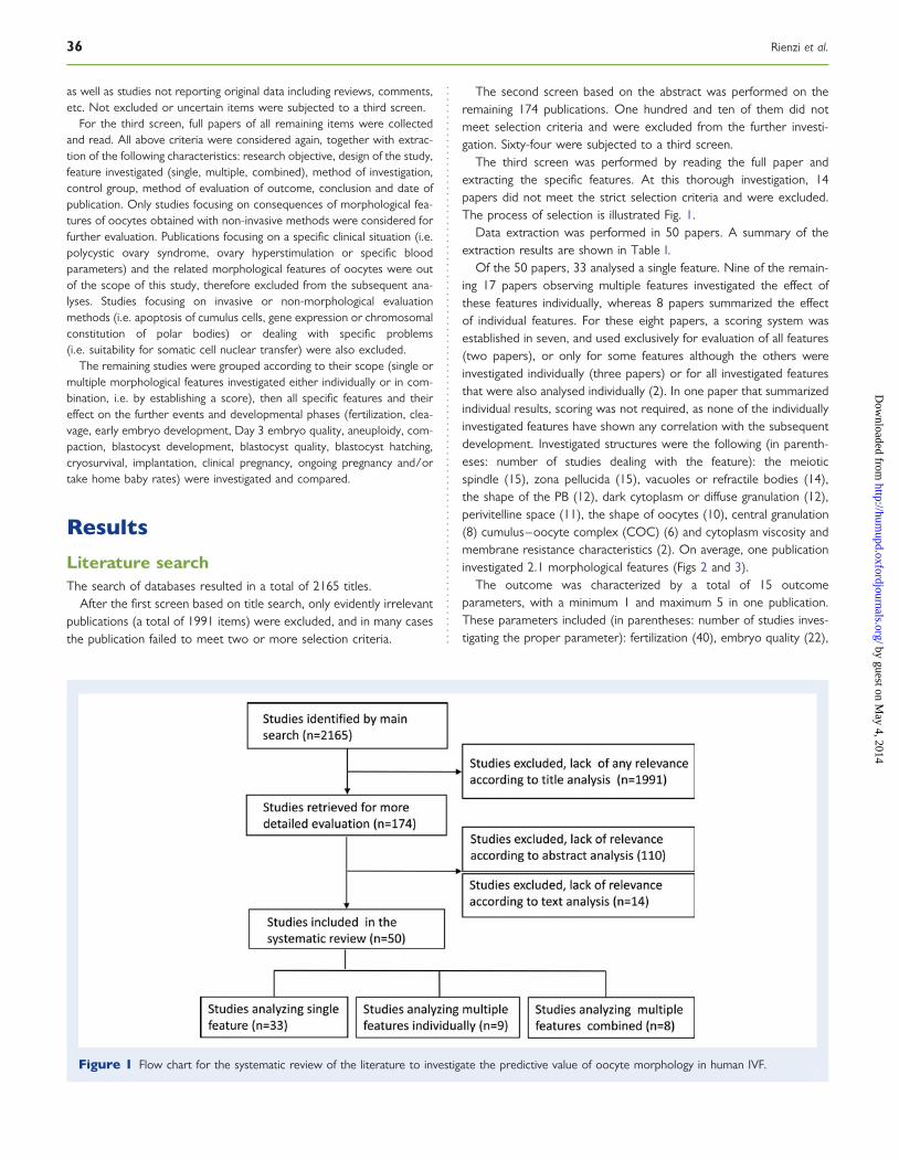
The third screen was performed by reading the full paper andextracting the specific features. At this thorough investigation, 14papers did not meet the strict selection criteria and were excluded.The process of selection is illustrated Fig. 1.
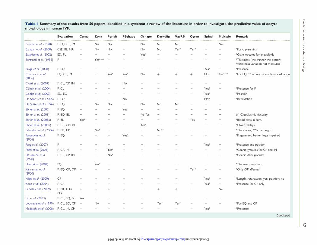
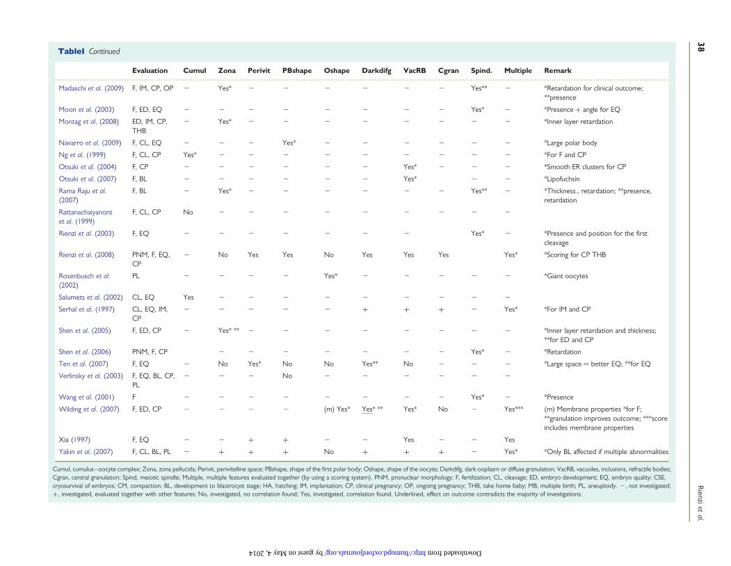
Data extraction was performed in 50 papers. A summary of theextraction results are shown in Table I.
Of the 50 papers, 33 analysed a single feature. Nine of the remain-ing 17 papers observing multiple features investigated the effect ofthese features individually, whereas 8 papers summarized the effectof individual features. For these eight papers, a scoring system wasestablished in seven, and used exclusively for evaluation of all features(two papers), or only for some features although the others wereinvestigated individually (three papers) or for all investigated featuresthat were also analysed individually (2). In one paper that summarizedindividual results, scoring was not required, as none of the individuallyinvestigated features have shown any correlation with the subsequentdevelopment. Investigated structures were the following (in parenth-eses: number of studies dealing with the feature): the meioticspindle (15), zona pellucida (15), vacuoles or refractile bodies (14),the shape of the PB (12), dark cytoplasm or diffuse granulation (12),perivitelline space (11), the shape of oocytes (10), central granulation(8) cumulus–oocyte complex (COC) (6) and cytoplasm viscosity andmembrane resistance characteristics (2). On average, one publicationinvestigated 2.1 morphological features (Figs 2 and 3).
The outcome was characterized by a total of 15 outcomeparameters, with a minimum 1 and maximum 5 in one publication.These parameters included (in parentheses: number of studies inves-tigating the proper parameter): fertilization (40), embryo quality (22),
Figure 1 Flow chart for the systematic review of the literature to investigate the predictive value of oocyte morphology in human IVF.
36 Rienzi et al.
by guest on May 4, 2014
http://humupd.oxfordjournals.org/
Dow
nloaded from
..........................................................................................................................................................................................................................................................
Table I Summary of the results from 50 papers identified in a systematic review of the literature in order to investigate the predictive value of oocytemorphology in human IVF.
Evaluation Cumul Zona Perivit PBshape Oshape Darkdifg VacRB Cgran Spind. Multiple Remark
Balaban et al. (1998) F, EQ, CP, IM 2 No No 2 No No No 2 2 No
Balaban et al. (2008) CSE, BL, HA 2 No No 2 No No Yes* Yes* 2 2 *For cryosurvival
Balakier et al. (2002) ED, PL 2 2 2 2 Yes* 2 2 2 2 2 *Giant oocytes for aneuploidy
Bertrand et al. (1995) F 2 Yes* ** 2 2 2 2 2 2 2 2 *Thickness (the thinner the better);**thickness variation not measured
Braga et al. (2008) F, EQ 2 2 2 2 2 2 2 2 Yes* 2 *Presence
Chamayou et al.(2006)
EQ, CP, IM 2 2 Yes* Yes* No + + + No Yes* ** *For EQ; **cumulative ooplasm evaluation
Ciotti et al. (2004) F, CL, CP, IM 2 2 2 No 2 2 2 2 2 2
Cohen et al. (2004) F, CL 2 2 2 2 2 2 2 2 Yes* 2 *Presence for F
Cooke et al. (2003) ED, EQ 2 2 2 2 2 2 2 2 Yes* *Position
De Santis et al. (2005) F, EQ 2 2 2 No 2 2 2 No* *Retardation
De Sutter et al. (1996) F, EQ 2 No No 2 No No No 2 2 2
Ebner et al. (2000) F, EQ 2 2 2 Yes 2 2 2 2 2 2
Ebner et al. (2003) F, EQ, BL 2 2 2 2 (v) Yes 2 2 2 2 2 (v) Cytoplasmic viscosity
Ebner et al. (2008a) F, BL Yes* 2 2 2 2 2 2 Yes 2 2 *Blood clots in cum.
Ebner et al. (2008b) F, CL, CM, BL 2 2 2 2 Yes* 2 2 2 2 2 *Ovoid: delays
Esfandiari et al. (2006) F, ED, CP 2 No* 2 2 2 No** 2 2 2 *Thick zona; **‘brown eggs’
Fancsovits et al.(2006)
F, EQ 2 2 2 Yes* 2 2 2 2 2 2 *Fragmented better large impaired
Fang et al. (2007) F 2 2 2 2 2 2 2 Yes* 2 *Presence and position
Farhi et al. (2002) F, CP, IM 2 2 Yes* 2 2 2 2 2 2 2 *Coarse granules for CP and IM
Hassan-Ali et al.(1998)
F, CL, CP, IM 2 2 No* 2 2 2 2 2 2 2 *Coarse dark granules
Høst et al. (2002) EQ 2 Yes* 2 2 2 2 2 2 2 2 *Thickness variation
Kahraman et al.(2000)
F, EQ, CP, OP 2 2 2 2 2 2 2 Yes* 2 2 *Only OP affected
Kilani et al. (2009) CP 2 2 2 2 2 2 2 Yes* 2 *Length, retardation: yes; position: no
Konc et al. (2004) F, CP 2 2 2 2 2 2 2 2 Yes* 2 *Presence for CP only
La Sala et al. (2009) F, PR, THB,MB
+ + + + 2 + + 2 2 No
Lin et al. (2003) F, CL, EQ, BL Yes 2 2 2 2 2 2 2 2 2
Loutradis et al. (1999) F, CL, EQ, CP 2 No 2 2 2 Yes* Yes* 2 2 2 *For EQ and CP
Madaschi et al. (2008) F, CL, IM, CP 2 2 2 2 2 2 2 2 Yes* *Presence
Continued
Predictivevalue
ofoocyte
morphology
37
by guest on May 4, 2014 http://humupd.oxfordjournals.org/ Downloaded from
..........................................................................................................................................................................................................................................................
TableI Continued
Evaluation Cumul Zona Perivit PBshape Oshape Darkdifg VacRB Cgran Spind. Multiple Remark
Madaschi et al. (2009) F, IM, CP, OP 2 Yes* 2 2 2 2 2 2 Yes** 2 *Retardation for clinical outcome;**presence
Moon et al. (2003) F, ED, EQ 2 2 2 2 2 2 2 2 Yes* 2 *Presence + angle for EQ
Montag et al. (2008) ED, IM, CP,THB
2 Yes* 2 2 2 2 2 2 2 2 *Inner layer retardation
Navarro et al. (2009) F, CL, EQ 2 2 2 Yes* 2 2 2 2 2 2 *Large polar body
Ng et al. (1999) F, CL, CP Yes* 2 2 2 2 2 2 2 2 2 *For F and CP
Otsuki et al. (2004) F, CP 2 2 2 2 2 2 Yes* 2 2 2 *Smooth ER clusters for CP
Otsuki et al. (2007) F, BL 2 2 2 2 2 2 Yes* 2 2 *Lipofuchsin
Rama Raju et al.(2007)
F, BL 2 Yes* 2 2 2 2 2 2 Yes** 2 *Thickness., retardation; **presence,retardation
Rattanachaiyanontet al. (1999)
F, CL, CP No 2 2 2 2 2 2 2 2 2
Rienzi et al. (2003) F, EQ 2 2 2 2 2 2 2 Yes* 2 *Presence and position for the firstcleavage
Rienzi et al. (2008) PNM, F, EQ,CP
2 No Yes Yes No Yes Yes Yes Yes* *Scoring for CP THB
Rosenbusch et al.(2002)
PL 2 2 2 2 Yes* 2 2 2 2 2 *Giant oocytes
Salumets et al. (2002) CL, EQ Yes 2 2 2 2 2 2 2 2 2
Serhal et al. (1997) CL, EQ, IM,CP
2 2 2 2 2 + + + 2 Yes* *For IM and CP
Shen et al. (2005) F, ED, CP 2 Yes* ** 2 2 2 2 2 2 2 2 *Inner layer retardation and thickness;**for ED and CP
Shen et al. (2006) PNM, F, CP 2 2 2 2 2 2 2 Yes* 2 *Retardation
Ten et al. (2007) F, EQ 2 No Yes* No No Yes** No 2 2 2 *Large space ¼ better EQ; **for EQ
Verlinsky et al. (2003) F, EQ, BL, CP,PL
2 2 2 No 2 2 2 2 2 2
Wang et al. (2001) F 2 2 2 2 2 2 2 2 Yes* 2 *Presence
Wilding et al. (2007) F, ED, CP 2 2 2 2 (m) Yes* Yes* ** Yes* No 2 Yes*** (m) Membrane properties *for F;**granulation improves outcome; ***scoreincludes membrane properties
Xia (1997) F, EQ 2 2 + + 2 2 Yes 2 2 Yes
Yakin et al. (2007) F, CL, BL, PL 2 + + + No + + + 2 Yes* *Only BL affected if multiple abnormalities
Cumul, cumulus–oocyte complex; Zona, zona pellucida; Perivit, perivitelline space; PBshape, shape of the first polar body; Oshape, shape of the oocyte; Darkdifg, dark ooplasm or diffuse granulation; VacRB, vacuoles, inclusions, refractile bodies;Cgran, central granulation; Spind, meiotic spindle; Multiple, multiple features evaluated together (by using a scoring system). PNM, pronuclear morphology; F, fertilization; CL, cleavage; ED, embryo development; EQ, embryo quality; CSE,cryosurvival of embryos; CM, compaction; BL, development to blastrocyst stage; HA, hatching; IM, implantation; CP, clinical pregnancy; OP, ongoing pregnancy; THB, take home baby; MB, multiple birth; PL, aneuploidy. 2, not investigated;+, investigated, evaluated together with other features; No, investigated, no correlation found; Yes, investigated, correlation found. Underlined, effect on outcome contradicts the majority of investigations.
38R
ienzietal.
by guest on May 4, 2014 http://humupd.oxfordjournals.org/ Downloaded from
clinical pregnancy rate (22), cleavage rate (13), development to blas-tocysts (9), implantation rate (9), embryo development (7), aneu-ploidy (4), ongoing pregnancy (2), take home babies (3),cryosurvival of embryos (1) compaction (1) and hatching (1). Onaverage, one publication investigated 2.7 outcome parameters.Exactly the same parameters were investigated in 8 papers (fertiliza-tion and embryo quality), 3 papers (fertilization and blastocyst rate),4 × 2 papers (fertilization; fertilization and clinical pregnancy rate; fer-tilization and embryo development; fertilization, cleavage, clinical preg-nancy and implantation rates, respectively).
Only two studies used exactly and exclusively the same parametersto investigate the effect of exactly and exclusively the same feature: in
these papers meiotic spindle morphology effect was characterized byfertilization and embryo quality.
In the most restricted investigations the consequence of one mor-phological feature was characterized by one outcome parameter (fivepapers), although the broadest study was performed in one paper,where the cumulative effect of seven features was compared withfive outcome parameters.
Owing to the heterogeneity of approaches, a single direct compari-son between the results of the 50 papers was impossible. Therefore,studies were grouped based on the investigated individual morphologi-cal features or score systems, and comparisons were performedaccordingly.
Cumulus–oocyte complexMorphological characteristics obtained by non-invasive methods inrelation to the further developmental competence were investigatedin 5 of the 50 papers. Rattanachaiyanont et al. (1999) have gradedthe expansion of both cumulus and corona radiata individually.According to their results, there was no correlation between the mor-phology of COCs and the fertilization, cleavage and clinical pregnancyrates. Ebner et al. (2008a) have performed similar grading and had asimilar conclusion; however, they found that presence of blood clots(but not that of amorphous clumps) were associated with densecentral granulation of oocytes and had a negative effect on fertilizationand blastocyst rates. Both studies have found a correlation between avery dense corona radiata layer and decreased maturity of oocytes.
In contrast, by using a 5-scale scoring system based mostly on themorphology of the cumulus–corona radiata cells, Lin et al. (2003)have found a correlation of the in vitro developmental potential andblastocyst quality. Another scoring system of the quality of theCOCs found associations between the observed quality and bothfertilization and subsequent pregnancy rates, but not cleavage rates
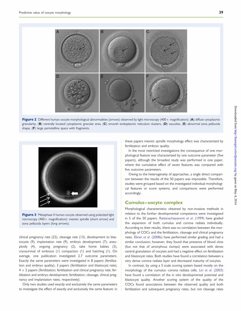
Figure 2 Different human oocyte morphological abnormalities (arrows) observed by light microscopy (400× magnification): (A) diffuse cytoplasmicgranularity, (B) centrally located cytoplasmic granular area, (C) smooth endoplasmic reticulum clusters, (D) vacuoles, (E) abnormal zona pellucidashape, (F) large perivitelline space with fragments.
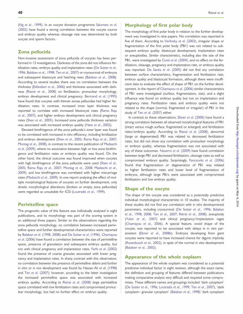
Figure 3 Metaphase II human oocyte observed using polarized lightmicroscopy (400× magnification): meiotic spindle (short arrow) andzona pellucida layers (long arrows).
Predictive value of oocyte morphology 39
by guest on May 4, 2014
http://humupd.oxfordjournals.org/
Dow
nloaded from
(Ng et al., 1999). In an oocyte donation programme Salumets et al.(2002) have found a strong correlation between the oocyte sourceand embryo quality whereas cleavage rate was determined by bothoocyte and sperm factors.
Zona pellucidaNon-invasive assessment of zona pellucida of oocytes has been per-formed in 13 investigations. Darkness of the zona did not influence fer-tilization rates, embryo quality and implantation rates (De Sutter et al.,1996; Balaban et al., 1998; Ten et al., 2007) or cryosurvival of embryosand subsequent blastocyst and hatching rates (Balaban et al., 2008).According to several studies there was no correlation between thethickness (Esfandiari et al., 2006) and thickness associated with dark-ness (Rienzi et al., 2008) on fertilization, pronuclear morphology,embryo development and clinical pregnancy. Bertrand et al. (1995)have found that oocytes with thinner zonae pellucidae had higher fer-tilization rates. In contrast, increased inner layer thickness wasreported to correlate with increased blastocyst rates (Rama Rajuet al., 2007), and higher embryo development and clinical pregnancyrates (Shen et al., 2005). Increased zona pellucida thickness variationwas associated with increased embryo quality (Høst et al., 2002).
Elevated birefringence of the zona pellucida’s inner layer was foundto be correlated with increased in vitro efficiency, including fertilizationand embryo development (Shen et al., 2005; Rama Raju et al., 2007;Montag et al., 2008), in contrast to the recent publication of Madaschiet al. (2009), where no association between high or low zona birefrin-gence and fertilization rates or embryo quality was found. On theother hand, the clinical outcome was found improved when oocyteswith high birefringence of the zona pellucida were used (Shen et al.,2005; Rama Raju et al., 2007; Montag et al., 2008; Madaschi et al.,2009), and low birefringence was correlated with higher miscarriagerates (Madaschi et al., 2009). In one report analysing the effect of mul-tiple morphological features of oocytes on further development, onlydrastic morphological alterations (broken or empty zona pellucidae)were regarded as unsuitable for ICSI (Loutradis et al., 1999).
Perivitelline spaceThe prognostic value of this feature was individually analysed in eightpublications, and its morphology was part of the scoring system inan additional three papers. Similar to the observations regarding thezona pellucida morphology, no correlation between increased perivi-telline space and further developmental characteristics were reportedby Balaban et al. (1998, 2008) and De Sutter et al. (1996). Chamayouet al. (2006) have found a correlation between the size of perivitellinespace, presence of granulation and subsequent embryo quality, butnot with clinical pregnancy and implantation rates. Farhi et al. (2002)found the presence of coarse granules associated with lower preg-nancy and implantation rates. In sharp contrast with this observation,no correlation between the presence of perivitelline debris and furtherin vitro or in vivo development was found by Hassan-Ali et al. (1998)and Ten et al. (2007); however, according to the latter investigationthe increased perivitelline space was associated with increasedembryo quality. According to Rienzi et al. (2008) large perivitellinespace correlated with low fertilization rates and compromised pronuc-lear morphology, but had no further effect on embryo quality.
Morphology of first polar bodyThe morphology of first polar body in relation to the further develop-ment was investigated in nine papers. No correlation was reported infive of them. According to Verlinsky et al. (2003), irregular shape orfragmentation of the first polar body (PB1) was not related to sub-sequent embryo quality, blastocyst development, implantation ratesor aneuploidies. Similar characteristics, including also the size of thePB1, were investigated by Ciotti et al. (2004), and no effect on the fer-tilization, cleavage, pregnancy and implantation rate, or embryo qualitywas reported. De Santis et al. (2005) did not find any correlationbetween surface characteristics, fragmentation and fertilization rate,embryo quality and blastocyst formation, although there were insuffi-cient data to evaluate the effect of shape of PB1 on the further devel-opment. In the report of Chamayou et al. (2006) similar characteristicsof PB1 were investigated (surface, fragmentation, size), and a slightinfluence was found on embryo quality but not on implantation andpregnancy rates. Fertilization rates and embryo quality were notrelated to the shape (normal, fragmented or irregular) of PB1 in thestudy of Ten et al. (2007) either.
In contrast to these observations, Ebner et al. (2000) have found astrong correlation between all observed morphological features of PB1(intact versus rough surface, fragmented or enlarged) and fertilizationrates/embryo quality. According to Rienzi et al. (2008), abnormal(large or degenerated) PB1 was related to decreased fertilizationrates, but did not show any correlation with pronuclear morphologyor embryo quality, whereas fragmentation was not associated withany of these outcomes. Navarro et al. (2009) have found correlationbetween large PB1 and decreased fertilization, cleavage rates as well ascompromised embryo quality. Surprisingly, Fancsovits et al. (2006)found that fragmentation or degeneration of PB1 was relatedto higher fertilization rates and lower level of fragmentation ofembryos, although large PB1s were associated with compromisedfertilization and low embryo quality.
Shape of the oocyteThe shape of the oocyte was considered as a potentially predictiveindividual morphological characteristic in 10 studies. The majority ofthese studies did not find any correlation with in vitro developmentalparameters, including cryosurvival (De Sutter et al., 1996; Balabanet al., 1998, 2008; Ten et al., 2007; Rienzi et al., 2008), aneuploidy(Yakin et al., 2007) and clinical pregnancy/implantation rages(Chamayou et al., 2006). A special feature, ovoid shape of theoocyte, was reported to be associated with delays in in vitro par-ameters (Ebner et al., 2008b). Embryos developing from giantoocytes were reported to have increased chance for digynic triploidy(Rosenbusch et al., 2002), in spite of the normal in vitro development(Balakier et al., 2002).
Appearance of the whole ooplasmThe appearance of the whole ooplasm was considered as a potentialpredictive individual factor in eight reviews, although the exact name,the definition and grouping of features differed between publicationsmaking comparative analysis very difficult and required some compro-mises. These different names and groupings included ‘dark cytoplasm’(De Sutter et al., 1996; Loutradis et al., 1999; Ten et al., 2007), ‘darkcytoplasm–granular cytoplasm’ (Balaban et al., 1998) ‘dark cytoplasm
40 Rienzi et al.
by guest on May 4, 2014
http://humupd.oxfordjournals.org/
Dow
nloaded from
with slight granulation’ (Balaban et al., 2008), ‘dark granular appear-ance of the cytoplasm’ (Esfandiari et al., 2006) and ‘diffused cyto-plasmic granularity’ (Rienzi et al. 2008). In one paper, homogenousgranulation was not mentioned, but two groups of heterogenous gran-ulations were distinguished: on one or two sides of the oocyte, or inthe centre (Wilding et al., 2007). Oocytes with heterogenous granula-tions had better fertilization rates than the control ones with clearcytoplasm. In another paper, the dark and the granular cytoplasm(either homogenous or centrally located) was considered as a separ-ate morphological feature (Ten et al., 2007), accordingly in the presentanalysis only the dark cytoplasm group was involved.
The different terminology may be the cause of the rather controver-sial outcome of investigation of the predictive value of this feature. Thedark cytoplasm, when analysed as an individual feature was found notto be a predictive factor in most investigated in vitro or in vivo par-ameters (De Sutter et al., 1996; Balaban et al., 1998, 2008; Esfandiariet al., 2006). Compromised quality of embryos which developed fromoocytes with dark cytoplasm was reported by Ten et al., (2007) andLoutradis et al. (1999). Diffuse peripheral granulation was found tobe associated with compromised pronuclear morphology (Rienziet al., 2008). According to Wilding et al. (2007), however, any typeof cytoplasmic granulation was associated with higher fertilizationrates than in oocytes with total absence of granularity.
Presence of vacuoles and/or cytoplasmicinclusionsThe presence of vacuoles and/or cytoplasmic inclusions was investi-gated in 10 publications. Although less heterogeneity in consideration,definition and classification was found than in the previous group, slightvariations included observation of vacuoles (saccules, smooth endo-plasmic reticulum clusters) only (Otsuki et al., 2004; Ten et al.,2007; Balaban et al., 2008); inclusions (refractile bodies, dark incorpor-ations, fragments, spots, dense granules; lipid droplets; lipofuchsin)only (De Sutter et al., 1996; Xia, 1997; Balaban et al., 1998; Otsukiet al., 2007); or both vacuoles and inclusions (Loutradis et al., 1999;Wilding et al., 2007). In two papers the presence of vacuoles,smooth endoplasmic reticulum clusters and refractile bodies wereindividually registered and their predictive value were individually ana-lysed (Rienzi et al., 2008). Although the aetiology, structure and poten-tial predictive value of the different structures may differ considerably,owing to the limitations of non-invasive investigation techniques as wellas the potential confusion at sorting different structures, here wefollow the everyday routine reflected also by these reviews bymaking tentative groups for analysis of their correlation with theoutcome.
Although fertilization rates and embryo quality were not affected(Ten et al., 2007), the presence of vacuoles in oocytes was negativelycorrelated with cryosurvival and developmental competence ofembryos after fertilization (Balaban et al., 2008). Increased biochemicalpregnancy rates were followed by decreased clinical pregnancy ratesafter transfer of embryos derived from oocytes with vacuoles(Otsuki et al., 2004). According to some investigators, cytoplasmicinclusions did not appear to affect fertilization, embryo quality andimplantation rates (De Sutter et al., 1996; Balaban et al., 1998).Others, however, reported decreased fertilization and embryo devel-opment (Xia, 1997; Otsuki et al., 2007). The presence of both
vacuoles and inclusions was related to compromised clinical pregnancyrates in the report by Loutradis et al. (1999). According to Wildinget al. (2007) these oocytes also had lower fertilization, embryo devel-opmental and higher aneuploidy rates. When analysed separately thepredictive role of presence of vacuoles, smooth endoplasmic reticulumclusters and refractile bodies, Rienzi et al. (2008) have only found aslight but significant decrease in fertilization rates of vacuolatedoocytes. However, none of the three factors affected pronuclear orembryo morphology.
Central granulation or centrally locatedgranular cytoplasmThe correlation between central granulation or centrally located gran-ular cytoplasm of oocytes and developmental competence was dis-cussed in five papers. Pronuclear morphology and embryo qualitywas compromised after fertilization of these oocytes (Ebner et al.,2008a; Rienzi et al., 2008). Decreased survival and impaired in vitrodevelopment after cryopreservation of embryos derived fromoocytes with central granulation was reported by Balaban et al.(2008). Centrally located granular cytoplasm was the only featureinvestigated by Kahraman et al. (2000) who have not found any corre-lation with fertilization rates, embryo development or pregnancy rates.However, ongoing pregnancy rates were seriously compromised whenembryos from centrally granulated oocytes were transferred. In con-trast, oocytes with centrally located granulation were not found tohave inferior fertilizing and in vitro developmental ability comparedwith those with completely absent granulation in the cytoplasm(Wilding et al., 2007).
Presence and morphology of the meioticspindleFifteen papers of the 50 selected were dealing with the correlationbetween the presence and morphology of the meiotic spindle andfurther development including fertilizing ability and in vitro/in vivodevelopmental competence. Computer assisted polarizationmicroscopy systems were used to evaluate the presence and othermorphological features of the spindle including position, length andbirefringence.
Only one study, that of Chamayou et al. (2006), found the presenceor absence of the meiotic spindle irrelevant regarding the furtherdevelopmental competence, including embryo quality and clinicalpregnancy rates. The presence of meiotic spindle has been associatedwith higher birefringence of the zona pellucida (Madaschi et al., 2009)and higher fertilization rates (Moon et al., 2003; Cohen et al., 2004;Konc et al., 2004; Fang et al., 2007; Rama Raju et al., 2007; Bragaet al., 2008, Madaschi et al., 2008; Wang et al., 2001). Results regard-ing the correlation between the presence of the spindle and earlyembryo development were controversial, with improved results(Moon et al., 2003; Rama Raju et al., 2007; Madaschi et al., 2008),versus no difference as observed by Cohen et al. (2004). Pregnancyrates were reported to be higher when embryos were derived fromoocytes with the presence of spindle (Konc et al., 2004; Madaschiet al., 2008, 2009). Position of the meiotic spindle close to the PBwas correlated with fertilization and cleavage rates (Fang et al.,2007) and early embryo development and quality (Cooke et al.,2003), whereas Moon et al. (2003) did not find any significant
Predictive value of oocyte morphology 41
by guest on May 4, 2014
http://humupd.oxfordjournals.org/
Dow
nloaded from
difference. According to Rienzi et al. (2003) high degrees of misalign-ment between the meiotic spindle and the first PB increased risk offertilization abnormalities. However, when normal fertilization hadoccurred, the cleavage potential of embryos developing from suchoocytes was not impaired. De Santis et al. (2005) did not find signifi-cant correlation between the spindle retardance and embryo quality;on the other hand, Shen et al. (2006) found that better pronuclearscores and higher pregnancy rates correlated with higher retardance.Higher blastocyst rates were also found to be related to higher retar-dance in the report of Rama Raju et al. (2007). Pregnancy rates werestrongly related to the normal morphology (complete, barrel shaped,with strong birefringence) and also to the retardance of the spindle(Kilani et al., 2009).
Viscosity of the cytoplasm and the resistanceof the cell membrane at ICSITwo special features, the viscosity of the cytoplasm and the resistanceof the cell membrane at ICSI, were analysed by Ebner et al. (2003) andWilding et al. (2007). Although these parameters were not strictlymorphological ones, resistance (together with two morphological fea-tures) was included into the grading system of Wilding et al. (2007)and does not seem to be sharply distinct from the viscosity analysedby Ebner et al. (2003), accordingly both of them were included inthis review. Both viscosity and resistance had a significant effect onsome investigated outcome parameters (fertilization, embyro quality,blastocyst rates and fertilization for viscosity and resistance,respectively).
Cumulative effect of multiple featuresInvestigations regarding the cumulative effect of multiple features wereperformed in eight studies. As the applied systems were different inalmost all of these investigations, here we summarize briefly themethod and the outcome.
Balaban et al. (1998) have compared the outcome of embryo trans-fers of cycles where all transferred embryos were derived from mor-phologically intact oocytes with those where all oocytes hadmorphological abnormalities, including extracytoplasmic, cytoplasmic,shape and multiple defects. No difference between the pregnancyand implantation rates was found.
A similar system was used and the same conclusion was obtained byLa Sala et al. (2009). Investigated features involved the morphology ofthe COC, zona pellucida, perivitelline space, PB, ooplasm and pres-ence of vacuoles or granulations. Outcomes (fertilization, pregnancy,take home baby and multiple pregnancy rates) were similar when allembryos were derived from intact oocytes, or all from morphologi-cally ‘handicapped’ oocytes.
According to Yakin et al. (2007), the impaired morphology ofoocytes characterized by almost all previously listed morphologicalfeatures—except for the morphology of the COC and the investi-gation of the meiotic spindle—affected only blastocyst development,but not fertilization, cleavage and aneuploidy rates.
Chamayou et al. (2006) have used a cumulative evaluation for cyto-plasmic features of oocytes including texture, inclusions, vacuoles andcentral granulation. The presence of these features was correlatedwith impaired embryo quality, but did not influence pregnancy andimplantation rates.
In the study of Serhal et al. (1997) similar features (excessive gran-ularity, cytoplasmic inclusions, smooth endoplasmic reticulum cluster-ing and refactile bodies) were tentatively investigated with acompletely different conclusion: in vitro developmental parameterswere not influenced, but implantation and pregnancy rates werelower, when embryos were derived from oocytes with cytoplasmicabnormalities.
The oocyte grading system of Xia (1997) was based on the evalu-ation of the perivitelline space, PB morphology and cytoplasmicinclusions, and a strong correlation with fertilization rates andembryo quality was found.
In the study of Wilding et al. (2007), two morphologic and one func-tional parameters were involved in a grading system: oocyte granular-ity, vacuoles and inclusions (as one parameter) and injectionproperties (the resistance and fragility of the plasma membrane atICSI). Fertilization, embryo development and clinical pregnancy rateswere significantly different between the high and low quality group.
A complex grading system (MOMS; MII oocyte morphologicalscore) has been established recently by Rienzi et al. (2008) accordingto the impact of different oocyte morphological features (vacuoles,abnormal PB1, large perivitelline space, diffused cytoplasmic granular-ity and/or centrally located granular area) on fertilization rate, andpronuclear and embryo morphology. A significant relationshipbetween the MOMS rate and clinical pregnancy was found.
In general, out of the 92 studies of different parameters (includingboth single features and cumulative scores) investigating direct associ-ation with the further prognosis, 57 resulted in a significant correlationwith the outcome, whereas in 35 no predictive value of the micro-scopic feature was found. The diversity of observations and resultsdid not allow statistical comparison; however, there was no clear ten-dency of improved accuracy regarding the predictive value in recentpublications compared with the earlier ones. Twenty four of 42 obser-vations performed between 1997 and 2005 have found correlationswith the outcome, whereas these values between 2006 and 2009were 33 of 50.
DiscussionIn a recent review Swain and Pool (2008) summarize the short list offeatures of an oocyte that is regarded healthy on the basis of morpho-logical investigations during the routine IVF programme: single PB,‘normal-looking’ cytoplasm, appropriate zona thickness, proper perivi-telline space. According to the authors, the common experience isthat these features often fail to predict the future fertilizing abilityand developmental competence. This seems to be a widely acceptedand shared opinion between human embryologists. On the otherhand, most embryologists may also agree that some morphologicallydetectable features of MII phase oocytes indicate seriously compro-mised developmental competence. Morphological alterations mayalso be related to the patient and the cycle characteristics, and mayinvolve most or all oocytes in the cohort.
The purpose of our review was to investigate if any non-invasivemorphological feature or group of features of MII phase humanoocytes has a strong predictive value for further development accord-ing to the literature between 1996 and 2009. Out of the 2159 titlesidentified by the initial search of relevant electronic databases, only50 met the selection criteria (full papers dealing with light or polarized
42 Rienzi et al.
by guest on May 4, 2014
http://humupd.oxfordjournals.org/
Dow
nloaded from
microscopic morphological features of in vivo matured, not cryopre-served MII phase human oocytes, and analysing the prognostic valueof these features for further development). Unfortunately the majority(27) of the papers restricted the outcome investigation to the in vitroperiod, with limited value regarding the primary outcome includingclinical pregnancy, implantation and ongoing pregnancy rates. Onlythree publications (Montag et al., 2008; Rienzi et al., 2008; La Salaet al., 2009) extended the investigation to the ultimate parameter,the number of take home babies. Moreover, because of the hetero-geneity of the approaches and the unavailability of clinical data, thecorrelation between oocyte morphology and patient and cycle charac-teristics was not included in the analysis.
The grouping of morphological features was the result of an una-voidable compromise, as the use of terms and description of altera-tions were inconsistent between papers. Experimental designs alsovaried considerably, with wide differences between papers regardingthe investigated features and outcome parameters. The only excep-tion was the morphology of the meiotic spindle, where the reasonablyhomogenous material (relatively well-defined morphological featuresand similar outcome parameters) would allow meta-analysis.However, for this feature a recent review provided an excellent com-parative analysis (Petersen et al., 2009): as the full papers investigatedby this review were the same that were recovered by our independentsearch, the results of Petersen et al. (2009) were acknowledged andconsidered when conclusions were made.
As a result of these facts, and the relatively small number of publi-cations investigating the predictive value of other morphological fea-tures, no strict selection according to the experimental designs(retrospective, prospective, randomized studies etc.) was applied;statistical calculations of authors were not re-evaluated or ranked.Each statistically supported result (correlation or no correlationbetween the feature and the outcome) was included and consideredas a proven individual observation.
The tentative summary of our work is rather disappointing: in the50 relevant papers recovered from databases from the past 15years, none of the investigated 9 single features was unanimously cor-related with normal or compromised development, when evaluatedby 15 outcome parameters.
Among extracytoplasmic features, only dysmorphism of the COCwas found to be indicative for further development in the majorityof papers (four of five) dealing with the subject. For the zona pellucida,only 6 of 13 papers found correlation between the morphology anddevelopmental competence including four papers dealing with thebirefringence of the zona (one of them did not find correlation withthe in vitro period; another one did not extend the investigation tothe in vivo period). The commonly assessed light microscopic altera-tions (thickness and thickness variations, other dysmorphism) werefound to be related to improved/compromised development almostrandomly by the relevant papers.
Other extracytoplasmic features, including the size and granulationsof the perivitelline space, and the shape of the oocyte or the PB, wererelated to developmental competence only in approximately half ofthe publications focusing on the issue, questioning their commonlysupposed indicative value.
Among intracytoplasmic features, the presence, position and retar-dance of the spindle were found to be related to the developmentalcompetence in 12 of 13 papers. In accordance with the recent
meta-analysis of Petersen et al. (2009), based essentially on thesame material, the significantly correlated outcome parameters wererestricted to the in vitro development, whereas in vivo outcome ana-lyses were relatively rare and the statistical analysis failed to show sig-nificant differences. Among features observable with simpletransmitted light, the presence of central granulation resulted inimpaired further development in four of five publications, and twoof these also found a correlation with in vivo development. Regardingother cytoplasmic features, the assessment of their predictive valuehas shown less consistent results.
Complex analyses of multiple features based on a scoring systemseems to be a promising alternative to single feature analysis, andthis seems to be supported by the fact that six of the eight papersapplying this strategy have found a correlation between the cumulativeresults and the prognosis. Unfortunately the applied scoring systemshave shown extreme variability regarding the type and individualscoring of investigated features including combination with functionaltests, and outcome parameters. Accordingly, at present, they canonly be regarded as diverse approaches to the same strategy, buttheir comparative evaluation is impossible. Paradoxically, two recentpublications investigating similar parameters and extending theoutcome evaluation to take home babies (Rienzi et al., 2008; LaSala et al., 2009) resulted in contradicting conclusions about the pre-dictive value of their system.
Additionally, the slight increase in IVF efficiency in recent publi-cations can mostly be attributed to the application of the Polscopemicroscope for detecting retardance of the zona, or presence, pos-ition and retardance of the spindle, a method that is applied in alimited number of assisted reproduction technology (ART) unitsworldwide.
ConclusionThe analysis of 50 papers published in the past 15 years about predic-tive value of non-invasive morphological parameters of MII phaseoocytes has produced contradicting results, and did not entirelysupport the average opinion about the features of ‘good’ and ‘bad’quality and respective developmental competence. This outcomemay be explained both with the common misjudgement of predictivevalue of some features, and the extremely diverse approaches thesepapers used for investigations regarding terms, features, outcome par-ameters and grouping. As oocyte selection before insemination mayhave important benefits in certain situations, including that in countrieswith legal restrictions, our finding underlines the importance of moreintensive and coordinated research to reach a consensus and exploitfully the predictive potential of morphological examination, particularlyas that is expected to remain, for several years to come, the only avail-able selection method for many ART units.
Supplementary dataSupplementary data are available at http://humupd.oxfordjournals.org/.
FundingFunding to pay the Open Access publication charges for this articlewas provided by G.EN.E.R.A.
Predictive value of oocyte morphology 43
by guest on May 4, 2014
http://humupd.oxfordjournals.org/
Dow
nloaded from
ReferencesArav A, Aroyo A, Yavin S, Roth Z. Prediction of embryonic developmental
competence by time-lapse observation and ‘shortest half’ analysis. ReprodBioMed Online 2008;17:669–675.
Balaban B, Urman B, Sertac A, Alatas C, Aksoy S, Mercan R. Oocyte morphologydoes not affect fertilization rate, embryo quality and implantation rate afterintracytoplasmic sperm injection. Hum Reprod 1998;13:3431–3433.
Balaban B, Ata B, Isiklar A, Yakin K, Urman B. Severe cytoplasmic abnormalities ofthe oocyte decrease cryosurvival and subsequent embryonic development ofcryopreserved embryos. Hum Reprod 2008;23:1778–1785.
Balakier H, Bouman D, Sojecki A, Librach C, Squire JA. Morphological andcytoenetic analysis of human giant oocytes and giant embryos. Hum Reprod2002;17:2394–2401.
Bertrand E, Van den Bergh M, Englert Y. Does zona pellucida thickness influence thefertilization rate? Hum Reprod 1995;10:1189–1193.
Braga DP, Figueira RC, Rodrigues D, Madaschi C, Pasqualotto FF, Iaconelli A Jr,Borges E Jr. Prognostic value of meiotic spindle imaging on fertilization rate andembryo development in in vitro-matured human oocytes. Fertil Steril 2008;90:429–433.
Chamayou S, Ragolia C, Alecci C, Storaci G, Maglia E, Russo E, Guglielmino A.Meiotic spindle presence and oocyte morphology do not predict clinical ICSIoutcomes: a study of 967 transferred embryos. Reprod Biomed Online 2006;13:661–667.
Ciotti PM, Notarangelo L, Morselli-Labate AM, Felletti V, Porcu E, Venturoli S. Firstpolar body morphology before ICSI is not related to embryo quality or pregnancyrate. Hum Reprod 2004;19:2334–2339.
Cohen Y, Malcov M, Schwartz T, Mey-Raz N, Carmon A, Cohen T, Lessing JB,Amit A, Azem F. Spindle imaging: a new marker for optimal timing of ICSI?Hum Reprod 2004;19:649–654.
Cooke S, Tyler JPP, Driscoll GL. Meiotic spindle location and identification and itseffect on embryonic cleavage plane and early development. Hum Reprod 2003;11:2397–2405.
Cummins JM, Breen TM, Harrison KL, Shaw JM, Wilson LM, Hennessey JF. Aformula for scoring human embryo growth rates in in-vitro fertilization: its valuein predicting pregnancy and in comparison with visual estimates of embryoquality. J In Vitro Fert Embryo Transf 1986;3:284–295.
De Santis L, Cino I, Rabellotti E, Calzi F, Persico P, Borini A, Coticchio G. Polar bodymorphology and spindle imaging as predictors of oocyte quality. Reprod BiomedOnline 2005;11:36–42.
De Sutter P, Dozortsev D, Qian C, Dhont M. Oocyte morphology does notcorrelate with fertilization rate and embryo quality after intracytoplasmic sperminjection. Hum Reprod 1996;11:595–597.
Ebner T, Yaman C, Moser M, Sommergruber M, Feichtinger O, Tews G. Prognosticvalue of first polar body morphology on fertilization rate and embryo quality inintracytoplasmic sperm injection. Hum Reprod 2000;15:427–430.
Ebner T, Moser M, Sommergruber M, Puchner M, Wiesinger R, Tews G.Developmental competence of oocytes showing increased cytoplasmicviscosity. Hum Reprod 2003;18:1294–1298.
Ebner T, Moser M, Shebl O, Sommergruber M, Yaman C, Tews G. Blood clots inthe cumulus–oocyte complex predict poor oocyte quality and post-fertilizationdevelopment. Reprod Biomed Online 2008a;16:801–807.
Ebner T, Shebl O, Moser M, Sommergruber M, Tews G. Developmental fate ofovoid oocytes. Hum Reprod 2008b;23:62–66.
Emiliani S, Fasano G, Vandamme B et al. Impact of the assessment of earlycleavage in a single embryo transfer policy. Reprod BioMed Online 2006;13:255–260.
Esfandiari N, Burjaq H, Gotlieb L, Casper RF. Brown oocytes: implications forassisted reproductive technology. Fertil Steril 2006;86:1522–1525.
Fancsovits P, Tothne ZG, Murber A, Takacs FZ, Papp Z, Urbancsek J. Correlationbetween first polar body morphology and further embryo development. Acta BiolHung 2006;57:331–338.
Fang C, Tang M, Li T, Peng WL, Zhou CQ, Zhuang GL, Leong M. Visualization ofmeiotic spindle and subsequent embryonic development in in vitro and in vivomatured human oocytes. J Assist Reprod Genet 2007;24:547–551.
Farhi J, Nahum H, Weissman A, Zahalka N, Glezerman M, Levran D. Coarsegranulation in the perivitelline space and IVF-ICSI outcome. J Assist ReprodGenet 2002;19:545–549.
Gilchrist RB, Lane M, Thompson JG. Oocyte-secreted factors: regulators of cumuluscell function and oocyte quality. Hum Reprod Update 2008;14:159–177.
Hassan-Ali H, Hisham-Saleh A, El-Gezeiry D, Baghdady I, Ismaeil I, Mandelbaum J.Perivitelline space granularity: a sign of human menopausal gonadotrophinoverdose in intracytoplasmic sperm injection. Hum Reprod 1998;13:3425–3430.
Høst E, Gabrielsen A, Lindenberg S, Smidt-Jensen S. Apoptosis in human cumuluscells in relation to zona pellucida thickness variation, maturation stage, andcleavage of the corresponding oocyte after intracytoplasmic sperm injection.Fertil Steril 2002;77:511–515.
Kahraman S, Yakin K, Donmez E, Samli H, Bahce M, Cengiz G, Sertyel S, Samli M,Imirzalioglu N. Relationship between granular cytoplasm of oocytes andpregnancy outcome following intracytoplasmic sperm injection. Hum Reprod2000;15:2390–2393.
Kilani S, Cooke S, Kan A, Chapman M. Are there non-invasive markers in humanoocytes that can predict pregnancy outcome? Reprod Biomed Online 2009;18:674–680.
Konc J, Kanyo K, Cseh S. Visualization and examination of the meiotic spindle inhuman oocytes with polscope. J Assist Reprod Genet 2004;21:349–353.
La Sala GB, Nicoli A, Villani MT, Di Girolamo R, Capodanno F, Blickstein I. Theeffect of selecting oocytes for insemination and transferring all resultantembryos without selection on outcomes of assisted reproduction. Fertil Steril2009;91:96–100.
Lemmen JG, Agerholm I, Ziebe S. Kinetic markers of human embryo quality usingtime-lapse recordings of IVF/ICSI-fertilized oocytes. Reprod Biomed Online 2008;17:385–391.
Lin YC, Chang SY, Lan KC, Huang HW, Chang CY, Tsai MY, Kung FT, Huang FJ.Human oocyte maturity in vivo determines the outcome of blastocystdevelopment in vitro. J Assist Reprod Genet 2003;20:506–512.
Loutradis D, Drakakis P, Kallianidis K, Milingos S, Dendrinos S, Michalas S. Oocytemorphology correlates with embryo quality and pregnancy rate afterintracytoplasmic sperm injection. Fertil Steril 1999;72:240–244.
Madaschi C, de Souza Bonetti TC, de Almeida Ferreira Braga DP, Pasqualotto FF,Iaconelli A Jr, Borges E Jr. Spindle imaging: a marker for embryo development andimplantation. Fertil Steril 2008;90:194–198.
Madaschi C, Aoki T, de Almeida Ferreira Braga DP, de Cassia Savio FR, Semiao FL,Iaconelli A Jr, Borges E Jr. Zona pellucida birefringence score and meiotic spindlevisualization in relation to embryo development and ICSI outcomes. ReprodBiomed Online 2009;18:681–686.
Marteil G, Richard-Parpaillon L, Kubiak JZ. Role of oocyte quality in meioticmaturation and embryonic development. Reprod Biol 2009;9:203–224.
Montag M, Schimming T, Koster M, Zhou C, Dorn C, Rosing B, van der Ven H, Vender Ven K. Oocyte zona birefringence intensity is associated with embryonicimplantation potential in ICSI cycles. Reprod Biomed Online 2008;16:239–244.
Moon JH, Hyun CS, Lee SW, Son WY, Yoon SH, Lim JH. Visualization of themetaphase II meiotic spindle in living human oocytes using the Polscopeenables the prediction of embryonic developmental competence after ICSI.Hum Reprod 2003;18:817–820.
Mtango NR, Potireddy S, Latham K. Oocyte quality and maternal control ofdevelopment. Int Rev Cell Mol Biol 2008;268:223–290.
Nagy ZP, Jones-Colon S, Roos P, Botros L, Greco E, Dasig J, Behr B. Metabolomicassessment of oocyte viability. Reprod Biomed Online 2009;18:219–225.
Navarro PA, de Araujo MM, de Araujo CM, Rocha M, dos RR, Martins W.Relationship between first polar body morphology before intracytoplasmicsperm injection and fertilization rate, cleavage rate, and embryo quality. Int JGynaecol Obstet 2009;104:226–229.
Ng ST, Chang TH, Jackson Wu TC. Prediction of the rates of fertilization, cleavage,and pregnancy success by cumulus-coronal morphology in an in vitro fertilizationprogram. Fertil Steril 1999;72:412–417.
Otsuki J, Okada A, Morimoto K, Nagai Y, Kubo H. The relationship betweenpregnancy outcome and smooth endoplasmic reticulum clusters in MII humanoocytes. Hum Reprod 2004;19:1591–1597.
Otsuki J, Nagai Y, Chiba K. Lipofuscin bodies in human oocytes as an indicator ofoocyte quality. J Assist Reprod Genet 2007;24:263–270.
Patrizio P, Fragouli E, Bianchi V, Borini A, Wells D. Molecular methods for selectionof the ideal oocyte. Reprod Biomed Online 2007;15:346–353.
Petersen CG, Oliveira JB, Mauri AL, Massaro FC, Baruffi RL, Pontes A, Franco JG Jr.Relationship between visualization of meiotic spindle in human oocytes and ICSIoutcomes: a meta-analysis. Reprod Biomed Online 2009;18:235–243.
44 Rienzi et al.
by guest on May 4, 2014
http://humupd.oxfordjournals.org/
Dow
nloaded from
Rama Raju GA, Prakash GJ, Krishna KM, Madan K. Meiotic spindle and zonapellucida characteristics as predictors of embryonic development: a preliminarystudy using PolScope imaging. Reprod Biomed Online 2007;14:166–174.
Rattanachaiyanont M, Leader A, Leveille MC. Lack of correlation between oocyte–corona–cumulus complex morphology and nuclear maturity of oocytes collectedin stimulated cycles for intracytoplasmic sperm injection. Fertil Steril 1999;71:937–940.
Revelli A, Delle Piane L, Casano S, Molinari E, Massobrio M, Rinaudo P. Follicularfluid content and oocyte quality: from single biochemical markers tometabolomics. Reprod Biol Endocrinol 2009;4:7–40.
Rienzi L, Ubaldi F, Martinez F, Iacobelli M, Minasi MG, Ferrero S, Tesarik J, Greco E.Relationship between meiotic spindle location with regard to the polar bodyposition and oocyte developmental potential after ICSI. Hum Reprod 2003;18:1289–1293.
Rienzi L, Ubaldi FM, Iacobelli M, Minasi MG, Romano S, Ferrero S, Sapienza F, Baroni E,Litwicka K, Greco E. Significance of metaphase II human oocyte morphology on ICSIoutcome. Fertil Steril 2008;90:1692–1700.
Rosenbusch B, Schneider M, Glaser B, Brucker C. Cytogenetic analysis of giantoocytes and zygotes to assess their relevance for the development of digynictriploidy. Hum Reprod 2002;17:2388–2393.
Salumets A, Suikkari AM, Mols T, Soderstrom-Anttila V, Tuuri T. Influence ofoocytes and spermatozoa on early embryonic development. Fertil Steril 2002;78:1082–1087.
Serhal PF, Ranieri DM, Kinis A, Marchant S, Davies M, Khadum IM. Oocytemorphology predicts outcome of intracytoplasmic sperm injection. Hum Reprod1997;12:1267–1270.
Shen Y, Stalf T, Mehnert C, Eichenlaub-Ritter U, Tinneberg HR. High magnitude oflight retardation by the zona pellucida is associated with conception cycles. HumReprod 2005;20:1596–1606.
Shen Y, Stalf T, Mehnert C, De SL, Cino I, Tinneberg HR, Eichenlaub-Ritter U. Lightretardance by human oocyte spindle is positively related to pronuclear score afterICSI. Reprod Biomed Online 2006;12:737–751.
Swain JE, Pool TB. ART failure: ooctye contributions to unsuccessful fertilization.Hum Reprod Update 2008;14:431–446.
Ten J, Mendiola J, Vioque J, de JJ, Bernabeu R. Donor oocyte dysmorphisms andtheir influence on fertilization and embryo quality. Reprod Biomed Online 2007;14:40–48.
Van Soom A, Vandaele L, Goossens K, de Kruif A, Peelman L. Gamete origin inrelation to early embryo development. Theriogenology 2007;68(Suppl.1):131–137.
Testart J, Lassalle B, Frydman R, Belaisch JC. A study of factors affectingthe successof human fertilization in vitro. II. Influence of semen quality and oocyte maturityon fertilization and cleavage. Biol Reprod 1983;28:425–431.
Verlinsky Y, Lerner S, Illkevitch N, Kuznetsov V, Kuznetsov I, Cieslak J, Kuliev A. Isthere any predictive value of first polar body morphology for embryo genotype ordevelopmental potential? Reprod Biomed Online 2003;7:336–341.
Wang WH, Meng L, Hackett RJ, Odenbourg R, Keefe DL. The spindle observationand its relationship with fertilization after intracytoplasmic sperm injection in livinghuman oocytes. Fertil Steril 2001;75:348–353.
Wilding M, Di ML, D’Andretti S, Montanaro N, Capobianco C, Dale B. Anoocyte score for use in assisted reproduction. J Assist Reprod Genet 2007;24:350–358.
Xia P. Intracytoplasmic sperm injection: correlation of oocyte grade based on polarbody, perivitelline space and cytoplasmic inclusions with fertilization rate andembryo quality. Hum Reprod 1997;12:1750–1755.
Yakin K, Balaban B, Isiklar A, Urman B. Oocyte dysmorphism is not associated withaneuploidy in the developing embryo. Fertil Steril 2007;88:811–816.
Predictive value of oocyte morphology 45
by guest on May 4, 2014
http://humupd.oxfordjournals.org/
Dow
nloaded from