Embed Size (px)
Citation preview

RESEARCH ARTICLE
Prefrontal Activity and Connectivity with the
Basal Ganglia during Performance of
Complex Cognitive Tasks Is Associated with
Apathy in Healthy Subjects
Leonardo Fazio1,4, Giancarlo Logroscino2, Paolo Taurisano1, Graziella Amico1,
Tiziana Quarto1, Linda Antonella Antonucci1, Maria Rosaria Barulli2, Marina Mancini1,
Barbara Gelao1, Laura Ferranti3, Teresa Popolizio4, Alessandro Bertolino1,5,
Giuseppe Blasi5*
1 University of Bari ‘Aldo Moro’—Department of Basic Medical Science, Neuroscience, and Sense Organs,
70124, Bari, Italy, 2 University of Bari ‘Aldo Moro’—Department of Basic Medicine, Neuroscience, and
Sense Organs, 70124 Bari, Italy and University of Bari ‘Aldo Moro’, “Pia Fondazione Cardinale G. Panico”—
Department of Clinical Research in Neurology, 73039 Tricase, Lecce, Italy, 3 University of Perugia—
Department of General and Experimental Medicine, 06123, Perugia, Italy, 4 IRCCS “Casa Sollievo della
Sofferenza”, 71013, San Giovanni Rotondo, Italy, 5 Bari University Hospital—Psychiatry Unit, 70124, Bari,
Italy
Abstract
Objective
Convergent evidence indicates that apathy affects cognitive behavior in different neurological
and psychiatric conditions. Studies of clinical populations have also suggested the primary
involvement of the prefrontal cortex and the basal ganglia in apathy. These brain regions are
interconnected at both the structural and functional levels and are deeply involved in cognitive
processes, such as working memory and attention. However, it is unclear how apathy modu-
lates brain processing during cognition and whether such a modulation occurs in healthy
young subjects. To address this issue, we investigated the link between apathy and prefrontal
and basal ganglia function in healthy young individuals. We hypothesized that apathy may be
related to sub-optimal activity and connectivity in these brain regions.
Methods
Three hundred eleven healthy subjects completed an apathy assessment using the Stark-
stein’s Apathy Scale and underwent fMRI during working memory and attentional perfor-
mance tasks. Using an ROI approach, we investigated the association of apathy with
activity and connectivity in the DLPFC and the basal ganglia.
Results
Apathy scores correlated positively with prefrontal activity and negatively with prefrontal-
basal ganglia connectivity during both working memory and attention tasks. Furthermore,
prefrontal activity was inversely related to attentional behavior.
PLOS ONE | DOI:10.1371/journal.pone.0165301 October 31, 2016 1 / 15
a11111
OPENACCESS
Citation: Fazio L, Logroscino G, Taurisano P,
Amico G, Quarto T, Antonucci LA, et al. (2016)
Prefrontal Activity and Connectivity with the Basal
Ganglia during Performance of Complex Cognitive
Tasks Is Associated with Apathy in Healthy
Subjects. PLoS ONE 11(10): e0165301.
doi:10.1371/journal.pone.0165301
Editor: Natasha M. Maurits, Universitair Medisch
Centrum Groningen, NETHERLANDS
Received: March 31, 2016
Accepted: October 10, 2016
Published: October 31, 2016
Copyright: © 2016 Fazio et al. This is an open
access article distributed under the terms of the
Creative Commons Attribution License, which
permits unrestricted use, distribution, and
reproduction in any medium, provided the original
author and source are credited.
Data Availability Statement: The data of this study
are available at the 4TU.ResearchData repository;
http://dx.doi.org/10.4121/uuid:d5ec1a8a-6c61-
4f19-8af1-b728bb07e8c0.
Funding: The authors received no specific funding
for this work.
Competing Interests: Prof. Bertolino is consultants
of Hoffman-LaRoche, Ltd. All other authors report
no potential conflicts of interest. This does not alter

Conclusions
These results suggest that in healthy young subjects, apathy is a trait associated with ineffi-
cient cognitive-related prefrontal activity, i.e., it increases the need for prefrontal resources
to process cognitive stimuli. Furthermore, apathy may alter the functional relationship
between the prefrontal cortex and the basal ganglia during cognition.
Introduction
Apathy is defined as reducedmotivation towards goal-directedbehavior, a flattened affect,emotional indifference and a restricted response to important life events [1–2]. It impliesdiminishedmotivation and effort to perform personal and social everyday activities [1, 3]. Inaddition to being associated with a broad range of brain disorders [4–7], apathy is also presentin elderly and young healthy subjects [8–12].
The brain circuitry that sustains apathy has been investigated primarily in clinical andelderly populations. Many of these studies have demonstrated an association between apathyand structural anomalies, such as lesions of lateral or medial areas of the prefrontal cortex(PFC) [13–14] and decreased PFC volume [15–18]. Furthermore, functional imaging studies indementia and in post-stroke patients corroborate the relationship between frontal alterationsand apathy. In particular, previous reports have indicated associations between apathy andreduced glucosemetabolism and frontal rCBF perfusion [19–20], as well as a negative correla-tion between rCBF perfusion and apathy scores [21]. Other studies also suggested a linkbetween apathy and abnormalities in the basal ganglia (BG). In particular, this trait has beenrelated to focal lesions of the globus pallidus, the caudate and the putamen [22–24], as well asto BG hypoperfusion [25–26] and reduced BG volume [27–28]. Moreover, apathy is a frequentsymptom in clinical conditions that are characterized by BG impairment [4, 29–30]. A smallnumber of studies have suggested the involvement of other brain regions, such as the cingulatecortex, the insula or the premotor cortex [31–32], in apathy, but these results require replica-tion. Overall, the current evidence is consistent in suggesting a role for both the PFC and theBG in apathy in clinical and elderly populations. Accordingly, it has been hypothesized thatapathy may be subtended by a dysfunction in the neuronal circuit that includes both BG andPFC [33–35], which are interconnected at both the structural and functional levels [36–38].
The loop between the BG and the dorsolateral prefrontal portion of the PFC (DLPFC) isdeeply involved in cognitive processing, as indicated repeatedly by studies that focused onworking memory and attentional processing [39–41]. Apathy has also been correlated withanomalies in cognitive functions, as revealed by findings in clinical populations; these findingshighlighted a relationship between high levels of apathy and poorer performance on tests forworking memory and attention [42–46]. Similarly, apathy has been associated with cognitiveimpairment in elderly individuals who are not affected by neurological or psychiatric condi-tions [9, 47–49].
Given that most of studies of apathy have been performed in elderly or clinical populations,it is important to investigate this trait in healthy young subjects, thus nullifying the possibleimpact of other illness-related variables or aging on brain physiology. To our knowledge, onlyone recent study has addressed the relationship between apathy and brain imaging phenotypesin healthy young individuals. In that study [32], the authors investigated behavioral apathy(i.e., a sub-domain of apathy that is characterized by a lack of physical engagement, productiv-ity and initiative). The results of this study indicated that behavioral apathy is associated with
Brain Cognitive Processing and Apathy in Healthy Subjects
PLOS ONE | DOI:10.1371/journal.pone.0165301 October 31, 2016 2 / 15
the authors’ adherence to PLOS ONE policies on
sharing data and materials.

greater recruitment of the supplemental motor area and the motor cingulate, as well as lowerfunctional connectivity between these brain regions during a reward task. However, to ourknowledge, no studies have been performed to investigate the relationship between apathy andbrain correlates of high order cognitive processing, such as working memory and attention, inhealthy young subjects.
Here, we aimed to investigate the association of apathy with brain activity and functionalconnectivity during working memory and attentional processing in healthy young individuals.Given our strong a priori hypothesis concerning the role of the basal ganglia and the PFC inapathy, we focused our analyses on those regions. Based on current knowledge, we hypothe-sized that greater levels of apathy would be associated with sub-optimal activity and connectiv-ity in the DLPFC and the BG during these cognitive processes.
Methods
Subjects
Three hundred eleven healthy subjects (146 males, mean age + SD = 27.3 + 6.79 years) wereincluded in the study. All subjects were evaluated using the Structured Clinical Interview [50]for the Diagnostic and Statistical Manual of Mental Disorders to exclude any actual or past psy-chiatric disorder. A standard MRI procedure was used to exclude brain structural alterations orillness. Other exclusion criteria were a history of drug or alcohol abuse, active drug use in thepast year, head trauma with loss of consciousness and any significantmedical condition. IQ(WAIS-R [51]), handedness (Edinburgh Inventory [52]), and socio-economic status (Hollings-head Four Factor Index, [53]) were also measured (Table 1). Handedness was determined byusing the Edinburgh Inventory [52], which assesses the dominance of a person's right or lefthand in everyday activities. The scores ranged from -1 (totally left handed) to 1 (totally right-handed). Socio-economic status was determined based on reports of paternal and maternaleducation and occupation by using the Hollingshead Four Factor Index [53]. This index rangesfrom 8 to 66 (8 = lowest level of occupational status and education). Furthermore, all subjectscompleted an apathy assessment and underwent one or more of the fMRI procedures describedbelow.
The present study was approved by the local institutional review board, i.e. the “Indepen-dent Ethical Committee” at the University of Bari ‘Aldo Moro,’ and written informed consentwas obtained from all subjects after a full explanation of all procedures was provided.
All the procedures were performed according to the Declaration of Helsinki. The data ofthis study are available at the 4TU.ResearchData repository; http://dx.doi.org/10.4121/uuid:d5ec1a8a-6c61-4f19-8af1-b728bb07e8c0.
Table 1. Demographic data and Apathy Scale (AS) data for the whole sample (All) and for the subsamples performing the working memory
(N-Back) and attentional control (VAC) tasks.
Demographic Data Apathy Scale
N Age Handedness Socio-economic status IQ
All 311 mean 27.3 0.72 41.13 108.3 9.46
146 ♂ sd 6.79 0.48 16.29 12.15 3.83
N-Back 247 mean 27.2 0.73 42.42 108.49 9.53
119 ♂ sd 6.96 0.47 16.18 12.40 3.85
VAC 201 mean 27 0.72 42.42 109.645 9.16
94 ♂ sd 5.4 0.48 16.7 11.81 3.25
doi:10.1371/journal.pone.0165301.t001
Brain Cognitive Processing and Apathy in Healthy Subjects
PLOS ONE | DOI:10.1371/journal.pone.0165301 October 31, 2016 3 / 15

Apathy Scale
Apathy was measured in all individuals using the self-administered form of the Starkstein’sApathy Scale (AS) [54], which is a reduced version of the Marin’s Apathy Evaluation Scale[55]. The AS was developed by removing redundant items from the Marin’s Apathy EvaluationScale based on a factor analysis and included a total of 14 questions (e.g., “Do you have plansand goals for the future?”). Each item is rated on a four-point scale. The total score rangedfrom 0 to 42, with higher scores indicating greater apathy levels. The AS has adequate reliabil-ity, as well as good one-week test-retest and inter-rater reliability [54].
The Shapiro-Wilk normality test was used to assess the normality of the distribution fordemographic characteristics and AS scores (p<0.05). Considering that the AS scores, age,handedness and socio-economic status were not normally distributed (see below), we usedSpearman’s correlations and Kruskal-Wallis tests as needed to investigate putative relation-ships between demographic characteristics and AS scores. The statistical threshold was set atp<0.05.
fMRI tasks
Two hundred forty-seven participants (119 males; mean age 27.20 + 6.96 years; Table 1) per-formed the N-Back task, which is a paradigm that has been used extensively to evaluate brainactivity during working memory (WM) tasks [56–58]. ‘‘N-Back” refers to how far back in asequence of stimuli the subject can recall. The stimuli consisted of numbers (1–4) shown inrandom sequence and displayed at the points of a diamond-shaped box. A non-memory-guided control condition (0-back) simply required the subjects to identify the stimulus cur-rently seen. In the working memory condition, the task required the recollection of a numberseen one (1-Back), two (2-Back) or three stimuli (3-Back) beforehand, while continuing toencode additional incoming stimuli. The stimuli were arranged in a block design, consisting ofeight 30 sec blocks: four blocks of the control condition alternating with four blocks of eachWM condition. Each run lasted 4 min 8 sec.
To extend our findings to another cognitive domain, 201 subjects (94 males; mean age27 ± 5.4 years; Table 1) performed the Variable Attentional Control (VAC) task, which is a par-adigm that has been used in several previous investigations [57, 59–63] and was designed toelicit increasing demands for attentional control processing. The stimuli were composed ofarrows of three different sizes pointing either to the right or to the left; small arrows wereembedded in medium-sized arrows, which were in turn embedded in a large arrow. The sub-jects were instructed by a cue word (big, medium or small) displayed above each stimulus topress a button corresponding to the direction of the large, medium or small arrows (right orleft). To increase the level of attentional control required, the direction of the arrows was con-gruent or incongruent across all three sizes. This approach resulted in the following conditions:low level of attentional control (Low), for which all 3 sizes of arrows were congruent in direc-tion, with the cue word BIG; intermediate level of attentional control (Int), for which two sti-muli used, with the big arrow incongruent in direction to the small and the medium arrows inboth stimuli and cue words of BIG in one stimulus and SMALL in the other stimulus; and highlevel of attentional control (High), for which two stimuli were used, with the medium-sizedarrows incongruent in direction to the big and the small arrows in both stimuli and cue wordsof SMALL in one stimulus and MEDIUM in the other stimulus. A simple bold arrow pointingeither to the left or right was used as a sensorimotor control condition.
The total number of stimuli was 241: 50 High (25 of each of the two stimuli that required ahigh degree of attentional control), 68 Int (34 of each of the two stimuli that required an inter-mediate degree of attentional control), 57 Low and 66 stimuli for the control condition. A
Brain Cognitive Processing and Apathy in Healthy Subjects
PLOS ONE | DOI:10.1371/journal.pone.0165301 October 31, 2016 4 / 15

fixation cross-hair was presented during the interstimulus interval, which ranged from 2 to 6sec. The total duration of the task was 10 min 8 sec. The subjects were instructed to respond totask stimuli with the right hand using a button box (right button for ‘right’ response, left buttonfor ‘left’ response) and to press the response button as rapidly and accurately as possible.
For both the N-Back and VAC tasks, the stimuli were presented via a back-projection sys-tem. The responses were recorded through a fiber optic response box, allowingmeasurementof the percent accuracy and the reaction time (in milliseconds).All subjects were trained toperform the task prior to the fMRI session. One hundred forty-one subjects performed both ofthe tasks used in this study.
fMRI Data Acquisition
Blood oxygen level-dependent (BOLD) fMRI was performed on a GE Signa 3T scanner (GEHealthcare) equipped with a standard quadrature head coil. A gradient-echo planar imagingsequence (repetition time, 2000 ms; echo time, 30 ms; thickness, 4 mm; gap, 1 mm; flip angle,90°; field of view, 24 cm; and matrix, 64 × 64) was used to acquire images while the subjectsperformed the tasks (N-back: 120 volumes for each run, 20 interleaved axial slices; VAC: 300volumes, 26 interleaved axial slices). The first four scans were discarded to allow for T1 equili-bration effects.
fMRI
BOLD response. The analysis of the fMRI data was completed using Statistical ParametricMapping 8 (SPM8; http://www.fil.ion.ucl.ac.uk/spm). Images of each subject were pre-pro-cessed and slice timing corrected using the centrally acquired slice as the reference slice. In par-ticular, standard procedures for realignment to the mean image were performed using theRealign and Unwarp algorithm provided in SPM8 in order to compensate for non-linear signaldistortions that may be induced by head motion. Furthermore, movement parameters wereextracted to eventually exclude subjects with excessive head motion (>2 mm translation,> 2°rotation). The realigned images were resliced to a 2 mm isotropic voxel size, spatially normal-ized into a standard space (Montreal Neurological Institute template) with a 12 parameteraffinemodel and smoothed using a 6 mm full-width half-maximum isotropic Gaussian kernelto minimize noise and to account for residual inter-subject differences.
In the N-Back first-level analysis, a box car model convolved with the hemodynamic responsefunction at each voxel was used. Linear contrasts were then computed, producing a t statisticalmap for the 1-, 2- and 3-Back conditions, assuming the 0-Back condition as a baseline. Thus, amultiple regression analysis of the N-Back-related brain activity was then performed at the grouplevel, using apathy as the continuous predictor, WM load (1-, 2-, and 3-Back) as the repeated-measures factor and age and gender as covariates of no interest (see below).
In the VAC task, the fMRI responses were modeled using a canonical hemodynamicresponse function. Vectors were created for each condition using the timing of correctresponses. Using a t statistic, linear contrasts were computed for the three levels of attentionalcontrol (High, Int, and Low). Thus, a multiple regression analysis was performed on VAC-related brain activity, using apathy as the continuous predictor, attentional control load (High,Int, and Low) as the repeated-measures factor and age and gender as covariates of no interest.Because of our strong a priori hypothesis about the involvement of the DLPFC and of BG inworking memory and attentional control processing [60, 64–66], as well as the putativeinvolvement of these regions in apathy [34, 67], we used a statistical threshold of p<0.05, fam-ily-wise error (FWE) small-volume correctedwithin a region of interest that included DLPFCBrodmann’s areas 9, 10 and 46, as well as the BG, as defined by theWFU_PickAtlas [68].
Brain Cognitive Processing and Apathy in Healthy Subjects
PLOS ONE | DOI:10.1371/journal.pone.0165301 October 31, 2016 5 / 15

To explore the association of brain activity with behavior, BOLD parameter estimates wereextracted for both the N-back and VAC from the cluster that showed a significant associationwith apathy usingMarsBaR (http://marsbar.sourceforge.net/) (see below).
Psychophysiological interactions (PPI). A psychophysiological interaction (PPI) analysis[69] was performed to evaluate the association of apathy scores with DLPFC functional con-nectivity. For both the N-Back and VAC tasks, 5 mm ROIs centered on the clusters whoseactivity was associated with apathy (see results) were used as seed regions. PPI was calculatedusing the first eigenvariate of individual raw activation time courses, which were extracted byusing a singular value decompositionmethod from a volume of interest (VOI) centered on thesubject-specificpeak cluster within the seed regions. These time courses were then mean-cen-tered, high-pass filtered and deconvolved. A general linear model was computed using threeregressors: a physiological regressor (the time course response in the VOI), a psychologicalregressor (task design) and a PPI term, which was calculated as the cross-product of the previ-ous two terms. Thus, subject-specific statistical PPI contrast images were entered in second-level random effectsmultiple regressions, using apathy as the continuous predictor, task loadas the repeated-measures factor and age and gender as covariates of no interest. Based on therelevance of the DLPFC/BG functional loop for both apathy and cognitive processing [34], wefocused our investigation on the BG, which included the caudate, the putamen and the globuspallidus, as identified using theWFU Pickatlas. A statistical threshold of p<0.05, family-wiseerror (FWE) small-volume correctedwithin this ROI, was used for these analyses.
To further explore the association of brain connectivity with behavior, PPI values wereextracted for both the N-back and VAC tasks from the cluster that showed a significant associ-ation with apathy usingMarsBaR (http://marsbar.sourceforge.net/) (see below).
Correlation analysis. To explore the relationship between apathy and cognitive behavior,we calculated a parametric cognitive efficiency (PCE) score for both the N-Back and the VACtasks, which takes into account the increase in cognitive load that is elicited by the workingmemory and attentional control tasks. In particular, we obtained an efficiency index as theratio between the percent accuracy and the reaction time for each of the three loads of the tasks(1-, 2- and 3-Back for the N-back and Low, Int and High for the VAC). Then, we ranked eachcognitive load (1-Back = 1; 2-Back = 2; 3-Back = 3 for the N-back; Low = 1; Int = 2; High = 3for the VAC) and multiplied the efficiency index and the rank of each load. Thus, we summedthe scores obtained for each cognitive load in order to obtain an individual PCE for each task.This procedure is simplified by the following formulas:
N� Back PCE ¼ ½ðpercent accuracy 1� Back = reaction time 1� BackÞ�1�þ ½ðpercent accuracy 2� Back = reaction time 2� BackÞ�2�þ ½ðpercent accuracy 3� Back = reaction time 3� BackÞ�3�
VAC PCE ¼ ½ðpercent accuracy Low = reaction time LowÞ�1�þ ½ðpercent accuracy Int = reaction time IntÞ�2�þ ½ðpercent accuracy High = reaction time HighÞ�3�
The Shapiro-Wilk normality test was used to assess the normality of the distribution of thePCE scores (p<0.05). Considering that the N-Back PCE score was not distributed normally(see below), for each task, we performed separate Spearman’s correlation analyses between thePCE score and the BOLD parameter estimates or the PPI values extracted from significant clus-ters obtained in the activity and connectivity analyses. A statistical threshold of p<0.05 wasused for these analyses.
Brain Cognitive Processing and Apathy in Healthy Subjects
PLOS ONE | DOI:10.1371/journal.pone.0165301 October 31, 2016 6 / 15

To assess the relationship between PCE and AS scores, we used robust regression models,which allowed us to control for age and gender (see below) without being affected by violationsof parametric assumptions. The statistical threshold was set at p<0.05.
Results
Apathy scores and Demographics
The Shapiro-Wilk normality test indicated that the AS scores, age, handedness and socio-eco-nomic status were not distributed normally (AS scoreW = 0.95, p<0.001; Age W = 0.83,p<0.001; handednessW = 0.97, p<0.001; socio-economic status W = 0.59, p<0.001). The ASscores (range = 2 to 26, mean ± SD = 9.46 ± 3.83; N-Back sample = 9.53 ± 3.85; VAC sam-ple = 9.16 + 3.25) were greater in males than in females (Kruskal-Wallis Chi-squared = 7.29;p = 0.006) and correlated with age (Spearman’s rho = -0.18; p = 0.002) but not with handed-ness, socio-economic status and IQ (all p>0.05). Age and gender were thus used as covariatesin all fMRI and behavioral analyses that included apathy.
fMRI
Regional activity. Analysis of the N-back data indicated a positive correlation betweenapathy scores and left middle frontal gyrus BOLD responses during the N-back task (BA 46; x-42, y 40, z 24; Z = 3.98; k = 145; p = 0.03 FWE-corrected) (Fig 1A). No interaction betweenapathy and working memory load was found. Similarly, analysis of the VAC data indicated apositive correlation between apathy scores and right middle frontal gyrus activity (BA 46; x 42,y 30, z 18; Z = 3.83; k = 228; p = 0.047 FWE-corrected) (Fig 1B). Again, no interaction wasobservedbetween apathy scores and attentional load.
PPI. A PPI analysis was performed using the DLPFC clusters associated with apathyscores during N-Back and VAC performance as seeds. A PPI analysis of the N-Back datarevealed that apathy scores correlated negatively with functional connectivity between theDLPFC and the right BG (x18,y -2,z 22; Z = 3.71; k = 68; p = 0.036 FWE-corrected) (Fig 2Aand 2C). No interaction betweenworking memory load and apathy scores was present.
Fig 1. Rendered image of the brain depicting the dorsolateral prefrontal clusters whose activity correlated positively with apathy scores during (a) working
memory and (b) attentional control tasks. See the text for statistics.
doi:10.1371/journal.pone.0165301.g001
Brain Cognitive Processing and Apathy in Healthy Subjects
PLOS ONE | DOI:10.1371/journal.pone.0165301 October 31, 2016 7 / 15
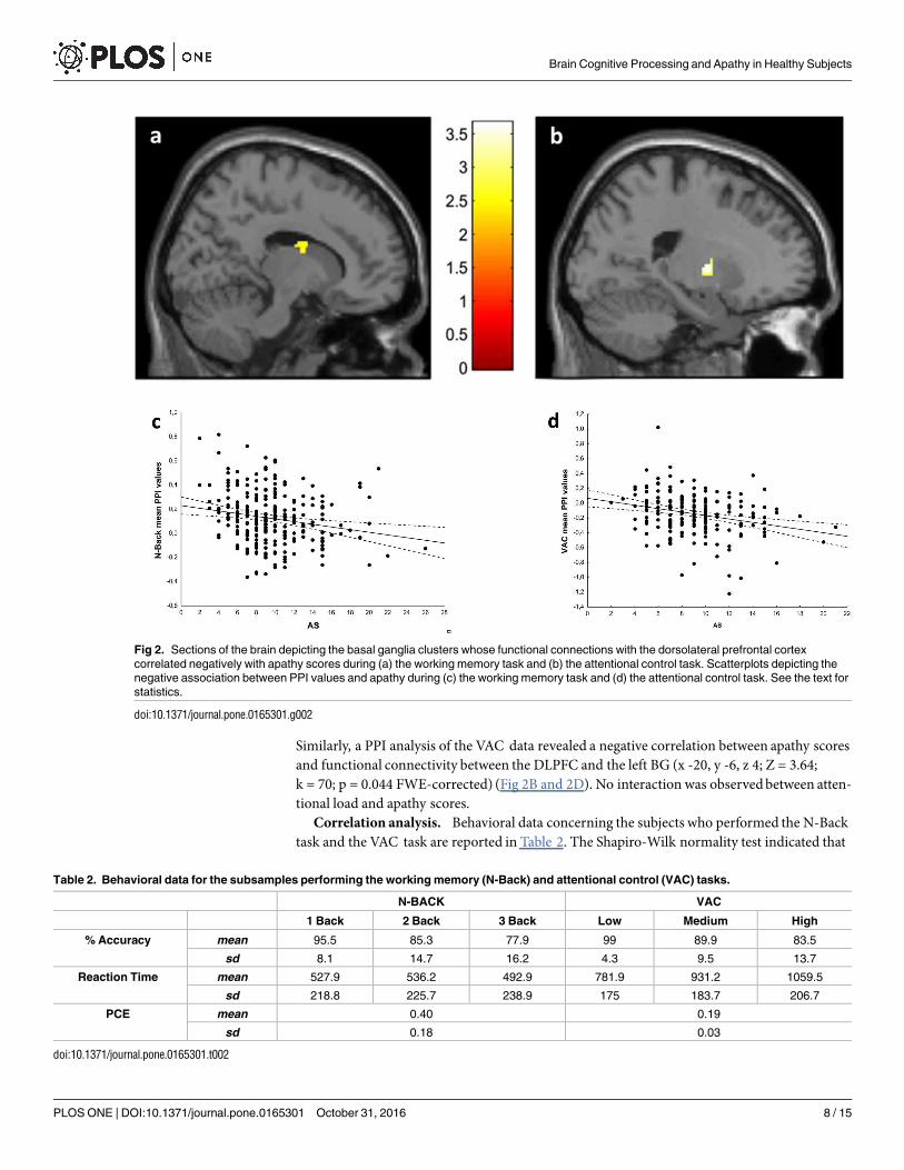
Similarly, a PPI analysis of the VAC data revealed a negative correlation between apathy scoresand functional connectivity between the DLPFC and the left BG (x -20, y -6, z 4; Z = 3.64;k = 70; p = 0.044 FWE-corrected) (Fig 2B and 2D). No interaction was observedbetween atten-tional load and apathy scores.
Correlation analysis. Behavioral data concerning the subjects who performed the N-Backtask and the VAC task are reported in Table 2. The Shapiro-Wilk normality test indicated that
Fig 2. Sections of the brain depicting the basal ganglia clusters whose functional connections with the dorsolateral prefrontal cortex
correlated negatively with apathy scores during (a) the working memory task and (b) the attentional control task. Scatterplots depicting the
negative association between PPI values and apathy during (c) the working memory task and (d) the attentional control task. See the text for
statistics.
doi:10.1371/journal.pone.0165301.g002
Table 2. Behavioral data for the subsamples performing the working memory (N-Back) and attentional control (VAC) tasks.
N-BACK VAC
1 Back 2 Back 3 Back Low Medium High
% Accuracy mean 95.5 85.3 77.9 99 89.9 83.5
sd 8.1 14.7 16.2 4.3 9.5 13.7
Reaction Time mean 527.9 536.2 492.9 781.9 931.2 1059.5
sd 218.8 225.7 238.9 175 183.7 206.7
PCE mean 0.40 0.19
sd 0.18 0.03
doi:10.1371/journal.pone.0165301.t002
Brain Cognitive Processing and Apathy in Healthy Subjects
PLOS ONE | DOI:10.1371/journal.pone.0165301 October 31, 2016 8 / 15

the N-Back PCE score was not distributed normally (N-back PCEW = 0.95, p<0.001). Spear-man’s correlations indicated a negative correlation betweenPCE scores and BOLD activity dur-ing the VAC task (Spearman’s Rho = -0.22; p = 0.002) (Fig 3B) but not during the N-back task(Spearman’s Rho = -0.08; p = 0.2) (Fig 3A). Spearman’s correlations betweenPPI and behavioraldata revealed no significant results (p>0.05). Robust regressionmodels did not indicate a rela-tionship between apathy and PCE scores during the N-back task or the VAC task (all p> 0.05).
Discussion
Here we investigated whether apathy is associatedwith DLPFC and BG activity and functionalconnectivity during cognitive processing in healthy individuals.We found a positive correlationbetween apathy scores and prefrontal responses during workingmemory tasks, such that subjectswith greater apathy also had greater DLPFC activity during this cognitive skill. Notably, wefound a similar relationship when investigating the domain of attentional control processing.Moreover, greater levels of apathy were also associatedwith lower functional connectivitybetween the DLPFC and the BG during both cognitive processes. These results suggest a relation-ship between patterns of brain cognitive processing and apathy in healthy subjects.
The observed association between dorsolateral prefrontal activity and AS scores in healthysubjects is consistent with previous results obtained in clinical populations, suggesting thatapathy may be subtended by damage that occurs primarily in the lateral prefrontal cortex [13].The DLPFC is a key region for working memory and attention processing [70–72], and previ-ous models suggested that for between-group comparisons, greater DLPFC activity despiteworse or unaffected behavior, may be an index of inefficient prefrontal processing during cog-nition [58, 60, 70, 73]. In light of this model, our results that demonstrate a positive correlationbetweenAS and DLPFC activity suggest that greater apathy in healthy subjects is linked withless efficient prefrontal processing of working memory and attentional stimuli. Consistent withthis finding, our correlation analysis indicates that greater DLPFC activity linked with greaterapathy predicts poorer behavioral performance in the attentional control task used in ourstudy. Indeed, we found no significant correlation between the PCE score and DLPFC activityduring working memory tasks. A possible explanation for this lack of a relationship is that theregion that we found to be associated with apathy during working memory is less modulated
Fig 3. Scatterplots of Spearman’s test on cognitive behavior as indexed by a parametric cognitive efficiency score (PCE) and BOLD parameter
estimates extracted from the dorsolateral prefrontal region associated with apathy, depicting (a) absence of correlation during working memory
task and (b) negative correlation during attentional control. See text for statistics.
doi:10.1371/journal.pone.0165301.g003
Brain Cognitive Processing and Apathy in Healthy Subjects
PLOS ONE | DOI:10.1371/journal.pone.0165301 October 31, 2016 9 / 15

by task load but is relevant for task execution [74]. Overall, our findings in healthy subjectsstrengthen previous evidence of an association between apathy and impaired cognition [42–45,75–78], further suggesting a biological basis for this phenomenon and uncoupling this relation-ship from the effects of confounding factors related to disease or aging. Moreover, our findingshighlight that the role of cognitive processing in apathy flanks those of emotion processing andmood [79–80].
Another finding of the present study is the relationship between apathy and DLPFC-BGconnectivity during both working memory and attentional control tasks. In particular, wefound that greater levels of apathy are associated with a weaker functional connection betweeninefficient clusters of prefrontal activity during cognitive processing and BG. These results areconsistent with previous models that posited a role for reduced DLPFC-BG connectivity inapathy [33–35, 67] and with previous findings that indicated that bilateral lesions of the BG areassociated with a severe form of apathy (auto-activation deficit) that is characterized by a com-plete loss of self-initiated goal-directedbehavior [81]. The DLPFC-BG loop plays a crucial rolein high-order cognitive processes, such as working memory and attentional control [64],andparticipates in a so-called “associative pathway,” in which information that arises from severalassociative areas is transmitted to the caudate and the anterior putamen and subsequentlyreaches the DLPFC through the thalamus [66, 82]. This pathway is involved in several cogni-tive processes and is crucial for the generation of context-dependent and goal-directedpatternsof behavior [66, 82–83]. Indeed, previous models postulated that a neurobiologicalmechanismsubtending apathy may imply a failure of the BG to engage the DLPFC, thus lowering the abil-ity of the DLPFC to support goal-directed cognitive processing [35, 67].
In our PPI analysis, we found that AS scores for both tasks are correlated with the functionalconnection between the DLPFC and the contralateral BG. Previous findings indicated the exis-tence of a bilateral interconnection between the DLPFC and the BG [84–85]. Furthermore, bylowering the statistical threshold of our analysis to an uncorrected threshold of p<0.005, wefound that AS scores in the N-Back and VAC tasks also correlated with the functional connec-tions between the DLPFC and the homolateral BG. Thus, it is possible that our PPI finding atthe corrected p value is related to the statistical threshold used.
A potential limitation of the present study is that apathy is considered to be an aspect ofdepression, and correlations between apathy and depression scales have been reported [2].Even if we excluded from the study all of the subjects with a current or past diagnosis of depres-sion, as well as individuals with first-degree relatives affected by a psychiatric disorder, itremains possible that subclinical levels of depression might have affected our results. However,previous studies have suggested good discriminability between apathy and depression scores inboth clinical [86–87] and non-clinical populations [11–12], as well as an adequate discriminantvalidity between apathy scales and depression scales [88]. Thus, it is possible that our resultsare not strongly impacted by subclinical levels of depression. Further studies should addressthis issue.
In conclusion, we provided evidence for a relationship between inefficient brain processingduring cognition and apathy in healthy subjects in the absence of confounding factors, such aspathophysiological conditions or pharmacological treatment. These findings shed new light onour understanding of the link between apathy and brain processes that may be relevant to neu-rological and psychiatric conditions for which apathy is a central feature.
Acknowledgments
We are grateful to Dr. Raffaella Romano and Dr. Annamaria Porcelli, for making data acquisi-tion possible. Moreover, we gratefully acknowledge the work by Ivan Abbrescia, Catia
Brain Cognitive Processing and Apathy in Healthy Subjects
PLOS ONE | DOI:10.1371/journal.pone.0165301 October 31, 2016 10 / 15
D’Agostino, Nunzio Langiulli, Pierluigi Selvaggi and Aldo Tomasicchio for their help with dataprocessing. Finally, we would like to acknowledge all people who participated to this study.
Author Contributions
Conceptualization:LF (first author) GB GL AB.
Data curation: LF (first author) GA.
Formal analysis: LF (first author) PT TQ LAA LF (tenth author).
Funding acquisition:GL AB.
Investigation: LF (first author) PT TQ LAAMMBG.
Methodology:LF (first author) PT TQMRB.
Project administration: LF (first author) GB GL AB.
Resources:TP.
Software: LF (first author) PT TQ.
Supervision:GB GL AB.
Visualization: LF (first author) GA.
Writing – original draft: LF (first author).
Writing – review& editing:GB.
References1. Marin RS. Differential diagnosis and classification of apathy. The American journal of psychiatry. 1990;
147(1):22–30. Epub 1990/01/01. PMID: 2403472. doi: 10.1176/ajp.147.1.22
2. Marin RS. Apathy: a neuropsychiatric syndrome. J Neuropsychiatry Clin Neurosci. 1991; 3(3):243–54.
Epub 1991/01/01. doi: 10.1176/jnp.3.3.243 PMID: 1821241.
3. Starkstein SE, Leentjens AF. The nosological position of apathy in clinical practice. Journal of neurol-
ogy, neurosurgery, and psychiatry. 2008; 79(10):1088–92. doi: 10.1136/jnnp.2007.136895 PMID:
18187477.
4. Leentjens AF, Dujardin K, Marsh L, Martinez-Martin P, Richard IH, Starkstein SE, et al. Apathy and
anhedonia rating scales in Parkinson’s disease: critique and recommendations. Movement disorders:
official journal of the Movement Disorder Society. 2008; 23(14):2004–14. doi: 10.1002/mds.22229
PMID: 18709683.
5. Roth RM, Koven NS, Pendergrass JC, Flashman LA, McAllister TW, Saykin AJ. Apathy and the pro-
cessing of novelty in schizophrenia. Schizophrenia research. 2008; 98(1–3):232–8. doi: 10.1016/j.
schres.2007.08.020 PMID: 17884352; PubMed Central PMCID: PMC2843546.
6. Bentley SM, Pagalilauan GL, Simpson SA. Major Depression. The Medical clinics of North America.
2014; 98(5):981–1005. Epub 2014/08/20. doi: 10.1016/j.mcna.2014.06.013 PMID: 25134869.
7. Starkstein SE, Petracca G, Chemerinski E, Kremer J. Syndromic validity of apathy in Alzheimer’s dis-
ease. The American journal of psychiatry. 2001; 158(6):872–7. Epub 2001/06/01. PMID: 11384893.
doi: 10.1176/appi.ajp.158.6.872
8. Pardini M, Cordano C, Guida S, Grafman J, Krueger F, Sassos D, et al. Prevalence and cognitive
underpinnings of isolated apathy in young healthy subjects. J Affect Disord. 2015; 189:272–5. Epub
2015/10/12. S0165-0327(15)30617-0 [pii] doi: 10.1016/j.jad.2015.09.062 PMID: 26454331.
9. Okura T, Plassman BL, Steffens DC, Llewellyn DJ, Potter GG, Langa KM. Prevalence of neuropsychi-
atric symptoms and their association with functional limitations in older adults in the United States: the
aging, demographics, and memory study. J Am Geriatr Soc. 2010; 58(2):330–7. Epub 2010/04/09.
JGS2680 [pii] doi: 10.1111/j.1532-5415.2009.02680.x PMID: 20374406; PubMed Central PMCID:
PMC2875937.
Brain Cognitive Processing and Apathy in Healthy Subjects
PLOS ONE | DOI:10.1371/journal.pone.0165301 October 31, 2016 11 / 15

10. Onyike CU, Sheppard JM, Tschanz JT, Norton MC, Green RC, Steinberg M, et al. Epidemiology of
apathy in older adults: the Cache County Study. Am J Geriatr Psychiatry. 2007; 15(5):365–75. Epub
2007/04/28. 15/5/365 [pii] doi: 10.1097/01.JGP.0000235689.42910.0d PMID: 17463187.
11. Spalletta G, Fagioli S, Caltagirone C, Piras F. Brain microstructure of subclinical apathy phenomenol-
ogy in healthy individuals. Hum Brain Mapp. 2013; 34(12):3193–203. Epub 2012/07/19. doi: 10.1002/
hbm.22137 PMID: 22807351.
12. Bonnelle V, Veromann KR, Burnett Heyes S, Lo Sterzo E, Manohar S, Husain M. Characterization of
reward and effort mechanisms in apathy. J Physiol Paris. 2015; 109(1–3):16–26. doi: 10.1016/j.
jphysparis.2014.04.002 PMID: 24747776; PubMed Central PMCID: PMCPMC4451957.
13. Paradiso S, Chemerinski E, Yazici KM, Tartaro A, Robinson RG. Frontal lobe syndrome reassessed:
comparison of patients with lateral or medial frontal brain damage. J Neurol Neurosurg Psychiatry.
1999; 67(5):664–7. Epub 1999/10/16. PMID: 10519877; PubMed Central PMCID: PMC1736625.
14. Barrash J, Tranel D, Anderson SW. Acquired personality disturbances associated with bilateral dam-
age to the ventromedial prefrontal region. Dev Neuropsychol. 2000; 18(3):355–81. Epub 2001/06/02.
doi: 10.1207/S1532694205Barrash PMID: 11385830.
15. Roth RM, Flashman LA, Saykin AJ, McAllister TW, Vidaver R. Apathy in schizophrenia: reduced frontal
lobe volume and neuropsychological deficits. Am J Psychiatry. 2004; 161(1):157–9. Epub 2004/01/02.
PMID: 14702265. doi: 10.1176/appi.ajp.161.1.157
16. Reijnders JS, Scholtissen B, Weber WE, Aalten P, Verhey FR, Leentjens AF. Neuroanatomical corre-
lates of apathy in Parkinson’s disease: A magnetic resonance imaging study using voxel-based mor-
phometry. Movement disorders: official journal of the Movement Disorder Society. 2010; 25(14):2318–
25. doi: 10.1002/mds.23268 PMID: 20669264.
17. Grool AM, Geerlings MI, Sigurdsson S, Eiriksdottir G, Jonsson PV, Garcia ME, et al. Structural MRI
correlates of apathy symptoms in older persons without dementia: AGES-Reykjavik Study. Neurology.
2014; 82(18):1628–35. Epub 2014/04/18. doi: 10.1212/WNL.0000000000000378 PMID: 24739783;
PubMed Central PMCID: PMC4013817.
18. Stanton BR, Leigh PN, Howard RJ, Barker GJ, Brown RG. Behavioural and emotional symptoms of
apathy are associated with distinct patterns of brain atrophy in neurodegenerative disorders. Journal of
neurology. 2013; 260(10):2481–90. doi: 10.1007/s00415-013-6989-9 PMID: 23793818.
19. Marshall GA, Monserratt L, Harwood D, Mandelkern M, Cummings JL, Sultzer DL. Positron emission
tomography metabolic correlates of apathy in Alzheimer disease. Arch Neurol. 2007; 64(7):1015–20.
Epub 2007/07/11. 64/7/1015 [pii] doi: 10.1001/archneur.64.7.1015 PMID: 17620493.
20. Okada K, Kobayashi S, Yamagata S, Takahashi K, Yamaguchi S. Poststroke apathy and regional
cerebral blood flow. Stroke; a journal of cerebral circulation. 1997; 28(12):2437–41. Epub 1997/12/31.
PMID: 9412628.
21. Benoit M, Clairet S, Koulibaly PM, Darcourt J, Robert PH. Brain perfusion correlates of the apathy
inventory dimensions of Alzheimer’s disease. International journal of geriatric psychiatry. 2004; 19
(9):864–9. Epub 2004/09/08. doi: 10.1002/gps.1163 PMID: 15352144.
22. Strub RL. Frontal lobe syndrome in a patient with bilateral globus pallidus lesions. Arch Neurol. 1989;
46(9):1024–7. Epub 1989/09/01. PMID: 2775008.
23. Bhatia KP, Marsden CD. The behavioural and motor consequences of focal lesions of the basal gan-
glia in man. Brain. 1994; 117 (Pt 4):859–76. Epub 1994/08/01. PMID: 7922471.
24. Mendez MF, Adams NL, Lewandowski KS. Neurobehavioral changes associated with caudate lesions.
Neurology. 1989; 39(3):349–54. Epub 1989/03/01. PMID: 2927642.
25. Onoda K, Kuroda Y, Yamamoto Y, Abe S, Oguro H, Nagai A, et al. Post-stroke apathy and hypoperfu-
sion in basal ganglia: SPECT study. Cerebrovasc Dis. 2011; 31(1):6–11. Epub 2010/10/29.
000319771 [pii] doi: 10.1159/000319771 PMID: 20980747.
26. Lopez OL, Zivkovic S, Smith G, Becker JT, Meltzer CC, DeKosky ST. Psychiatric symptoms associ-
ated with cortical-subcortical dysfunction in Alzheimer’s disease. The Journal of neuropsychiatry and
clinical neurosciences. 2001; 13(1):56–60. Epub 2001/02/24. PMID: 11207330. doi: 10.1176/jnp.13.1.
56
27. Josephs KA, Whitwell JL, Eggers SD, Senjem ML, Jack CR, Jr. Gray matter correlates of behavioral
severity in progressive supranuclear palsy. Mov Disord. 2011; 26(3):493–8. Epub 2011/04/05. doi: 10.
1002/mds.23471 PMID: 21462261.
28. Bruen PD, McGeown WJ, Shanks MF, Venneri A. Neuroanatomical correlates of neuropsychiatric
symptoms in Alzheimer’s disease. Brain. 2008; 131(Pt 9):2455–63. Epub 2008/08/02. awn151 [pii] doi:
10.1093/brain/awn151 PMID: 18669506.
29. Craufurd D, Thompson JC, Snowden JS. Behavioral changes in Huntington Disease. Neuropsychiatry,
neuropsychology, and behavioral neurology. 2001; 14(4):219–26. Epub 2001/11/29. PMID: 11725215.
Brain Cognitive Processing and Apathy in Healthy Subjects
PLOS ONE | DOI:10.1371/journal.pone.0165301 October 31, 2016 12 / 15

30. Litvan I, Mega MS, Cummings JL, Fairbanks L. Neuropsychiatric aspects of progressive supranuclear
palsy. Neurology. 1996; 47(5):1184–9. Epub 1996/11/01. PMID: 8909427.
31. Yuen GS, Gunning-Dixon FM, Hoptman MJ, AbdelMalak B, McGovern AR, Seirup JK, et al. The
salience network in the apathy of late-life depression. Int J Geriatr Psychiatry. 2014; 29(11):1116–24.
Epub 2014/07/06. doi: 10.1002/gps.4171 PMID: 24990625; PubMed Central PMCID: PMC4197060.
32. Bonnelle V, Manohar S, Behrens T, Husain M. Individual Differences in Premotor Brain Systems
Underlie Behavioral Apathy. Cereb Cortex. 2016; 26(2):807–19. doi: 10.1093/cercor/bhv247 PMID:
26564255; PubMed Central PMCID: PMCPMC4712805.
33. Brown RG, Pluck G. Negative symptoms: the ’pathology’ of motivation and goal-directed behaviour.
Trends in neurosciences. 2000; 23(9):412–7. Epub 2000/08/15. PMID: 10941190.
34. Levy R, Dubois B. Apathy and the functional anatomy of the prefrontal cortex-basal ganglia circuits.
Cereb Cortex. 2006; 16(7):916–28. doi: 10.1093/cercor/bhj043 PMID: 16207933.
35. Levy R. Apathy: a pathology of goal-directed behaviour: a new concept of the clinic and pathophysiol-
ogy of apathy. Revue neurologique. 2012; 168(8–9):585–97. doi: 10.1016/j.neurol.2012.05.003 PMID:
22921248.
36. Middleton FA, Strick PL. Basal-ganglia ’projections’ to the prefrontal cortex of the primate. Cereb Cor-
tex. 2002; 12(9):926–35. Epub 2002/08/17. PMID: 12183392.
37. Lehericy S, Ducros M, Van de Moortele PF, Francois C, Thivard L, Poupon C, et al. Diffusion tensor
fiber tracking shows distinct corticostriatal circuits in humans. Annals of neurology. 2004; 55(4):522–9.
Epub 2004/03/30. doi: 10.1002/ana.20030 PMID: 15048891.
38. Kemp JM, Powell TP. The cortico-striate projection in the monkey. Brain: a journal of neurology. 1970;
93(3):525–46. Epub 1970/01/01. PMID: 4990231.
39. Lindgren HS, Wickens R, Tait DS, Brown VJ, Dunnett SB. Lesions of the dorsomedial striatum impair
formation of attentional set in rats. Neuropharmacology. 2013; 71:148–53. Epub 2013/04/16. S0028-
3908(13)00130-5 [pii] doi: 10.1016/j.neuropharm.2013.03.034 PMID: 23583929.
40. McNab F, Klingberg T. Prefrontal cortex and basal ganglia control access to working memory. Nat
Neurosci. 2008; 11(1):103–7. Epub 2007/12/11. nn2024 [pii] doi: 10.1038/nn2024 PMID: 18066057.
41. Baier B, Karnath HO, Dieterich M, Birklein F, Heinze C, Muller NG. Keeping memory clear and stable
—the contribution of human basal ganglia and prefrontal cortex to working memory. J Neurosci. 2010;
30(29):9788–92. Epub 2010/07/28. 30/29/9788 [pii] doi: 10.1523/JNEUROSCI.1513-10.2010 PMID:
20660261.
42. Andersson S, Bergedalen AM. Cognitive correlates of apathy in traumatic brain injury. Neuropsychia-
try, neuropsychology, and behavioral neurology. 2002; 15(3):184–91. PMID: 12218711.
43. McPherson S, Fairbanks L, Tiken S, Cummings JL, Back-Madruga C. Apathy and executive function
in Alzheimer’s disease. J Int Neuropsychol Soc. 2002; 8(3):373–81. Epub 2002/04/10. PMID:
11939696.
44. Zgaljardic DJ, Borod JC, Foldi NS, Rocco M, Mattis PJ, Gordon MF, et al. Relationship between self-
reported apathy and executive dysfunction in nondemented patients with Parkinson disease. Cognitive
and behavioral neurology: official journal of the Society for Behavioral and Cognitive Neurology. 2007;
20(3):184–92. doi: 10.1097/WNN.0b013e318145a6f6 PMID: 17846518.
45. Konstantakopoulos G, Ploumpidis D, Oulis P, Patrikelis P, Soumani A, Papadimitriou GN, et al. Apa-
thy, cognitive deficits and functional impairment in schizophrenia. Schizophrenia research. 2011; 133
(1–3):193–8. Epub 2011/07/27. doi: 10.1016/j.schres.2011.07.003 PMID: 21788116.
46. Varanese S, Perfetti B, Ghilardi MF, Di Rocco A. Apathy, but not depression, reflects inefficient cogni-
tive strategies in Parkinson’s disease. PLoS One. 2011; 6(3):e17846. Epub 2011/03/26. doi: 10.1371/
journal.pone.0017846 PMID: 21437255; PubMed Central PMCID: PMC3060914.
47. Yamashita K, Iijima K, Kobayashi S. Relationship among activities of daily living, apathy, and subjec-
tive well-being in elderly people living alone in a rural town. Gerontology. 1999; 45(5):279–82. Epub
1999/08/26. doi: ger45279 [pii]. PMID: 10460990.
48. Clarke DE, Ko JY, Lyketsos C, Rebok GW, Eaton WW. Apathy and cognitive and functional decline in
community-dwelling older adults: results from the Baltimore ECA longitudinal study. International psy-
chogeriatrics / IPA. 2010; 22(5):819–29. Epub 2010/05/19. doi: 10.1017/S1041610209991402 PMID:
20478091; PubMed Central PMCID: PMC2893259.
49. Brodaty H, Heffernan M, Draper B, Reppermund S, Kochan NA, Slavin MJ, et al. Neuropsychiatric
symptoms in older people with and without cognitive impairment. Journal of Alzheimer’s disease: JAD.
2012; 31(2):411–20. Epub 2012/05/11. doi: 10.3233/JAD-2012-120169 PMID: 22571979.
50. First MB, Gibbon M, Spitzer RL, Williams JBW. Guide for the structured clinical interview for DSM-IV
axis I disorders-Research version. New York: Biometrics Research; 1996.
Brain Cognitive Processing and Apathy in Healthy Subjects
PLOS ONE | DOI:10.1371/journal.pone.0165301 October 31, 2016 13 / 15

51. Orsini A, Laicardi C. Factor structure of the Italian version of the WAIS-R compared with the American
standardization. Percept Mot Skills. 2000; 90(3 Pt 2):1091–100. Epub 2000/08/12. doi: 10.2466/pms.
2000.90.3c.1091 PMID: 10939053.
52. Oldfield RC. The assessment and analysis of handedness: the Edinburgh inventory. Neuropsycholo-
gia. 1971; 9(1):97–113. Epub 1971/03/01. PMID: 5146491.
53. Hollingshead AA. Four-factor index of social status. (Unpublished Manuscript) Yale University, New
Haven, CT. 1975.
54. Starkstein SE, Mayberg HS, Preziosi TJ, Andrezejewski P, Leiguarda R, Robinson RG. Reliability,
validity, and clinical correlates of apathy in Parkinson’s disease. The Journal of neuropsychiatry and
clinical neurosciences. 1992; 4(2):134–9. Epub 1992/01/01. PMID: 1627973. doi: 10.1176/jnp.4.2.134
55. Marin RS, Biedrzycki RC, Firinciogullari S. Reliability and validity of the Apathy Evaluation Scale. Psy-
chiatry research. 1991; 38(2):143–62. Epub 1991/08/01. PMID: 1754629.
56. Bertolino A, Caforio G, Blasi G, De Candia M, Latorre V, Petruzzella V, et al. Interaction of COMT (Val
(108/158)Met) genotype and olanzapine treatment on prefrontal cortical function in patients with
schizophrenia. The American journal of psychiatry. 2004; 161(10):1798–805. Epub 2004/10/07. doi:
10.1176/appi.ajp.161.10.1798 PMID: 15465976.
57. Blasi G, Napolitano F, Ursini G, Di Giorgio A, Caforio G, Taurisano P, et al. Association of GSK-3beta
genetic variation with GSK-3beta expression, prefrontal cortical thickness, prefrontal physiology, and
schizophrenia. Am J Psychiatry. 2013; 170(8):868–76. Epub 2013/04/20. 1680035 [pii] doi: 10.1176/
appi.ajp.2012.12070908 PMID: 23598903.
58. Gelao B, Fazio L, Selvaggi P, Di Giorgio A, Taurisano P, Quarto T, et al. DRD2 genotype predicts pre-
frontal activity during working memory after stimulation of D2 receptors with bromocriptine. Psycho-
pharmacology (Berl). 2014; 231(11):2361–70. Epub 2014/01/16. doi: 10.1007/s00213-013-3398-9
PMID: 24424781.
59. Blasi G, Taurisano P, Papazacharias A, Caforio G, Romano R, Lobianco L, et al. Nonlinear response
of the anterior cingulate and prefrontal cortex in schizophrenia as a function of variable attentional con-
trol. Cereb Cortex. 2010; 20(4):837–45. Epub 2009/07/28. bhp146 [pii] doi: 10.1093/cercor/bhp146
PMID: 19633177.
60. Blasi G, Goldberg TE, Elvevag B, Rasetti R, Bertolino A, Cohen J, et al. Differentiating allocation of
resources and conflict detection within attentional control processing. Eur J Neurosci. 2007; 25(2):594–
602. Epub 2007/02/08. EJN5283 [pii] doi: 10.1111/j.1460-9568.2007.05283.x PMID: 17284202.
61. Rasetti R, Mattay VS, Stankevich B, Skjei K, Blasi G, Sambataro F, et al. Modulatory effects of modafi-
nil on neural circuits regulating emotion and cognition. Neuropsychopharmacology. 2010; 35
(10):2101–9. Epub 2010/06/18. npp201083 [pii] doi: 10.1038/npp.2010.83 PMID: 20555311; PubMed
Central PMCID: PMC3013347.
62. Blasi G, De Virgilio C, Papazacharias A, Taurisano P, Gelao B, Fazio L, et al. Converging evidence for
the association of functional genetic variation in the serotonin receptor 2a gene with prefrontal function
and olanzapine treatment. JAMA Psychiatry. 2013; 70(9):921–30. Epub 2013/07/12. 1710489 [pii] doi:
10.1001/jamapsychiatry.2013.1378 PMID: 23842608.
63. Blasi G, Selvaggi P, Fazio L, Antonucci LA, Taurisano P, Masellis R, et al. Variation in Dopamine D2
and Serotonin 5-HT2A Receptor Genes is Associated with Working Memory Processing and
Response to Treatment with Antipsychotics. Neuropsychopharmacology. 2015; 40(7):1600–8. Epub
2015/01/08. npp20155 [pii] doi: 10.1038/npp.2015.5 PMID: 25563748.
64. O’Reilly RC. Biologically based computational models of high-level cognition. Science. 2006; 314
(5796):91–4. Epub 2006/10/07. 314/5796/91 [pii] doi: 10.1126/science.1127242 PMID: 17023651.
65. Bertolino A, Taurisano P, Pisciotta NM, Blasi G, Fazio L, Romano R, et al. Genetically determined
measures of striatal D2 signaling predict prefrontal activity during working memory performance. PLoS
One. 2010; 5(2):e9348. Epub 2010/02/25. doi: 10.1371/journal.pone.0009348 PMID: 20179754;
PubMed Central PMCID: PMC2825256.
66. Arsalidou M, Duerden EG, Taylor MJ. The centre of the brain: topographical model of motor, cognitive,
affective, and somatosensory functions of the basal ganglia. Hum Brain Mapp. 2013; 34(11):3031–54.
Epub 2012/06/20. doi: 10.1002/hbm.22124 PMID: 22711692.
67. Levy R, Czernecki V. Apathy and the basal ganglia. J Neurol. 2006; 253 Suppl 7:VII54–61. doi: 10.
1007/s00415-006-7012-5 PMID: 17131230.
68. Maldjian JA, Laurienti PJ, Kraft RA, Burdette JH. An automated method for neuroanatomic and
cytoarchitectonic atlas-based interrogation of fMRI data sets. Neuroimage. 2003; 19(3):1233–9. Epub
2003/07/26. S1053811903001691 [pii]. PMID: 12880848.
69. Friston KJ, Buechel C, Fink GR, Morris J, Rolls E, Dolan RJ. Psychophysiological and modulatory
interactions in neuroimaging. Neuroimage. 1997; 6(3):218–29. PMID: 9344826. doi: 10.1006/nimg.
1997.0291
Brain Cognitive Processing and Apathy in Healthy Subjects
PLOS ONE | DOI:10.1371/journal.pone.0165301 October 31, 2016 14 / 15

70. Callicott JH, Mattay VS, Bertolino A, Finn K, Coppola R, Frank JA, et al. Physiological characteristics
of capacity constraints in working memory as revealed by functional MRI. Cereb Cortex. 1999; 9
(1):20–6. PMID: 10022492
71. Fuster JM, Alexander GE. Neuron activity related to short-term memory. Science. 1971; 173
(997):652–4. Epub 1971/08/13. PMID: 4998337.
72. Goldman-Rakic PS. Cellular and circuit basis of working memory in prefrontal cortex of nonhuman pri-
mates. Prog Brain Res. 1990; 85:325–35. PMID: 2094903
73. Taurisano P, Romano R, Mancini M, Giorgio AD, Antonucci LA, Fazio L, et al. Prefronto-striatal physi-
ology is associated with schizotypy and is modulated by a functional variant of DRD2. Front Behav
Neurosci. 2014; 8:235. Epub 2014/07/30. doi: 10.3389/fnbeh.2014.00235 PMID: 25071490; PubMed
Central PMCID: PMC4089730.
74. Callicott JH, Mattay VS, Bertolino A, Finn K, Coppola R, Frank JA, et al. Physiological characteristics
of capacity constraints in working memory as revealed by functional MRI. Cereb Cortex. 1999; 9
(1):20–6. Epub 1999/02/18. PMID: 10022492.
75. Baudic S, Maison P, Dolbeau G, Boisse MF, Bartolomeo P, Dalla Barba G, et al. Cognitive impairment
related to apathy in early Huntington’s disease. Dementia and geriatric cognitive disorders. 2006; 21
(5–6):316–21. doi: 10.1159/000091523 PMID: 16484810.
76. Pluck GC, Brown RG. Apathy in Parkinson’s disease. Journal of neurology, neurosurgery, and psychi-
atry. 2002; 73(6):636–42. PMID: 12438462; PubMed Central PMCID: PMC1757348. doi: 10.1136/
jnnp.73.6.636
77. Isella V, Melzi P, Grimaldi M, Iurlaro S, Piolti R, Ferrarese C, et al. Clinical, neuropsychological, and
morphometric correlates of apathy in Parkinson’s disease. Movement disorders: official journal of the
Movement Disorder Society. 2002; 17(2):366–71. PMID: 11921125.
78. Faerden A, Vaskinn A, Finset A, Agartz I, Ann Barrett E, Friis S, et al. Apathy is associated with execu-
tive functioning in first episode psychosis. BMC psychiatry. 2009; 9:1. doi: 10.1186/1471-244X-9-1
PMID: 19133132; PubMed Central PMCID: PMC2640391.
79. Robert G, Le Jeune F, Dondaine T, Drapier S, Peron J, Lozachmeur C, et al. Apathy and impaired
emotional facial recognition networks overlap in Parkinson’s disease: a PET study with conjunction
analyses. J Neurol Neurosurg Psychiatry. 2014; 85(10):1153–8. Epub 2014/01/10. jnnp-2013-307025
[pii] doi: 10.1136/jnnp-2013-307025 PMID: 24403280.
80. Bayard S, Jacus JP, Raffard S, Gely-Nargeot MC. Apathy and emotion-based decision-making in
amnesic mild cognitive impairment and Alzheimer’s disease. Behav Neurol. 2014; 2014:231469. Epub
2014/07/23. doi: 10.1155/2014/231469 PMID: 25049450; PubMed Central PMCID: PMC4090525.
81. Laplane D, Dubois B. Auto-Activation deficit: a basal ganglia related syndrome. Movement disorders:
official journal of the Movement Disorder Society. 2001; 16(5):810–4. Epub 2001/12/18. PMID:
11746609.
82. Parent A, Hazrati LN. Functional anatomy of the basal ganglia. I. The cortico-basal ganglia-thalamo-
cortical loop. Brain Res Brain Res Rev. 1995; 20(1):91–127. Epub 1995/01/01. 016501739400007C
[pii]. PMID: 7711769.
83. Alexander GE, DeLong MR, Strick PL. Parallel organization of functionally segregated circuits linking
basal ganglia and cortex. Annu Rev Neurosci. 1986; 9:357–81. Epub 1986/01/01. doi: 10.1146/
annurev.ne.09.030186.002041 PMID: 3085570.
84. Postuma RB, Dagher A. Basal ganglia functional connectivity based on a meta-analysis of 126 posi-
tron emission tomography and functional magnetic resonance imaging publications. Cereb Cortex.
2006; 16(10):1508–21. Epub 2005/12/24. bhj088 [pii] doi: 10.1093/cercor/bhj088 PMID: 16373457.
85. Jarbo K, Verstynen TD. Converging structural and functional connectivity of orbitofrontal, dorsolateral
prefrontal, and posterior parietal cortex in the human striatum. The Journal of neuroscience: the official
journal of the Society for Neuroscience. 2015; 35(9):3865–78. Epub 2015/03/06. 35/9/3865 [pii] doi:
10.1523/JNEUROSCI.2636-14.2015 PMID: 25740516; PubMed Central PMCID: PMC4461697.
86. Levy ML, Cummings JL, Fairbanks LA, Masterman D, Miller BL, Craig AH, et al. Apathy is not depres-
sion. J Neuropsychiatry Clin Neurosci. 1998; 10(3):314–9. Epub 1998/08/26. doi: 10.1176/jnp.10.3.
314 PMID: 9706539.
87. Starkstein SE, Ingram L, Garau ML, Mizrahi R. On the overlap between apathy and depression in
dementia. J Neurol Neurosurg Psychiatry. 2005; 76(8):1070–4. Epub 2005/07/19. 76/8/1070 [pii] doi:
10.1136/jnnp.2004.052795 PMID: 16024880; PubMed Central PMCID: PMC1739766.
88. Clarke DE, Ko JY, Kuhl EA, van Reekum R, Salvador R, Marin RS. Are the available apathy measures
reliable and valid? A review of the psychometric evidence. J Psychosom Res. 2011; 70(1):73–97.
Epub 2011/01/05. S0022-3999(10)00017-6 [pii] doi: 10.1016/j.jpsychores.2010.01.012 PMID:
21193104; PubMed Central PMCID: PMC3902773.
Brain Cognitive Processing and Apathy in Healthy Subjects
PLOS ONE | DOI:10.1371/journal.pone.0165301 October 31, 2016 15 / 15