Embed Size (px)
Citation preview

Pregnancy of Unknown Location: The New Rules
James M. Shwayder, M.D., J.D.Professor and Chair
Department of Obstetrics and Gynecology University of Mississippi School of Medicine
Jackson, Mississippi
Pregnancy of Unknown Location: The New Rules
James M. Shwayder, M.D., J.D.
Disclosures: None
Learning Objectives
• Nomenclature regarding pregnancy of unknown location (PUL)
• Alternative approaches the diagnostic dilemma of evaluating a patient with a possible ectopic pregnancy.
• Understand the value of various diagnostic tests.
• Gain insight into the ultrasound findings in patients with an ectopic pregnancy

Consensus Nomenclature
1. Definite ectopic pregnancy (EP)
2. Probable EP
3. PUL
4. Probable intrauterine pregnancy
5. Definite IUP
Barnhart et al. Fertil Steril 2011; 95: 857-866
Consensus Nomenclature
• Definite ectopic pregnancy (EP)– Extrauterine gestational sac with yolk sac
and/or embryo (with or without cardiac activity)
• Probable EP– Inhomogeneous adnexal mass or
extrauterine sac-like structure
Barnhart et al. Fertil Steril 2011; 95: 857-866
Consensus Nomenclature
• Probable intrauterine pregnancy– Intrauterine echogenic sac-like structure
• Definite IUP– Intrauterine gestational sac with yolk sac
and/or embryo (with or without cardiac activity)
Barnhart et al. Fertil Steril 2011; 95: 857-866

Consensus Nomenclature
• PUL– no signs of either EP or IUP
Barnhart et al. Fertil Steril 2011; 95: 857-866
Pregnancy of Unknown Location
Barnhart et al. Fertil Steril 2011; 95: 857-866
Case Presentation
• 28 y.o. G1P0 presents with pelvic pain and scant vaginal spotting.
• LMP ~ 4-5 weeks ago
• + UPT at home
• Exam: VSS
Uterus AV, NT, TNS
Adnexa: NT, without masses
• hCG = 874 IU/L
CP 1

Increase in hCG in early pregnancy
Sampling Interval (days) % Increase1 292 663 1144 1755 255
Kadar et. al. Obstet Gynecol 1981; 58: 162 (Yale)
Increase in hCG in early pregnancy
• Doubling time = 2.98 days
• 15% of normal pregnancies had abnormal ß-hCG increases
Kadar et. al. Obstet Gynecol 1981; 58: 162 (Yale)
Increase in hCG in early pregnancy
Days Range Median
1 1.24 – 1.81 1.50
2 1.53 – 3.28 2.24
3 1.88 – 5.94 3.35
4 2.33 – 10.76 5.00
7 4.38 – 63.88 16.73
Barnhart et al. Obstet Gynecol 2004; 104: 50-55.
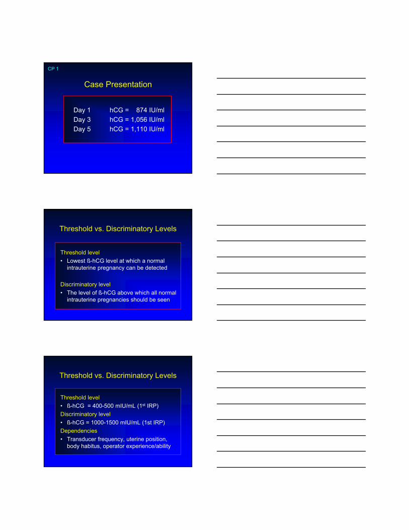
Case Presentation
Day 1 hCG = 874 IU/ml
Day 3 hCG = 1,056 IU/ml
Day 5 hCG = 1,110 IU/ml
CP 1
Threshold vs. Discriminatory Levels
Threshold level
• Lowest ß-hCG level at which a normal intrauterine pregnancy can be detected
Discriminatory level
• The level of ß-hCG above which all normal intrauterine pregnancies should be seen
Threshold vs. Discriminatory Levels
Threshold level
• ß-hCG = 400-500 mIU/mL (1st IRP)
Discriminatory level
• ß-hCG = 1000-1500 mIU/mL (1st IRP)
Dependencies
• Transducer frequency, uterine position, body habitus, operator experience/ability

Evidence Against the hCG Discriminatory Level
• January 1, 2000 - December 31, 2010
• TVS and β-hCG on same day
• No intrauterine fluid collection
• Subsequent embryonic or fetal cardiac activity
Doubilet and Benson, J Ultrasound Med 2011; 30:1637-1642
Evidence Against the hCG Discriminatory Level
Doubilet and Benson, J Ultrasound Med 2011; 30:1637-1642
hCG (3-4th IS) # (202) %
<1000 162 80.2
1000-1499 19 9.4
1500-1999 12 5.9
2000 9 4.5
Reevaluation of Discriminatory and Threshold Levels
• 651 patients
• TVS and β-hCG within 6 hours of each other
• Known intrauterine pregnancies
• Findings visualized 99% of the time
• 1st, 3rd, or 4th International Standard
– 2nd I.S.~ ½ that of others
Connolly et al. Obstet Gynecol 2013;121:65-70.

Reevaluation of Discriminatory and Threshold Levels
hCG = mIU/mLGestational
SacYolk Sac
Embryo
Threshold level 390 1094 1394
Discriminatorylevel
3510 17,716 47,685
Connolly et al. Obstet Gynecol 2013;121:65-70.
Reevaluation of Discriminatory and Threshold Levels
Connolly et al. Obstet Gynecol 2013;121:65-70.

Case Presentation
• TVS– Uterus
• No evidence of IUP– Ovaries
• Corpus luteum - left– Adnexa
• No definite adnexal pathology• Current terminology PUL• Treatment MTX 50 mg/m2
CP 1
Case Presentation
• 24 y.o. G2P0010 presents with scant vaginal spotting and pain
• LMP ~ 5 weeks ago
• Exam: VSS
Uterus NSSC, NT; Adnexa: NT
• Initial: hCG = 710 IU/L
• Repeat in 2 days: hCG = 980 IU/L

7.4 mm
Endometrial Thickness in Ectopic Pregnancy when hCG < Discriminatory Zone
OutcomeMean (mm)
Range (mm)
Intrauterine pregnancy 13.42 + 0.68
Spontaneous abortion 9.28 + 0.88
Ectopic pregnancy 5.95 + 0.35
Abnormal pregnancy (97%) < 8
Spandorfer and Barnhart. Fertil Steril 1996; 474-47.


Case Presentation - #3
• 28 y.o. G1P0 presents with pelvic pain and scant vaginal spotting.
• LMP ~ 7 weeks ago
• Exam: VSS
Uterus TNS;
Mild adnexal discomfort
• hCG = 4,634 IU/L

Intrauterine Fluid with Ectopic Pregnancy
229 patients with ectopic # %
• No intrauterine fluid 191 83.4
• Intrauterine fluid 38 16.6– Adnexal mass 33 86.8
Benson et al. J Ultrasound Med 2013;32:389-393.
Intrauterine Fluid with Ectopic Pregnancy
38 patients # %
• Type A 31 81.6– Pointy edged 30 78.9– Echoes 28 73.7
– Located with the cavity 21 55.3
• Type B 7 18.4– Smooth walled
– Located in decidua or uncertain
Benson et al. J Ultrasound Med 2013;32:389-393.

Pointed edge Echoes or debris
Within the uterine cavityBenson et al. J Ultrasound Med 2013;32:389-393.
Type A
Type B
Benson et al. J Ultrasound Med 2013;32:389-393.
Ectopic Pregnancy Intrauterine Pregnancy
Conclusions
• Findings– A smooth-walled anechoic intrauterine
cystic structure
– No adnexal mass
• Probability– Intrauterine pregnancy 99.8%
– Ectopic pregnancy 0.02%
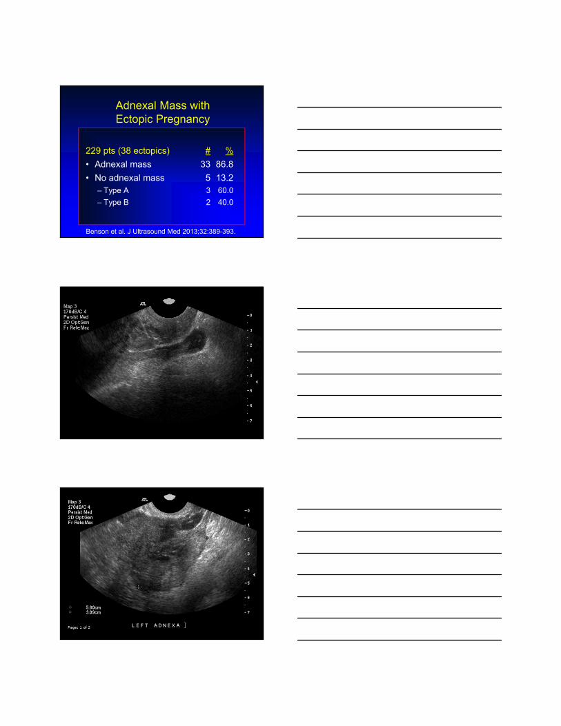
Adnexal Mass with Ectopic Pregnancy
229 pts (38 ectopics) # %
• Adnexal mass 33 86.8
• No adnexal mass 5 13.2– Type A 3 60.0
– Type B 2 40.0
Benson et al. J Ultrasound Med 2013;32:389-393.

TVS for Diagnosing Ectopics
Reviewed 10 studies
• 2216 patients
• Ectopic = 565 25.5%
• No ectopic = 1651 74.5%
Brown and Doubilet, J Ultrasound Med 1994; 13: 259 (Harvard)
*

TVS for Diagnosing Ectopics
Inclusion criteria
• Clinical suspicion of ectopic pregnancy
• All patients underwent TVS
• All cases of EP were surgically confirmed
• No adnexal masses were excluded, except simple cysts
Brown and Doubilet, J Ultrasound Med 1994; 13: 259 (Harvard)
TVS for Diagnosing Ectopics
Criteria for ectopic pregnancy
A: Adnexal embryo with heartbeat
B: Adnexal mass containing yolk sac or embryo
C: Adnexal mass with central anechoic area and hyperechoic rim (“tubal ring”)
D: Any adnexal mass other than a simple cyst or an intraovarian lesion
Brown and Doubilet, J Ultrasound Med 1994; 13: 259
TVS Criteria for Ectopic Pregnancy
Brown and Doubilet, J Ultrasound Med 1994; 13: 259
Adnexal Findings
TVS FindingLikelihood of
Ectopic
Extrauterine embryo + heartbeat
100%
Adnexal mass with yolk sac
or embryo without heartbeat100%
Tubal ring 95%
Complex or solid adnexal mass
No tubal ring, yolk sac, embryo92%

Embryo without cardiac activity
100%
Adnexal mass with yolk sac (100%)

Tubal ring (95%)
95%
95%
Tubal ring (95%)
OVARYOVARY
MASSMASS
92%

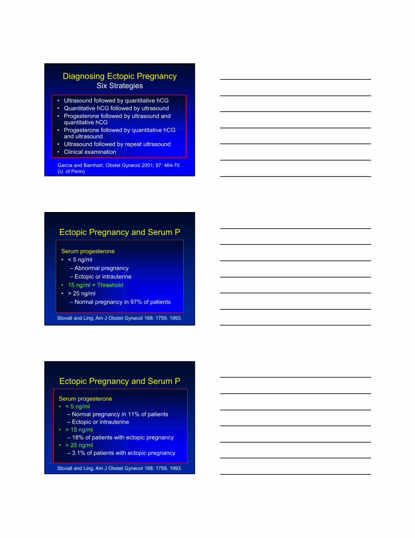
Diagnosing Ectopic PregnancySix Strategies
• Ultrasound followed by quantitative hCG• Quantitative hCG followed by ultrasound• Progesterone followed by ultrasound and
quantitative hCG• Progesterone followed by quantitative hCG
and ultrasound• Ultrasound followed by repeat ultrasound• Clinical examination
Garcia and Barnhart. Obstet Gynecol 2001; 97: 464-70 (U. of Penn)
Ectopic Pregnancy and Serum P
Serum progesterone
• < 5 ng/ml
– Abnormal pregnancy
– Ectopic or intrauterine
• 15 ng/ml = Threshold
• > 25 ng/ml
– Normal pregnancy in 97% of patients
Stovall and Ling, Am J Obstet Gynecol 168: 1759, 1993.
Ectopic Pregnancy and Serum P
Serum progesterone• < 5 ng/ml
– Normal pregnancy in 11% of patients– Ectopic or intrauterine
• > 15 ng/ml– 18% of patients with ectopic pregnancy
• > 25 ng/ml– 3.1% of patients with ectopic pregnancy
Stovall and Ling, Am J Obstet Gynecol 168: 1759, 1993.

Diagnosing Ectopic PregnancySix Strategies-Outcomes
StrategyLS/
10,000
DC/
10,000
US/
10,000
H/
10,000
US hCG 940 2581 10,250 0
hCG US 940 2463 8276 0
P US hCG 916 3547 3272 0
P hCG US 916 3191 2555 0
US US 940 3319 11,760 1760
Clinical Exam 0 0 0 0
Garcia and Barnhart. Obstet Gynecol 2001; 97: 464-70
Diagnosing Ectopic PregnancySix Strategies-Presumptions
• Hypothetical cohort of 10,000 women
• Charges
– Laparoscopy = $5000
– D&C = $2500
– Ultrasound = $ 784
– P4 or hCG = $ 75
– Admission x 24 h = $1500
Garcia and Barnhart. Obstet Gynecol 2001; 97: 464-70
Diagnosing Ectopic PregnancySix Strategies-Outcomes
Strategy Days to DxBlood
draws/10,000Total
Charge/Pt
US hCG 1.46 5,227 $1958
hCG US 1.66 14,375 $1842
P US hCG 1.25 12,108 $1692
P hCG US 1.26 15,003 $1569
US US 1.21 0 $2486
Clinical Exam 1.0 0 0
Garcia and Barnhart. Obstet Gynecol 2001; 97: 464-70

Strategy Missed EP/10,000
Interrupted IUP/10,000
US hCG 0 70
hCG US 0 122
P US hCG 24 25
P hCG US 24 39
US US 0 121
Clinical Exam 940 0
Diagnosing Ectopic PregnancySix Strategies-Outcomes
Garcia and Barnhart. Obstet Gynecol 2001; 97: 464-70
Diagnosing Ectopic PregnancySix Strategies-Recommendations
• Ultrasound followed by hCG• hCG followed by ultrasound• Either progesterone protocol
– More missed ectopic pregnancies
• Ultrasound followed by repeat ultrasound– May be applicable in poorly compliant patient
• Clinical exam only – NOT recommended
Garcia and Barnhart. Obstet Gynecol 2001; 97: 464-70
Case Presentation
• 41 G2P0010 with LMP 3 weeks ago
• c/o vaginal bleeding and abdominal pain
• Unprotected intercourse x 10 years
• + UCG

Quantitative hCG = 78
hCG Dynamics with Spontaneous Resolution of Ectopic
Helsinki, Finland118 patientsEntry criteria– Decreasing or stable hCG– No signs of rupture/intraperitoneal hemorrhage– Adnexal mass < 4 cm– No cardiac activity
Korhonen, Stenman, Ylostalo. Fertil Steril 1994; 61: 632-36 (Finland)
Ectopic Pregnancy
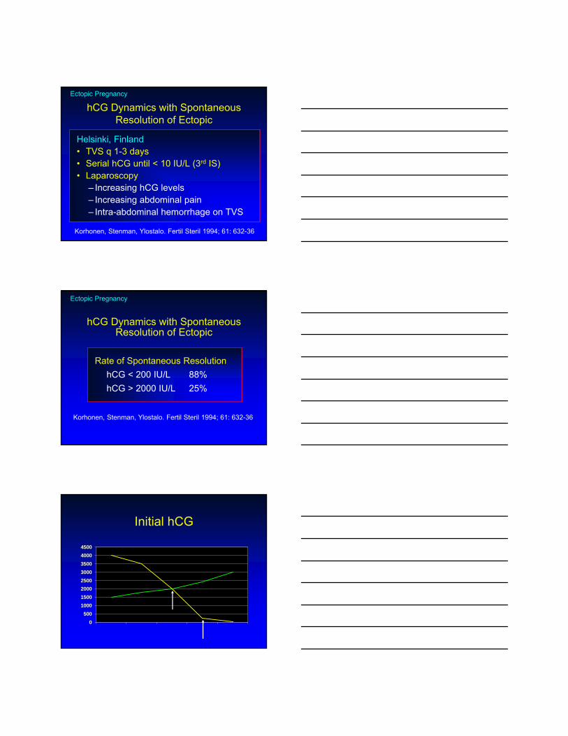
hCG Dynamics with Spontaneous Resolution of Ectopic
Helsinki, Finland• TVS q 1-3 days• Serial hCG until < 10 IU/L (3rd IS)• Laparoscopy
– Increasing hCG levels– Increasing abdominal pain– Intra-abdominal hemorrhage on TVS
Korhonen, Stenman, Ylostalo. Fertil Steril 1994; 61: 632-36
Ectopic Pregnancy
hCG Dynamics with Spontaneous Resolution of Ectopic
Rate of Spontaneous Resolution
hCG < 200 IU/L 88%
hCG > 2000 IU/L 25%
Korhonen, Stenman, Ylostalo. Fertil Steril 1994; 61: 632-36
Ectopic Pregnancy
Initial hCG
0
500
1000
1500
2000
2500
3000
3500
4000
4500

Case Presentation
• 36 y.o. G3P0020 seen in ER with c/o slight spotting and mild abdominal discomfort
• Uterus: Mid-position, TNS
• Adnexa: No definite masses
• hCG = 357 IU/L
• Hct = 36.4
• D/C home with F/U 2 days in WCC
Case Presentation
• WCC– c/o increasing pain and weakness
BLOOD
hCG = 465 IU/L

hCG = 465 IU/L
Serum hCG and Tubal Rupture
ß-hCG (IU/L) Unruptured Ruptured
< 100 9.2% 11.4%
100 – 999 47.3% 38.6%
1000 - 9,999 38.2% 38.6%
> 10,000 5.3% 11.4%
Saxon et al. Obstet Gynecol 1997; 90: 46 (McGill, Cleveland Clinic)
Serum hCG and Tubal Rupture
Frates et al. J Ultrasound Med 2014; 33:697-703.
hCG, mIU/mL Unruptured RupturedRupture Rate
%
< 1000 53 14 (41.2%) 20.9
1000-1999 14 6 (17.6%) 30.0
> 2000 38 14 (41.2%) 26.9

Case Presentation
21 yo G1P0 at 6w3d by LMP
• c/o vaginal bleeding x 1 day
• LLQ pain x 1 day
• + home pregnancy test 3 days ago
• BC: progestin oral contraceptives
• Negative past gyn history
• Quantitative hCG = 25,340

Case Presentation
• Ultrasound• Uterus
– IUP with + yolk sac, CRL c/w 5w6d
– + cardiac activity
• Left adnexa– Mass = 4 x 3 x 4 cm, with gestational sac
• Diagnosis: heterotopic pregnancy

Heterotopic Pregnancy
• More common with ART
• Incidence– Spontaneous 1:30,000
– ART 1:110-1:667
Clayton et al. Obstet Gynecol 2006; 107:595-604.
Case presentation
• 28 y.o. G2P0010
• Presents with pelvic pain and vaginal spotting
• LMP = 7 weeks ago
• hCG: positive

Courtesy of William W. Brown, III, M.D.

Ultrasound Diagnosis of Interstitial Pregnancy
• Empty uterine cavity
• Chorionic sac > 1 cm from the lateral edge of the uterine cavity (endometrium)
• Thin (<5 mm) layer of myometrium surrounding the chorionic sac
Timor et al. Obstet Gynecol 1992;79:1044


Terminology
• Interstitial pregnancy– Embryo implants in the interstitial or intramural
portion of the Fallopian tube
• Cornual pregnancy– Pregnancies that occur in a rudimentary horn,
unicornuate uterus, cornual region of a septate uterus, a bicornuate uterus, or a uterus didelphys
• Angular pregnancy– Embryo implants in one of the lateral angles of the
uterine cavity, medial to the utero-tubal junction

Uterine HornPregnancy in Uterine Horn
Non-Communicating Uterine Horn
Bicornuate with Non-communicating Horn
Uterine HornPregnancy in Uterine Horn
Terminology
• Interstitial pregnancy– Embryo implants in the interstitial or intramural
portion of the Fallopian tube
• Cornual pregnancy– Pregnancies that occur in a rudimentary horn,
unicornuate uterus, cornual region of a septate uterus, a bicornuate uterus, or a uterus didelphys
• Angular pregnancy– Embryo implants in one of the lateral angles of the
uterine cavity, medial to the utero-tubal junction


Case Presentation
• 29 y.o. G3P1011 presents to ED with c/o vaginal bleeding with clots. No tissue passed.
• hCG = 50,637 one week prior
• hCG = 71,460
• Hct = 40.9


Case Presentation
• Cervical pregnancy
• hCG = 25,9789
• Under transvaginal ultrasound, transcervical injection of MTX into the placenta and intracardiac KCl
• Ultrasound direction in the OR
• Admitted for observation
Cervical EctopicMTX + KCl
Cervical Pregnancy: MTX + KCL
62,011
2,564
11,222
53
50,637
71,460
6 3
-2 0 2 4 6 8
Weeks from treatment
hC
G le
vels
hCG

Case Presentation
• 24 y.o. G2P1001 presents with pelvic pain and scant spotting
• LMP ~ 5 weeks ago
• Exam: VSS
Uterus: TNS, moderate tenderness
Adnexa: Slight fullness,
? Left mass
• Initial: hCG = 1,340 IU/L

1 WEEK POST MTX
1 WEEK POST MTX

1 WEEK POST MTX
9 WEEKS POST MTX
9 WEEKS POST MTX

9 WEEKS POST MTX
Cesarean Scar Pregnancy
Timor-Tritsch et al. Am J Obstet Gynecol 2012;207(1):14-29.
Treatment C-Section Scar Ectopic
Timor-Tritsch et al. Am J Obstet Gynecol 2012;207(1):14-29.

Case Presentation
• 23 y.o. G2P1001
• Enters c/o slight spotting and cramping
• LMP = Unknown
• UCG = positive
• hCG = 2,392
hCG = 2392
12/30/10
ET = 17.84 mm


hCG = 2392
12/30/10
ET = 17.84 mm
Consensus Nomenclature
• Pregnancy of unknown location (PUL)
– Possible IUP
• Increased endometrial thickness
Barnhart et al. Fertil Steril 2011; 95: 857-866
Quantitative hCG
12/30/10 2392
1/01/11 7721

hCG = 7721
1/01/11
01/01/11

01/04/11
hCG = 16,371
01/18/11
FHR = 140 bpm
Ectopic Pregnancy-Summary
• Ultrasound can be justified prior to obtaining a quantitative hCG
~ 50% of ruptured ectopics had hCG levels below the discriminatory zone (<1000 IU)
• Endometrial thickness when hCG < discriminatory level
• An endometrial thickness < 8 mm is associated with an abnormal pregnancy 97% of the time

Ectopic Pregnancy-Summary
• The discriminatory level has changed
• It may be as high as 2500-3500 IU/L
• A cystic structure within the endometrium, in the absence of an adnexal mass
• Is associated with an IUP in > 99% of patients
Ectopic Pregnancy-Summary
• Finding an IUP r/o ectopic pregnancy• Exception: heterotopic pregnancy
• (1:667-1:30,000)• Finding of embryo + heart beat or yolk sac
in adnexa• Diagnostic of ectopic pregnancy
• No IUP. Complex/solid mass, sep from ovary• 92% likelihood of ectopic
Thank You