Embed Size (px)
Citation preview
Effective Jan. 2016
Prehospital Care Manual
2021

Contra Costa County Emergency Medical Services
Treatment Guideline Index
Field Treatment GuideReference
Page 1 of 3
Section 1 General Treatment GuidelinesG01 Universal Patient CareG02 Death with DignityG03 End of Life CareG04 Fever/Infection ControlG05 IV and IO AccessG06 Ventricular Assist Devices
Section 2 Interfacility Transport Treatment GuidelinesI01 STEMI TransferI02 Intubated Patient TransferI03 Stroke Transfer
Section 3 Adult Cardiac Treatment GuidelinesAC01 Cardiac ArrestAC02 Asystole and PEAAC03 V-Fib and Pulseless V-TachAC04 Post Resuscitation (ROSC)AC05 Symptomatic BradycardiaAC06 Narrow Complex TachycardiaAC07 Wide Complex TachycardiaAC08 Chest Pain: Suspected Cardiac or STEMI
Section 4 Adult Medical Treatment GuidelinesA01 Abdominal PainA02 AirwayA03 BehavioralA04 Allergic Reaction/AnaphylaxisA05 Altered Mental StatusA06 Childbirth/LaborA07 DiabeticA08 Dialysis/Renal FailureA09 Shortness of BreathA10 Dystonic ReactionA11 Emergencies Involving Central LinesA12 Hypotension/ShockA13 Overdose/Toxic IngestionA13a Suspected Opioid OverdoseA14 Pain Control A15 Respiratory Distress With a Tracheostomy TubeA16 SeizureA17 Suspected Sepsis
Effective Jan. 2021

Contra Costa County Emergency Medical Services
Treatment Guideline Index
Field Treatment GuideReference
Page 2 of 3
Section 4 Adult Medical Treatment Guidelines (continued)A18 Suspected StrokeA19 SyncopeA20 Vomiting and Diarrhea
Section 5 Pediatric Cardiac Treatment GuidelinesPC01 Pediatric Cardiac ArrestPC02 Asystole/PEAPC03 V-Fib and Pulseless V-TachPC04 Post ResuscitationPC05 BradycardiaPC06 Tachycardia
Section 6 Pediatric Treatment GuidelinesP01 AirwayP02 Allergic Reaction/AnaphylaxisP03 Altered Mental StatusP04 BehavioralP05 Brief Unexplained Resolved Event (BRUE)P06 DiabeticP07 Hypotension/ShockP08 Newly BornP09 Overdose/Toxic IngestionP10 Pain ControlP11 Respiratory DistressP12 SeizureP13 Vomiting and Diarrhea
Section 7 Trauma and Environmental Treatment GuidelinesT01 Trauma TriageT02 Bites and EnvenomationT03 BurnsT04 Extremity TraumaT05 Head TraumaT06 Multi-System TraumaT07 Heat Illness/HyperthermiaT08 Hypothermia
Section 8 Field ProceduresFP01 12-Lead ECGFP02 Airway: BLS ManagementFP03 Airway: Bougie
Effective Jan. 2020

Section 8 Field Procedures (continued)FP04 Airway: Endotracheal IntubationFP05 Airway: Foreign Body RemovalFP06 Airway: Supraglottic AirwayFP07 Airway: Stomal IntubationFP08 Airway: Tracheostomy Tube ReplacementFP09 Cardiac Arrest ManagementFP10 ChildbirthFP11 CPAPFP12 End Tidal CO2 MonitoringFP13 External PacingFP14 Helmet Removal FP15 Intraosseous AccessFP16 Needle DecompressionFP17 Pediatric AssessmentFP18 Spinal Injury AssessmentFP19 Spinal Motion RestrictionFP20 Taser Dart RemovalFP21 TourniquetFP22 Valsalva (Modified) ManeuverFP23 Vascular AccessFP24 Pediatric Medication AdministrationFP25 Finger Stick Blood Glucose AnalysisFP26 EMT Epinephrine AdministrationFP27 EMT Naloxone AdministrationFP28 Hemostatic Dressing ApplicationFP29 Vaccination by EMS Personnel
Section 9 ReferencePediatric Dosing GuideApproved AbbreviationsDrug Reference
Contra Costa County Emergency Medical Services
Treatment Guideline Index
Field Treatment GuideReference
Page 3 of 3 Effective Jan. 2020

This page intentionally left blank

Treatment Guideline G01Page 1 of 2
Contra Costa County Emergency Medical Services
Universal Patient CareRequired Vital Signs:Blood pressure as age appropriatePalpated pulse rateRespiratory ratePulse oximetry
If indicated:Blood glucose12‐Lead ECGLung soundsTemperaturePain scaleEtCO2 monitoring
Bring all necessary equipment to patientDemonstrate professionalism and courtesy
For mass assembly, consider WMD
Utilize appropriate PPE
E
Consider airborne or droplet isolation if indicated
Exit to appropriate TG
Evaluate mechanism of injuryConsider spinal motion restriction
if indicated
High Risk Mechanism
MedicalPatient
Repeat assessment while preparing for transport
Continue on‐going assessmentRepeat VS
Evaluate interventions / procedures
Patient transfer to hospital staffInclude summary of care and patient
propertyMedical
patient does not fit specific
TG
Initial assessmentBLS care
Initiate oxygen if indicated
Adult assessment procedure
Pediatric assessment procedureUse PediaTape for measurement
TraumaPatient
Low Risk Mechanism
Primary and secondary trauma
assessment
Obtain VS
Obtain SAMPLE
Primary and secondary trauma
assessment
Focused assessment on specific injury
Mental status
Unresponsive Responsive
Primary and secondary assessment
Chief complaintObtain SAMPLE
Obtain history of present illness from available sources / scene
survey
Primary and secondary assessment
Focused assessment on
specific complaint
Obtain VS
Trauma patient does not fit specific
TG
Exit to appropriate TG
Notify receiving facility. Contact Base Hospital for
medical direction, as needed.
Effective Jan. 2020
Effective Jan. 2016
Contra Costa County Emergency Medical Services
Universal Patient Care
Treatment Guideline G01Page 2 of 2
Pearls• A pediatric patient is defined as being <15 years of age.• Timing of transport should be based on the patient’s condition and the destination policy.• Never hesitate to contact the Base Hospital as a high risk refusal resource for any patient who refuses transport.• SAMPLE: Signs / Symptoms; Allergies; Medications; PMH; Last oral intake; Events leading to injury/illness.
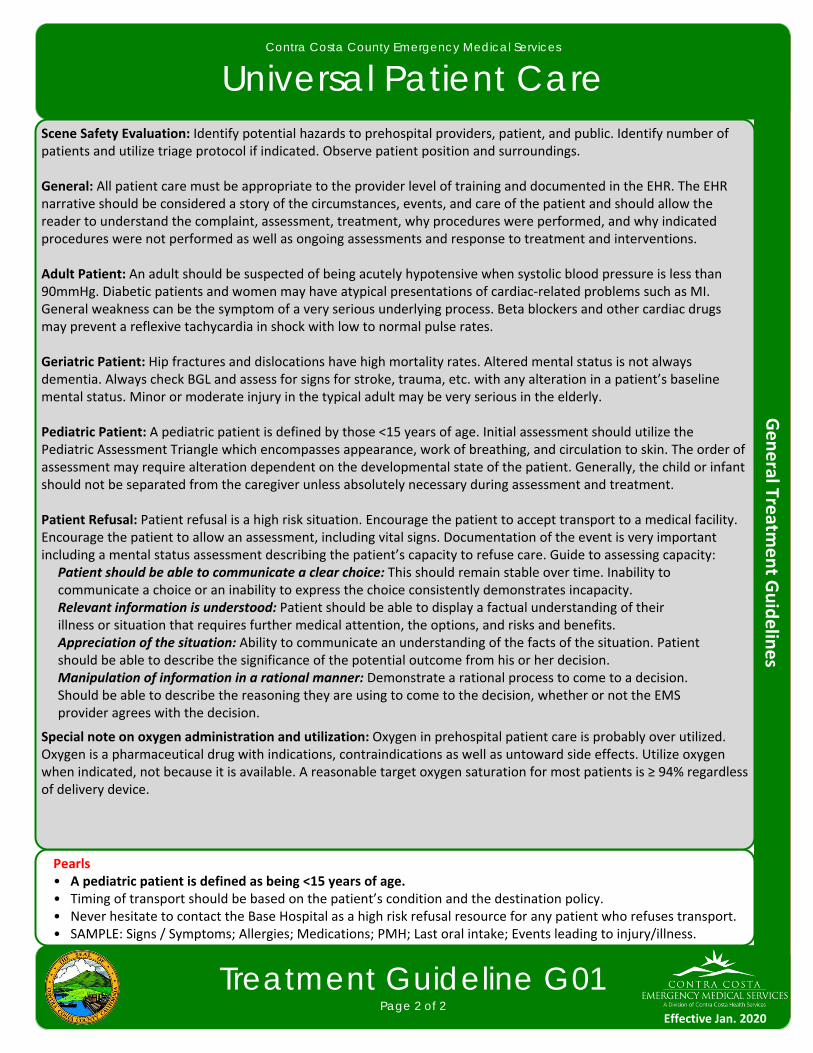
Scene Safety Evaluation: Identify potential hazards to prehospital providers, patient, and public. Identify number of patients and utilize triage protocol if indicated. Observe patient position and surroundings.
General: All patient care must be appropriate to the provider level of training and documented in the EHR. The EHR narrative should be considered a story of the circumstances, events, and care of the patient and should allow the reader to understand the complaint, assessment, treatment, why procedures were performed, and why indicated procedures were not performed as well as ongoing assessments and response to treatment and interventions.
Adult Patient: An adult should be suspected of being acutely hypotensive when systolic blood pressure is less than 90mmHg. Diabetic patients and women may have atypical presentations of cardiac‐related problems such as MI. General weakness can be the symptom of a very serious underlying process. Beta blockers and other cardiac drugs may prevent a reflexive tachycardia in shock with low to normal pulse rates.
Geriatric Patient: Hip fractures and dislocations have high mortality rates. Altered mental status is not always dementia. Always check BGL and assess for signs for stroke, trauma, etc. with any alteration in a patient’s baseline mental status. Minor or moderate injury in the typical adult may be very serious in the elderly.
Pediatric Patient: A pediatric patient is defined by those <15 years of age. Initial assessment should utilize the Pediatric Assessment Triangle which encompasses appearance, work of breathing, and circulation to skin. The order of assessment may require alteration dependent on the developmental state of the patient. Generally, the child or infant should not be separated from the caregiver unless absolutely necessary during assessment and treatment.
Patient Refusal: Patient refusal is a high risk situation. Encourage the patient to accept transport to a medical facility. Encourage the patient to allow an assessment, including vital signs. Documentation of the event is very important including a mental status assessment describing the patient’s capacity to refuse care. Guide to assessing capacity: Patient should be able to communicate a clear choice: This should remain stable over time. Inability to communicate a choice or an inability to express the choice consistently demonstrates incapacity. Relevant information is understood: Patient should be able to display a factual understanding of their illness or situation that requires further medical attention, the options, and risks and benefits. Appreciation of the situation: Ability to communicate an understanding of the facts of the situation. Patient should be able to describe the significance of the potential outcome from his or her decision. Manipulation of information in a rational manner: Demonstrate a rational process to come to a decision. Should be able to describe the reasoning they are using to come to the decision, whether or not the EMS provider agrees with the decision.
Special note on oxygen administration and utilization: Oxygen in prehospital patient care is probably over utilized. Oxygen is a pharmaceutical drug with indications, contraindications as well as untoward side effects. Utilize oxygen when indicated, not because it is available. A reasonable target oxygen saturation for most patients is ≥ 94% regardless of delivery device.
Effective Jan. 2020
Effective Jan. 2016
Contra Costa County Emergency Medical Services
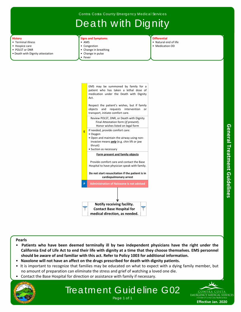
Death with Dignity
Treatment Guideline G02Page 1 of 1
History• Terminal illness• Hospice care• POLST or DNR• Death with Dignity attestation
Differential• Natural end of life • Medication OD
Signs and Symptoms• AMS• Congestion• Change in breathing• Change in pulse• Fever
Review POLST, DNR, or Death with Dignity Final Attestation form (if present).Honor wishes listed on legal form
P
E If needed, provide comfort care:• Oxygen• Open and maintain the airway using non‐ invasive means only (e.g. chin lift or jaw thrust)• Suction as necessary
Administration of Naloxone is not advised
EMS may be summoned by family for a patient who has taken a lethal dose of medication under the Death with Dignity Act.
Respect the patient’s wishes, but if family objects and requests intervention or transport, initiate comfort care.
Pearls• Patients who have been deemed terminally ill by two independent physicians have the right under the
California End of Life Act to end their life with dignity at a time that they choose themselves. EMS personnel should be aware of and familiar with this act. Refer to Policy 1003 for additional information.
• Naxolone will not have an affect on the drugs prescribed for death with dignity patients. • It is important to recognize that families may be educated on what to expect with a dying family member, but
no amount of preparation can eliminate the stress and grief of watching a loved one die. • Contact the Base Hospital for direction or assistance with family if necessary.
Form present and family objects
Provide comfort care and contact the Base Hospital to have physician speak with family.
Do not start resuscitation if the patient is in cardiopulmonary arrest
Notify receiving facility. Contact Base Hospital for
medical direction, as needed.
Effective Jan. 2020

Effective Jan. 2016
Contra Costa County Emergency Medical Services
End of Life Care
Treatment Guideline G03Page 1 of 2
History• Terminal illness• Hospice care• POLST or DNR
Differential• Natural end of life • Medication OD
Signs and Symptoms• AMS• Congestion• Change in breathing• Change in pulse• Fever
Review POLST or DNR formHonor wishes listed on legal form
Transport to appropriate receiving center or requested facility of choice
If family member with decision‐making authority is not present, ask family for the
phone number for their Hospice Nurse if not already on scene
Contact Hospice Nurse
P
If an existing patent IV is availableFentanyl 25 – 200mcg IV
titrated in 25 – 50mcg increments to pain relief. Consider 25mcg increments in elderly
patients
If no IV accessFentanyl 100mcg IN
May repeat once after 15 minutes
If no IV access and IN route not advisableFentanyl 50 – 100mcg IM
May repeat once after 15 minutes
Max of 200mcg totalMonitor and reassess 5 minutes following
administration
Fentanyl Contact Base Hospital for
additional orderN
E
Honor and respect patient/family wishesfor transport after discussion
Let Hospice Nurse discuss options with family, even if only over the phone
The family and Hospice Nurse should decide on an appropriate course of treatment or
decision to transport
If needed, provide immediate supportive care• Oxygen• Open and maintain the airway using non‐ invasive means only (e.g. chin lift or jaw thrust)• Suction as necessary• Position for comfort• Control external hemorrhaging• Immobilize obvious fractures using techniques to minimize pain
Determine level of pain and treat if indicated
If transport is declined, complete Refusal and thoroughly document encounter
Administration of Naloxone is not advised
Is the patient on hospice?
No
Yes
Notify receiving facility. Contact Base Hospital for
medical direction, as needed.
Effective Jan. 2020

Effective Jan. 2016
Contra Costa County Emergency Medical Services
End of Life Care
Treatment Guideline G03Page 2 of 2
Pearls• Hospice patients and those on palliative end of life care are often heavily medicated with pain medications.
Administration of Naxolone, even in small amounts, can result in unnecessary suffering.• Follow the wishes outlined in a signed POLST or DNR order. A competent patient or designated decision
maker acting on behalf of the patient can override POLST.• If a POLST or DNR order is not immediately available, immediately initiate BLS supportive care. Do not delay
care while waiting for the form.• If transport is initiated at the request of the family and the patient subsequently goes into cardiac or
respiratory arrest during transport, continue to the closest approved receiving facility.• Always involve the patient’s assigned Hospice Nurse, even if it is by phone. It is important to recognize that
families may be educated on what to expect with a dying family member, but no amount of preparation can eliminate the stress and grief of watching a loved one die.
• Contact the Base Hospital for direction or assistance with family in the absence of a Hospice Nurse if necessary.
POLST forms are generally copied on pink paper to help ensure that the document stands out and is followed. However, POLST on any paper color is valid.
Unlike POLST, there is no standardized DNR order form. If you have doubt of a DNR order authenticity, initiate BLS care and contact the Base Hospital for guidance.
Effective Jan. 2020
Effective Jan. 2016
Contra Costa County Emergency Medical Services
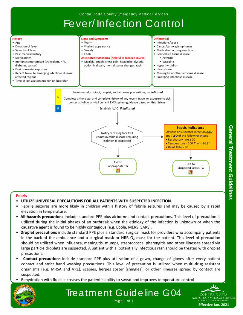
Fever/Infection Control
Treatment Guideline G04Page 1 of 1
History• Age• Duration of fever• Severity of fever• Past medical history• Medications• Immunocompromised (transplant, HIV, diabetes, cancer)• Environmental exposure• Recent travel to emerging infectious disease affected regions• Time of last acetaminophen or ibuprofen
Differential• Infections/sepsis• Cancer/tumors/lymphomas • Medication or drug reaction• Connective tissue disease • Arthritis • Vasculitis• Hyperthyroidism• Heat stroke • Meningitis or other airborne disease• Emerging infectious disease
Signs and Symptoms• Warm• Flushed appearance• Sweaty• Chills Associated symptoms (helpful to localize source)• Myalgia, cough, chest pain, headache, dysuria, abdominal pain, mental status changes, rash
Use universal, contact, droplet, and airborne precautions, as indicated
E
Notify receiving facility if communicable disease requiring
isolation is suspected
Complete a thorough and complete history of any recent travel or exposure to sick contacts; follow any/all current EMS system guidance based on this history
Establish IV/IO, if indicatedP
Exit to appropriate TG
Pearls• UTILIZE UNIVERSAL PRECAUTIONS FOR ALL PATIENTS WITH SUSPECTED INFECTION.• Febrile seizures are more likely in children with a history of febrile seizures and may be caused by a rapid
elevation in temperature.• All‐hazards precautions include standard PPE plus airborne and contact precautions. This level of precaution is
utilized during the initial phases of an outbreak when the etiology of the infection is unknown or when the causative agent is found to be highly contagious (e.g. Ebola, MERS, SARS).
• Droplet precautions include standard PPE plus a standard surgical mask for providers who accompany patients in the back of the ambulance and a surgical mask or NRB O2 mask for the patient. This level of precaution should be utilized when influenza, meningitis, mumps, streptococcal pharyngitis and other illnesses spread via large particle droplets are suspected. A patient with a potentially infectious rash should be treated with droplet precautions.
• Contact precautions include standard PPE plus utilization of a gown, change of gloves after every patient contact and strict hand washing precautions. This level of precaution is utilized when multi‐drug resistant organisms (e.g. MRSA and VRE), scabies, herpes zoster (shingles), or other illnesses spread by contact are suspected.
• Rehydration with fluids increases the patient’s ability to sweat and improves temperature control.
Sepsis IndicatorsObvious or suspected infection AND any TWO of the following criteria:• Respiratory rate ≥ 20• Temperature > 100.4° or < 96.8° • Heart Rate > 90
Exit to Suspected Sepsis TG
Effective Jan. 2021

Effective Jan. 2016
Contra Costa County Emergency Medical Services
IV and IO Access
Treatment Guideline G05Page 1 of 1
First circulatory access for cardiac arrest?
Assess need for IVEmergent or potentially emergent
medical or trauma condition
Establish IV/IOP
Pearls• In the setting of cardiac arrest, any preexisting dialysis shunt or external central venous catheter may be used.• In patients who are hemodynamically unstable, pre‐existing indwelling central lines can be used to deliver fluid
and medications.• Approved prehospital IV solutions include: Isotonic (balanced) saline solution, Ringer’s lactate, and glucose
solutions with no additional additives.• Any working venous catheter already accessed prior to EMS arrival may be used for EMS IV fluids and medications.• Use Chlorhexidine for all IV/IO attempts.• Intraosseous access should be obtained only with the appropriate adult or pediatric device (e.g. IO drill).• Any prehospital fluids or medications approved for IV use may also be given through IO.• External jugular access is only indicated for patients ≥ 15 years of age.• All IV rates should be kept at TKO (minimal rate to keep the vein open) unless administering fluid bolus.• Use micro drip sets for all patients 6 years of age and less.• Upper extremity IV sites are preferable to lower extremity sites; lower extremity sites are discouraged in
patients with vascular disease or diabetes.• In post‐mastectomy patients and patients with a working dialysis fistula, avoid IV attempts, injections and blood
pressure measurements in the upper extremity on the affected side.
Yes
No
Peripheral IV
P
External jugular IV for unstable patients needing emergent IV medication or fluids AND no peripheral site is available AND IO
is not obtainable
Intraosseous IV for life‐threatening event at most suitable site available
Saline lock
Monitor infusionP
Monitor saline lock/existing approved prehospital fluids
E
Effective Jan. 2020
Effective Jan. 2016
Contra Costa County Emergency Medical Services
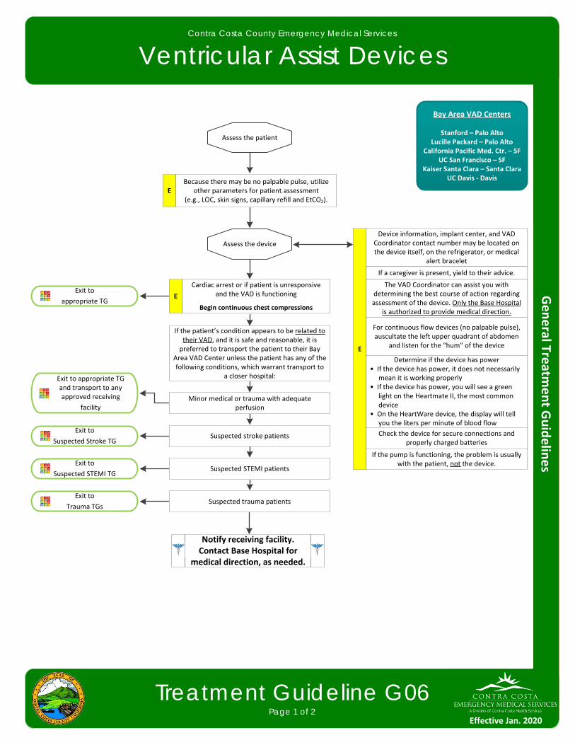
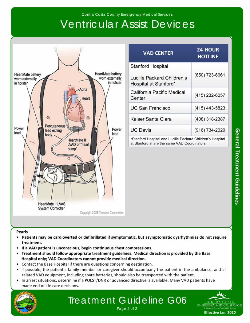
Ventricular Assist Devices
Treatment Guideline G06Page 1 of 2
Because there may be no palpable pulse, utilize other parameters for patient assessment
(e.g., LOC, skin signs, capillary refill and EtCO2).
Exit to
Suspected Stroke TG
E
Assess the patient
Assess the device
Device information, implant center, and VAD Coordinator contact number may be located on the device itself, on the refrigerator, or medical
alert bracelet
E
If a caregiver is present, yield to their advice.
The VAD Coordinator can assist you with determining the best course of action regarding assessment of the device. Only the Base Hospital
is authorized to provide medical direction.
For continuous flow devices (no palpable pulse), auscultate the left upper quadrant of abdomen
and listen for the “hum” of the device
Determine if the device has power• If the device has power, it does not necessarily mean it is working properly• If the device has power, you will see a green light on the Heartmate II, the most common device• On the HeartWare device, the display will tell you the liters per minute of blood flow
Check the device for secure connections and properly charged batteries
If the pump is functioning, the problem is usually with the patient, not the device.
If the patient’s condition appears to be related to their VAD, and it is safe and reasonable, it is preferred to transport the patient to their Bay
Area VAD Center unless the patient has any of the following conditions, which warrant transport to
a closer hospital:
Minor medical or trauma with adequate perfusion
Suspected stroke patients
Suspected STEMI patients
Suspected trauma patients
Cardiac arrest or if patient is unresponsive and the VAD is functioning
Begin continuous chest compressions
Exit to appropriate TG and transport to any approved receiving
facility
Exit to
Suspected STEMI TG
Exit to
Trauma TGs
Exit to
appropriate TG
Bay Area VAD Centers
Stanford – Palo AltoLucille Packard – Palo Alto
California Pacific Med. Ctr. – SF UC San Francisco – SF
Kaiser Santa Clara – Santa ClaraUC Davis ‐ Davis
E
Notify receiving facility. Contact Base Hospital for
medical direction, as needed.
Effective Jan. 2020
Effective Jan. 2016
Contra Costa County Emergency Medical Services
Ventricular Assist Devices
Treatment Guideline G06Page 2 of 2
Pearls• Patients may be cardioverted or defibrillated if symptomatic, but asymptomatic dysrhythmias do not require
treatment.• If a VAD patient is unconscious, begin continuous chest compressions.• Treatment should follow appropriate treatment guidelines. Medical direction is provided by the Base Hospital only; VAD Coordinators cannot provide medical direction.• Contact the Base Hospital if there are questions concerning destination.• If possible, the patient’s family member or caregiver should accompany the patient in the ambulance, and all
related VAD equipment, including spare batteries, should also be transported with the patient.• In arrest situations, determine if a POLST/DNR or advanced directive is available. Many VAD patients have made end of life care decisions.
VAD CENTER24‐HOUR HOTLINE
Stanford Hospital
Lucille Packard Children’s Hospital at Stanford*
(650) 723-6661
California Pacific Medical Center
(415) 232-6057
UC San Francisco (415) 443-5823
Kaiser Santa Clara (408) 318-2387
UC Davis (916) 734-2020
*Stanford Hospital and Lucille Packard Children’s Hospital at Stanford share the same VAD Coordinators
Effective Jan. 2020
Effective Jan. 2016
Contra Costa County Emergency Medical Services
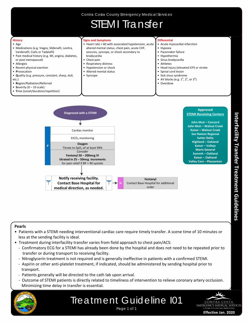
STEMI Transfer
Treatment Guideline I01Page 1 of 1
Diagnosed with a STEMI
Cardiac monitor
ConsiderFentanyl 50 – 200mcg IV
titrated in 25 – 50mcg increments for pain relief if BP > 90 systolic
P
Fentanyl Contact Base Hospital for additional
orderN
ApprovedSTEMI Receiving Centers
John Muir – ConcordJohn Muir – Walnut CreekKaiser – Walnut CreekSan Ramon Regional
Sutter DeltaHighland – OaklandKaiser – VallejoMarin General
Summit – OaklandKaiser – Oakland
Valley Care – Pleasanton
OxygenTitrate to SpO2 of at least 94%
History• Age• Medications (e.g. Viagra, Sildenafil, Levitra, Vardenafil, Cialis or Tadalafil)• Past medical history (e.g. MI, angina, diabetes, or post menopausal)• Allergies• Recent physical exertion• Provocation• Quality (e.g. pressure, constant, sharp, dull, etc.)• Region/Radiation/Referred• Severity (0 – 10 scale)• Time (onset/duration/repetition)
Differential• Acute myocardial infarction• Hypoxia • Pacemaker failure• Hypothermia• Sinus bradycardia• Athletes• Head injury (elevated ICP) or stroke• Spinal cord lesion• Sick sinus syndrome• AV blocks (e.g. 1o, 2o, or 3o)• Overdose
Signs and Symptoms• Heart rate < 60 with associated hypotension, acute altered mental status, chest pain, acute CHF, seizures, syncope, or shock secondary to bradycardia• Chest pain• Respiratory distress• Hypotension or shock• Altered mental status• Syncope
Pearls• Patients with a STEMI needing interventional cardiac care require timely transfer. A scene time of 10 minutes or less at the sending facility is ideal.• Treatment during interfacility transfer varies from field approach to chest pain/ACS: ‐ Confirmatory ECG for a STEMI has already been done by the hospital and does not need to be repeated prior to transfer or during transport to receiving facility. ‐ Nitroglycerin treatment is not required and is generally ineffective in patients with a confirmed STEMI. ‐ Aspirin or other anti‐platelet treatment, if indicated, should be administered by sending hospital prior to transport. ‐ Patients generally will be directed to the cath lab upon arrival. ‐ Outcome of STEMI patients is directly related to timeliness of intervention to relieve coronary artery occlusion. Minimizing time delay in transfer is essential.
EtCO2 monitoring
Notify receiving facility. Contact Base Hospital for
medical direction, as needed.
Effective Jan. 2020

Effective Jan. 2016
Contra Costa County Emergency Medical Services
Intubated Patient Transfer
Treatment Guideline I02Page 1 of 1
Patient with an advanced airway
Cardiac monitor
ConsiderMidazolam 2‐5mg IV/IOMaximum 5mg total
P
MidazolamContact Base Hospital for additional
orderN
Oxygen 100%
History• Age• Medications (e.g. Viagra, Sildenafil, Levitra, Vardenafil, Cialis or Tadalafil)• Past medical history • Allergies
Differential• Acute myocardial infarction• Hypoxia • Hypothermia• Head injury (elevated ICP) or stroke• Spinal cord lesion• Overdose
Signs and Symptoms• Cardiac arrest• Respiratory distress or respiratory failure• Hypotension or shock• Altered mental status
Pearls• Patients requiring specialty care, most commonly trauma or neurological care, may be transferred with an established endotracheal tube. Sedation may be required if patient agitation is present because of risk of inadvertent extubation.• Some patients may need chemical paralysis and require additional nursing or physician staff to administer these medications.• If inadvertent extubation occurs, manage with basic airway maneuvers unless ventilation cannot be adequately maintained.
Ventilate at 6 breaths per minutes
EtCO2 monitoring
Notify receiving facility. Contact Base Hospital for
medical direction, as needed.
Effective Jan. 2020
Effective Jan. 2016
Contra Costa County Emergency Medical Services
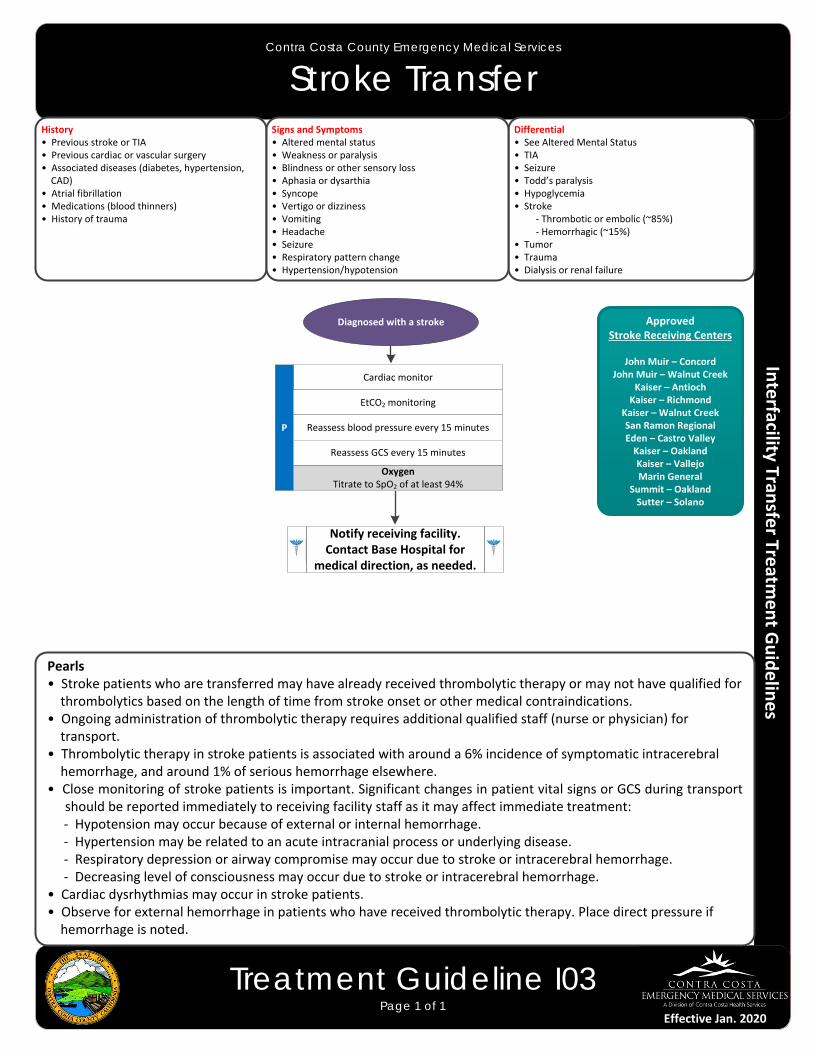
Stroke Transfer
Treatment Guideline I03Page 1 of 1
Pearls• Stroke patients who are transferred may have already received thrombolytic therapy or may not have qualified for thrombolytics based on the length of time from stroke onset or other medical contraindications.• Ongoing administration of thrombolytic therapy requires additional qualified staff (nurse or physician) for transport.• Thrombolytic therapy in stroke patients is associated with around a 6% incidence of symptomatic intracerebral hemorrhage, and around 1% of serious hemorrhage elsewhere.• Close monitoring of stroke patients is important. Significant changes in patient vital signs or GCS during transport
should be reported immediately to receiving facility staff as it may affect immediate treatment: ‐ Hypotension may occur because of external or internal hemorrhage. ‐ Hypertension may be related to an acute intracranial process or underlying disease. ‐ Respiratory depression or airway compromise may occur due to stroke or intracerebral hemorrhage. ‐ Decreasing level of consciousness may occur due to stroke or intracerebral hemorrhage.• Cardiac dysrhythmias may occur in stroke patients.• Observe for external hemorrhage in patients who have received thrombolytic therapy. Place direct pressure if hemorrhage is noted.
History• Previous stroke or TIA• Previous cardiac or vascular surgery• Associated diseases (diabetes, hypertension, CAD) • Atrial fibrillation• Medications (blood thinners)• History of trauma
Differential• See Altered Mental Status• TIA• Seizure• Todd’s paralysis• Hypoglycemia• Stroke ‐ Thrombotic or embolic (~85%) ‐ Hemorrhagic (~15%)• Tumor• Trauma• Dialysis or renal failure
Signs and Symptoms• Altered mental status• Weakness or paralysis• Blindness or other sensory loss• Aphasia or dysarthia• Syncope• Vertigo or dizziness• Vomiting• Headache• Seizure• Respiratory pattern change• Hypertension/hypotension
ApprovedStroke Receiving Centers
John Muir – ConcordJohn Muir – Walnut Creek
Kaiser – AntiochKaiser – Richmond
Kaiser – Walnut CreekSan Ramon RegionalEden – Castro ValleyKaiser – OaklandKaiser – VallejoMarin General
Summit – OaklandSutter – Solano
Diagnosed with a stroke
Cardiac monitor
P
OxygenTitrate to SpO2 of at least 94%
EtCO2 monitoring
Reassess blood pressure every 15 minutes
Reassess GCS every 15 minutes
Notify receiving facility. Contact Base Hospital for
medical direction, as needed.
Effective Jan. 2020

This page intentionally left blank
Effective Jan. 2016
Contra Costa County Emergency Medical Services
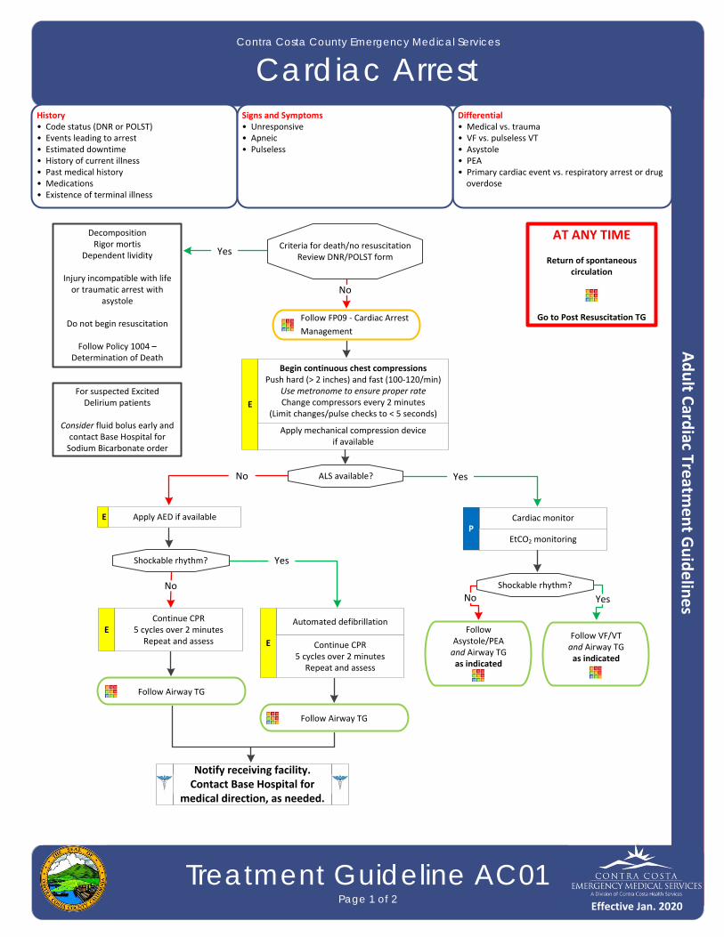
Cardiac Arrest
Treatment Guideline AC01Page 1 of 2
History• Code status (DNR or POLST)• Events leading to arrest• Estimated downtime• History of current illness• Past medical history• Medications• Existence of terminal illness
Differential• Medical vs. trauma• VF vs. pulseless VT • Asystole• PEA• Primary cardiac event vs. respiratory arrest or drug overdose
Signs and Symptoms• Unresponsive• Apneic• Pulseless
Follow Airway TG
Criteria for death/no resuscitationReview DNR/POLST form
Cardiac monitorP
AT ANY TIME
Return of spontaneous circulation
Go to Post Resuscitation TG
ALS available?
Shockable rhythm?
Continue CPR5 cycles over 2 minutes
Repeat and assessE
Automated defibrillation
E Continue CPR5 cycles over 2 minutes
Repeat and assess
Follow Airway TG
Shockable rhythm?
Follow Asystole/PEAand Airway TG as indicated
Follow VF/VTand Airway TG as indicated
DecompositionRigor mortis
Dependent lividity
Injury incompatible with life or traumatic arrest with
asystole
Do not begin resuscitation
Follow Policy 1004 – Determination of Death
Yes
Yes
Yes
YesNo
No
No
Apply AED if available
EtCO2 monitoring
No
E
E
For suspected Excited Delirium patients
Consider fluid bolus early andcontact Base Hospital for Sodium Bicarbonate order
Begin continuous chest compressions Push hard (> 2 inches) and fast (100‐120/min)
Use metronome to ensure proper rateChange compressors every 2 minutes
(Limit changes/pulse checks to < 5 seconds)
Apply mechanical compression deviceif available
Follow FP09 ‐ Cardiac Arrest
Management
Notify receiving facility. Contact Base Hospital for
medical direction, as needed.
Effective Jan. 2020

Effective Jan. 2016
Contra Costa County Emergency Medical Services
Cardiac Arrest
Treatment Guideline AC01Page 2 of 2
Pearls• Efforts should be directed at high quality and continuous chest compressions with limited interruptions.
Consider early IO placement if available or direct IV access if anticipated. • Passive ventilation for the first three cycles (6 minutes) of CPR. After that time, the patient should be
ventilated using a BLS airway and BVM at a rate of 6 ventilation/minute (1:10 seconds) with continuous CPR.
• Placement of an advanced airway should be deferred unless a provider is unable to ventilate the patient with a BLS airway and BVM.
• Do not delay chest compressions while applying any device or intervention.• Use a metronome during chest compression to ensure proper rate.• In cases of clear-cut traumatic arrest, epinephrine is not indicated in PEA or asystole. Epinephrine will not
correct arrest caused by a tension pneumothorax, cardiac tamponade, or hemorrhagic shock. If there is any doubt as to the cause of arrest, treat as a non-traumatic arrest.
• If a non-shockable rhythm persists for 30 minutes despite aggressive resuscitative efforts, consider cessation of efforts as outlined in the Determination of Death policy.• The AutoPulse device is limited to 80 compressions/minute, which is acceptable when using this device
during cardiac arrest.• Resuscitation is based on proper planning and organized execution. Procedures require space and patient
access. Make room to work. Utilize a team focused approach assigning responders to predetermined tasks.
• Reassess and document ETT placement and EtCO2 frequently, after every move, and at transfer of care.• Maternal arrest: Treat mother per appropriate TG with immediate notification to the Base Hospital along
with rapid transport. Place pillows or padding underneath mother to displace fetus from inferior vena cava as to ensure continued fetal blood circulation; left lateral position. IV/IO access should be preferably placed above the diaphragm. Defibrillation is safe at all energy levels.
Effective Jan. 2020
Effective Jan. 2016
Contra Costa County Emergency Medical Services
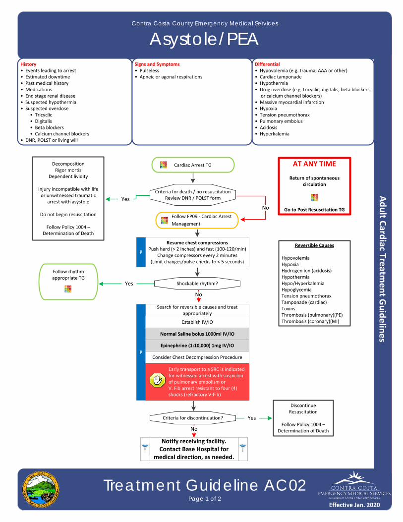
Asystole/PEA
Treatment Guideline AC02Page 1 of 2
History• Events leading to arrest• Estimated downtime• Past medical history• Medications• End stage renal disease• Suspected hypothermia• Suspected overdose • Tricyclic • Digitalis • Beta blockers • Calcium channel blockers• DNR, POLST or living will
Differential• Hypovolemia (e.g. trauma, AAA or other)• Cardiac tamponade • Hypothermia• Drug overdose (e.g. tricyclic, digitalis, beta blockers, or calcium channel blockers)• Massive myocardial infarction• Hypoxia• Tension pneumothorax• Pulmonary embolus• Acidosis• Hyperkalemia
Signs and Symptoms• Pulseless• Apneic or agonal respirations
Cardiac Arrest TG
Search for reversible causes and treat appropriately
P
Establish IV/IO
AT ANY TIME
Return of spontaneous circulation
Go to Post Resuscitation TG
Normal Saline bolus 1000ml IV/IO
Epinephrine (1:10,000) 1mg IV/IO
Criteria for death / no resuscitationReview DNR / POLST form
Resume chest compressions Push hard (> 2 inches) and fast (100‐120/min)
Change compressors every 2 minutes(Limit changes/pulse checks to < 5 seconds)
P
Shockable rhythm?
Consider Chest Decompression Procedure
Criteria for discontinuation?
Reversible Causes
HypovolemiaHypoxiaHydrogen ion (acidosis)HypothermiaHypo/HyperkalemiaHypoglycemiaTension pneumothoraxTamponade (cardiac)ToxinsThrombosis (pulmonary)(PE)Thrombosis (coronary)(MI)
DecompositionRigor mortis
Dependent lividity
Injury incompatible with life or unwitnessed traumatic
arrest with asystole
Do not begin resuscitation
Follow Policy 1004 – Determination of Death
DiscontinueResuscitation
Follow Policy 1004 – Determination of Death
Follow rhythm appropriate TG
Yes
No
No
Yes
No
Yes
Early transport to a SRC is indicated for witnessed arrest with suspicion of pulmonary embolism or V. Fib arrest resistant to four (4) shocks (refractory V‐Fib)
Follow FP09 ‐ Cardiac Arrest
Management
Notify receiving facility. Contact Base Hospital for
medical direction, as needed.
Effective Jan. 2020
Effective Jan. 2016
Contra Costa County Emergency Medical Services
Asystole/PEA
Treatment Guideline AC02Page 2 of 2
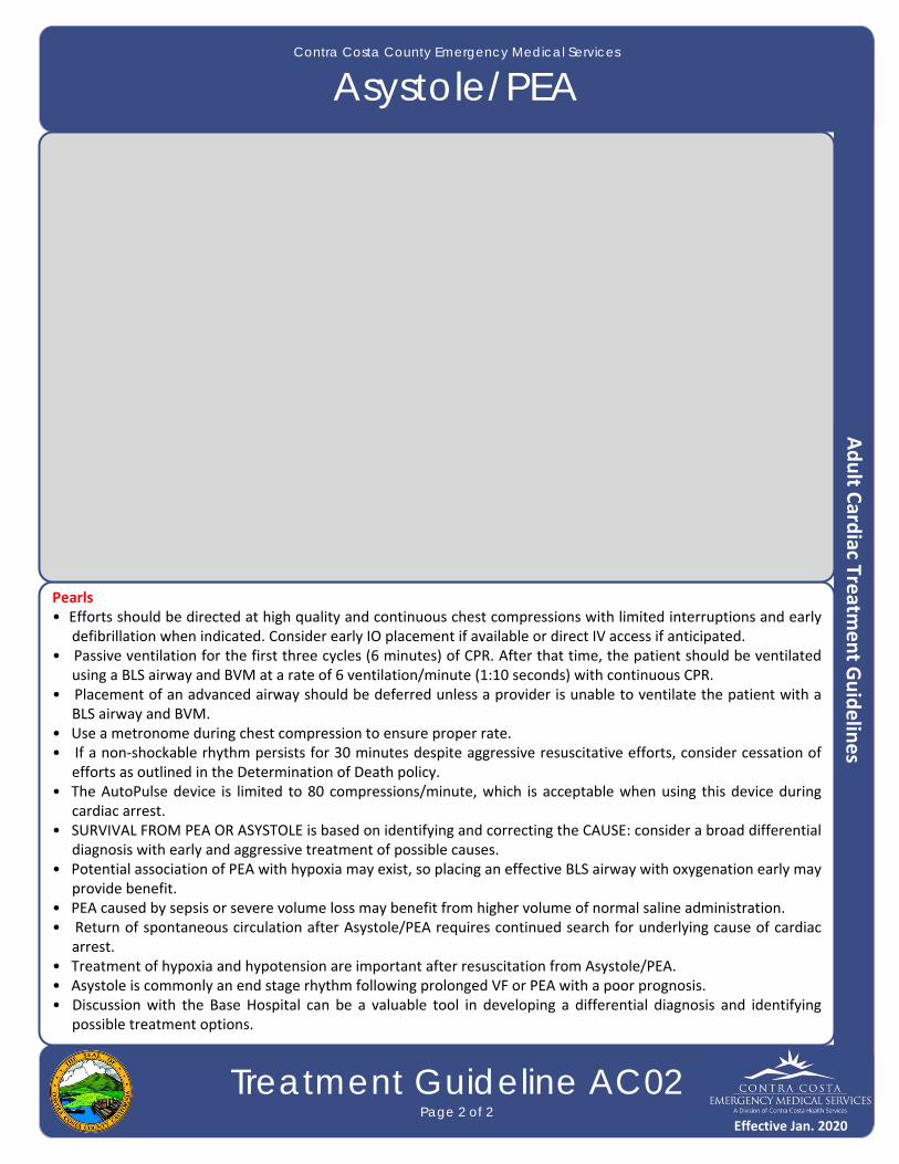
Pearls• Efforts should be directed at high quality and continuous chest compressions with limited interruptions and early
defibrillation when indicated. Consider early IO placement if available or direct IV access if anticipated. • Passive ventilation for the first three cycles (6 minutes) of CPR. After that time, the patient should be ventilated
using a BLS airway and BVM at a rate of 6 ventilation/minute (1:10 seconds) with continuous CPR. • Placement of an advanced airway should be deferred unless a provider is unable to ventilate the patient with a
BLS airway and BVM.• Use a metronome during chest compression to ensure proper rate.• If a non‐shockable rhythm persists for 30 minutes despite aggressive resuscitative efforts, consider cessation of
efforts as outlined in the Determination of Death policy.• The AutoPulse device is limited to 80 compressions/minute, which is acceptable when using this device during
cardiac arrest.• SURVIVAL FROM PEA OR ASYSTOLE is based on identifying and correcting the CAUSE: consider a broad differential
diagnosis with early and aggressive treatment of possible causes.• Potential association of PEA with hypoxia may exist, so placing an effective BLS airway with oxygenation early may
provide benefit.• PEA caused by sepsis or severe volume loss may benefit from higher volume of normal saline administration.• Return of spontaneous circulation after Asystole/PEA requires continued search for underlying cause of cardiac
arrest.• Treatment of hypoxia and hypotension are important after resuscitation from Asystole/PEA.• Asystole is commonly an end stage rhythm following prolonged VF or PEA with a poor prognosis.• Discussion with the Base Hospital can be a valuable tool in developing a differential diagnosis and identifying
possible treatment options.
Effective Jan. 2020
Effective Jan. 2016
Contra Costa County Emergency Medical Services
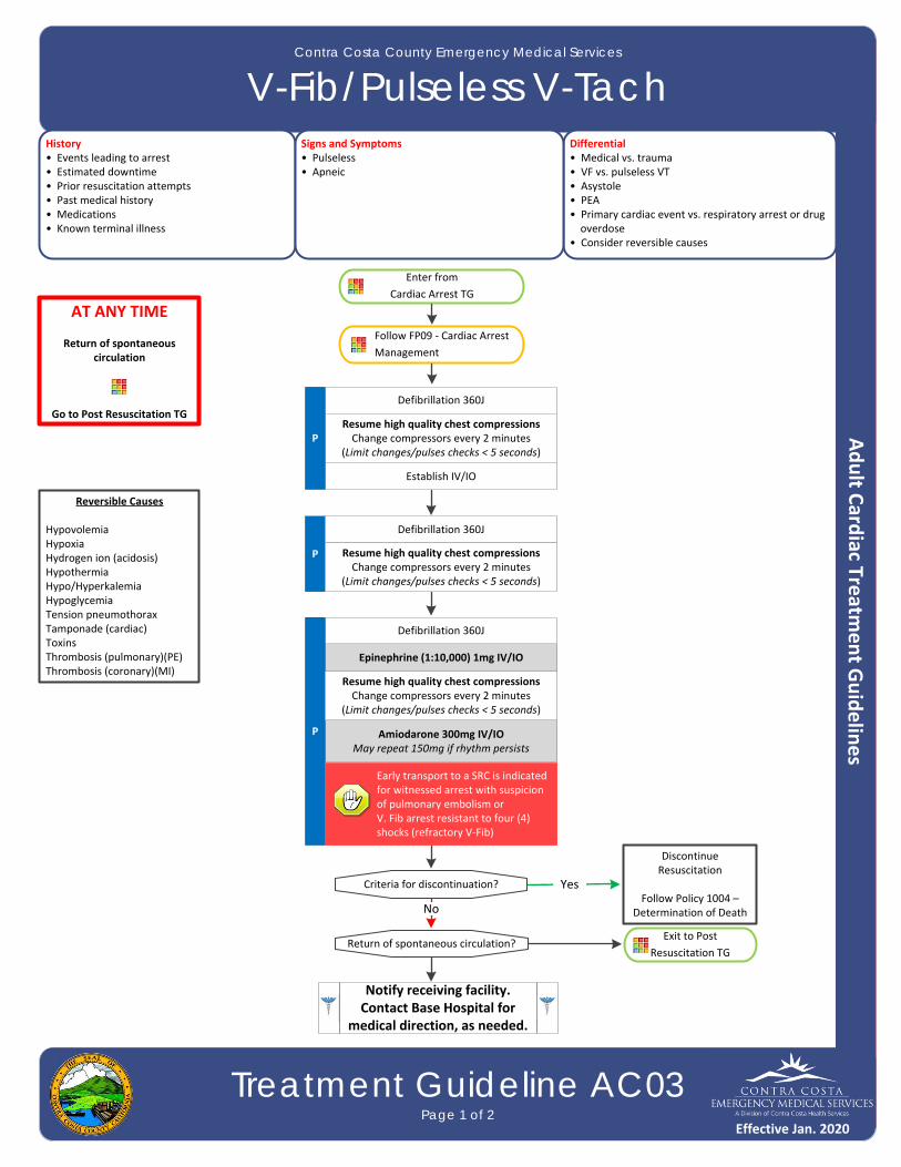
V-Fib/Pulseless V-Tach
Treatment Guideline AC03Page 1 of 2
History• Events leading to arrest• Estimated downtime• Prior resuscitation attempts• Past medical history• Medications• Known terminal illness
Differential• Medical vs. trauma• VF vs. pulseless VT • Asystole• PEA• Primary cardiac event vs. respiratory arrest or drug overdose• Consider reversible causes
Signs and Symptoms• Pulseless• Apneic
Enter from
Cardiac Arrest TG
Establish IV/IO
P
Epinephrine (1:10,000) 1mg IV/IO
Defibrillation 360J
AT ANY TIME
Return of spontaneous circulation
Go to Post Resuscitation TGResume high quality chest compressionsChange compressors every 2 minutes
(Limit changes/pulses checks < 5 seconds)P
Defibrillation 360J
Defibrillation 360J
Amiodarone 300mg IV/IOMay repeat 150mg if rhythm persists
Exit to Post
Resuscitation TG Return of spontaneous circulation?
Resume high quality chest compressionsChange compressors every 2 minutes
(Limit changes/pulses checks < 5 seconds)
P
Resume high quality chest compressionsChange compressors every 2 minutes
(Limit changes/pulses checks < 5 seconds)
Criteria for discontinuation?
DiscontinueResuscitation
Follow Policy 1004 – Determination of DeathNo
Yes
Reversible Causes
HypovolemiaHypoxiaHydrogen ion (acidosis)HypothermiaHypo/HyperkalemiaHypoglycemiaTension pneumothoraxTamponade (cardiac)ToxinsThrombosis (pulmonary)(PE)Thrombosis (coronary)(MI)
Early transport to a SRC is indicated for witnessed arrest with suspicion of pulmonary embolism or V. Fib arrest resistant to four (4) shocks (refractory V‐Fib)
Follow FP09 ‐ Cardiac Arrest
Management
Notify receiving facility. Contact Base Hospital for
medical direction, as needed.
Effective Jan. 2020
Effective Jan. 2016
Contra Costa County Emergency Medical Services
V-Fib/Pulseless V-Tach
Treatment Guideline AC03Page 2 of 2
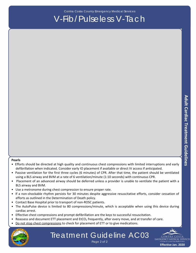
Pearls• Efforts should be directed at high quality and continuous chest compressions with limited interruptions and early
defibrillation when indicated. Consider early IO placement if available or direct IV access if anticipated. • Passive ventilation for the first three cycles (6 minutes) of CPR. After that time, the patient should be ventilated
using a BLS airway and BVM at a rate of 6 ventilation/minute (1:10 seconds) with continuous CPR. • Placement of an advanced airway should be deferred unless a provider is unable to ventilate the patient with a
BLS airway and BVM.• Use a metronome during chest compression to ensure proper rate.• If a non‐shockable rhythm persists for 30 minutes despite aggressive resuscitative efforts, consider cessation of
efforts as outlined in the Determination of Death policy.• Contact Base Hospital prior to transport of non‐ROSC patients.• The AutoPulse device is limited to 80 compressions/minute, which is acceptable when using this device during
cardiac arrest.• Effective chest compressions and prompt defibrillation are the keys to successful resuscitation.• Reassess and document ETT placement and EtCO2 frequently, after every move, and at transfer of care.• Do not stop chest compressions to check for placement of ETT or to give medications.
Effective Jan. 2020
Effective Jan. 2016
Contra Costa County Emergency Medical Services
Post Resuscitation (ROSC)
Treatment Guideline AC04Page 1 of 1
History• Respiratory arrest• Cardiac arrest
Differential• Continue to address specific differentials associated with the original dysrhythmia
Signs and Symptoms• Return of spontaneous circulation
Repeat primary assessment
E
Optimize ventilation and oxygenation Maintain SpO2 ≥ 94% Maintain respiratory rate between 6 –
10/minute for EtCO2 35 – 45 DO NOT HYPERVENTILATE
P
Establish IO/IV
Obtain 12‐Lead ECG
Monitor vital signs
Normal Saline bolus 500ml IV/IOMay repeat as needed if lungs are clear
Advanced airway placement, if indicated
Transport to STEMI Receiving Center
ApprovedSTEMI Receiving Centers
John Muir – ConcordJohn Muir – Walnut CreekKaiser – Walnut CreekSan Ramon Regional
Sutter DeltaHighland – OaklandKaiser ‐ OaklandKaiser – VallejoMarin General
Summit – OaklandValley Care ‐ Pleasanton
Notify receiving facility. Contact Base Hospital for
medical direction, as needed.
Systolic BP < 90Push Dose Epi 1 ml (10 mcg) IV/IO
every 3 minTitrate to a Systolic BP > 90
Cycle Blood Pressure every 3 minutes
Worsening bradycardia in ROSC patients may indicate impending
rearrest
1:1000 Epinephrine Mixing Instructions
NEED:1:1000 Epineprhine ampuletuberculin syringe10ml Normal Saline flush
1. Draw up 0.1ml (1 ml/mg) of 1:1000 Epi in the tuberculin syringe
2. Add the 1:1000 Epi from the tuberculin syringe into the Normal Saline flush ‐ mix gently
3. Now you have 10mL of Epinephrine at a 0.01mg/mL (10mcg/mL) concentration
4. Label the syringe
1:10,000 Epinephrine Mixing Instructions
NEED:1:10000 Epinephrine pre‐load10 ml Normal Saline flush
1. Waste 1 ml from Normal Saline Flush2. Draw 1 ml of 0.1mg/mL (Epi 1:10000) from
pre‐load into Normal Saline Flush – mix gently
3. Now you have 10 mL of Epinephrine at a 0.01mg/mL (10mcg/mL) concentration
4. Label the syringe
Effective Jan. 2021

This page intentionally left blank
Contra Costa County Emergency Medical Services
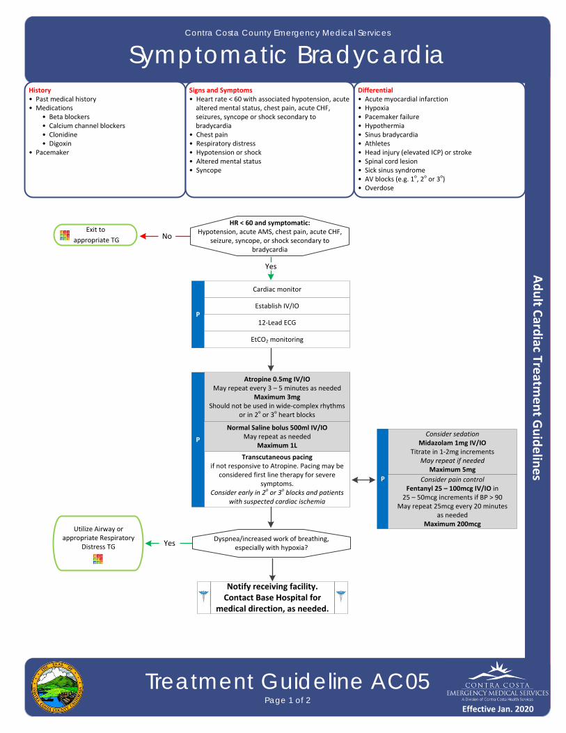
Symptomatic Bradycardia
Treatment Guideline AC05Page 1 of 2
History• Past medical history• Medications • Beta blockers • Calcium channel blockers • Clonidine • Digoxin• Pacemaker
Differential• Acute myocardial infarction• Hypoxia • Pacemaker failure• Hypothermia• Sinus bradycardia• Athletes• Head injury (elevated ICP) or stroke• Spinal cord lesion• Sick sinus syndrome• AV blocks (e.g. 1o, 2o or 3o)• Overdose
Signs and Symptoms• Heart rate < 60 with associated hypotension, acute altered mental status, chest pain, acute CHF, seizures, syncope or shock secondary to bradycardia• Chest pain• Respiratory distress• Hypotension or shock• Altered mental status• Syncope
HR < 60 and symptomatic:Hypotension, acute AMS, chest pain, acute CHF,
seizure, syncope, or shock secondary to bradycardia
Exit to
appropriate TG
Cardiac monitor
Establish IV/IOP
12‐Lead ECG
Transcutaneous pacingif not responsive to Atropine. Pacing may be
considered first line therapy for severe symptoms.
Consider early in 2o or 3o blocks and patients with suspected cardiac ischemia
No
Utilize Airway or appropriate Respiratory
Distress TG Dyspnea/increased work of breathing,
especially with hypoxia?
Yes
Yes
Atropine 0.5mg IV/IOMay repeat every 3 – 5 minutes as needed
Maximum 3mgShould not be used in wide‐complex rhythms
or in 2o or 3o heart blocks
P
Normal Saline bolus 500ml IV/IOMay repeat as needed
Maximum 1L
Consider sedationMidazolam 1mg IV/IO
Titrate in 1‐2mg incrementsMay repeat if needed
Maximum 5mg
P
EtCO2 monitoring
Consider pain control Fentanyl 25 – 100mcg IV/IO in
25 – 50mcg increments if BP > 90May repeat 25mcg every 20 minutes
as neededMaximum 200mcg
Notify receiving facility. Contact Base Hospital for
medical direction, as needed.
Effective Jan. 2020
Effective Jan. 2016
Contra Costa County Emergency Medical Services
Symptomatic Bradycardia
Treatment Guideline AC05Page 2 of 2
Pearls• Bradycardia causing symptoms is typically < 50/minutes. Rhythm should be interpreted in the context of symptoms and pharmacological treatment given only when symptomatic, otherwise monitor and reassess
frequently.• Identifying signs and symptoms of poor perfusion caused by bradycardia is paramount.• Atropine vs. pacing: Caution should be exercised in the setting of a suspected acute MI. The use of Atropine for
PVCs in the presence of an acute MI may worsen heart damage. Providers should NOT DELAY transcutaneous pacing for patients with poor perfusion in the setting of an acute MI or 2o or 3o heart block.
• For patients who are not in 2o or 3o heart block, pacing may be considered for bradycardia not responsive to Atropine. Prepare to utilize transcutaneous pacing early if the patient does not respond to Atropine.
• For wide complex, bizarre appearance of QRS complexes with slow rhythm, consider hyperkalemia.• Consider treatable causes for bradycardia (e.g. beta blocker OD, calcium channel blocker OD, etc.)• Hypoxemia is a common cause of bradycardia. Be sure to oxygenate the patient and support respiratory effort.• Sinus bradycardia in the absence of key symptoms requires no specific treatment; monitor and observe.• Sinus bradycardia is often seen in patients with STEMI or ischemia. An early 12‐Lead ECG should be obtained to
assess for STEMI.• A fluid bolus may address hypotension and lessen the need for pacing or treatment with Atropine.• Sedation prior to starting pacing is not required. Patients with urgent needs should be paced first and sedated
afterwards.• The objective of sedation with pacing is to decrease discomfort, not to decrease level of consciousness. Patients
who are in need of pacing are unstable and sedation should be used with extreme caution.• Monitor respiratory status closely and support ventilation as necessary.• Atropine is not effective for bradycardia in heart transplant patients as there is no vagus nerve innervation in
these patients.• Patients with wide QRS or 2o or 3o heart blocks will not have a response to Atropine because they heart rates are
not based on vagal tone. An increase in ventricular arrhythmias may occur.
Effective Jan. 2020

Contra Costa County Emergency Medical Services
Narrow Complex Tachycardia
Treatment Guideline AC06Page 1 of 2
History• Medications (e.g. Aminophylline, Adderall, diet pills, thyroid supplements, decongestants, and Digoxin)• Diet• Drugs (e.g. nicotine and illegal drugs)• Past medical history• History of palpations/heart racing• Syncope/near syncope
Differential• Heart disease (e.g. WPW or valvular)• Sick sinus syndrome • Myocardial infarction• Electrolyte imbalance• Exertion, pain, or emotional stress• Fever• Hypoxia• Hypovolemia or anemia• Drug effect/overdose (see History)• Hypothyroidism• Pulmonary embolus
Signs and Symptoms• Heart rate > 150 with narrow, regular complexes• Systolic BP < 90• Dizziness, chest pain, shortness of breath, altered mental status, or diaphoresis• CHF• Potential presenting rhythm: • Atrial/sinus tachycardia • Atrial fibrillation/flutter • Multifocal atrial tachycardia • Ventricular tachycardia
Assess symptom severity
Cardiac monitor
Consider IV/IO
P 12‐Lead ECG
Regular rhythm (SVT)(QRS ≤ 0.09 sec)
Irregular rhythm(A‐Fib/A‐Flutter)
P
Adenosine 6mg IV/IO rapid push
May repeat 12mg IV/IO
EtCO2 monitoring
Attempt Valsalva maneuver
Consider sedation pre‐cardioversionMidazolam 1mg IV/IO
May repeat if needed in 1‐2mg incrementsMaximum 5mg
Cardiac monitor
Establish IV/IO
P
EtCO2 monitoring
StableUnstable
(HR typically > 150)
If rhythm change, repeat 12‐Lead ECG
Regular rhythm (SVT)(QRS ≤ 0.09 sec)
Irregular rhythm(A‐Fib/A‐Flutter)
P
Synchronized cardioversion 100J
May repeat in escalating energy (200J, 300J, 360J)
12‐Lead ECGor
repeat if rhythm change
P
Synchronized cardioversion 200J
May repeat in escalating energy (300J, 360J)
12‐Lead ECGor
repeat if rhythm change
Notify receiving facility. Contact Base Hospital for
medical direction, as needed.
Effective Jan. 2020
Effective Jan. 2016
Contra Costa County Emergency Medical Services
Narrow Complex Tachycardia
Treatment Guideline AC06Page 2 of 2
Pearls• Most important goal is to differentiate the type of tachycardia and if STABLE or UNSTABLE.• If at any point the patient becomes unstable, move to the unstable arm of the algorithm.• For ASYMPTOMATIC patients (or those with only minimal symptoms, such as palpitations) and any tachycardia
with a rate of approximately 100 – 120 with a normal blood pressure, consider CLOSE OBSERVATION or fluid bolus rather than immediate treatment with an anti‐arrhythmic medication. For example, a patient’s “usual” atrial fibrillation may not require emergent treatment.
• All Adenosine administrations should be immediately followed by a 20ml rapid flush.• Typical sinus tachycardia is in the range of 100 to (200 – the patient’s age) beats per minute.• Symptomatic tachycardia usually occurs at rates of 120 – 150 and typically ≥ 150 beats per minute. Patients who are symptomatic with heart rates < 150 likely have impaired cardiac function, such as CHF.• Serious Signs / Symptoms include: Hypotension; acutely altered mental status; signs of shock/poor perfusion;
chest pain with evidence of ischemia (e.g. STEMI, T‐wave inversions or depressions); and acute CHF.• Search for underlying cause of tachycardia such as fever, sepsis, dyspnea, etc.• If patient has a history or if 12‐Lead ECG reveals Wolfe Parkinson White (WPW), use caution with Adenosine and give only with a defibrillator immediately available.• Regular Narrow‐Complex Tachycardias: • Adenosine may be considered to assist with rhythm diagnosis or if a patient has a history of Adenosine conversion. • Adenosine may not be effective with atrial fibrillation/flutter, yet is not harmful and may help identify rhythm.• Synchronized Cardioversion is recommended to treat UNSTABLE atrial fibrillation/flutter and monomorphic‐
regular tachycardia (SVT).• Monitor for respiratory depression and hypotension associated with Midazolam.• Continue pulse oximetry and EtCO2 monitoring is required for all narrow complex tachycardia patients. • Providers must export all monitor data to EHR when caring for and treating narrow complex tachycardia patients.
Effective Jan. 2020
Effective Jan. 2016
Contra Costa County Emergency Medical Services
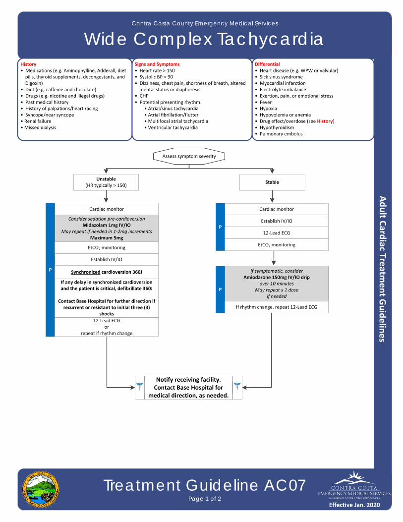
Wide Complex Tachycardia
Treatment Guideline AC07Page 1 of 2
History• Medications (e.g. Aminophylline, Adderall, diet pills, thyroid supplements, decongestants, and Digoxin)• Diet (e.g. caffeine and chocolate)• Drugs (e.g. nicotine and illegal drugs)• Past medical history• History of palpations/heart racing• Syncope/near syncope• Renal failure• Missed dialysis
Differential• Heart disease (e.g. WPW or valvular)• Sick sinus syndrome • Myocardial infarction• Electrolyte imbalance• Exertion, pain, or emotional stress• Fever• Hypoxia• Hypovolemia or anemia• Drug effect/overdose (see History)• Hypothyroidism• Pulmonary embolus
Signs and Symptoms• Heart rate > 150• Systolic BP < 90• Dizziness, chest pain, shortness of breath, altered mental status or diaphoresis• CHF• Potential presenting rhythm: • Atrial/sinus tachycardia • Atrial fibrillation/flutter • Multifocal atrial tachycardia • Ventricular tachycardia
Cardiac monitor
Establish IV/IOP
12‐Lead ECG
If symptomatic, considerAmiodarone 150mg IV/IO drip
over 10 minutesMay repeat x 1 dose
if needed
P
EtCO2 monitoring
Assess symptom severity
StableUnstable
(HR typically > 150)
If rhythm change, repeat 12‐Lead ECG
Consider sedation pre‐cardioversionMidazolam 1mg IV/IO
May repeat if needed in 1‐2mg incrementsMaximum 5mg
Cardiac monitor
Establish IV/IO
P
EtCO2 monitoring
Synchronized cardioversion 360J
12‐Lead ECGor
repeat if rhythm change
If any delay in synchronized cardioversion and the patient is critical, defibrillate 360J
Contact Base Hospital for further direction if recurrent or resistant to initial three (3)
shocks
Notify receiving facility. Contact Base Hospital for
medical direction, as needed.
Effective Jan. 2020
Effective Jan. 2016
Contra Costa County Emergency Medical Services
Wide Complex Tachycardia
Treatment Guideline AC07Page 2 of 2
Pearls• Most important goal is to differentiate the type of tachycardia and if STABLE or UNSTABLE.• If at any point the patient becomes unstable, move to the unstable arm of the algorithm.• For ASYMPTOMATIC patients (or those with only minimal symptoms, such as palpitations) and any tachycardia with
a rate of approximately 100 – 120 with a normal blood pressure, consider CLOSE OBSERVATION or fluid bolus rather than immediate treatment with an anti‐arrhythmic medication. For example, a patient’s “usual” atrial fibrillation may not require emergent treatment.
• In unstable patients with fever or other signs of sepsis, the underlying cause of the rapid heart rate is more likely fever and hypovolemia. This is particularly true in wide irregular tachycardia which is frequently underlying A fib with a bundle branch block. Initial efforts should focus on treating appropriately for underlying sepsis.
• In Polymorphic VT: (torsade de pointes) etc., the variation in QRS morphology may make it difficult to synchronize. If you cannot synchronize, move to defibrillation.
• In wide irregular rhythm such as A Fib with left or right bundle etc, synchronized cardioversion at 360J is more likely to yield first shock conversion.
• Typical sinus tachycardia is in the range of 100 to (200 – the patient’s age) beats per minute.• Symptomatic tachycardia usually occurs at rates of 120 – 150 and typically ≥ 150 beats per minute. Patients who are symptomatic with heart rates < 150 likely have impaired cardiac function, such as CHF.• Serious Signs/Symptoms include: Hypotension; acutely altered mental status; signs of shock/poor perfusion; chest
pain with evidence of ischemia (e.g. STEMI, T‐wave inversions, or depressions); and acute CHF.• Search for underlying cause of tachycardia such as fever, sepsis, dyspnea, etc.• Monitor for respiratory depression and hypotension associated with Midazolam.• Continue pulse oximetry and EtCO2 monitoring is required for all wide complex tachycardia patients. • Providers must export all monitor data to EHR when caring for and treating wide complex tachycardia patients.
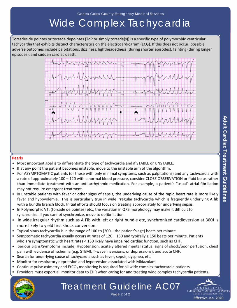
Torsades de pointes or torsade depointes (TdP or simply torsade(s)) is a specific type of polymorphic ventricular tachycardia that exhibits distinct characteristics on the electrocardiogram (ECG). If this does not occur, possible adverse outcomes include palpitations, dizziness, lightheadedness (during shorter episodes), fainting (during longer episodes), and sudden cardiac death.
Effective Jan. 2020
Effective Jan. 2016
Contra Costa County Emergency Medical Services
Chest Pain: Suspected Cardiac or STEMI
Treatment Guideline AC08Page 1 of 2
History• Age• Medications (e.g. Viagra, Sildenafil, Levitra, Vardenafil, Cialis or Tadalafil)• Past medical history (e.g. MI, angina, diabetes, or post menopausal)• Allergies• Recent physical exertion• Provocation• Quality (e.g. pressure, constant, sharp, dull, etc.)• Region/Radiation/Referred• Severity (0 – 10 scale)• Time (onset/duration/repetition)
Differential• Acute myocardial infarction• Hypoxia • Pacemaker failure• Hypothermia• Sinus bradycardia• Athletes• Head injury (elevated ICP) or stroke• Spinal cord lesion• Sick sinus syndrome• AV blocks (e.g. 1o, 2o, or 3o)• Overdose
Signs and Symptoms• Heart rate < 60 with associated hypotension, acute altered mental status, chest pain, acute CHF, seizures, syncope, or shock secondary to bradycardia• Chest pain• Respiratory distress• Hypotension or shock• Altered mental status• Syncope
Cardiac monitor
Aspirin 324mg PO
P
Chest pain?Signs/symptoms consistent with
cardiac etiology?
ConsiderFentanyl 25 – 200mcg IV titrated in
25 – 50mcg increments for pain relief if BP > 90 systolic
12‐Lead ECG
Acute MI/STEMI(STEMI = 1mm ST segment elevation ≥
2 contiguous leads; See 12‐Lead Procedure)
Establish IV/IO
P
Nitroglycerin 0.4mg sublingualif systolic BP is > 90
May repeat ever 5 minutes until pain subsides.
Max 3 doses
Yes
No
Declare a STEMI Alert
Fentanyl Contact Base Hospital for additional
orderN
ApprovedSTEMI Receiving Centers
John Muir – ConcordJohn Muir – Walnut CreekKaiser – Walnut CreekSan Ramon Regional
Sutter DeltaHighland – OaklandKaiser – VallejoMarin General
Summit – OaklandKaiser – Oakland
Valley Care – Pleasanton
Yes
If indicated,Normal Saline bolus 500ml IV/IO
May repeat as needed Maximum 1L
ConsiderFentanyl 25 – 200mcg IV titrated in
25 – 50mcg increments for pain relief if BP > 90 systolic
Establish IV/IO
P
If indicated,Normal Saline bolus 500ml IV/IO
May repeat as needed Maximum 1L
Notify receiving facility. Contact Base Hospital for
medical direction, as needed.
Effective Jan. 2021
Effective Jan. 2016
Contra Costa County Emergency Medical Services
Chest Pain: Suspected Cardiac or STEMI
Treatment Guideline AC08Page 2 of 2
Pearls• Avoid Nitroglycerin in any patient who has used Viagra (Sildenafil) or Levitra (Vardenafil) in the past 24 hours or
Cialis (Tadalafil) in the past 36 hours due to the potential of severe hypotension.• Patients with a STEMI should be transported to the closest most appropriate STEMI receiving center.• Many STEMIs evolve during prehospital care and may not be noted on the initial 12‐Lead ECG.• An ECG should be obtained prior to treatment for bradycardia if patient condition permits.• Transmit all 12‐Lead ECGs whether STEMI is detected or not.• If a patient has taken their own Nitroglycerin without relief, consider potency of medication. Provider maximum
doses do not include patient administered doses.• Monitor for hypotension after administration of nitroglycerin and opioids.• Diabetics, geriatric, and female patients often have atypical pain, or only generalized complaints. Suspect cardiac
etiology in these patients, and perform a 12‐Lead ECG.• Document the time of the 12‐Lead ECG in the EHR as a procedure along with the interpretation.
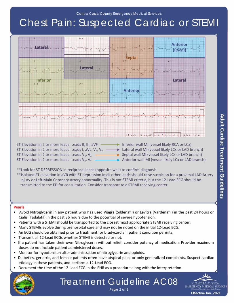
ST Elevation in 2 or more leads: Leads II, III, aVF Inferior wall MI (vessel likely RCA or LCx) ST Elevation in 2 or more leads: Leads I, aVL, V5, V6 Lateral wall MI (vessel likely LCx or LAD branch) ST Elevation in 2 or more leads: Leads V1, V2 Septal wall MI (vessel likely LCx or LAD branch) ST Elevation in 2 or more leads: Leads V3, V4 Anterior wall MI (vessel likely LCx or LAD branch)
**Look for ST DEPRESSION in reciprocal leads (opposite wall) to confirm diagnosis. **Isolated ST elevation in aVR with ST depression in all other leads should raise suspicion for a proximal LAD Artery injury or Left Main Coronary Artery abnormality. This is not STEMI criteria, but the 12‐Lead ECG should be transmitted to the ED for consultation. Consider transport to a STEMI receiving center.
Lateral
LateralInferior
Lateral
Septal
Anterior
Anterior(RVMI)
Effective Jan. 2021

No
Yes
Effective Jan. 2016
Contra Costa County Emergency Medical Services
Abdominal Pain
Treatment Guideline A01Page 1 of 2
History• Age• Past medical/surgical history• Medications • Onset• Provocation• Quality (e.g. crampy, constant, sharp, dull, etc.)• Region / radiation/referred• Severity (0 – 10 scale)• Time (duration/repetition)• Fever• Last meal eaten• Last bowel movement/emesis• Menstrual history (pregnancy)
Differential• Pneumonia or pulmonary embolus• Liver (hepatitis) • Peptic ulcer disease/gastritis• Gallbladder• MI• Pancreatitis• Kidney stone• Abdominal aneurysm• Appendicitis• Bladder/prostate disorder• Pelvic (PID, ectopic pregnancy, or ovarian cyst)• Spleen enlargement• Diverticulitis• Bowel obstruction• Gastroenteritis (infectious)• Ovarian or testicular torsion
Signs and Symptoms• Pain (location/migration)• Tenderness• Nausea• Vomiting• Diarrhea• Dysuria (painful or difficult urination)• Constipation• Vaginal bleeding/discharge• Pregnancy
Associated symptoms: (Helpful to localize source)Fever, headache, weakness, malaise, myalgia, cough, headache, mental status change, or rash
P
P
If patient has nausea or vomitingOndansetron 4mg IV/IO/IM/ODT
May repeat x 1 after 15 minutes
Normal Saline bolus 500mlRepeat as needed
Titrate to systolic BP > 90Maximum 1L
Establish IV/IO
Exit to Pain Control TG
if indicated
P
Establish IV/IO
Cardiac monitor
Cardiac monitor
Exit to Appropriate Cardiac TG
Hypotensive, Signs and Symptoms of shock?
Signs/symptomssuggesting cardiac
Etiology?
Consider 12‐Lead ECG
Assess symptom severity
StableUnstable
(Hypotension/poor perfusion)
Notify receiving facility. Contact Base Hospital for
medical direction, as needed.
Exit to Hypotension/Shock TG
if indicated
Effective Jan. 2021

Effective Jan. 2016
Contra Costa County Emergency Medical Services
Abdominal Pain
Treatment Guideline A01Page 2 of 2
Pearls• Diabetic, females, and geriatric patients often have atypical pain, or only generalized complaints. Suspect cardiac
etiology in these patients, perform a 12‐Lead ECG, and investigate until proven otherwise.• For chronic abdominal pain, consider non‐narcotic pain control.• Zofran is not indicated or useful for motion sickness.• Document the mental status and vital signs prior to administration of anti‐emetics.• Abdominal pain in women of childbearing age should be treated as pregnancy‐related until proven otherwise.• An impression of abdominal aneurysm should be considered with severe abdominal or non‐traumatic back pain,
especially in patients > 50 years of age or patients with shock/poor perfusion.
Effective Jan. 2021
Effective Jan. 2016
Contra Costa County Emergency Medical Services
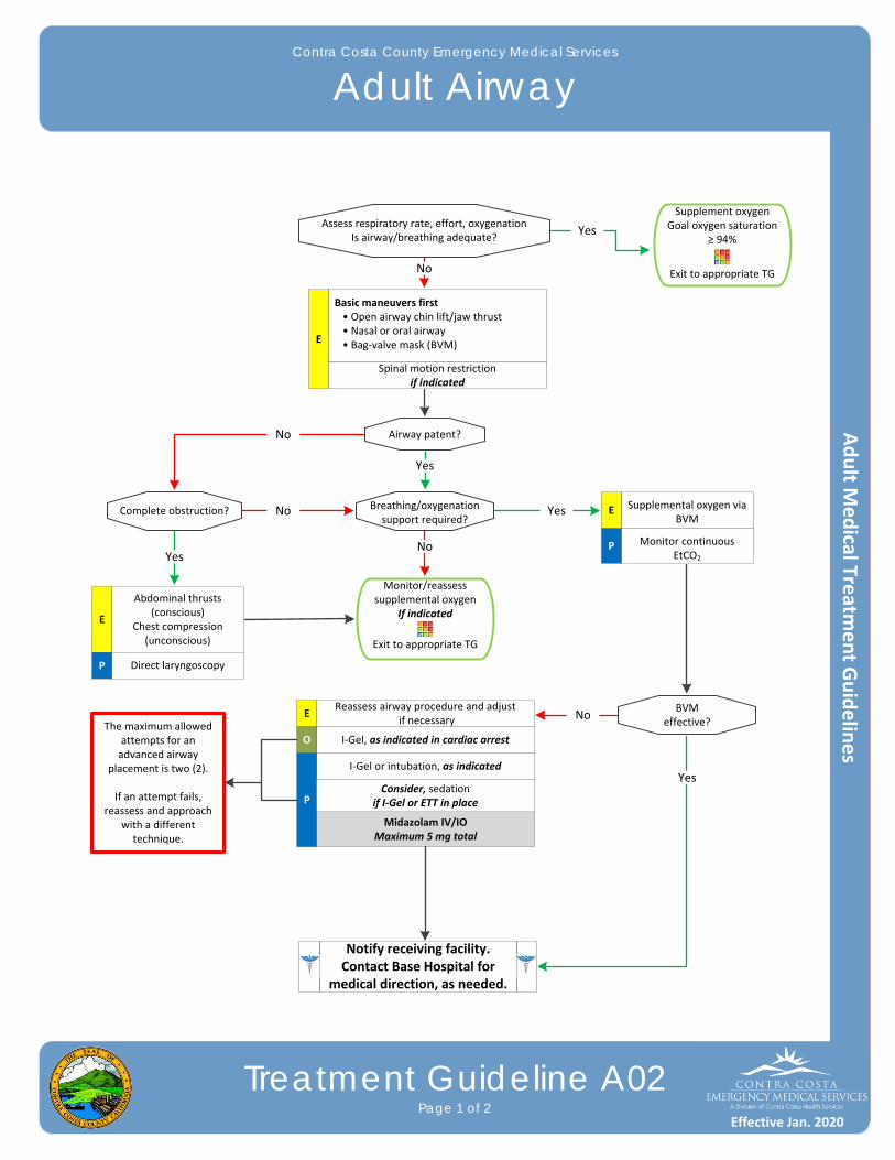
Adult Airway
Treatment Guideline A02Page 1 of 2
Basic maneuvers first • Open airway chin lift/jaw thrust • Nasal or oral airway • Bag‐valve mask (BVM)E
Spinal motion restrictionif indicated
Yes
Supplement oxygenGoal oxygen saturation
≥ 94%
Exit to appropriate TG
Monitor/reassess supplemental oxygen
If indicated
Exit to appropriate TG
Abdominal thrusts (conscious)
Chest compression (unconscious)
E
Direct laryngoscopyP
Supplemental oxygen via BVM
E
The maximum allowed attempts for an advanced airway
placement is two (2).
If an attempt fails, reassess and approach
with a different technique.
No
No
No
Yes
Yes
NoYes
Yes
No
Assess respiratory rate, effort, oxygenationIs airway/breathing adequate?
Airway patent?
Breathing/oxygenationsupport required?
Complete obstruction?
BVM effective?
Monitor continuous EtCO2
P
Reassess airway procedure and adjust if necessary
E
I‐Gel, as indicated in cardiac arrestO
I‐Gel or intubation, as indicated
PConsider, sedation
if I‐Gel or ETT in place
Midazolam IV/IOMaximum 5 mg total
Notify receiving facility. Contact Base Hospital for
medical direction, as needed.
Effective Jan. 2020
Effective Jan. 2016
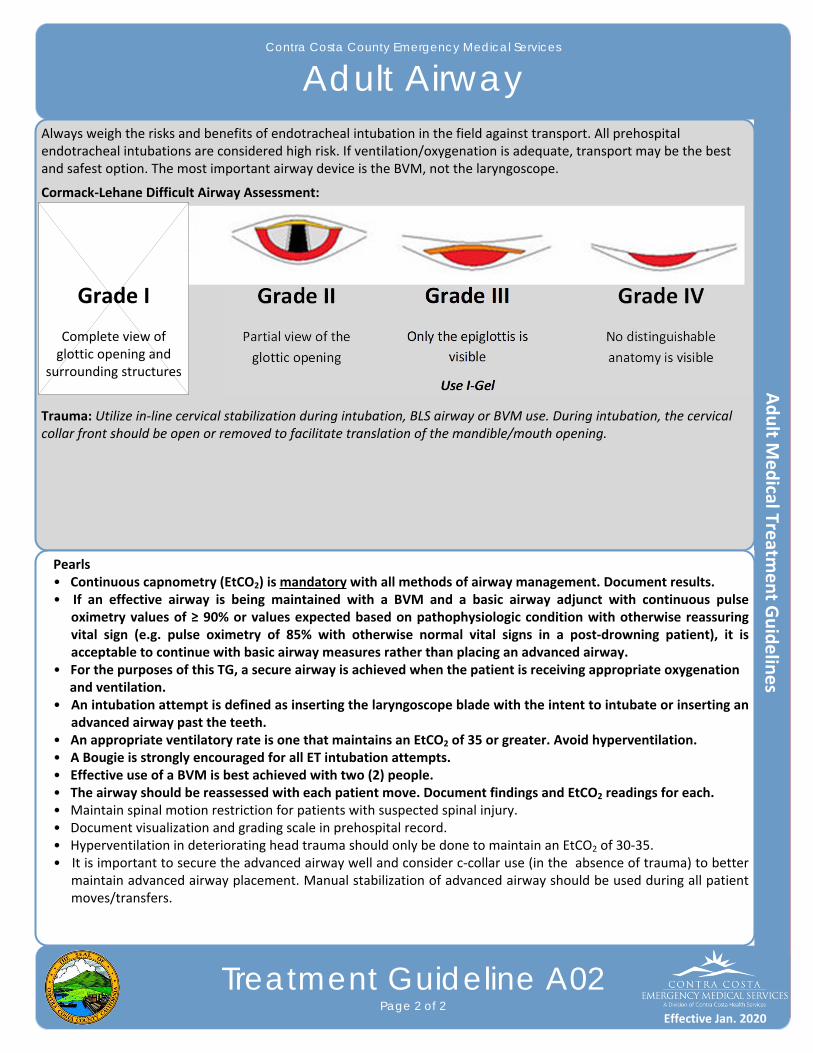
Always weigh the risks and benefits of endotracheal intubation in the field against transport. All prehospital endotracheal intubations are considered high risk. If ventilation/oxygenation is adequate, transport may be the best and safest option. The most important airway device is the BVM, not the laryngoscope.
Cormack‐Lehane Difficult Airway Assessment:
Trauma: Utilize in‐line cervical stabilization during intubation, BLS airway or BVM use. During intubation, the cervical collar front should be open or removed to facilitate translation of the mandible/mouth opening.
Contra Costa County Emergency Medical Services
Adult Airway
Treatment Guideline A02Page 2 of 2
Pearls• Continuous capnometry (EtCO2) is mandatory with all methods of airway management. Document results.• If an effective airway is being maintained with a BVM and a basic airway adjunct with continuous pulse
oximetry values of ≥ 90% or values expected based on pathophysiologic condition with otherwise reassuring vital sign (e.g. pulse oximetry of 85% with otherwise normal vital signs in a post‐drowning patient), it is acceptable to continue with basic airway measures rather than placing an advanced airway.
• For the purposes of this TG, a secure airway is achieved when the patient is receiving appropriate oxygenation and ventilation.• An intubation attempt is defined as inserting the laryngoscope blade with the intent to intubate or inserting an
advanced airway past the teeth.• An appropriate ventilatory rate is one that maintains an EtCO2 of 35 or greater. Avoid hyperventilation.• A Bougie is strongly encouraged for all ET intubation attempts.• Effective use of a BVM is best achieved with two (2) people.• The airway should be reassessed with each patient move. Document findings and EtCO2 readings for each.• Maintain spinal motion restriction for patients with suspected spinal injury.• Document visualization and grading scale in prehospital record.• Hyperventilation in deteriorating head trauma should only be done to maintain an EtCO2 of 30‐35.• It is important to secure the advanced airway well and consider c‐collar use (in the absence of trauma) to better
maintain advanced airway placement. Manual stabilization of advanced airway should be used during all patient moves/transfers.
Grade III
Only the epiglottis is
visible
Use I‐Gel
Grade I
Complete view of glottic opening and
surrounding structures
Effective Jan. 2020
Effective Jan. 2016
Contra Costa County Emergency Medical Services
Adult Behavioral
Treatment Guideline A03Page 1 of 2
History• Situational crisis• Psychiatric illness/medications• Injury to self or threats to others• Medical alert tag• Substance abuse/overdose• Diabetes
Differential• Altered mental status• Alcohol intoxication • Toxin / substance abuse• Medication effect/overdose• Withdrawal symptoms• Depression • Bipolar (manic‐depressive)• Schizophrenia• Anxiety disorders• Hypoglycemia
Signs and Symptoms• Anxiety, agitation or confusion• Affect change or hallucinations• Delusional thoughts or bizarre behavior• Combative or violent • Expression of suicidal/homicidal thoughts
Exit to appropriate TG, if indicated
Altered Mental Status TGOverdose/Toxic Ingestion TG
Head Trauma TG
Assume patient has medical cause of
behavioral change
Consider IV
E
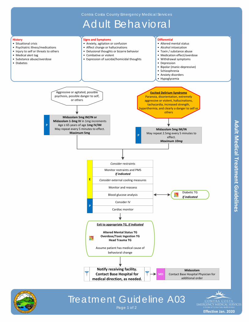
Excited Delirium SyndromeParanoia, disorientation, extremely aggressive or violent, hallucinations, tachycardia, increased strength,
hyperthermia, and clearly a danger to self or others
Consider restraints
Monitor restraints and PMSif indicated
P
Midazolam 5mg IM/INMay repeat 2.5mg every 5 minutes to
effect. Maximum 10mg
Diabetic TG
if indicated
Monitor and reassess
Aggressive or agitated, possible psychosis, possible danger to self,
or others
Blood glucose analysis
P
Midazolam 5mg IM/IN orMidazolam 1‐3mg IV in 1mg increments
Age ≥ 65 years of age 1mg IV/IM May repeat every 5 minutes to effect.
Maximum 5mg
Midazolam Contact Base Hospital Physician for
additional orderMD
Cardiac monitor
P
Consider external cooling measures
Notify receiving facility. Contact Base Hospital for
medical direction, as needed.
Effective Jan. 2020
Effective Jan. 2016
Contra Costa County Emergency Medical Services
Adult Behavioral
Treatment Guideline A03Page 2 of 2
Pearls• Crew/responder safety is the main priority. See Policy 1008 – Managing Assaultive Behavior/Patient Restraint.• Avoid using benzodiazepines for patients with alcohol intoxication.• Limit IN administrations to ½ dose in each nare.• All patients who receive either physical restraint or chemical sedation must be continuously observed by EMS
personnel. This includes direct visualization of the patient as well as cardiac and pulse oximetry monitoring.• Consider all possible medical/trauma causes for behavior (e.g. hypoglycemia, overdose, substance abuse, hypoxia, seizure, head injury, etc.).• Use caution when considering the use of Midazolam with postictal patients. • Do not irritate the patient with a prolonged exam. Be thorough but quick.• Do not overlook the possibility of associated domestic violence or child abuse.• If patient suspected of excited delirium and suffers cardiac arrest, consider fluid bolus and sodium bicarbonate early.• Do not position or transport any restrained patient in a way that negatively affects the patient’s respiratory or circulatory status (e.g. hog‐tied or prone positions). Do not place backboards, splints or other devices on top of
the patient.• If restrained, the extremities that are restrained will have a circulation check at least every 15 minutes. The first
of these checks should occur as soon after placement of the restraints as possible. This shall be documented in the EHR.
Excited Delirium Syndrome:
This is a medical emergency. The condition is a combination of delirium, psychomotor agitation, anxiety, hallucinations, speech disturbances, disorientation, violent/bizarre behavior, insensitivity to pain, hyperthermia and increased strength. The condition is life‐threatening and is often associated with use of physical control measures, including physical restraints, and tasers. Most commonly seen in male patients with a history of serious mental illness or drug abuse, particularly stimulant drugs such as cocaine, crack cocaine, methamphetamine, amphetamines, bath salts, or similar agents. Alcohol withdrawal or head injury may also contribute to the condition.
Effective Jan. 2020
Effective Jan. 2016
Contra Costa County Emergency Medical Services
Allergic Reaction/Anaphylaxis
Treatment Guideline A04Page 1 of 2
History• Onset and location• Insect sting or bite• Food allergy / exposure • Medication allergy / exposure• New clothing, soap or detergent• Past history of reactions• Past medical history• Medication history
Differential• Urticaria (rash only)• Anaphylaxis (systemic effect)• Shock (vascular effect)• Angioedema (drug induced)• Aspiration or airway obstruction• Vasovagal event• Asthma or COPD• CHF
Signs and Symptoms• Itching or hives• Coughing, wheezing or respiratory distress• Chest or throat restriction• Difficulty swallowing• Hypotension or shock• Edema• Nausea or vomiting• Feeling of impending doom
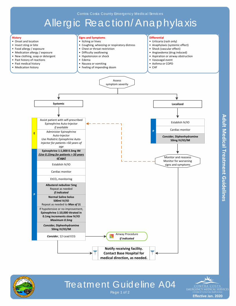
Assess symptom severity
P
Establish IV/IO
Cardiac monitor
Monitor and reassessMonitor for worsening signs and symptoms
Airway Procedure
if indicated
LocalizedSystemic
Consider, Diphenhydramine 50mg IV/IO/IM
If hypotensive or no improvement,Epinephrine 1:10,000 titrated in 0.1mg increments slow IV/IO
Maximum 0.5mg
P
Establish IV/IO
Cardiac monitor
Consider, 12‐Lead ECG
Assist patient with self‐prescribed Epinephrine Auto‐Injector
if available
E
Albuterol nebulizer 5mgRepeat as needed
if indicated
Normal Saline bolus 500ml IV/IO
Repeat as needed to Max of 1L
EtCO2 monitoring
Epinephrine 1:1,000 0.3mg IM(Use 0.15mg for patients > 50 years
of age)
Administer Epinephrine Auto‐Injector
Use Pediatric Epinephrine Auto‐Injector for patients >50 years of
age
Consider, Diphenhydramine 50mg IV/IO/IM
Notify receiving facility. Contact Base Hospital for
medical direction, as needed.
Effective Jan. 2020
Effective Jan. 2016
Contra Costa County Emergency Medical Services
Allergic Reaction/Anaphylaxis
Treatment Guideline A04Page 2 of 2
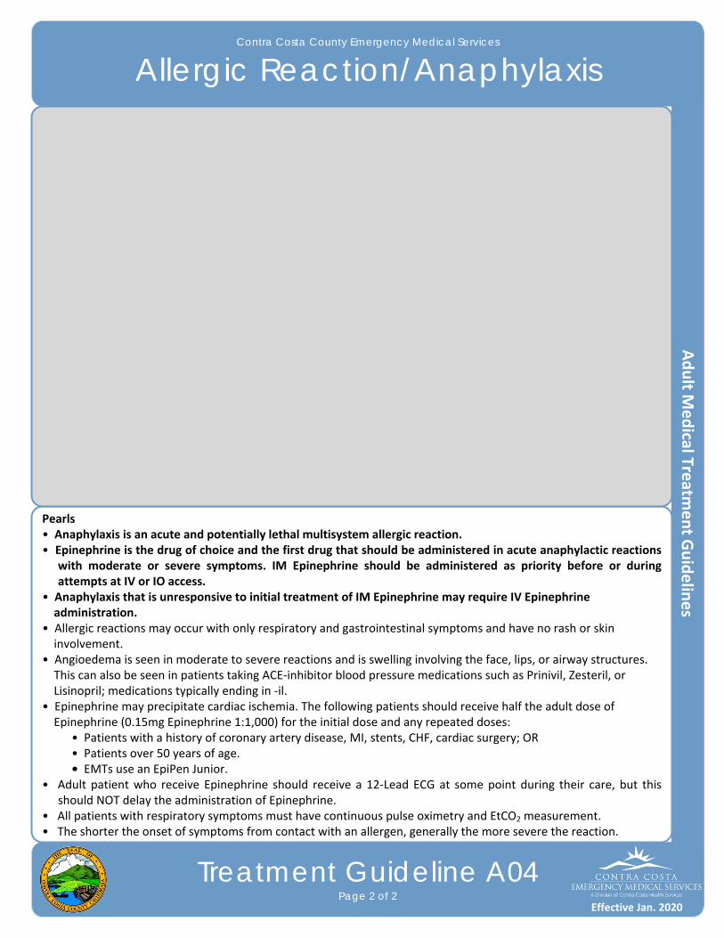
Pearls• Anaphylaxis is an acute and potentially lethal multisystem allergic reaction. • Epinephrine is the drug of choice and the first drug that should be administered in acute anaphylactic reactions
with moderate or severe symptoms. IM Epinephrine should be administered as priority before or during attempts at IV or IO access.
• Anaphylaxis that is unresponsive to initial treatment of IM Epinephrine may require IV Epinephrine administration.• Allergic reactions may occur with only respiratory and gastrointestinal symptoms and have no rash or skin involvement.• Angioedema is seen in moderate to severe reactions and is swelling involving the face, lips, or airway structures. This can also be seen in patients taking ACE‐inhibitor blood pressure medications such as Prinivil, Zesteril, or Lisinopril; medications typically ending in ‐il.• Epinephrine may precipitate cardiac ischemia. The following patients should receive half the adult dose of Epinephrine (0.15mg Epinephrine 1:1,000) for the initial dose and any repeated doses: • Patients with a history of coronary artery disease, MI, stents, CHF, cardiac surgery; OR • Patients over 50 years of age. • EMTs use an EpiPen Junior.• Adult patient who receive Epinephrine should receive a 12‐Lead ECG at some point during their care, but this
should NOT delay the administration of Epinephrine.• All patients with respiratory symptoms must have continuous pulse oximetry and EtCO2 measurement.• The shorter the onset of symptoms from contact with an allergen, generally the more severe the reaction.
Effective Jan. 2020

Effective Jan. 2016
Contra Costa County Emergency Medical Services
Altered Mental Status
Treatment Guideline A05Page 1 of 2
History• Known diabetic or medical alert tag• Drugs or drug paraphernalia• Report of illicit drug use or toxic ingestion • Past medical history• Medications• History of trauma or traumatic brain injury• Change in condition• Changes in feeding or sleep habits
Differential• Head trauma• CNS (stroke, tumor, seizure, infection)• Cardiac (MI, CHF)• Hypothermia• Infection or sepsis• Thyroid• Shock• Diabetes• Toxicological or ingestion • Acidosis or alkalosis• Environmental exposure• Hypoxia• Electrolyte abnormality• Psychiatric disorder• Sepsis
Signs and Symptoms• Change in baseline mental status• Decrease mental status or lethargy• Bizarre behavior• Hypoglycemia (cool, diaphoretic skin)• Hyperglycemia (warm, dry skin; fruity breath; Kussmaul respirations; signs of dehydration)• Irritability
P
Blood glucose analysis
Cardiac monitor
Blood glucose ≤ 60 or ≥ 350Exit to
Diabetic TG
12‐Lead ECG
Airway TG, if indicated
IV/IO Procedure
Signs of shock/poor perfusion
Signs of OD/toxic exposure
Signs of stroke or seizure
Signs of hypo/hyperthermia
Signs of sepsis
No
No
No
No
Exit to
Hypotension/Shock TG
Exit to Overdose/Toxic
Exposure TG
Exit to Stroke or Seizure TG
as indicated
Exit to Hypo / Hyperthermia TG
as indicated
Exit to Suspected Sepsis TG
as indicated
Yes
Yes
Yes
Yes
Yes
Yes
No
Arrhythmia/STEMI
No
Exit to Appropriate Cardiac TG
as indicatedYes
No
E
Notify receiving facility. Contact Base Hospital for
medical direction, as needed.
Effective Jan. 2020

Contra Costa County Emergency Medical Services
Altered Mental Status
Treatment Guideline A05Page 2 of 2
Pearls• Pay careful attention to the head exam for signs of bruising or other injury. • Be aware of AMS as a presenting sign of an environmental toxin or Haz‐mat exposure and protect personal safety and that of other responders who may already be exposed.• It is safer to assume hypoglycemia than hyperglycemia if doubt exists. Recheck blood glucose after D‐10 or Glucagon administration.• Do not let alcohol confuse the clinical picture. Alcoholics frequently develop hypoglycemia and may have
unrecognized injuries.• Consider restraints if necessary for patient or personnel protection.
Effective Jan. 2020
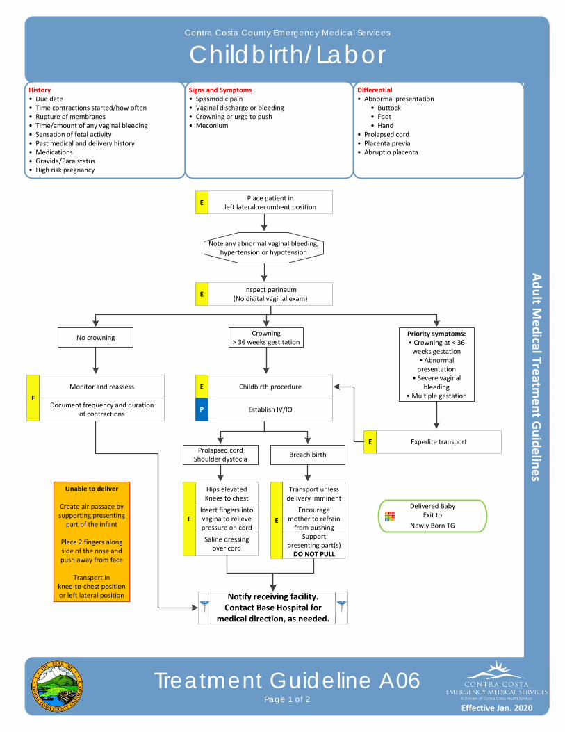
Effective Jan. 2016
Contra Costa County Emergency Medical Services
Childbirth/Labor
Treatment Guideline A06Page 1 of 2
History• Due date• Time contractions started/how often• Rupture of membranes• Time/amount of any vaginal bleeding• Sensation of fetal activity• Past medical and delivery history• Medications• Gravida/Para status• High risk pregnancy
Differential• Abnormal presentation • Buttock • Foot • Hand• Prolapsed cord• Placenta previa• Abruptio placenta
Signs and Symptoms• Spasmodic pain• Vaginal discharge or bleeding• Crowning or urge to push• Meconium
Note any abnormal vaginal bleeding, hypertension or hypotension
No crowning
Delivered BabyExit to
Newly Born TG
EPlace patient in
left lateral recumbent position
EInspect perineum
(No digital vaginal exam)
Crowning> 36 weeks gestitation
Priority symptoms:• Crowning at < 36 weeks gestation• Abnormal presentation
• Severe vaginal bleeding
• Multiple gestationE
Monitor and reassess
Document frequency and duration of contractions
E Childbirth procedure
Establish IV/IOP
E Expedite transportProlapsed cord
Shoulder dystociaBreach birth
E
Hips elevatedKnees to chest
Insert fingers into vagina to relieve pressure on cord
Saline dressing over cord
E
Transport unless delivery imminent
Encourage mother to refrain from pushing
Support presenting part(s)DO NOT PULL
Unable to deliver
Create air passage by supporting presenting
part of the infant
Place 2 fingers along side of the nose and push away from face
Transport in knee‐to‐chest position or left lateral position Notify receiving facility.
Contact Base Hospital for medical direction, as needed.
Effective Jan. 2020
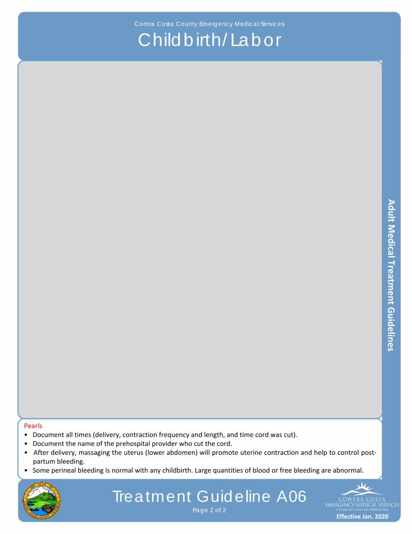
Effective Jan. 2016
Contra Costa County Emergency Medical Services
Childbirth/Labor
Treatment Guideline A06Page 2 of 2
Pearls• Document all times (delivery, contraction frequency and length, and time cord was cut).• Document the name of the prehospital provider who cut the cord.• After delivery, massaging the uterus (lower abdomen) will promote uterine contraction and help to control post‐
partum bleeding.• Some perineal bleeding is normal with any childbirth. Large quantities of blood or free bleeding are abnormal.
Effective Jan. 2020
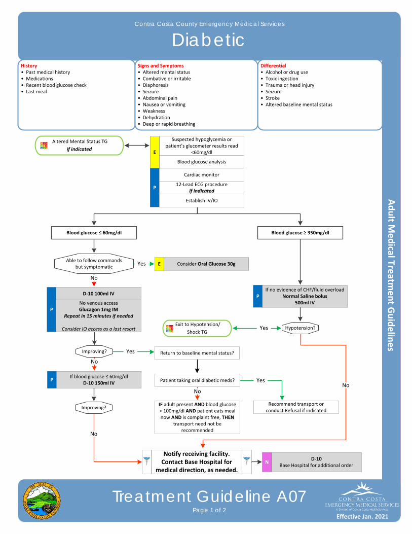
Effective Jan. 2016
Contra Costa County Emergency Medical Services
Diabetic
Treatment Guideline A07Page 1 of 2
History• Past medical history• Medications• Recent blood glucose check • Last meal
Differential• Alcohol or drug use• Toxic ingestion• Trauma or head injury• Seizure• Stroke• Altered baseline mental status
Signs and Symptoms• Altered mental status• Combative or irritable• Diaphoresis• Seizure• Abdominal pain• Nausea or vomiting• Weakness• Dehydration• Deep or rapid breathing
P
Blood glucose analysis
Cardiac monitor
12‐Lead ECG procedureif indicated
Establish IV/IO
Altered Mental Status TG
if indicated
Able to follow commandsbut symptomatic
Exit to Hypotension/
Shock TG
Consider Oral Glucose 30gE
Blood glucose ≤ 60mg/dl Blood glucose ≥ 350mg/dl
D‐10 100ml IV
PNo venous accessGlucagon 1mg IM
Repeat in 15 minutes if needed
Consider IO access as a last resort
Improving?
PIf blood glucose ≤ 60mg/dl
D‐10 150ml IV
No
PIf no evidence of CHF/fluid overload
Normal Saline bolus500ml IV
Hypotension?
Return to baseline mental status?
Patient taking oral diabetic meds?
Recommend transport orconduct Refusal if indicated
IF adult present AND blood glucose > 100mg/dl AND patient eats meal now AND is complaint free, THEN
transport need not be recommended
Yes
Yes
No
Yes
No
Yes
Improving?
No
D‐10 Base Hospital for additional order
N
No
Suspected hypoglycemia or patient’s glucometer results read
<60mg/dlE
Notify receiving facility. Contact Base Hospital for
medical direction, as needed.
Effective Jan. 2021

Effective Jan. 2016
Contra Costa County Emergency Medical Services
Diabetic
Treatment Guideline A07Page 2 of 2
Pearls• It is safer to assume hypoglycemia than hyperglycemia if doubt exists.• Recheck BGL after each D‐10 or Glucagon administration.• Patients with prolonged hypoglycemia may not respond to Glucagon.• Response to Glucagon can take 15‐20 minutes. Consider the entire clinical picture when treating hypoglycemia,
including a patient’s overall clinical condition and other vital signs. It may be safe to wait for some time for Glucagon to work instead of pursing the more aggressive course of performing IO access to give faster acting D‐10 solution. Diabetics may have poor wound healing capabilities, and IO access may present a greater risk for infection or complicate the patient’s long‐term condition due to poor wound healing. IO access may also present a greater risk for infection. On the other hand, consider IO access to give D‐10 solution early in patients who are critically ill or peri‐arrest and hypoglycemic.
• Do not administer oral glucose to patients that are not able to swallow or protect their airway.• Quality control checks should be maintained per manufacturer’s recommendation for all glucometers. • Patients refusing transport to a hospital after treatment of hypoglycemia: ‐ Oral agents: Patients taking oral diabetic medications should be strongly encouraged to allow ambulance transportation to a hospital. They are at risk of recurrent hypoglycemia that can be delayed for hours and require close monitoring even after a prehospital blood glucose level of greater than 60mg/dl has been achieved. Patients who meet criteria to refuse care should be instructed to contact their physician immediately and consume a meal with complex carbohydrates and protein now. ‐ Insulin agents: Many forms of Insulin now exist. Longer acting Insulin places the patient at risk of recurrent hypoglycemia even after a prehospital blood glucose level of greater than 60mg/dl has been achieved. Patient who meet criteria to refuse care should be instructed to contact their physician immediately and consume a meal with complex carbohydrates and protein now.
Effective Jan. 2021
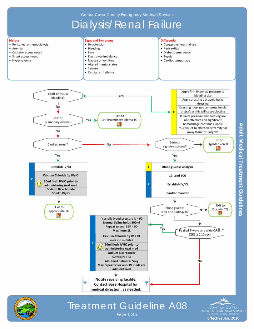
Contra Costa County Emergency Medical Services
Trauma Triage Algorithm
Field Treatment GuidelinesPage 3 of 3
Effective Jan. 2016
Contra Costa County Emergency Medical Services
Dialysis/Renal Failure
Treatment Guideline A08Page 1 of 2
History• Peritoneal or hemodialysis• Anemia• Catheter access noted • Shunt access noted• Hyperkalemia
Differential• Congestive heart failure• Pericarditis • Diabetic emergency• Sepsis• Cardiac tamponade
Signs and Symptoms• Hypotension• Bleeding• Fever• Electrolyte imbalance• Nausea or vomiting• Altered mental status• Seizure• Cardiac arrhythmia
E
Apply firm finger tip pressure to bleeding site
Apply dressing but avoid bulky dressing
Dressing must not compress fistula or graft as this will cause clotting
If direct pressure and dressing are not effective and significant hemorrhage continues, apply
tourniquet to affected extremity far away from fistula/graft
Exit to CHF/Pulmonary Edema TG
Graft or fistulableeding?
Exit to appropriate TG
Calcium Chloride 1g IV/IOP
Sodium Bicarbonate50mEq IV/IO
P
Blood glucose analysis
Yes
No
CHF orpulmonary edema?
Yes
No
Cardiac arrest?
Yes
NoSerious
signs/symptoms?
Yes
12‐Lead ECG
Establish IV/IO
Cardiac monitor
Blood glucose≤ 60 or ≥ 350mg/dl?
Peaked T‐wave and wide QRS?(QRS ≥ 0.12 sec)Calcium Chloride 1g IV / IO
over 2‐3 minutes
Sodium Bicarbonate50mEq IV / IO
Albuterol nebulizer 5mgMay repeat x3 or until IV meds are
administered
Yes
P
No
Exit to Diabetic TG
Exit to Hypotension TG
If systolic blood pressure is < 90,Normal Saline bolus 500mlRepeat to goal SBP > 90
Maximum 1L
Establish IV/IO
20ml flush IV/IO prior to administering next med
20ml flush IV/IO prior to administering next med
Notify receiving facility. Contact Base Hospital for
medical direction, as needed.
E
Effective Jan. 2020
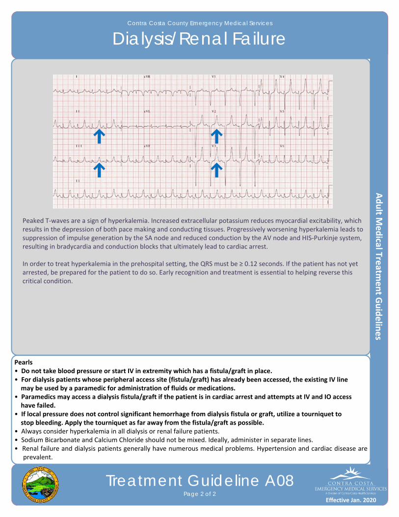
Contra Costa County Emergency Medical Services
Trauma Triage Algorithm
Field Treatment GuidelinesPage 3 of 3
Effective Jan. 2016
Contra Costa County Emergency Medical Services
Dialysis/Renal Failure
Treatment Guideline A08Page 2 of 2
Pearls• Do not take blood pressure or start IV in extremity which has a fistula/graft in place.• For dialysis patients whose peripheral access site (fistula/graft) has already been accessed, the existing IV line may be used by a paramedic for administration of fluids or medications.• Paramedics may access a dialysis fistula/graft if the patient is in cardiac arrest and attempts at IV and IO access have failed.• If local pressure does not control significant hemorrhage from dialysis fistula or graft, utilize a tourniquet to stop bleeding. Apply the tourniquet as far away from the fistula/graft as possible.• Always consider hyperkalemia in all dialysis or renal failure patients. • Sodium Bicarbonate and Calcium Chloride should not be mixed. Ideally, administer in separate lines.• Renal failure and dialysis patients generally have numerous medical problems. Hypertension and cardiac disease are
prevalent.
Peaked T‐waves are a sign of hyperkalemia. Increased extracellular potassium reduces myocardial excitability, which results in the depression of both pace making and conducting tissues. Progressively worsening hyperkalemia leads to suppression of impulse generation by the SA node and reduced conduction by the AV node and HIS‐Purkinje system, resulting in bradycardia and conduction blocks that ultimately lead to cardiac arrest.
In order to treat hyperkalemia in the prehospital setting, the QRS must be ≥ 0.12 seconds. If the patient has not yet arrested, be prepared for the patient to do so. Early recognition and treatment is essential to helping reverse this critical condition.
Effective Jan. 2020
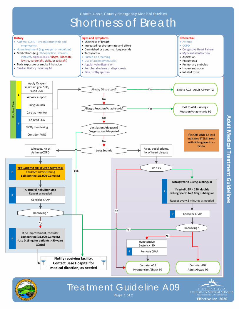
Effective Jan. 2016
Contra Costa County Emergency Medical Services
Shortness of Breath
Treatment Guideline A09Page 1 of 2
History Asthma; COPD – chronic bronchitis and
emphysema Home treatment (e.g. oxygen or nebulizer)Medications (e.g. Theophylline, steroids,
inhalers, digoxin, lasix, Viagra, Sildenafil, levitra, vardenafil, cialis, or tadalafil)
Toxic exposure or smoke inhalation Cardiac History including MI
Differential Asthma COPD Congestive Heart FailureMyocardial Infarction Aspiration Pneumonia Pulmonary embolusHyperventilation Inhaled toxin
Signs and Symptoms Shortness of breath Increased respiratory rate and effort Diminished or abnormal lung sounds Tachycardia Pursed lip breathing Use of accessory muscles Jugular vein distension Peripheral edema or diaphoresis Pink, frothy sputum
Cardiac monitor
EtCO2 monitoringP
12‐Lead ECG
Airway Obstructed?
Albuterol nebulizer 5mgRepeat as needed
If no improvement, considerEpinephrine 1:1,000 0.3mg IM
(Use 0.15mg for patients > 50 years of age)
Consider CPAP
P
P
Apply Oxygen maintain goal SpO2
93 to 95%
E
Notify receiving facility. Contact Base Hospital for
medical direction, as needed
Consider IV/IO
PERI‐ARREST OR SEVERE DISTRESS?Consider administering
Epinephrine 1:1,000 0.3mg IM
Airway support
Lung Sounds
Exit to A02 ‐ Adult Airway TG
Ventilation Adequate?Oxygenation Adequate?
Allergic Reaction/Anaphylaxis? Exit to A04 – Allergic
Reaction/Anaphylaxis TG
Wheezes, Hx of Asthma/COPD
Rales, pedal edema, hx of heart disease
Routine Care
Yes
No
No
No
Yes
Lung Sounds
P
No
Improving?
Improving?
Consider A02
Adult Airway TG
Yes
Nitroglycerin 0.4mg sublingual
If systolic BP > 150, doubleNitroglycerin to 0.8mg sublingual
Repeat every 5 minutes as needed
P
Consider CPAPP
BP > 90
Remove CPAPP
HypotensiveSystolic < 90
No
Consider A12
Hypotension/Shock TG
No
Yes
If in CHF AND 12 lead indicates STEMI, treat with Nitroglycerin as
below
Effective Jan. 2020
Effective Jan. 2016
Contra Costa County Emergency Medical Services
Shortness of Breath
Treatment Guideline A09Page 2 of 2
Pearls
If a patient is in CHF AND the 12 lead indicates STEMI, treat with nitroglycerine as on this treatment guideline. Nitro is withheld if the patient has a STEMI and CHF symptoms are not present.
Patients receiving epinephrine should receive a 12‐Lead ECG at some point in their care in the prehospital setting, but this should NOT delay the administration of Epinephrine.
Epinephrine may precipitate cardiac ischemia. The following patients should receive half the adult dose of Epinephrine (0.15mg Epinephrine 1:1,000) for the initial dose and any repeated doses:
1) Patients with a history of coronary artery disease, MI, stents, CHF, cardiac surgery; OR2) Patients over 50 years of age and has a heart rate ≥ 150.
Pulse oximetry and continuous EtCO2 monitoring is required for all respiratory patients.
A silent chest in respiratory distress is a pre‐respiratory arrest sign.
CPAP is not a ventilation device. Patients with an inadequate respiratory rate or depth of respiration will need assistance with a BVM.
Opioids have NOT been shown to improve the outcomes of EMS patients with pulmonary edema. Even though this has historically been a mainstay of EMS treatment, it is no longer routinely recommended.
Avoid Nitroglycerin in any patient who has used Viagra (Sildenafil) or Levitra (Vardenafil) in the past 24 hours or Cialis (Tadalafil) in the past 36 hours due to potential for severe hypotension.
Carefully monitor the patient’s level of consciousness, chest pain, and respiratory status with the above
interventions.
If a patient has taken their own nitroglycerin without relief, consider potency of medication. Provider maximum doses do not include patient administered doses.
Consider MI in all of these patients: Diabetic, geriatric, and female patients often have atypical pain or only generalized complaints.
Document CPAP application using the CPAP procedure in the EHR. Document the 12‐Lead ECG in the EHR as a procedure along with the interpretation.
Effective Jan. 2020

Effective Jan. 2016
Contra Costa County Emergency Medical Services
Dystonic Reaction
Treatment Guideline A10Page 1 of 1
History• Medical history • Medications• Abuse or recreational use of prescription medications
Differential• Trauma • Stroke• Tumor• Hypoxia• Infection • Drug reactions• Poisoning
Signs and Symptoms• Restleness• Muscle spasms of the neck, jaw and back• Oculogyric crisis• A “dragging leg”• Speech difficulties
Consider, Oxygen (low flow unless altered or in respiratory distress
or shock)
Pulse oximetry
E
P
Pearls• Common drugs implicated in dystonic reactions include many anti‐emetics and anti‐psychotic medications including, but not limited to: • Prochlorperazine (Compazine) • Fluphenazine (Prolixin) • Haloperidol (Haldol) • Chlorpromazine (Thorazine) • Metoclopromide (Reglan) • Many other anti‐psychotic and anti‐emetic drugs • Promethazine (Phenergan)• Rarely, benzodiazepine drugs have been implicated as a cause of dystonic reaction.
Obtain vital signs
Cardiac montior
Establish IV/IO
Diphenhydramine 50mg IV/IO/IM
Monitor and reassessE
Exit to
AMS TG
Exit to appropriate
respiratory TG
Exit to
Hypotension / Shock TG
Notify receiving facility. Contact Base Hospital for
medical direction, as needed.
Effective Jan. 2020
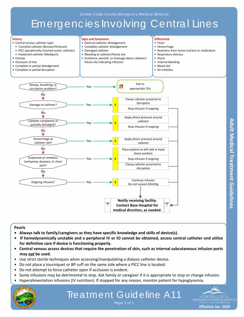
Effective Jan. 2016
Contra Costa County Emergency Medical Services
Emergencies Involving Central Lines
Treatment Guideline A11Page 1 of 1
History• Central venous catheter type: • Tunneled catheter (Broviac/Hickman) • PICC (peripherally inserted center catheter) • Implanted catheter (Mediport)• Fistulas• Occlusion of line• Complete or partial dislodgement • Complete or partial disruption
Differential• Fever• Hemorrhage• Reactions from home nutrient or medication• Respiratory distress• Shock• Internal bleeding• Blood clot• Air embolus
Signs and Symptoms• External catheter dislodgement• Complete catheter dislodgement• Damaged catheter• Bleeding at catheter/fistula site• Erythema, warmth, or drainage about catheter/ fistula site indicating infection
Airway, breathing, or circulation problem?
Exit to
appropriate TGs
Clamp catheter proximal to disruption
Damage to catheter?
Catheter completely or partially dislodged?
Hemorrhage atcatheter site?
Suspected air embolus, tachypnea, dyspnea, or chest
pain?
No
No
No
No
Yes
Yes
Yes
Yes
Yes
Ongoing infusion?
No
Yes
EStop infusion if ongoing
Apply direct pressure around catheter
EStop infusion if ongoing
Apply direct pressure around catheter
E
Place patient on left side in head down position
E Stop infusion if ongoing
Clamp catheter proximal to disruption
Continue infusionDo not exceed 20ml/kg
E
Pearls• Always talk to family/caregivers as they have specific knowledge and skills of device(s).• If hemodynamically unstable and a peripheral IV or IO cannot be obtained, access central catheter and utilize
for definitive care if device is functioning properly.• Central venous access devices that require the penetration of skin, such as internal subcutaneous infusion ports
may not be used.• Use strict sterile techniques when accessing/manipulating a dialysis catheter device.• Do not place a tourniquet or BP cuff on the same side where a PICC line is located.• Do not attempt to force catheter open if occlusion is evident.• Some infusions may be detrimental to stop. Ask family or caregiver if it is appropriate to stop or change infusion.• Hyperalimentation infusions (IV nutrition): If stopped for any reason, monitor patient for hypoglycemia.
Notify receiving facility. Contact Base Hospital for
medical direction, as needed.
Effective Jan. 2020
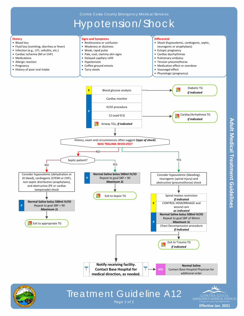
Effective Jan. 2016
Contra Costa County Emergency Medical Services
Hypotension/Shock
Treatment Guideline A12Page 1 of 2
History• Blood loss• Fluid loss (vomiting, diarrhea or fever)• Infection (e.g., UTI, cellulitis, etc.)• Cardiac ischemia (MI or CHF)• Medications• Allergic reaction• Pregnancy• History of poor oral intake
Differential• Shock (hypovolemic, cardiogenic, septic, neurogenic or anaphylaxis)• Ectopic pregnancy • Cardiac dysrhythmias• Pulmonary embolus• Tension pneumothorax• Medication effect or overdose• Vasovagal effect• Physiologic (pregnancy)
Signs and Symptoms• Restlessness or confusion• Weakness or dizziness• Weak, rapid pulse• Pale, cool, clammy skin signs• Delayed capillary refill• Hypotension• Coffee‐ground emesis• Tarry stools
Diabetic TG
if indicated
P
Blood glucose analysis
History, exam and circumstances often suggest (type of shock)WAS TRAUMA INVOLVED?
Cardiac monitor
IV/IO procedure
12‐Lead ECG
Airway TGs, if indicated
Cardiac/Arrhythmia TG
if indicated
Consider hypovolemic (dehydration or GI bleed), cardiogenic (STEMI or CHF), non‐septic distributive (anaphylaxis),
and obstructive (PE or cardiac tamponade) shock
NO
PNormal Saline bolus 500ml IV/IO
Repeat to goal SBP > 90Maximum 1L
Normal SalineContact Base Hospital Physician for
additional orderMD
Exit to appropriate TG
Notify receiving facility. Contact Base Hospital for
medical direction, as needed.
E
Spinal motion restrictionif indicated
E CONTROL HEMORRHAGE and wound careas indicated
P
Normal Saline bolus 500ml IV/IORepeat to goal SBP of 90mm
Maximum 1L
Consider hypovolemic (bleeding), neurogenic (spinal injury) and
obstructive (pneumothorax) shock
Chest Decompression procedureif indicated
Exit to Trauma TG
if indicated
PNormal Saline bolus 500ml IV/IO
Repeat to goal SBP > 90Maximum 2L
Exit to Sepsis TG
YESNO
YESSeptic patient?
Effective Jan. 2021
Effective Jan. 2016
Contra Costa County Emergency Medical Services
Hypotension/Shock
Treatment Guideline A12Page 2 of 2
Pearls• Hypotension can be defined as a systolic blood pressure of less than 90mmHg. This is not always reliable and
should be interpreted in context with the patient’s typical BP, if known. Shock may be present with a seemingly normal blood pressure initially.
• Shock is often present with normal vital signs and may develop insidiously. Tachycardia may be the only manifestation.
• Consider all causes of shock and treat per appropriate Treatment Guideline.• Hypovolemic shock: ‐ Hemorrhage, trauma, GI bleeding, ruptured AAA, or pregnancy‐related bleeding • Cardiogenic shock: ‐ Heart failure, MI, cardiomyopathy, myocardial contusion, ruptured ventricle/septum/valve or toxins• Distributive shock: ‐ Sepsis, anaphylactic, neurogenic, or toxins ‐ Neurogenic shock generally presents with warm, dry, and pink skin with normal capillary refill time; patient typically alert• Obstructive shock: ‐ Pericardial tamponade, PE, or tension pneumothorax ‐ Signs may include hypotension with distended neck veins, tachycardia, unilateral decreased breath sounds or muffled heart tones
Effective Jan. 2021

Effective Jan. 2016
Contra Costa County Emergency Medical Services
Overdose/Toxic Ingestion
Treatment Guideline A13Page 1 of 2
History• Ingestion or suspected ingestion of a potentially toxic substance• Substance ingested, route, and quantity• Time of ingestion • Reason (suicidal, accidental or criminal)• Available medications in home• Past medical history and medications
Differential• Tricyclic antidepressants (TCAs)• Acetaminophen (Tylenol) • Aspirin• Depressants• Stimulants• Anticholinergics• Cardiac medications• Solvents, alcohols or cleaning agents• Insecticides (organophosphates)
Signs and Symptoms• Mental status changes• Hypo or hypertension• Decreased respiratory rate• Tachycardia or dysrhythmias• Seizures• S.L.U.D.G.E.
P IV/IO procedure
Cardiac monitor
Exit to Diabetic/Behavioral TGs
as indicated
PSodium Bicarbonate
1mEq/kg IV/IO
Yes
Adequate respirations (> 8)and oxygenation?
12‐Lead ECG
No
Altered mental status?E Blood glucose analysis Yes
Exit to Opioid Overdose TG
as indicated
Systolic BP < 90
No
Exit to Hypotension/Shock TG
as indicated
Organophosphate OD
Tricyclic Antidepressant
OD
If bradycardic and symptomatic
Contact Base Hospital Physician for treatment or additional Fentanyl orders
MD
Hydrofluoric AcidExposure
QRS ≥ 0.12 sec and BP < 90?
Yes
P
Atropine 1‐2mg IV/IORepeat every 3‐5
minutes until relief of symptoms achieved
P
Calcium Chloride 500mg IV/IO
(5ml of 10% solution)For tetany or cardiac
arrestConsider Fentanyl
50‐200mcg IV Titrated in 25‐50mcg increments to pain
relief
OR
Fentanyl 100mcg IN/IM if no IV
accessMay repeat once in 15
minutes
California Poison Control Center (800) 222‐1222
Exit to Symptomatic Bradycardia
TG as indicated
Notify receiving facility. Contact Base Hospital for
medical direction, as needed.
Effective Jan. 2021
Effective Jan. 2016
Contra Costa County Emergency Medical Services
Overdose/Toxic Ingestion
Treatment Guideline A13Page 2 of 2
Pearls• Overdose or toxic ingestion patients with significant ingestion/exposures should be monitored very closely and aggressively treated as indicated. Do not hesitate to contact the Base Hospital for advice as certain critically ill overdose patients may quickly overwhelm medication supplies. For example, a tricyclic overdose with a wide QRS and altered mental status may need to receive multiple Sodium Bicarbonate boluses until QRS narrowing and clinical improvement; patients with organophosphate toxicity with SLUDGE syndrome may require more Atropine than is usually available on an ambulance.• Do not rely on patient history of ingestion, especially in suicide attempts. Make sure patient is still not carrying other medications or has any weapons. • Bring medication bottles, contents, and emesis to the Emergency Department.• S.L.U.D.G.E.: Salivation, Lacrimation, Urination, Defecation, GI distress, and Emesis• Tricyclic: 4 major areas of toxicity include decreased mental status, dysrythmias, seizures, hypotension then coma
and death. • Acetaminophen: Initially normal or with nausea/vomiting. If not detected and treated, causes irreversible liver
failure. • Aspirin: Early sign consist of abdominal pain and vomiting. Tachypnea and altered mental status may occur later.
Renal dysfunction, liver failure or cerebral edema among other things can present later. • Depressants: Decreased heart rate, blood pressure or temperature, decreased respirations, and non‐specific pupils.• Stimulants: Increased heart rate, blood pressure or temperature, dilated pupils, and seizures. • Anticholinergics: Increased heart rate or temperature, dilated pupils, and mental status changes. • Cardiac medications: Dysrhythmias and mental status changes. • Solvents: Nausea, vomiting, coughing, and mental status changes. • Insecticides: Increased or decreased heart rate, increased secretions, nausea, vomiting, diarrhea, and pinpoint pupils. Consider restraints if necessary for patient’s or personnel’s protection per Restraint Procedure.• Consider contacting the California Poison Control Center for Guidance.
Effective Jan. 2021
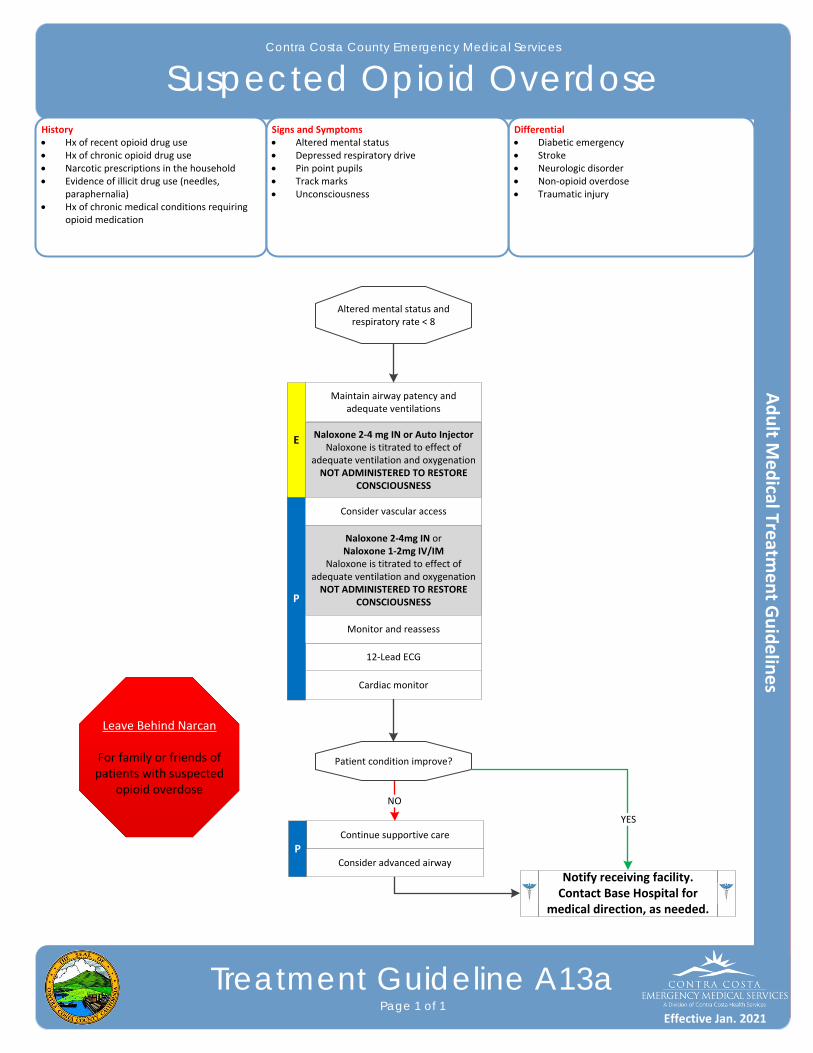
Effective Jan. 2016
Contra Costa County Emergency Medical Services
Suspected Opioid Overdose
Treatment Guideline A13aPage 1 of 1
History Hx of recent opioid drug use Hx of chronic opioid drug use Narcotic prescriptions in the household Evidence of illicit drug use (needles,
paraphernalia) Hx of chronic medical conditions requiring
opioid medication
Differential Diabetic emergency Stroke Neurologic disorder Non‐opioid overdose Traumatic injury
Signs and Symptoms Altered mental status Depressed respiratory drive Pin point pupils Track marks Unconsciousness
Cardiac monitor
12‐Lead ECG
P
Naloxone 2‐4mg IN orNaloxone 1‐2mg IV/IM
Naloxone is titrated to effect of adequate ventilation and oxygenationNOT ADMINISTERED TO RESTORE
CONSCIOUSNESS
E Naloxone 2‐4 mg IN or Auto Injector Naloxone is titrated to effect of
adequate ventilation and oxygenationNOT ADMINISTERED TO RESTORE
CONSCIOUSNESS
Monitor and reassess
Maintain airway patency and adequate ventilations
Consider vascular access
Notify receiving facility. Contact Base Hospital for
medical direction, as needed.
Continue supportive care
P
Altered mental status and respiratory rate < 8
NO
Patient condition improve?
Consider advanced airway
Leave Behind Narcan
For family or friends of patients with suspected
opioid overdose
YES
Effective Jan. 2021
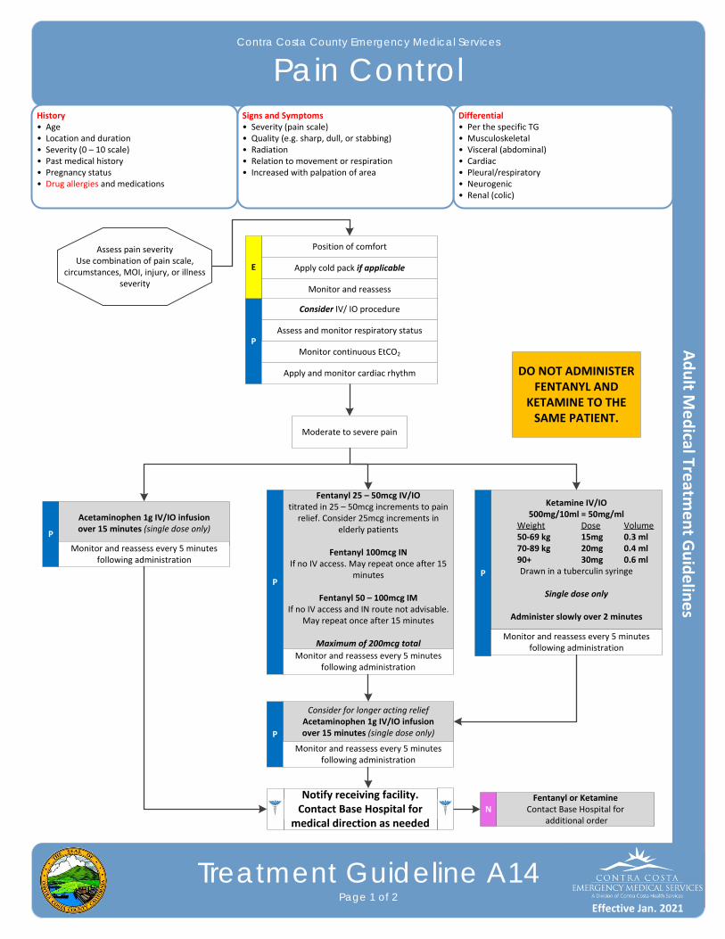
Effective Jan. 2016
Contra Costa County Emergency Medical Services
Pain Control
Treatment Guideline A14Page 1 of 2
History• Age• Location and duration• Severity (0 – 10 scale)• Past medical history• Pregnancy status• Drug allergies and medications
Differential• Per the specific TG• Musculoskeletal • Visceral (abdominal)• Cardiac• Pleural/respiratory• Neurogenic• Renal (colic)
Signs and Symptoms• Severity (pain scale)• Quality (e.g. sharp, dull, or stabbing)• Radiation• Relation to movement or respiration• Increased with palpation of area
Position of comfortAssess pain severityUse combination of pain scale,
circumstances, MOI, injury, or illness severity
Moderate to severe pain
Apply cold pack if applicable
Monitor and reassess
Consider IV/ IO procedure
Assess and monitor respiratory statusP
Fentanyl 25 – 50mcg IV/IOtitrated in 25 – 50mcg increments to pain relief. Consider 25mcg increments in
elderly patients
Fentanyl 100mcg INIf no IV access. May repeat once after 15
minutes
Fentanyl 50 – 100mcg IMIf no IV access and IN route not advisable.
May repeat once after 15 minutes
Maximum of 200mcg total
Monitor continuous EtCO2
Monitor and reassess every 5 minutes following administration
Apply and monitor cardiac rhythm
Fentanyl or KetamineContact Base Hospital for
additional orderN
E
Acetaminophen 1g IV/IO infusion over 15 minutes (single dose only)
P
P
Ketamine IV/IO500mg/10ml = 50mg/ml
Weight Dose Volume50‐69 kg 15mg 0.3 ml70‐89 kg 20mg 0.4 ml90+ 30mg 0.6 mlDrawn in a tuberculin syringe
Single dose only
Administer slowly over 2 minutes
P
Monitor and reassess every 5 minutes following administration
Monitor and reassess every 5 minutes following administration
Consider for longer acting relief Acetaminophen 1g IV/IO infusion over 15 minutes (single dose only)P
Monitor and reassess every 5 minutes following administration
Notify receiving facility. Contact Base Hospital for
medical direction as needed
DO NOT ADMINISTER FENTANYL AND
KETAMINE TO THE SAME PATIENT.
Effective Jan. 2021
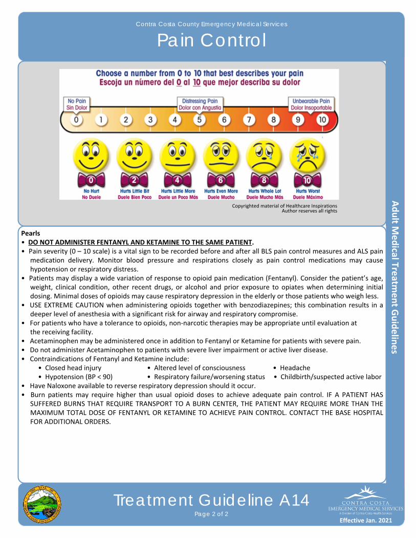
Effective Jan. 2016
Contra Costa County Emergency Medical Services
Pain Control
Treatment Guideline A14Page 2 of 2
Pearls• DO NOT ADMINISTER FENTANYL AND KETAMINE TO THE SAME PATIENT.• Pain severity (0 – 10 scale) is a vital sign to be recorded before and after all BLS pain control measures and ALS pain
medication delivery. Monitor blood pressure and respirations closely as pain control medications may cause hypotension or respiratory distress.
• Patients may display a wide variation of response to opioid pain medication (Fentanyl). Consider the patient’s age, weight, clinical condition, other recent drugs, or alcohol and prior exposure to opiates when determining initial dosing. Minimal doses of opioids may cause respiratory depression in the elderly or those patients who weigh less.
• USE EXTREME CAUTION when administering opioids together with benzodiazepines; this combination results in a deeper level of anesthesia with a significant risk for airway and respiratory compromise.
• For patients who have a tolerance to opioids, non‐narcotic therapies may be appropriate until evaluation at the receiving facility.
• Acetaminophen may be administered once in addition to Fentanyl or Ketamine for patients with severe pain.• Do not administer Acetaminophen to patients with severe liver impairment or active liver disease.• Contraindications of Fentanyl and Ketamine include: • Closed head injury • Altered level of consciousness • Headache • Hypotension (BP < 90) • Respiratory failure/worsening status • Childbirth/suspected active labor• Have Naloxone available to reverse respiratory depression should it occur.• Burn patients may require higher than usual opioid doses to achieve adequate pain control. IF A PATIENT HAS
SUFFERED BURNS THAT REQUIRE TRANSPORT TO A BURN CENTER, THE PATIENT MAY REQUIRE MORE THAN THE MAXIMUM TOTAL DOSE OF FENTANYL OR KETAMINE TO ACHIEVE PAIN CONTROL. CONTACT THE BASE HOSPITAL FOR ADDITIONAL ORDERS.
Copyrighted material of Healthcare Inspirations Author reserves all rights
Effective Jan. 2021
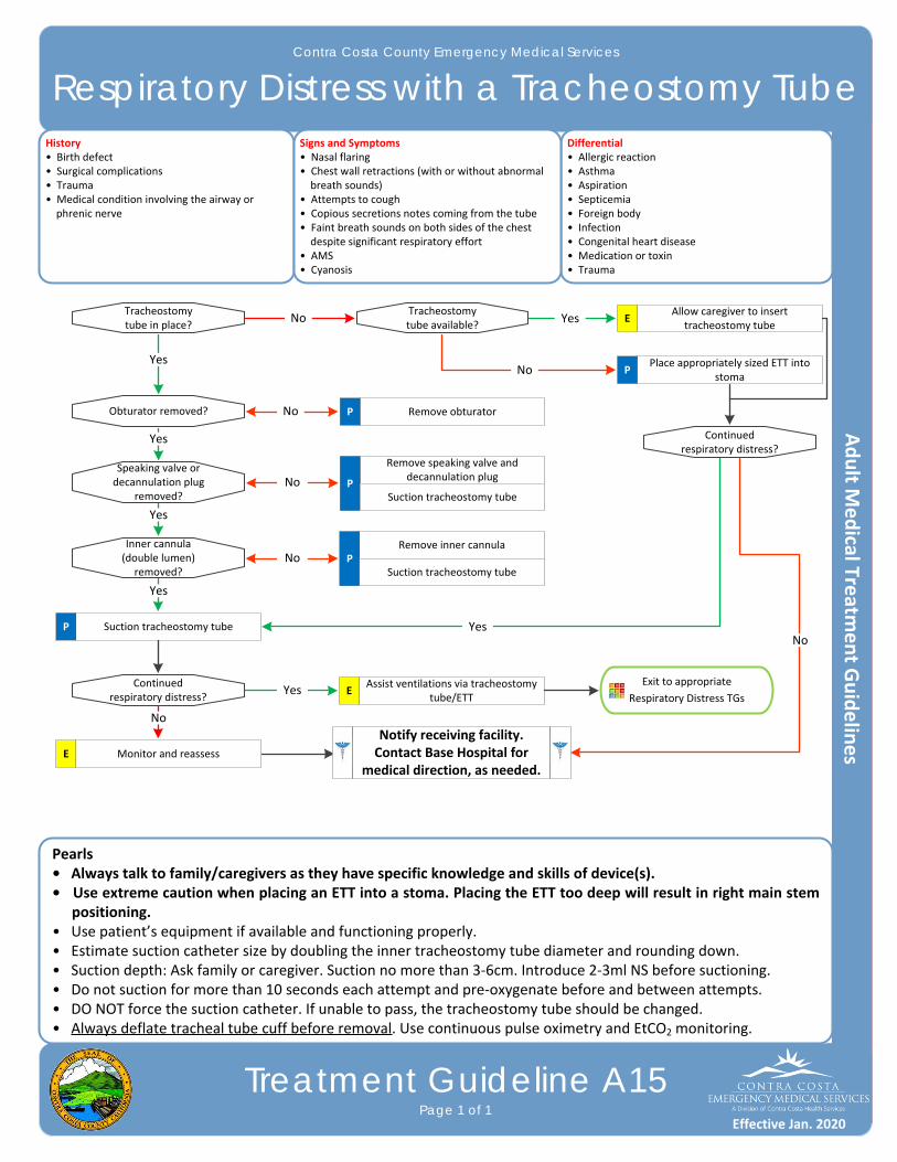
Effective Jan. 2016
Contra Costa County Emergency Medical Services
Respiratory Distress with a Tracheostomy Tube
Treatment Guideline A15Page 1 of 1
History• Birth defect• Surgical complications• Trauma • Medical condition involving the airway or phrenic nerve
Differential• Allergic reaction• Asthma• Aspiration• Septicemia• Foreign body• Infection• Congenital heart disease• Medication or toxin• Trauma
Signs and Symptoms• Nasal flaring• Chest wall retractions (with or without abnormal breath sounds)• Attempts to cough• Copious secretions notes coming from the tube• Faint breath sounds on both sides of the chest despite significant respiratory effort• AMS• Cyanosis
Tracheostomy tube in place?
Exit to appropriate
Respiratory Distress TGs
Allow caregiver to inserttracheostomy tube
Obturator removed?
Speaking valve ordecannulation plug
removed?
Inner cannula (double lumen)
removed?
Yes
Yes
Continued respiratory distress?
E
Place appropriately sized ETT into stoma
Remove speaking valve and decannulation plug
PSuction tracheostomy tube
Assist ventilations via tracheostomy tube/ETT
E
Pearls• Always talk to family/caregivers as they have specific knowledge and skills of device(s).• Use extreme caution when placing an ETT into a stoma. Placing the ETT too deep will result in right main stem
positioning.• Use patient’s equipment if available and functioning properly.• Estimate suction catheter size by doubling the inner tracheostomy tube diameter and rounding down.• Suction depth: Ask family or caregiver. Suction no more than 3‐6cm. Introduce 2‐3ml NS before suctioning.• Do not suction for more than 10 seconds each attempt and pre‐oxygenate before and between attempts.• DO NOT force the suction catheter. If unable to pass, the tracheostomy tube should be changed.• Always deflate tracheal tube cuff before removal. Use continuous pulse oximetry and EtCO2 monitoring.
Continued respiratory distress?
Tracheostomy tube available?
P
Remove obturatorP
No Yes
No
No
No
No
Yes
Remove inner cannulaP
Suction tracheostomy tube
Yes
Yes
Suction tracheostomy tubeP
No
Monitor and reassessE
YesNo
Notify receiving facility. Contact Base Hospital for
medical direction, as needed.
Effective Jan. 2020

This page intentionally left blank

If BGL ≤ 60mg/dl
Exit to Diabetic TG
YES
Effective Jan. 2016
Contra Costa County Emergency Medical Services
Seizure
Treatment Guideline A16Page 1 of 2
History• Reported or witnessed seizure• Previous seizure history• Medical alert tag • Seizure medications• History of trauma• History of diabetes• History of pregnancy• Time of seizure onset• Document number of seizures• Alcohol use, abuse, or abrupt cessation• Fever
Differential• Head trauma• Metabolic, hepatic or renal failure• Tumor• Hypoxia• Electrolyte abnormality• Drugs or medication non‐compliance• Infection or sepsis• Alcohol withdrawal• Eclampsia • Stroke • Hyperthermia • Hypoglycemia
Signs and Symptoms• Altered mental status• Sleepiness• Incontinence• Observed seizure activity• Evidence of trauma• Unconscious• Incontinence
P
Status Epilepticus?(Confirmed or Suspected)
Monitor and reassess
Midazolam IM: 10mg (preferred route)IN: 10mg (5mg in each nare)
IV/IO(if already established): 6mg – 8mg
If patient continues to seize, consider additional Midazolam with base contact
Loosen any constrictive clothing
Ensure scene safety
EAssess and manage ABCs
Initiate BVM ventilation for apnea or ongoing hypoxia
Apply high flow 02 via NRB
E
Cardiac monitor
Check blood glucose
SMR if indicated
Manage any traumatic injuries
E
Notify receiving facility. Contact Base Hospital for
medical direction, as needed.
NO
Status Epilepticus 2 or more seizures in ≤ 5 minutes
Or Any seizure lasting > 5 minutes
If duration cannot be confirmed, patient should be assumed to be in status if actively seizing when you arrived.
Effective Jan. 2021
Effective Jan. 2016
Contra Costa County Emergency Medical Services
Seizure
Treatment Guideline A16Page 2 of 2
Pearls
Status Epilepticus is defined as two or more seizures without a period of consciousness or recovery, or one prolonged seizure lasting longer than 5 minutes. If patient is seizing upon EMS arrival this is likely status epilepticus. This is a true emergency requiring rapid airway control, treatment, and transport.
Midazolam 10mg IM is effective in the termination of status epileptic. Do not delay IM administration to obtain IV or IO access in an actively seizing patient.
Limit IN administrations to ½ dose in each nare.
Be prepared to assist ventilations or manage the airway, especially if Midazolam is used.
For a seizure that begins in the presence of EMS, if the patient was previously conscious, alert and oriented, take the time to assess and protect the patient and providers and CONSIDER THE CAUSE. The seizure may stop, especially in patients who have prior history of self‐limiting seizures. However, do not hesitate to treat recurrent or prolonged (> 5 minute) seizure activity.
Assess the possibility of occult trauma and substance abuse.
Grand Mal seizures (generalized) are associated with a loss of consciousness, incontinence, and oral trauma.
Focal seizures (Petit Mal) affect only a part of the body and are not associated with a loss of consciousness.
Effective Jan. 2021
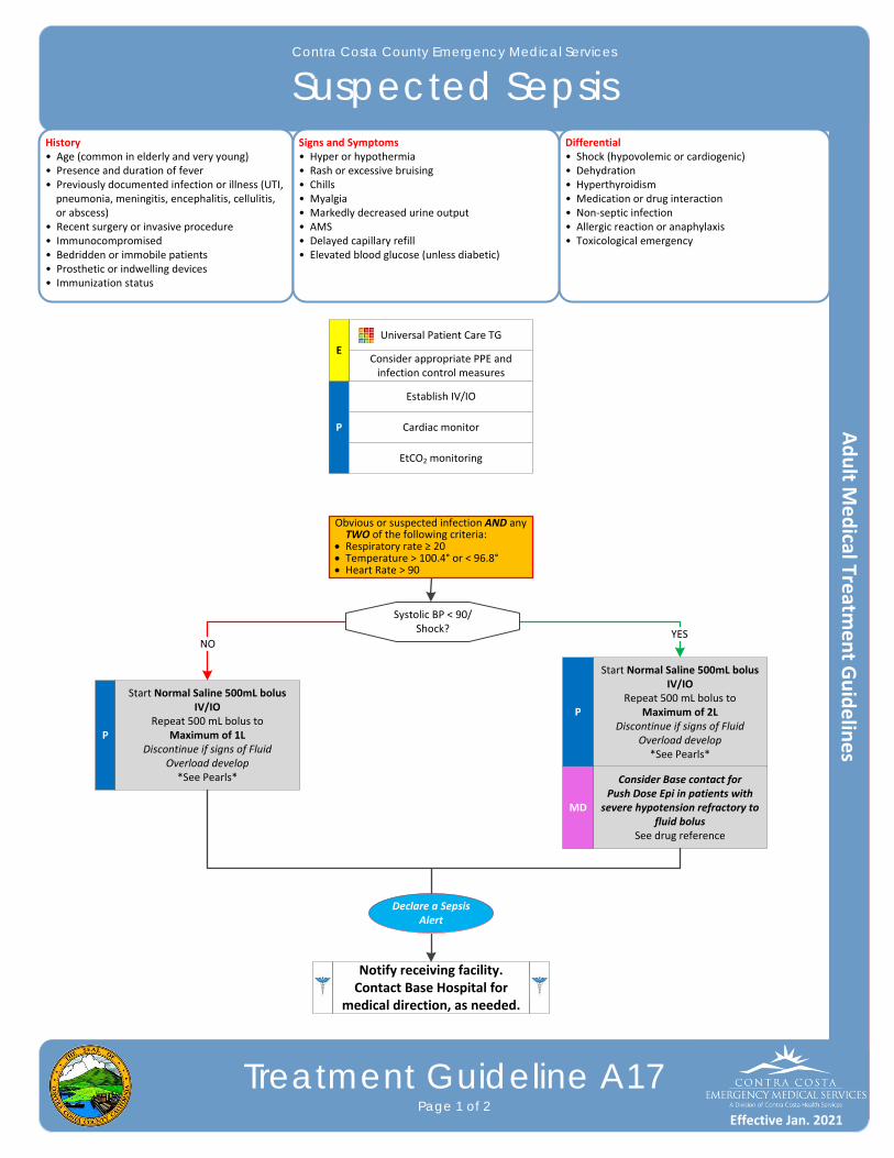
YES
Effective Jan. 2016
Contra Costa County Emergency Medical Services
Suspected Sepsis
Treatment Guideline A17Page 1 of 2
History• Age (common in elderly and very young)• Presence and duration of fever• Previously documented infection or illness (UTI, pneumonia, meningitis, encephalitis, cellulitis, or abscess) • Recent surgery or invasive procedure• Immunocompromised• Bedridden or immobile patients• Prosthetic or indwelling devices• Immunization status
Differential• Shock (hypovolemic or cardiogenic)• Dehydration • Hyperthyroidism• Medication or drug interaction• Non‐septic infection• Allergic reaction or anaphylaxis• Toxicological emergency
Signs and Symptoms• Hyper or hypothermia• Rash or excessive bruising• Chills• Myalgia• Markedly decreased urine output• AMS• Delayed capillary refill• Elevated blood glucose (unless diabetic)
P
Consider appropriate PPE and infection control measures
Establish IV/IO
Cardiac monitor
Universal Patient Care TG
Start Normal Saline 500mL bolus IV/IO
Repeat 500 mL bolus to Maximum of 2L
Discontinue if signs of Fluid Overload develop*See Pearls*
NO
E
Declare a Sepsis Alert
EtCO2 monitoring
Notify receiving facility. Contact Base Hospital for
medical direction, as needed.
Consider Base contact forPush Dose Epi in patients with
severe hypotension refractory to fluid bolus
See drug reference
P
Start Normal Saline 500mL bolus IV/IO
Repeat 500 mL bolus to Maximum of 1L
Discontinue if signs of Fluid Overload develop*See Pearls*
Systolic BP < 90/Shock?
Obvious or suspected infection AND any TWO of the following criteria:
Respiratory rate ≥ 20 Temperature > 100.4° or < 96.8° Heart Rate > 90
MD
P
Effective Jan. 2021
Effective Jan. 2016
Contra Costa County Emergency Medical Services
Suspected Sepsis
Treatment Guideline A17Page 2 of 2
Pearls• Early recognition of sepsis allows for attentive care and early administration of antibiotics.• Aggressive IV fluid therapy is the most important prehospital treatment for sepsis. Suspected sepsis patients
should receive repeated fluid boluses (to a Maximum of 2L) while being checked frequently for signs of pulmonary edema, especially in patients with a known history of CHF or ESRD on dialysis. STOP fluid administration in the setting of pulmonary edema.
• Septic patients are especially susceptible to traumatic lung injury and ARDS. If artificial ventilation is necessary, avoid ventilating with excessive tidal volumes. Use only enough tidal volume to see the chest rise. If CPAP is utilized, airway pressure should be limited to 7.5cm H2O if using a rate adjustable device.
• Attempt to identify source of infection (e.g. skin, respiratory, etc.) and relay previous treatments and related history to receiving ED physician.
• Disseminated Intravascular Coagulation (DIC) is an ominous, late stage manifestation of sepsis characterized by frank, extensive bruising, bleeding from multiple sites, and finally tissue death.
Effective Jan. 2021
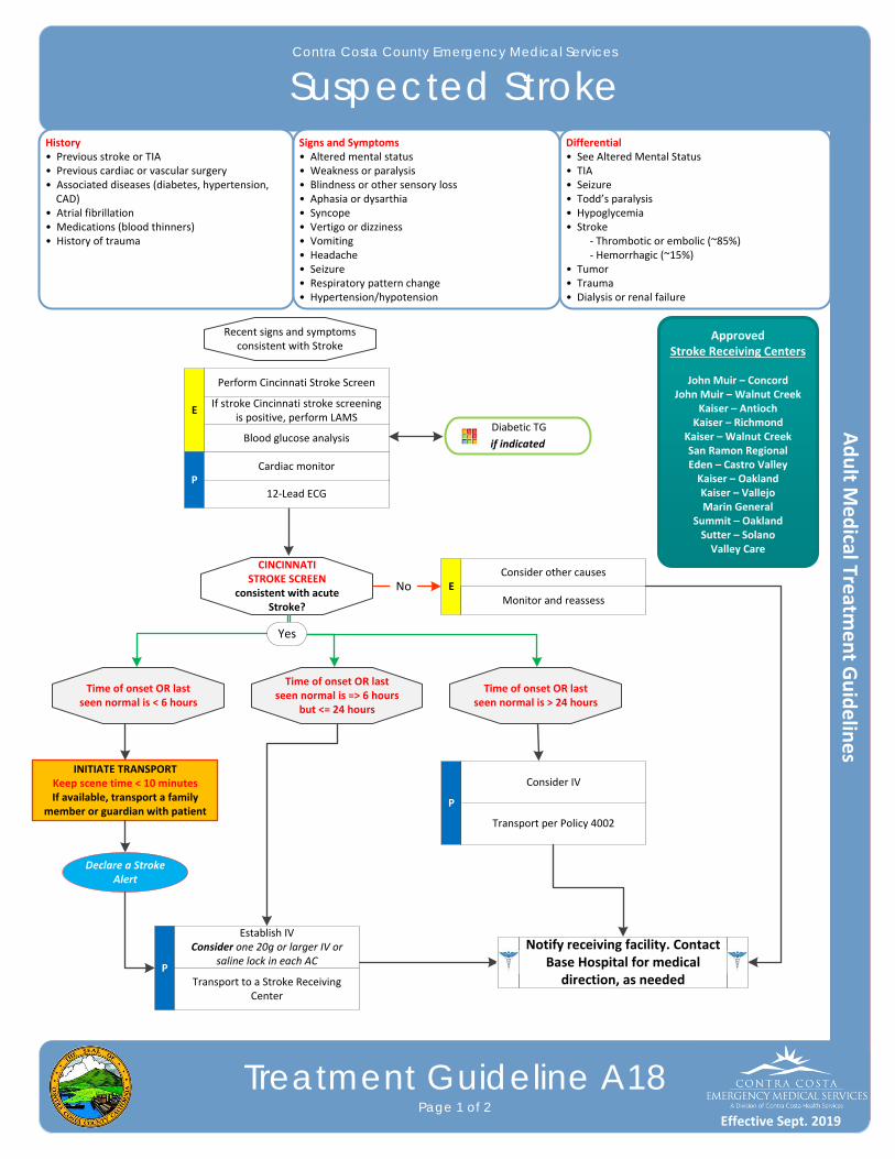
Effective Jan. 2016
Contra Costa County Emergency Medical Services
Suspected Stroke
Treatment Guideline A18Page 1 of 2
History• Previous stroke or TIA• Previous cardiac or vascular surgery• Associated diseases (diabetes, hypertension, CAD) • Atrial fibrillation• Medications (blood thinners)• History of trauma
Differential• See Altered Mental Status• TIA• Seizure• Todd’s paralysis• Hypoglycemia• Stroke ‐ Thrombotic or embolic (~85%) ‐ Hemorrhagic (~15%)• Tumor• Trauma• Dialysis or renal failure
Signs and Symptoms• Altered mental status• Weakness or paralysis• Blindness or other sensory loss• Aphasia or dysarthia• Syncope• Vertigo or dizziness• Vomiting• Headache• Seizure• Respiratory pattern change• Hypertension/hypotension
Blood glucose analysis
Recent signs and symptomsconsistent with Stroke
No
Perform Cincinnati Stroke Screen
E
CINCINNATI STROKE SCREEN
consistent with acute Stroke?
Time of onset OR last seen normal is < 6 hours
If stroke Cincinnati stroke screening is positive, perform LAMS
Diabetic TG
if indicated
INITIATE TRANSPORT Keep scene time < 10 minutesIf available, transport a family
member or guardian with patient
Declare a Stroke Alert
P
Notify receiving facility. Contact Base Hospital for medical
direction, as needed
ApprovedStroke Receiving Centers
John Muir – ConcordJohn Muir – Walnut Creek
Kaiser – AntiochKaiser – Richmond
Kaiser – Walnut CreekSan Ramon RegionalEden – Castro ValleyKaiser – OaklandKaiser – VallejoMarin General
Summit – OaklandSutter – SolanoValley Care
Consider other causesE
Monitor and reassess
Time of onset OR last seen normal is => 6 hours
but <= 24 hours
Time of onset OR last seen normal is > 24 hours
P
Transport per Policy 4002
12‐Lead ECG
Cardiac monitor
PTransport to a Stroke Receiving
Center
Establish IVConsider one 20g or larger IV or
saline lock in each AC
Yes
Consider IV
Effective Sept. 2019

Effective Jan. 2016
Contra Costa County Emergency Medical Services
Suspected Stroke
Treatment Guideline A18Page 2 of 2
Pearls• Acute stroke care is evolving rapidly. • Time last seen normal: One of the most important items that prehospital providers can obtain, on which all
treatment decisions are based. Be very precise in gathering data to establish the time of onset and report as an actual time (i.e. 13:45 NOT “about 45 minutes ago”). Without this information, patients may not be able to receive thrombolytics at the hospital. For patients with “woke up and noticed stroke symptoms,” time starts when the patient was last awake.
• The differential listed on the Altered Mental Status TG should also be considered.• Be alert for airway problems (difficulty swallowing, vomiting and aspiration).• Hypoglycemia can present as a LOCALIZED neurologic deficit, especially in the elderly. • Document the Cincinnati Stroke Screen and LAMS in the EHR.
A Stroke Alert is indicated when the Cincinnati Stoke Screen findings are abnormal and onset (time last seen normal) is less than 6 hours from time of patient contact. Report time last seen normal (clock time), the medical record number or name and birthdate, results of the Cincinnati Stroke Screen and the LAMS score, physical exam, and ETA using an approved report format.
If a family member or guardian is available, assure their availability by either transporting them in the ambulance or obtain their name and phone number to allow the receiving physician to contact them. Encourage a family member to be available to speak with hospital staff.
If any of portion of the Cincinnati Stroke Screen is abnormal and it is a new finding, the stroke screen is positive and may indicate an acute stroke.
Pre‐arrival information is necessary for the receiving hospital to make rapid treatment and potential transfer decisions.
Because the patient may need to receive thrombolytic therapy, avoid multiple IV attempts. Avoid distal placement of IVs, if possible, as this is a preferred access site by Interventionalists. When turning over patient care to hospital staff, make sure to include common anticoagulants taken by the
patient. Known use of these medications may affect the course of hospital treatment:‐ Warfarin (Coumadin) ‐ Enaoxaparin (Lovenox) ‐ Apixaban (Eliquis)
‐ Heparin ‐ Dabigatran (Pradaxa)‐ Fondaparinux (Arixtra) ‐ Rivaroxaban (Xarelto)
Cincinnati Stroke Screen LAMS Assessment
Finding Interpretation
Facial Droop
Arm Weakness
Speech Abnormality
Normal: Symmetrical smile or faceAbnormal: Asymmetry
Normal: Both arms move symmetricallyAbnormal: Asymmetrical arm movement
Normal: Correct words; no slurringAbnormal: Slurred or incorrect words
Finding Interpretation
Facial Droop
Arm Weakness
Grip
Normal: Symmetrical smile or faceAbnormal: Asymmetry
Normal: Both arms move symmetricallyAbnormal: Asymmetrical arm movementFalls rapidly: some or no effort
Normal: Equal grip in both handsWeak: Unequal grip in one handNo grip: no muscle strength or contraction
Scoring
Absent ‐ 0 pointsPresent ‐ 1 point
Absent ‐ 0 pointsDrifts ‐ 1 pointFalls rapidly ‐ 2 points
Normal ‐ 0 pointsWeak ‐ 1 pointNo grip ‐ 2 points
A LAMS score of ≥ 4 indicates a high likelihood of a LVO stroke
Effective Sept. 2019

Effective Jan. 2016
Contra Costa County Emergency Medical Services
Syncope
Treatment Guideline A19Page 1 of 2
History• History of cardiac, stroke or seizures• Occult blood loss (GI or ectopic)• Females: LMP or vaginal bleeding • Fluid loss: nausea, vomiting or diarrhea• Past medical history• Medications
Differential• Vasovagal• Orthostatic hypotension• Cardiac syncope• Micturition or defecation syncope• Psychiatric• Stroke• Hypoglycemia• Seizure• Shock • Toxicological (alcohol) • Medication effect (hypertension • Pulmonary embolism• AAA
Signs and Symptoms• Loss of consciousness with recovery• Lightheadedness or dizziness• Palpitations, slow or rapid• Pulse irregularity• Hypotension
P
Blood glucose analysis
Cardiac monitor
12‐Lead ECG
Consider IV/IO
Diabetic TG
if indicated
Consider orthostatic vital signs
Suspected or evident trauma
No
Hypotension orpoor perfusion
Altered mental status
Appropriate Cardiac TG
if indicated
Spinal Immobilization TGMultiple Trauma TG
if indicated
Altered Mental Status TG
if indicated
Yes
Yes
Hypotension / Shock TG
if indicatedYes
E
Contact the Base Hospital for all AMA requests
No
No
Notify receiving facility. Contact Base Hospital for
medical direction, as needed.
Effective Jan. 2020
Effective Jan. 2016
Contra Costa County Emergency Medical Services
Syncope
Treatment Guideline A19Page 2 of 2
Pearls• Utilize the Base Hospital for syncopal patients who do not want transport to a hospital.• Assess for signs and symptoms of trauma or head injury if associated with a fall or if it is questionable whether the patient fell due to syncope.• Consider dysrhythmias, GI bleed, ectopic pregnancy, and seizure as possible cause of syncope.• Syncope patients should be transported to a hospital for physician evaluation.• More than 25% of geriatric syncope is cardiac dysrhythmia based.
Effective Jan. 2020
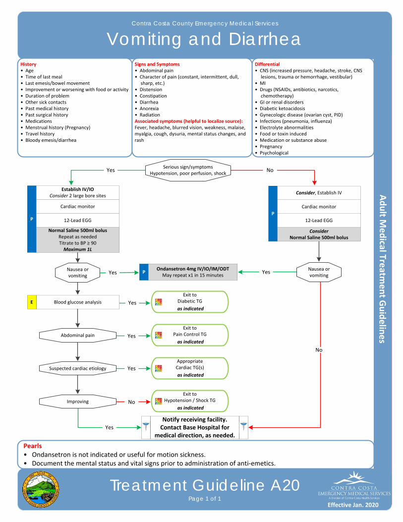
Effective Jan. 2016Effective Jan. 2017
Contra Costa County Emergency Medical Services
Vomiting and DiarrheaHistory• Age• Time of last meal• Last emesis/bowel movement• Improvement or worsening with food or activity• Duration of problem• Other sick contacts• Past medical history• Past surgical history• Medications• Menstrual history (Pregnancy)• Travel history• Bloody emesis/diarrhea
Differential• CNS (increased pressure, headache, stroke, CNS lesions, trauma or hemorrhage, vestibular)• MI • Drugs (NSAIDs, antibiotics, narcotics, chemotherapy)• GI or renal disorders• Diabetic ketoacidosis• Gynecologic disease (ovarian cyst, PID)• Infections (pneumonia, influenza)• Electrolyte abnormalities• Food or toxin induced• Medication or substance abuse• Pregnancy• Psychological
Signs and Symptoms• Abdominal pain• Character of pain (constant, intermittent, dull, sharp, etc.)• Distension• Constipation• Diarrhea• Anorexia• RadiationAssociated symptoms (helpful to localize source):Fever, headache, blurred vision, weakness, malaise, myalgia, cough, dysuria, mental status changes, and rash
Pearls• Ondansetron is not indicated or useful for motion sickness.• Document the mental status and vital signs prior to administration of anti‐emetics.
P
ConsiderNormal Saline 500ml bolus
Cardiac monitor
Serious sign/symptomsHypotension, poor perfusion, shock
Consider, Establish IV
POndansetron 4mg IV/IO/IM/ODT
May repeat x1 in 15 minutes
NoYes
Nausea or vomiting
P
Normal Saline 500ml bolusRepeat as neededTitrate to BP ≥ 90Maximum 1L
Cardiac monitor
Establish IV/IOConsider 2 large bore sites
12‐Lead EGG 12‐Lead EGG
Nausea or vomiting
Yes Yes
Blood glucose analysisE
Abdominal pain
Exit to Diabetic TG
as indicatedYes
Suspected cardiac etiology
Improving
Exit to Pain Control TG
as indicated
Appropriate Cardiac TG(s)
as indicated
Exit to Hypotension / Shock TG
as indicated
Yes
Yes
No
Treatment Guideline A20Page 1 of 1
Yes
No
Notify receiving facility. Contact Base Hospital for
medical direction, as needed.
Effective Jan. 2020

This page intentionally left blank
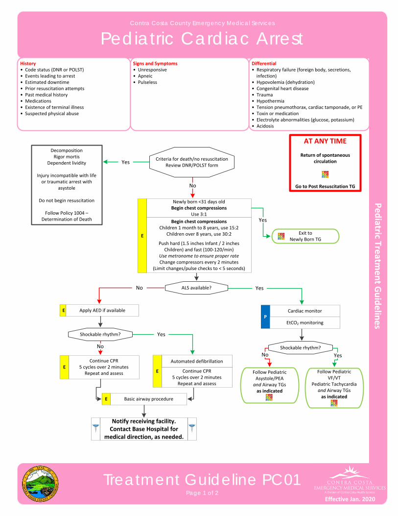
Effective Jan. 2016
Contra Costa County Emergency Medical Services
Pediatric Cardiac Arrest
Treatment Guideline PC01Page 1 of 2
History• Code status (DNR or POLST)• Events leading to arrest• Estimated downtime• Prior resuscitation attempts• Past medical history• Medications• Existence of terminal illness• Suspected physical abuse
Differential• Respiratory failure (foreign body, secretions, infection)• Hypovolemia (dehydration)• Congenital heart disease • Trauma• Hypothermia• Tension pneumothorax, cardiac tamponade, or PE• Toxin or medication• Electrolyte abnormalities (glucose, potassium)• Acidosis
Signs and Symptoms• Unresponsive• Apneic• Pulseless
Begin chest compressionsChildren 1 month to 8 years, use 15:2
Children over 8 years, use 30:2
Push hard (1.5 inches Infant / 2 inches Children) and fast (100‐120/min)
Use metronome to ensure proper rateChange compressors every 2 minutes
(Limit changes/pulse checks to < 5 seconds)
E
Criteria for death/no resuscitationReview DNR/POLST form
Cardiac monitorP
AT ANY TIME
Return of spontaneous circulation
Go to Post Resuscitation TG
ALS available?
Shockable rhythm?
Continue CPR5 cycles over 2 minutes
Repeat and assessE
Automated defibrillation
E Continue CPR5 cycles over 2 minutes
Repeat and assess
Shockable rhythm?
Follow Pediatric Asystole/PEAand Airway TGs as indicated
Follow Pediatric VF/VT
Pediatric Tachycardiaand Airway TGs as indicated
DecompositionRigor mortis
Dependent lividity
Injury incompatible with life or traumatic arrest with
asystole
Do not begin resuscitation
Follow Policy 1004 – Determination of Death
Yes
No
Yes
Yes
YesNo
No
Apply AED if available
EtCO2 monitoring
Yes
Exit toNewly Born TG
E
Basic airway procedureE
No
Newly born <31 days oldBegin chest compressions
Use 3:1
Notify receiving facility. Contact Base Hospital for
medical direction, as needed.
Effective Jan. 2020
Effective Jan. 2016
Contra Costa County Emergency Medical Services
Pediatric Cardiac Arrest
Treatment Guideline PC01Page 2 of 2
Pearls• Efforts should be directed at high quality chest compressions with limited interruptions and early defibrillation
when indicated. Compress 1.5 inches in infants and 2 inches in children. Consider early IO placement if available or direct IV access if anticipated.
• DO NOT HYPERVENTILATE. • Do not delay chest compressions while applying any device or intervention.• In cases of clear‐cut traumatic arrest, epinephrine is not indicated in PEA or asystole. Epinephrine will not correct
arrest caused by a tension pneumothorax, cardiac tamponade, or hemorrhagic shock. If there is any doubt as to the cause of arrest, treat as a non‐traumatic arrest.
• Use a metronome during chest compression to ensure proper rate.• Airway is a more important intervention in pediatric arrests. This should be accomplished quickly with a BVM and
appropriately sized mask. Patient survival is often dependent on proper ventilation and oxygenation.• Resuscitation is based on proper planning and organized execution. Procedures require space and patient access.
Make room to work. Utilize team focused approach assigning responders to predetermined tasks.• Prevent hypothermia by moving to a warm environment and avoid unnecessary exposure.
Effective Jan. 2020

Effective Jan. 2016
Contra Costa County Emergency Medical Services
Pediatric Asystole / PEA
Treatment Guideline PC02Page 1 of 1
History• Events leading to arrest• Estimated downtime• Past medical history• Medications• Existence of terminal illness• Airway obstruction• Hypothermia• Suspected abuse (shaken baby syndrome, pattern of injuries)• SIDS
Differential• Respiratory failure• Foreign body • Hypothermia• Infection• Congenital heart disease• Trauma• Tension pneumothorax• Toxin or medication• Acidosis• Hyperkalemia• Hypoglycemia
Signs and Symptoms• Apneic • Pulseless
Pediatric Cardiac Arrest TG
Search for reversible causes
P
Establish IV/IO
AT ANY TIME
Return of spontaneous circulation
Go to Post Resuscitation TG
Epinephrine (1:10,000) IO/IV Repeat every 3 to 5 minutes
Use Pediatape and refer to dosing guide
Criteria for death/no resuscitationReview DNR/POLST form
Resume chest compressionsPush hard (1.5 inches Infant / 2 inches
Children) and fast (100‐120/min)Change compressors every 2 minutes
(Limit changes / pulse checks to < 5 seconds)
P
Shockable rhythm?
Consider Chest Decompression Procedure
Criteria for discontinuation?
Reversible Causes
HypovolemiaHypoxiaHydrogen ion (acidosis)HypothermiaHypo / HyperkalemiaHypoglycemiaTension pneumothoraxTamponade (cardiac)ToxinsThrombosis (pulmonary)(PE)Thrombosis (coronary)(MI)
DecompositionRigor mortis
Dependent lividity
Injury incompatible with life or unwitnessed traumatic
arrest with asystole
Do not begin resuscitation
Follow Policy 1004 – Determination of Death
DiscontinueResuscitation
Follow Policy 1004 – Determination of Death
Follow rhythm
appropriate TG
YesNo
No
Yes
No
Yes
Pearls• Patients with a rapid pulseless rate are most likely hypovolemic. Fluid will likely reverse this condition.• In order to be successful in pediatric arrests, a cause must be identified and corrected.• Respiratory arrest is a common case of cardiac arrest. Unlike adults, early airway intervention is critical.• In most cases, pediatric airways can be maintained with basic interventions.
Normal saline fluid bolus IO/IV Use Pediatape and refer to dosing guide
Notify receiving facility. Contact Base Hospital for
medical direction, as needed.
Effective Jan. 2020

This page intentionally left blank
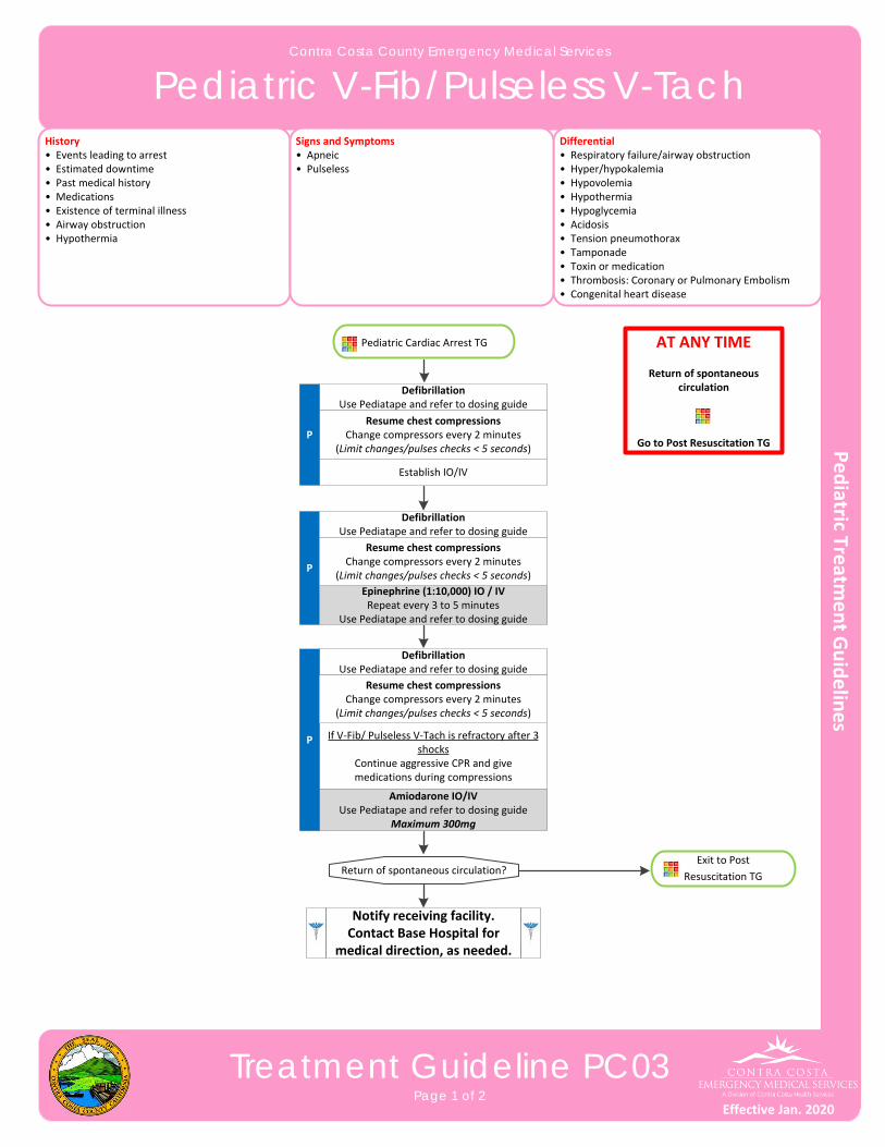
Effective Jan. 2016
Contra Costa County Emergency Medical Services
Pediatric V-Fib/Pulseless V-Tach
Treatment Guideline PC03Page 1 of 2
History• Events leading to arrest• Estimated downtime• Past medical history• Medications• Existence of terminal illness• Airway obstruction• Hypothermia
Differential• Respiratory failure/airway obstruction• Hyper/hypokalemia • Hypovolemia • Hypothermia• Hypoglycemia• Acidosis • Tension pneumothorax• Tamponade• Toxin or medication• Thrombosis: Coronary or Pulmonary Embolism• Congenital heart disease
Signs and Symptoms• Apneic • Pulseless
Establish IO/IV
P
Epinephrine (1:10,000) IO / IVRepeat every 3 to 5 minutes
Use Pediatape and refer to dosing guide
DefibrillationUse Pediatape and refer to dosing guide
AT ANY TIME
Return of spontaneous circulation
Go to Post Resuscitation TG
Resume chest compressionsChange compressors every 2 minutes
(Limit changes/pulses checks < 5 seconds)P
DefibrillationUse Pediatape and refer to dosing guide
If V‐Fib/ Pulseless V‐Tach is refractory after 3 shocks
Continue aggressive CPR and give medications during compressions
DefibrillationUse Pediatape and refer to dosing guide
Amiodarone IO/IVUse Pediatape and refer to dosing guide
Maximum 300mg
Exit to Post
Resuscitation TG Return of spontaneous circulation?
Resume chest compressionsChange compressors every 2 minutes
(Limit changes/pulses checks < 5 seconds)
P
Resume chest compressionsChange compressors every 2 minutes
(Limit changes/pulses checks < 5 seconds)
Pediatric Cardiac Arrest TG
Notify receiving facility. Contact Base Hospital for
medical direction, as needed.
Effective Jan. 2020
Effective Jan. 2016
Contra Costa County Emergency Medical Services
Pediatric V-Fib/Pulseless V-Tach
Treatment Guideline PC03Page 2 of 2
Pearls• Efforts should be directed at high quality chest compressions with limited interruptions and early defibrillation
when indicated. Compress 1.5 inches in infants and 2 inches in children. Consider early IO placement if available or direct IV access if anticipated.
• DO NOT HYPERVENTILATE.• Use a metronome during chest compression to ensure proper rate.• Airway is a more important intervention in pediatric arrests. This should be accomplished quickly with a BVM.
Patient survival is often dependent on proper ventilation and oxygenation with airway intervention.• In order to be successful in pediatric arrests, a cause must be identified and corrected.• Respiratory arrest is a common cause of cardiac arrest. Unlike adults, early ventilation intervention is critical.• In most cases, pediatric airways can be managed by basic intervention with a BVM.• Reassess and document ETT placement (patients above 40kg) and EtCO2 frequently, after every move, and at transfer of care.• Do not stop chest compressions to check for placement of ETT or to give medications.
Effective Jan. 2020
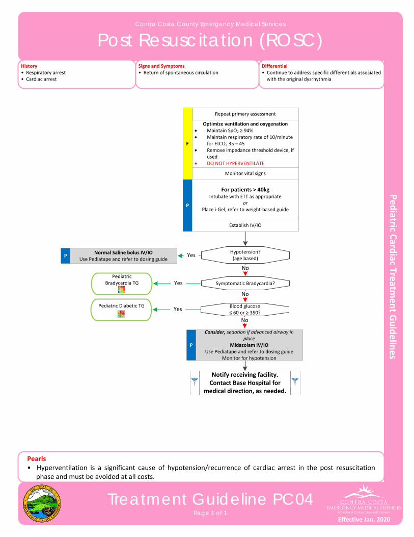
Effective Jan. 2016
Contra Costa County Emergency Medical Services
Post Resuscitation (ROSC)
Treatment Guideline PC04Page 1 of 1
History• Respiratory arrest• Cardiac arrest
Differential• Continue to address specific differentials associated with the original dysrhythmia
Signs and Symptoms• Return of spontaneous circulation
P
Consider, sedation if advanced airway in place
Midazolam IV/IO Use Pediatape and refer to dosing guide
Monitor for hypotension
Hypotension?(age based)
Repeat primary assessment
E
Optimize ventilation and oxygenation Maintain SpO2 ≥ 94% Maintain respiratory rate of 10/minute
for EtCO2 35 – 45 Remove impedance threshold device, if
used DO NOT HYPERVENTILATE
P
Pediatric Bradycardia TG Yes
No
Establish IV/IO
Monitor vital signs
Symptomatic Bradycardia?
Normal Saline bolus IV/IOUse Pediatape and refer to dosing guide
YesP
No
Pediatric Diabetic TGYes Blood glucose
≤ 60 or ≥ 350?
No
Pearls• Hyperventilation is a significant cause of hypotension/recurrence of cardiac arrest in the post resuscitation
phase and must be avoided at all costs.
Notify receiving facility. Contact Base Hospital for
medical direction, as needed.
For patients > 40kgIntubate with ETT as appropriate
orPlace i‐Gel, refer to weight‐based guide
Effective Jan. 2020
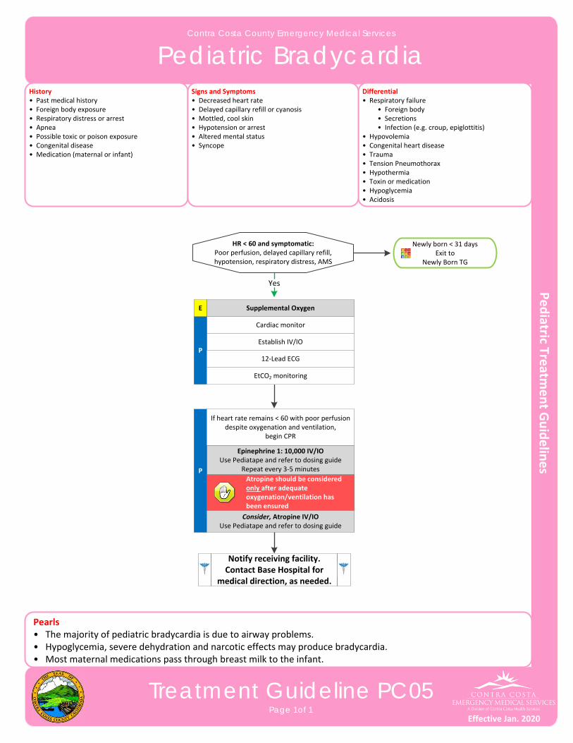
Effective Jan. 2016
Contra Costa County Emergency Medical Services
Pediatric Bradycardia
Treatment Guideline PC05Page 1of 1
History• Past medical history• Foreign body exposure• Respiratory distress or arrest• Apnea• Possible toxic or poison exposure• Congenital disease• Medication (maternal or infant)
Differential• Respiratory failure • Foreign body • Secretions • Infection (e.g. croup, epiglottitis)• Hypovolemia• Congenital heart disease• Trauma• Tension Pneumothorax• Hypothermia• Toxin or medication• Hypoglycemia• Acidosis
Signs and Symptoms• Decreased heart rate• Delayed capillary refill or cyanosis• Mottled, cool skin• Hypotension or arrest• Altered mental status• Syncope
HR < 60 and symptomatic:Poor perfusion, delayed capillary refill, hypotension, respiratory distress, AMS
Cardiac monitor
Establish IV/IOP
12‐Lead ECG
Yes
Epinephrine 1: 10,000 IV/IOUse Pediatape and refer to dosing guide
Repeat every 3‐5 minutesP
EtCO2 monitoring
If heart rate remains < 60 with poor perfusion despite oxygenation and ventilation,
begin CPR
Consider, Atropine IV/IOUse Pediatape and refer to dosing guide
Atropine should be considered only after adequate oxygenation/ventilation has been ensured
Pearls• The majority of pediatric bradycardia is due to airway problems.• Hypoglycemia, severe dehydration and narcotic effects may produce bradycardia.• Most maternal medications pass through breast milk to the infant.
Supplemental OxygenE
Newly born < 31 daysExit to
Newly Born TG
Notify receiving facility. Contact Base Hospital for
medical direction, as needed.
Effective Jan. 2020
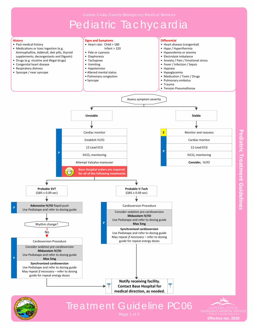
Effective Jan. 2016
Contra Costa County Emergency Medical Services
Pediatric Tachycardia
Treatment Guideline PC06Page 1 of 2
History• Past medical history• Medications or toxic ingestion (e.g. Aminophylline, Adderall, diet pills, thyroid supplements, decongestants and Digoxin)• Drugs (e.g. nicotine and illegal drugs)• Congenital heart disease• Respiratory distress• Syncope / near syncope
Differential• Heart disease (congenital)• Hypo / Hyperthermia • Hypovolemia or anemia• Electrolyte imbalance• Anxiety / Pain / Emotional stress• Fever / Infection / Sepsis• Hypoxia• Hypoglycemia• Medication / Toxin / Drugs• Pulmonary embolus• Trauma• Tension Pneumothorax
Signs and Symptoms• Heart rate: Child > 180 Infant > 220• Pale or cyanosis• Diaphoresis• Tachypnea• Vomiting• Hypotension • Altered mental status• Pulmonary congestion• Syncope
Assess symptom severity
Cardioversion Procedure
Synchronized cardioversionUse Pediatape and refer to dosing guideMay repeat if necessary – refer to dosing
guide for repeat energy doses
P
Consider sedation pre‐cardioversionMidazolam IV/IO
Use Pediatape and refer to dosing guideMax 5mg
Cardiac monitor
Establish IV/IO
P
12‐Lead ECG
Probable SVT(QRS ≤ 0.09 sec)
Probable V‐Tach(QRS ≥ 0.09 sec)
PAdenosine IV/IO Rapid push
Use Pediatape and refer to dosing guide
Base Hospital orders are required for all of the following treatments
No
EtCO2 monitoring
Attempt Valsalva maneuver
Rhythm change?
Cardioversion Procedure
Synchronized cardioversionUse Pediatape and refer to dosing guideMay repeat if necessary – refer to dosing
guide for repeat energy doses
P
Consider sedation pre‐cardioversionMidazolam IV/IO
Use Pediatape and refer to dosing guideMax 5mg
Cardiac monitor
Consider, IV/IO
P12‐Lead ECG
EtCO2 monitoring
Monitor and reassessE
StableUnstable
Notify receiving facility. Contact Base Hospital for
medical direction, as needed.
Effective Jan. 2020
Effective Jan. 2016
Contra Costa County Emergency Medical Services
Pediatric Tachycardia
Treatment Guideline PC06Page 2 of 2
Pearls• Most important goal is to differentiate the type of tachycardia and if STABLE or UNSTABLE.• Unstable is defined by poor perfusion with AMS, abnormal pulses, delayed capillary refill, or difficult or unable to
palpate a blood pressure.• If at any point the patient becomes unstable, move to the unstable arm of the algorithm.• Early transport is always appropriate in unstable patients.• For ASYMPTOMATIC patients (or those with only minimal symptoms, such as palpitations) and any tachycardia
with a rate of < 150 with a normal blood pressure, consider CLOSE OBSERVATION or fluid bolus rather than immediate treatment with an anti‐arrhythmic medication.
• Use Pediatape for pediatric weight measurement. ALWAYS use the weight‐based dosing guide.• Separating the child from the caregiver may worsen the child’s clinical condition.• Pediatric pads should be used in children < 10kg or Pediatape measurement of Purple.• Monitor for respiratory depression and associated hypotension associated if Midazolam is used.
Effective Jan. 2020
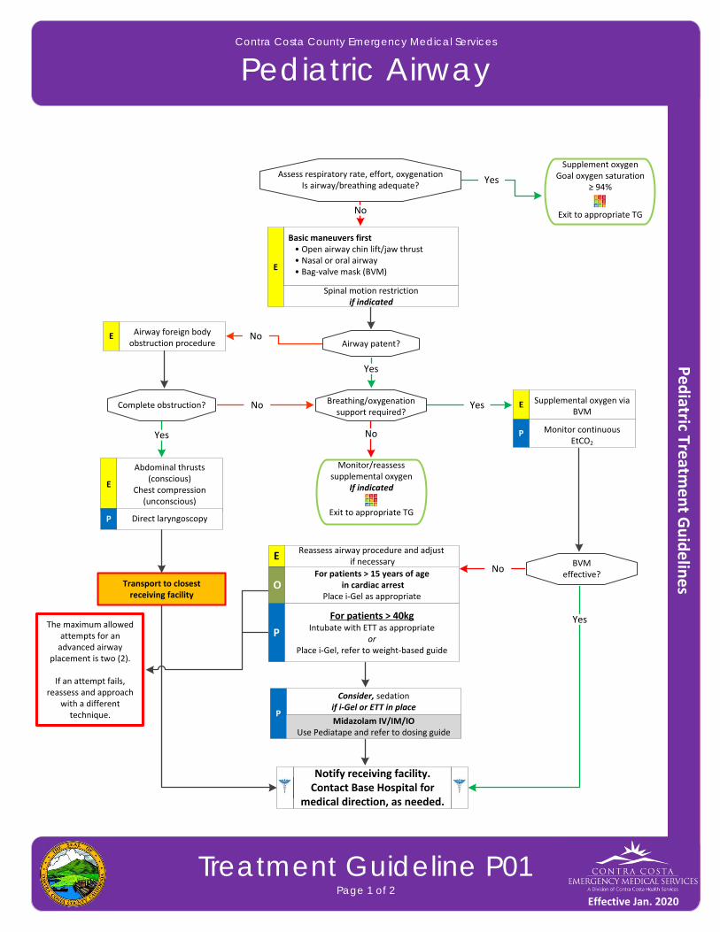
Effective Jan. 2016
Contra Costa County Emergency Medical Services
Pediatric Airway
Treatment Guideline P01Page 1 of 2
Basic maneuvers first • Open airway chin lift/jaw thrust • Nasal or oral airway • Bag‐valve mask (BVM)E
Spinal motion restrictionif indicated
Yes
Supplement oxygenGoal oxygen saturation
≥ 94%
Exit to appropriate TG
Monitor/reassess supplemental oxygen
If indicated
Exit to appropriate TG
Supplemental oxygen via BVM
E
Airway foreign body obstruction procedure
E
No
No
No
Yes
Yes
No
Assess respiratory rate, effort, oxygenationIs airway/breathing adequate?
Airway patent?
Breathing/oxygenationsupport required?
Complete obstruction?
Transport to closestreceiving facility
Yes
Consider, sedation if i‐Gel or ETT in place
PMidazolam IV/IM/IO
Use Pediatape and refer to dosing guide
The maximum allowed attempts for an advanced airway
placement is two (2).
If an attempt fails, reassess and approach
with a different technique.
NoBVM
effective?
Yes
Abdominal thrusts (conscious)
Chest compression (unconscious)
E
Monitor continuous EtCO2
P
Reassess airway procedure and adjust if necessary
For patients > 15 years of age in cardiac arrest
Place i‐Gel as appropriateO
For patients > 40kgIntubate with ETT as appropriate
orPlace i‐Gel, refer to weight‐based guide
P
E
Direct laryngoscopyP
Notify receiving facility. Contact Base Hospital for
medical direction, as needed.
Effective Jan. 2020
Effective Jan. 2016
Contra Costa County Emergency Medical Services
Pediatric Airway
Treatment Guideline P01Page 2 of 2
Pearls• Placement of an advanced airway is not a priority during the first five minutes of resuscitation unless ventilation is unable to be maintained with basic maneuvers.• Endotracheal intubation is only approved for patients over 40kg. • Capnometry is mandatory with all methods of airway management. Document results.• Continuous capnometry (EtCO2) is mandatory for the monitoring of all respiratory patients.• If an effective airway is being maintained with a BVM and a basic airway adjunct with continuous pulse oximetry values of ≥ 90% or values expected based on pathophysiologic condition with otherwise reassuring vital sign (e.g. pulse oximetry of 85% with otherwise normal vital signs in a post‐drowning patient), it is expected to continue with basic airway measures.• For the purposes of this TG, a secure airway is achieved when the patient is receiving appropriate oxygenation and ventilation.• An intubation attempt is defined as inserting the laryngoscope blade with the intent to intubate or inserting advanced airway past the teeth.• An appropriate ventilatory rate is one that maintains an EtCO2 of 35 or greater. Avoid hyperventilation.• Patients with perfusing pulses should be managed with a BLS airway unless unable to successfully ventilate.• Contraindications for i_Gel: • Presence of gag reflex • Laryngectomy with stoma (alternatively place ET in stoma) • Caustic ingestion • Known esophageal disease• Effective use of a BVM requires two (2) people.• Airway is a more important intervention in pediatric arrests. This should be accomplished quickly with a BVM and appropriately sized mask. Patient survival is often dependent on proper ventilation and oxygenation.• Maintain spinal immobilization for patients with suspected spinal injury.• Hyperventilation in deteriorating head trauma should only be done to maintain an EtCO2 of 30‐35.• It is important to secure the advanced airway well and consider c‐collar (in the absence of trauma) to better maintain advanced airway placement. Manual stabilization of advanced airway should be used during all patient moves/transfers.
Effective Jan. 2020
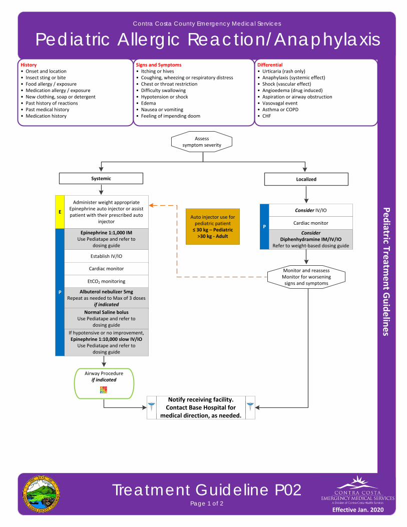
Effective Jan. 2016
Contra Costa County Emergency Medical Services
Pediatric Allergic Reaction/Anaphylaxis
Treatment Guideline P02Page 1 of 2
History• Onset and location• Insect sting or bite• Food allergy / exposure • Medication allergy / exposure• New clothing, soap or detergent• Past history of reactions• Past medical history• Medication history
Differential• Urticaria (rash only)• Anaphylaxis (systemic effect)• Shock (vascular effect)• Angioedema (drug induced)• Aspiration or airway obstruction• Vasovagal event• Asthma or COPD• CHF
Signs and Symptoms• Itching or hives• Coughing, wheezing or respiratory distress• Chest or throat restriction• Difficulty swallowing• Hypotension or shock• Edema• Nausea or vomiting• Feeling of impending doom
P
Consider IV/IO
Cardiac monitor
Monitor and reassessMonitor for worsening signs and symptoms
Airway Procedureif indicated
Consider Diphenhydramine IM/IV/IO
Refer to weight‐based dosing guide
If hypotensive or no improvement,Epinephrine 1:10,000 slow IV/IO
Use Pediatape and refer to dosing guide
P
Establish IV/IO
Cardiac monitor
Administer weight appropriate Epinephrine auto injector or assist patient with their prescribed auto
injector
E
Albuterol nebulizer 5mgRepeat as needed to Max of 3 doses
if indicated
Normal Saline bolus Use Pediatape and refer to
dosing guide
EtCO2 monitoring
Epinephrine 1:1,000 IMUse Pediatape and refer to
dosing guide
Assess symptom severity
LocalizedSystemic
Notify receiving facility. Contact Base Hospital for
medical direction, as needed.
Auto injector use for pediatric patient≤ 30 kg – Pediatric>30 kg ‐ Adult
Effective Jan. 2020

Effective Jan. 2016
Contra Costa County Emergency Medical Services
Pediatric Allergic Reaction/Anaphylaxis
Treatment Guideline P02Page 2 of 2
Pearls• Anaphylaxis is an acute and potentially lethal multisystem allergic reaction. • Epinephrine is the drug of choice and the first drug that should be administered in acute anaphylaxis reactions with moderate or severe symptoms. IM Epinephrine should be administered as priority before or during attempts at IV or IO access. • Anaphylaxis unresponsive to repeat doses of IM Epinephrine may require IV Epinephrine administration. Contact the Base Hospital for refractory anaphylaxis.• Allergic reactions may occur with only respiratory and gastrointestinal symptoms and have no rash or skin involvement.• All patients with respiratory symptoms must have continuous pulse oximetry and EtCO2 measurement.• The shorter the onset of symptoms from contact with an allergen, generally the more severe the reaction.
Effective Jan. 2020
Effective Jan. 2016
Contra Costa County Emergency Medical Services
Pediatric Altered Mental Status
Treatment Guideline P03Page 1 of 1
History• Past medical history• Medications• Recent illness • Irritability• Lethargy• Changes in feeding/sleeping• Diabetes• Potential ingestion• Trauma
Differential• Hypoxia• CNS (stroke, tumor, seizure, infection)• Thyroid• Shock • Diabetes• Toxicological• Acidosis or alkalosis• Diabetes• Environmental exposure• Psychiatric disorder• Sepsis
Signs and Symptoms• Decrease/change in mentation• Decrease/increase in blood sugar• Cool, diaphoretic skin• Warm, dry skin; fruity breath; Kussmaul respirations; signs of dehydration
P
Blood glucose analysis
Cardiac monitor
Blood glucose ≤ 60 or ≥ 350Exit to
Diabetic TG
12‐Lead ECG, if indicated
Airway TG, if indicated
Establish IV/IO
Signs of shock/poor perfusion
Signs of OD/toxic exposure
Signs of stroke or seizure
Signs of hypo/hyperthermia
No
No
No
No
Exit to
Hypotension/Shock TG
Exit to Overdose/Toxic
Exposure TG
Exit to Stroke or Seizure TG
as indicated
Exit to Hypo/Hyperthermia TG
as indicated
Yes
Yes
Yes
Yes
Yes
No
Arrhythmia/STEMI
No
Exit to Appropriate Cardiac TG
as indicatedYes
E
Notify receiving facility. Contact Base Hospital for
medical direction, as needed.
Effective Jan. 2020

Effective Jan. 2016
Contra Costa County Emergency Medical Services
Pediatric Behavioral
Treatment Guideline P04Page 1 of 2
History• Situational crisis• Psychiatric illness/medications• Injury to self or threats to others• Medical alert tag• Substance abuse/overdose• Diabetes
Differential• Altered mental status• Alcohol intoxication • Toxin/substance abuse• Medication effect/overdose• Withdrawal symptoms• Depression • Bipolar (manic‐depressive)• Schizophrenia• Anxiety disorders• Hypoglycemia
Signs and Symptoms• Anxiety, agitation or confusion• Affect change or hallucinations• Delusional thoughts or bizarre behavior• Combative or violent • Expression of suicidal/homicidal thoughts
Exit to appropriate TG, if indicated
Altered Mental Status TGOverdose/Toxic Ingestion TG
Head Trauma TG
Assume patient has medical cause of
behavioral change
Establish IV
E
Excited Delirium SyndromeParanoia, disorientation, extremely aggressive or violent, hallucinations, tachycardia, increased strength,
hyperthermia, and clearly a danger to self or others
Consider restraints
Monitor restraints and PMSif indicated
P
For patients ≥ 12 years of ageMidazolam
Use Pediatape and refer to dosing guide
Diabetic TG
if indicated
Monitor and reassess
Aggressive or agitated, possible psychosis, possible danger to self,
or others
Blood glucose analysis
P
For patients ≥ 12 years of ageMidazolam
Use Pediatape and refer to dosing guide
Midazolam Contact Base Hospital for additional
orderN
Cardiac monitor
P
Consider external cooling measures
Notify receiving facility. Contact Base Hospital for
medical direction, as needed.
Effective Jan. 2020

Effective Jan. 2016
Contra Costa County Emergency Medical Services
Pediatric Behavioral
Treatment Guideline P04Page 2 of 2
Pearls• Crew/responder safety is the main priority. See Policy 1008 – Managing Assaultive Behavior/Patient Restraint.• Avoid using benzodiazepines for patients with alcohol intoxication.• Limit IN administrations to ½ dose in each nare.• All patients who receive either physical restraint or chemical sedation must be continuously observed by EMS
personnel. This includes direct visualization of the patient as well as cardiac and pulse oximetry monitoring.• Consider all possible medical/trauma causes for behavior (e.g. hypoglycemia, overdose, substance abuse, hypoxia, seizure, head injury, etc.).• Use caution when considering the use of Midazolam with postictal patients. • Do not irritate the patient with a prolonged exam. Be thorough but quick.• Do not overlook the possibility of associated domestic violence or child abuse.• If patient suspected of excited delirium and suffers cardiac arrest, consider fluid bolus and sodium bicarbonate early.• Do not position or transport any restrained patient in a way that negatively affects the patient’s respiratory or circulatory status (e.g. hog‐tied or prone positions). Do not place backboards, splints or other devices on top of
the patient.• If restrained, the extremities that are restrained will have a circulation check at least every 15 minutes. The first
of these checks should occur as soon after placement of the restraints as possible. This shall be documented in the EHR.
Excited Delirium Syndrome:
This is a medical emergency. The condition is a combination of delirium, psychomotor agitation, anxiety, hallucinations, speech disturbances, disorientation, violent/bizarre behavior, insensitivity to pain, hyperthermia and increased strength. The condition is life‐threatening and is often associated with use of physical control measures, including physical restraints, and tasers. Most commonly seen in male patients with a history of serious mental illness or drug abuse, particularly stimulant drugs such as cocaine, crack cocaine, methamphetamine, amphetamines, bath salts, or similar agents. Alcohol withdrawal or head injury may also contribute to the condition.
Effective Jan. 2020
Contact the Base Hospital for all AMA requests
Effective Jan. 2016
Contra Costa County Emergency Medical Services
Pediatric Brief Resolved Unexplained Event (BRUE)
Treatment Guideline P05Page 1 of 1
History• Recent trauma, infection (e.g., fever, cough)• GERD• Congenital heart disease• Seizures • Medications
Differential• GERD• Pertussis• Respiratory infection• Seizure • Infection• Abuse
Signs and Symptoms• Brief decrease/change in mentation• Brief period of cyanosis or pallor• Brief absence, decrease or irregular respirations• Brief marked change in muscle tone • Brief altered responsiveness
P
Blood glucose analysis
Cardiac monitor
12‐Lead ECG, if indicated
Airway TG, if indicatedE
An infant ≤ 1 year who experienced an episode frightening to the observer, which is characterized by:• cyanosis or pallor• absent, decreased, or irregular breathing• choking or gagging• change in muscle tone• altered level of consciousness
Patients experiencing a BRUE should be transported to an appropriate hospital
for further evaluation
Pearls• BRUE was formally known as Apparent Life Threatening Event (ALTE).• BRUE is formally diagnosed in the ED only when there is no explanation for a qualifying event after a physician
conducts an appropriate history and physical examination.• Always consider non‐accidental trauma in any infant who presents with BRUE.• Even with a normal physical examination at the time of EMS contact, patients that have experienced BRUE should
be transported for further evaluation.• It is important to document sleeping position as parent co‐sleeping with child is associated with infant deaths.
Notify receiving facility. Contact Base Hospital for
medical direction, as needed.
Effective Jan. 2020
Effective Jan. 2016
Contra Costa County Emergency Medical Services
Pediatric Diabetic
Treatment Guideline P06Page 1 of 1
History• Past medical history• Medications• Recent blood glucose check • Last meal
Differential• Alcohol or drug use• Toxic ingestion• Trauma or head injury• Seizure• Stroke• Altered baseline mental status
Signs and Symptoms• Altered mental status• Combative or irritable• Diaphoresis• Seizure• Abdominal pain• Nausea or vomiting• Weakness• Dehydration• Deep or rapid breathing
P
Blood glucose analysis
Cardiac monitor
12‐Lead ECG procedureif indicated
Establish IV/IO
Altered Mental Status TG
if indicated
Able to follow commandsbut symptomatic
Exit to
Hypotension/ Shock TG
Consider Oral Glucose 1 tube (30g)E
Blood glucose ≤ 60mg/dl Blood glucose ≥ 350mg/dl
D‐10 IVUse Pediatape and refer to
dosing guide
PNo venous accessGlucagon IM
Use Pediatape and refer to dosing guide
Repeat in 15 minutes if neededConsider IO access as a last resort
Improving?
P
If blood glucose < 60mg/dlRepeat D‐10 IV
Use Pediatape and refer to dosing guide
No
PNormal Saline bolus IV
Use Pediatape and refer to dosing guide
Hypotension?Yes
Yes
No
Yes
Improving?
D‐10Contact Base Hospital for additional
orderN
No
Pearls• Do not administer oral glucose to patients that are not able to swallow or protect their airway.• Patients with prolonged hypoglycemia may not respond to Glucagon.
Notify receiving facility. Contact Base Hospital for
medical direction, as needed
Suspected hypoglycemia or patient’s glucometer results read
<60mg/dlE
No
Effective Jan. 2020
Effective Jan. 2016
Contra Costa County Emergency Medical Services
Pediatric Hypotension/Shock
Treatment Guideline P07Page 1 of 2
History• Blood loss • Vomiting• Diarrhea • Fever• Infection
Differential• Shock (hypovolemic, cardiogenic, septic, neurogenic, or anaphylaxis)• Trauma• Infection• Dehydration• Congenital heart disease• Medication or Toxin
Signs and Symptoms• Restlessness or confusion• Weakness or dizziness• Weak, rapid pulse• Pale, cool, clammy skin signs• Delayed capillary refill• Hypotension• Tarry stools
Diabetic TG
if indicated
Spinal motion restriction,if indicated
E CONTROL HEMORRHAGE and wound care,as indicated
P
Cardiac monitor
History, exam and circumstances often suggest (type of shock)WAS TRAUMA INVOLVED?
Yes
IV/IO procedure
Blood glucose analysis
Consider 12‐Lead ECG
P
Normal Saline bolus IV/IOUse Pediatape and refer to
dosing guideRepeat to age dependent goal SBP
Maximum 1L
Airway TGsif indicated
Cardiac/Arrhythmia TG
if indicated
Consider hypovolemic (bleeding), neurogenic (spinal injury) and
obstructive (pneumothorax) shock
Consider hypovolemic (dehydration or GI bleed), cardiogenic (STEMI or CHF), distributive (sepsis or anaphylaxis) and obstructive (PE or cardiac tamponade)
shock
No
Chest Decompression procedureif indicated
P
Normal Saline bolus IV/IOUse Pediatape and refer to
dosing guideRepeat to age dependent goal SBP
Maximum 1L
Exit to Trauma TG
if indicatedNormal Saline
Contact Base Hospital Physician for additional order
MD
Exit to appropriate TG
E
Notify receiving facility. Contact Base Hospital for
medical direction, as needed.
Effective Jan. 2020
Effective Jan. 2016
Contra Costa County Emergency Medical Services
Pediatric Hypotension/Shock
Treatment Guideline P07Page 2 of 2
Pearls• Hypotension is age dependent. This is not always reliable and should be interpreted in context with the patient’s typical BP, if known. Shock may be present with a seemingly normal blood pressure initially. Hypotension is defined as:
▫ Neonate: < 60mmHg or weak pulses ▫ Infant: < 70mmHg or weak pulses
▫ 1‐10 years: < 70mmHg + (age in years x2)▫ Over 10 years: < 90mmHg
• Systemic BP goals are defined as:▫ Neonate: > 60mmHg
▫ Infant: > 70mmHg ▫ 1‐10 years: > 70mmHg + (age in years x2)▫ Over 10 years: > 90mmHg
• Common pediatric terms used to describe children are defined as: ▫ Newly born are ≤ 24 hours old ▫ Neonates are ≤ 28 days old ▫ Infants are ≤ 1 year old
• Normal blood pressure, delayed capillary refill, diminished peripheral pulses, and tachycardia indicates compensated shock in children.• Hypotension and delayed capillary refill > 4 seconds indicates impending circulatory failure. • Systolic blood pressure in children may not drop until the patient is 25‐30% volume depleted. This may occur through dehydration, blood loss, or an increase in vascular capacity (e.g. anaphylaxis).• Decompensated shock (hypotension with capillary refill > 5 seconds) may present as PEA in children.• Sinus tachycardia is the most common cardiac rhythm in encountered in children.• SVT should be suspected in the heart rate is greater than 180 in children ages (1‐8) or greater than 220 in infants.• Hypoglycemia may be found in pediatric shock, especially in infants.• Pediatric shock victims are at risk for hypothermia due to their increased body surface area, exposure, and rapid
administration of IV/IO fluids.
Effective Jan. 2020
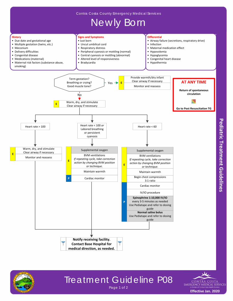
Effective Jan. 2016
Contra Costa County Emergency Medical Services
Newly Born
Treatment Guideline P08Page 1 of 2
History• Due date and gestational age• Multiple gestation (twins, etc.)• Meconium• Delivery difficulties• Congenital disease• Medications (maternal)• Maternal risk factors (substance abuse, smoking)
Differential• Airway failure (secretions, respiratory drive)• Infection• Maternal medication effect • Hypovolemia• Hypoglycemia• Congenital heart disease• Hypothermia
Signs and Symptoms• Just born• Uncut umbilical cord• Respiratory distress• Peripheral cyanosis or mottling (normal)• Central cyanosis or mottling (abnormal)• Altered level of responsiveness• Bradycardia
Term gestation?Breathing or crying?Good muscle tone?
AT ANY TIME
Return of spontaneous circulation
Go to Post Resuscitation TG
No
Warm, dry, and stimulateClear airway if necessary
E
Provide warmth/dry infantClear airway if necessary
EYesMonitor and reassess
BVM ventilationsIf repeating cycle, take correction action by changing BVM position
or technique.
E
Cardiac monitorP
Supplemental oxygen
Maintain warmth
Begin chest compressions 3:1 ratio
IV/IO procedure
Epinephrine 1:10,000 IV/IOevery 3‐5 minutes as needed
Use Pediatape and refer to dosing guide
Normal saline bolusUse Pediatape and refer to dosing
guide
Heart rate > 100 Heart rate < 100 orLabored breathing
or persistent cyanosis
Heart rate < 60
BVM ventilationsIf repeating cycle, take correction action by changing BVM position
or technique.E
Cardiac monitor
P
Supplemental oxygen
Maintain warmth
Warm, dry, and stimulateClear airway if necessary
EMonitor and reassess
Notify receiving facility. Contact Base Hospital for
medical direction, as needed.
Effective Jan. 2020
Effective Jan. 2016
Contra Costa County Emergency Medical Services
Newly Born
Treatment Guideline P08Page 2 of 2
Pearls• Most newborns requiring resuscitation will respond to ventilation/BVM, compressions, or Epinephrine. If not
responding, consider hypovolemia, pneumothorax, or hypoglycemia (< 40mg/dl). • Transport mother WITH infant whenever possible. • Do not place hot packs directly on baby’s skin as it may cause severe burns. • Common pediatric terms used to describe children are defined as:
▫ Newly born are ≤ 24 hours old ▫ Neonates are ≤ 28 days old ▫ Infants are ≤ 1 year old
• Term gestation, strong cry/breathing and with good muscle tone generally will need no resuscitation.• Most important vital signs in the newly born are respirations/respiratory effort and heart rate.• Place baby skin‐to‐skin on mother.• It is extremely important to keep an infant warm.• Maternal sedation or narcotics will sedate an infant. • Naloxone is no longer recommended for use in the newly born who may be sedated from maternal medications.
Effective Jan. 2020
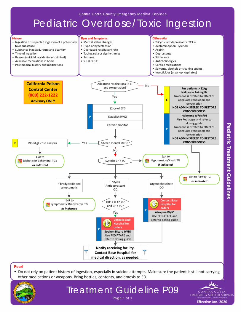
Effective Jan. 2016
Contra Costa County Emergency Medical Services
Pediatric Overdose/Toxic Ingestion
Treatment Guideline P09Page 1 of 1
History• Ingestion or suspected ingestion of a potentially toxic substance• Substance ingested, route and quantity• Time of ingestion • Reason (suicidal, accidental or criminal)• Available medications in home• Past medical history and medications
Differential• Tricyclic antidepressants (TCAs)• Acetaminophen (Tylenol) • Aspirin• Depressants• Stimulants• Anticholinergics• Cardiac medications• Solvents, alcohols or cleaning agents• Insecticides (organophosphates)
Signs and Symptoms• Mental status changes• Hypo or hypertension• Decreased respiratory rate• Tachycardia or dysrhythmias• Seizures• S.L.U.D.G.E.
P Establish IV/IO
Cardiac monitor
Exit to Diabetic or Behavioral TGs
as indicated
Yes
Adequate respirations (> 8)and oxygenation?
12‐Lead ECG
P
Naloxone IV/IM/INUse Pediatape and refer to
dosing guideNaloxone is titrated to effect of
adequate ventilation and oxygenation
NOT ADMINISTERED TO RESTORE CONSCIOUSNESS
No
Altered mental status?E Blood glucose analysis Yes
Exit to Airway TG
as indicated
Systolic BP < 90
No
Exit to Hypotension/Shock TG
if indicated
Organophosphate OD
Tricyclic Antidepressant
OD
If bradycardic and symptomatic
QRS ≥ 0.12 sec and BP < 90?
YesP
California Poison Control Center (800) 222‐1222Advisory ONLY
Exit to Symptomatic Bradycardia TG
as indicated
Pearl• Do not rely on patient history of ingestion, especially in suicide attempts. Make sure the patient is still not carrying other medications or weapons. Bring bottles, contents, and emesis to ED.
Contact Base Hospital for orders
P
Contact Base Hospital for orders
Atropine IV/IOUse PEDIATAPE and refer to dosing guide
Sodium Bicarb IV/IOUse PEDIATAPE and refer to dosing guide
E
For patients > 22kgNaloxone 2‐4 mg IN
Naloxone is titrated to effect of adequate ventilation and
oxygenationNOT ADMINISTERED TO RESTORE
CONSCIOUSNESS
Notify receiving facility. Contact Base Hospital for
medical direction, as needed.
Effective Jan. 2020

This page intentionally left blank
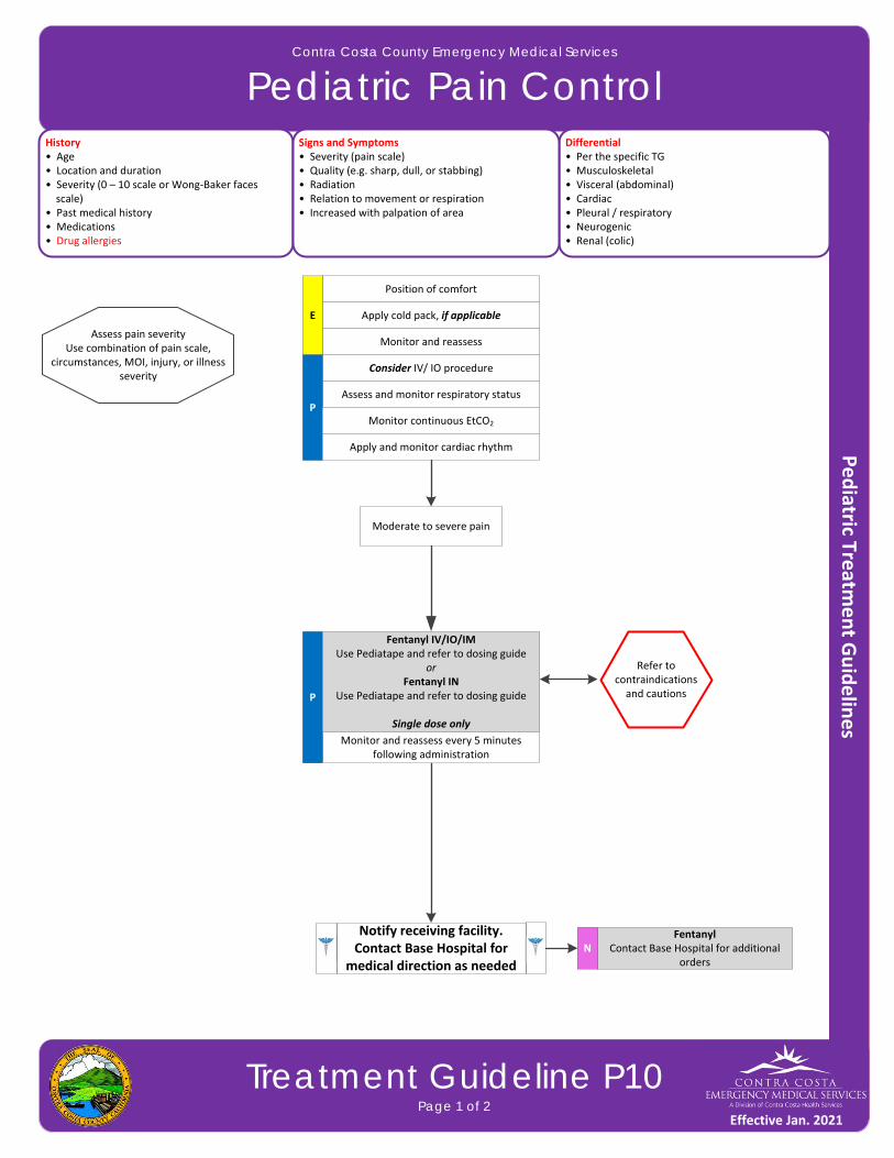
Effective Jan. 2016
Contra Costa County Emergency Medical Services
Pediatric Pain Control
Treatment Guideline P10Page 1 of 2
History• Age• Location and duration• Severity (0 – 10 scale or Wong‐Baker faces scale)• Past medical history• Medications• Drug allergies
Differential• Per the specific TG• Musculoskeletal • Visceral (abdominal)• Cardiac• Pleural / respiratory• Neurogenic• Renal (colic)
Signs and Symptoms• Severity (pain scale)• Quality (e.g. sharp, dull, or stabbing)• Radiation• Relation to movement or respiration• Increased with palpation of area
Position of comfort
Apply cold pack, if applicable
Monitor and reassess
FentanylContact Base Hospital for additional
ordersN
E
Assess pain severityUse combination of pain scale,
circumstances, MOI, injury, or illness severity
Moderate to severe pain
Consider IV/ IO procedure
Assess and monitor respiratory statusP
Monitor continuous EtCO2
Monitor and reassess every 5 minutes following administration
Apply and monitor cardiac rhythm
P
Fentanyl IV/IO/IMUse Pediatape and refer to dosing guide
orFentanyl IN
Use Pediatape and refer to dosing guide
Single dose only
Notify receiving facility. Contact Base Hospital for
medical direction as needed
Refer to contraindications and cautions
Effective Jan. 2021
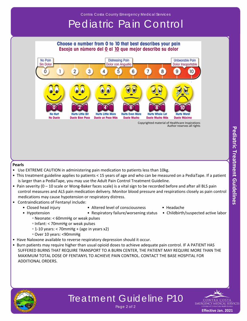
Effective Jan. 2016
Contra Costa County Emergency Medical Services
Pediatric Pain Control
Treatment Guideline P10Page 2 of 2
Pearls• Use EXTREME CAUTION in administering pain medication to patients less than 10kg.• This treatment guideline applies to patients < 15 years of age and who can be measured on a PediaTape. If a patient
is larger than a PediaTape, you may use the Adult Pain Control Treatment Guideline.• Pain severity (0 – 10 scale or Wong‐Baker faces scale) is a vital sign to be recorded before and after all BLS pain
control measures and ALS pain medication delivery. Monitor blood pressure and respirations closely as pain control medications may cause hypotension or respiratory distress.
• Contraindications of Fentanyl include: • Closed head injury • Altered level of consciousness • Headache • Hypotension • Respiratory failure/worsening status • Childbirth/suspected active labor
▫ Neonate: < 60mmHg or weak pulses ▫ Infant: < 70mmHg or weak pulses
▫ 1‐10 years: < 70mmHg + (age in years x2) ▫ Over 10 years: <90mmHg
• Have Naloxone available to reverse respiratory depression should it occur.• Burn patients may require higher than usual opioid doses to achieve adequate pain control. IF A PATIENT HAS
SUFFERED BURNS THAT REQUIRE TRANSPORT TO A BURN CENTER, THE PATIENT MAY REQUIRE MORE THAN THE MAXIMUM TOTAL DOSE OF FENTANYL TO ACHIEVE PAIN CONTROL. CONTACT THE BASE HOSPITAL FOR ADDITIONAL ORDERS.
Copyrighted material of Healthcare Inspirations Author reserves all rights
Effective Jan. 2021
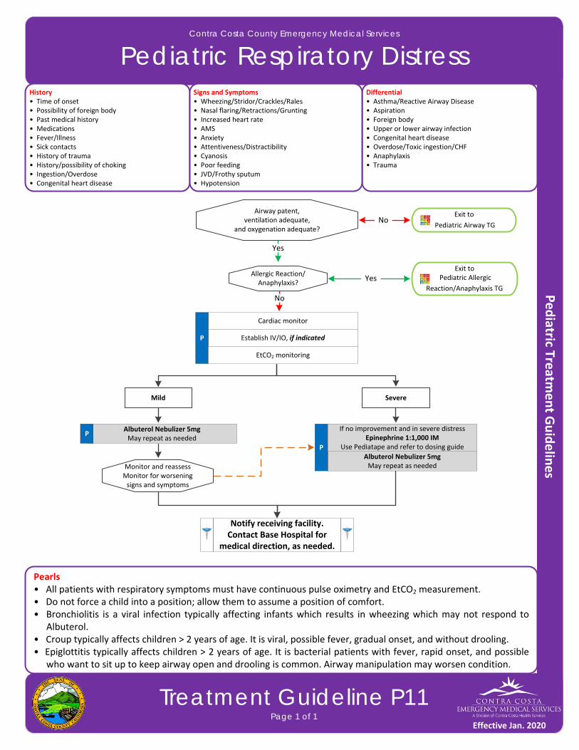
Effective Jan. 2016
Contra Costa County Emergency Medical Services
Pediatric Respiratory Distress
Treatment Guideline P11Page 1 of 1
History• Time of onset• Possibility of foreign body• Past medical history• Medications• Fever/Illness• Sick contacts• History of trauma• History/possibility of choking• Ingestion/Overdose• Congenital heart disease
Differential• Asthma/Reactive Airway Disease• Aspiration • Foreign body• Upper or lower airway infection• Congenital heart disease• Overdose/Toxic ingestion/CHF• Anaphylaxis• Trauma
Signs and Symptoms• Wheezing/Stridor/Crackles/Rales• Nasal flaring/Retractions/Grunting• Increased heart rate• AMS• Anxiety• Attentiveness/Distractibility• Cyanosis• Poor feeding• JVD/Frothy sputum• Hypotension
Airway patent,ventilation adequate,
and oxygenation adequate?
Exit to
Pediatric Airway TG
Cardiac monitor
Establish IV/IO, if indicatedP
No
Yes
Albuterol Nebulizer 5mgMay repeat as needed
P
EtCO2 monitoring
Exit to Pediatric Allergic
Reaction/Anaphylaxis TG
Allergic Reaction/ Anaphylaxis?
Yes
No
If no improvement and in severe distressEpinephrine 1:1,000 IM
Use Pediatape and refer to dosing guide
Mild
Pearls• All patients with respiratory symptoms must have continuous pulse oximetry and EtCO2 measurement.• Do not force a child into a position; allow them to assume a position of comfort. • Bronchiolitis is a viral infection typically affecting infants which results in wheezing which may not respond to
Albuterol. • Croup typically affects children > 2 years of age. It is viral, possible fever, gradual onset, and without drooling.• Epiglottitis typically affects children > 2 years of age. It is bacterial patients with fever, rapid onset, and possible
who want to sit up to keep airway open and drooling is common. Airway manipulation may worsen condition.
Severe
Albuterol Nebulizer 5mgMay repeat as needed
P
Monitor and reassessMonitor for worsening signs and symptoms
Notify receiving facility. Contact Base Hospital for
medical direction, as needed.
Effective Jan. 2020

This page intentionally left blank
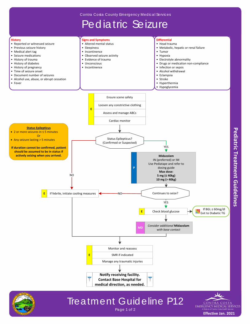
Effective Jan. 2016
Contra Costa County Emergency Medical Services
Pediatric Seizure
Treatment Guideline P12Page 1 of 2
History• Reported or witnessed seizure• Previous seizure history• Medical alert tag • Seizure medications• History of trauma• History of diabetes• History of pregnancy• Time of seizure onset• Document number of seizures• Alcohol use, abuse, or abrupt cessation• Fever
Differential• Head trauma• Metabolic, hepatic or renal failure• Tumor• Hypoxia• Electrolyte abnormality• Drugs or medication non‐compliance• Infection or sepsis• Alcohol withdrawal• Eclampsia • Stroke • Hyperthermia • Hypoglycemia
Signs and Symptoms• Altered mental status• Sleepiness• Incontinence• Observed seizure activity• Evidence of trauma• Unconscious• Incontinence
If BGL ≤ 60mg/dl Exit to Diabetic TG
YES
P
Status Epilepticus?(Confirmed or Suspected)
Monitor and reassess
Midazolam IN (preferred) or IM
Use Pediatape and refer to dosing guide Max dose:
5 mg (≤ 40kg)10 mg (> 40kg)
Loosen any constrictive clothing
Ensure scene safety
EAssess and manage ABCs
Cardiac monitor
SMR if indicated
Manage any traumatic injuries
E
Consider additional Midazolam with base contact
MD
Notify receiving facility. Contact Base Hospital for
medical direction, as needed.
NO
Status Epilepticus 2 or more seizures in ≤ 5 minutes
Or Any seizure lasting > 5 minutes
If duration cannot be confirmed, patient should be assumed to be in status if actively seizing when you arrived.
E If febrile, initiate cooling measures Continues to seize?
E
NO
YES
Check blood glucose
Effective Jan. 2021

Effective Jan. 2016
Contra Costa County Emergency Medical Services
Pediatric Seizure
Treatment Guideline P12Page 2 of 2
Pearls• Simple febrile seizures are most common in ages 6 months to 5 years of age. They are, by definition, generalized seizures with no seizure history in the setting of any grade of fever, with an otherwise normal neurologic and physical exam. Any seizure confirmed to last for more than five (5) minutes should be treated with medication.• Be prepared to assist ventilations, especially if Midazolam is used. Avoiding hypoxemia is extremely important.• In an infant, a seizure may be the only evidence of a closed head injury.• Status epilepticus is defined as two or more successive seizures without a period of consciousness or recovery OR
seizures lasting greater than 5 minutes. This is a true emergency requiring rapid airway control, treatment and transport.
• Assess for the possibility of occult trauma and substance abuse, overdose, or ingestion/toxins.
Effective Jan. 2021

Effective Jan. 2016
Contra Costa County Emergency Medical Services
Pediatric Vomiting and Diarrhea
Treatment Guideline P13Page 1 of 1
History• Age• Time of last meal• Last emesis/bowl movement• Improvement or worsening with food or activity• Duration of problem• Other sick contacts• Past medical history• Past surgical history• Medications• Menstrual history (Pregnancy)• Travel history• Bloody emesis/diarrhea
Differential• CNS (increased pressure, headache, stroke, CNS lesions, trauma or hemorrhage, vestibular)• MI • Drugs (NSAIDs, antibiotics, narcotics, chemotherapy)• GI or renal disorders• Diabetic ketoacidosis• Gynecologic disease (ovarian cyst, PID)• Infections (pneumonia, influenza)• Electrolyte abnormalities• Food or toxin induced• Medication or substance abuse• Pregnancy• Psychological
Signs and Symptoms• Abdominal pain• Character of pain (constant, intermittent, dull, sharp, etc.)• Distension• Constipation• Diarrhea• Anorexia• RadiationAssociated symptoms (helpful to localize source):Fever, headache, blurred vision, weakness, malaise, myalgia, cough, dysuria, mental status changes, and rash
PConsider
Normal Saline bolusUse Pediatape and refer to
dosing guide
Cardiac monitor
NoSerious sign/symptoms
Hypotension, poor perfusion, shock
Consider, IV/IO
P
Ondansetron IV/IO/IM/ODTUse Pediatape and refer to
dosing guide May repeat x1 in 15 minutes for
patients > 40kg
Yes
Nausea or vomiting
P
Normal Saline bolusUse Pediatape and refer to
dosing guide
Cardiac monitor
Establish IV/IO
Nausea or vomiting
Yes Yes
Blood glucose analysisE
Exit to Diabetic TG
as indicatedYes
Improving
Exit to Hypotension/Shock TG
as indicatedNo
Yes
Pearls• Heart rate: One of the first clinical signs of dehydration is almost always an increased heart rate. Tachycardia
increases as dehydration becomes more severe, very unlikely to be significantly dehydrated if heart rate is close to normal.
• Beware of only vomiting (without diarrhea) in children. Pyloric stenosis, bowel obstruction, and CNS processes (bleeding, tumors, or increased CSF pressures) all often present with isolated vomiting.• Ondansetron is not indicated for motion sickness.
Notify receiving facility. Contact Base Hospital for
medical direction, as needed.
Effective Jan. 2020

This page intentionally left blank

Measure vital signs and level of consciousness
Trauma Center transport
with early notification
Meets Destination / Call‐in Criteria?
Assess anatomy of injury
Assess mechanism of injury and evidence of high‐energy impact
No
Trauma Center transport
with early notificationNo
Call for destination decision
Trauma Center transport
with early notification
No
Unmanageable airway orTraumatic arrest not meeting field determination
Closest facilityYesNo
1
3
2
GCS ≤ 13
Systolic blood pressure < 90mmHg
Adult respiratory rate < 10 or > 29 or need for ventilatory support
Infant (< l year of age) respiratory rate < 20
All penetrating injuries to head, neck, torso, groin, pelvis, buttocks, and
extremities above the elbow or knee
Chest wall instability or deformity (e.g. flail chest)
Two or more proximal long bone fractures
Crushed, degloved, mangled, or pulseless extremity
Amputation above the wrist or ankle
Pelvic instability
Open or depressed skull deformity
Traumatic paralysis
Adult fall > 20 feet
Pediatric fall > 10 feet or 2‐3 times height of child
High risk auto crash with > 12 inches intrusion on patient side or > 18 inches at any site of passenger compartment
Ejection (partial or complete)
Death in same passenger compartment
Auto vs. pedestrian/bicyclist thrown, run over or with significant impact >
20mph
Any unenclosed vehicle crash > 20mph (e.g. motorcycle, bicycle,
ATV, etc.)
Contra Costa County Emergency Medical Services
Trauma Triage
Treatment Guideline T01Page 1 of 3
Yes
Yes
Yes
Major burn associated with trauma
ACTIVATION
Uncontrolled bleeding with tourniquet applied and tightened
Effective Jan. 2020

High energy mechanism
For situations not described above, consider Base Hospital contact if paramedic has concern that a serious injury may exist
Contra Costa County Emergency Medical Services
Trauma Triage
Treatment Guideline T01Page 2 of 3
4
Risk Factor Advisory
Patients with either high energy or low energy mechanisms are more prone to serious injury if they have one or more of the following risk factors:
Pregnancy over 20 weeks Communication barrier (e.g., age, language, psychiatric, or developmental issues) Age 55 or older Patient taking anticoagulants or with known bleeding disorder
Motor vehicle crash Estimated impact speed of > 40mph Mechanical extrication required by fire
department personnel Rollover with unrestrained occupant
Low energy mechanism
Person struck by a vehicle at < 20mph
Person ejected /fell from other object (e.g., motorcycle, horse, or ATV)
Blunt assault with weapon (e.g., pipe, bat, or golf club)
Falls > 10 but < 20 feet
This list is not all‐inclusive and other high energy mechanisms encountered also merit Base Hospital contact
Contact Base Hospital for destination
Low energy mechanisms should merit Base Hospital contact if symptoms, physical findings, or concern are encountered
Examples include, but are not limited to ground level or short falls, blunt assault without a weapon (e.g., closed fist), low speed motor vehicle crash, or other blunt trauma (e.g., sports injury)
Consider Base Hospital contact for destination
Effective Jan. 2020

Field Treatment GuidelinesPage 3 of 3
Effective Jan. 2016
Contra Costa County Emergency Medical Services
Trauma Triage
Treatment Guideline T01Page 3 of 3
Pearls• Do not let alcohol confuse the clinical picture. Alcoholics may have unrecognized injuries, particularly head bleeds.• A complete hands on head‐to‐toe assessment is required for all trauma patients. • Transport should be initiated within 10 minutes of ambulance arrival unless patient requires extrication. Age Categories Adult Patient – Trauma patients 15 years of age and older. Pediatric Patients – Trauma patients under the age of 15 years.
Trauma Receiving Facilities Adult Trauma Centers – John Muir Medical Center – Walnut Creek is the designated trauma center for adults in
Contra Costa County. In some circumstances, patients may be transported to other trauma receiving facilities. Alameda County Medical Center (Highland) and Eden Medical Center are trauma receiving facilities that, when they are the closest trauma receiving facility, may be appropriate for ground transport of trauma patients.
Pediatric Trauma Centers – UCSF Benioff Children’s Hospital of Oakland (CHO) is the most appropriate destination for the majority of pediatric trauma patients.
• John Muir Medical Center – Walnut Creek may be an appropriate trauma receiving facility for critically injured pediatric trauma patients who are near arrest or have a very prolonged transport time. UC Davis Medical Center is also a pediatric trauma receiving facility and may be utilized when helicopter transport is involved. Receiving Facilities – Local hospitals that are not trauma receiving facilities are destinations for patients who are
triaged by the Base Hospital at the time of report as not requiring trauma center care. A trauma receiving facility may also serve as the receiving facility when it is the patient’s facility of choice.
Low Energy Mechanism Trauma Low energy mechanism trauma may reveal significant trauma. Examples include, but are not limited to ground level or short falls, blunt assault without a weapon (e.g. closed fist), low speed motor vehicle crash, or other blunt trauma (e.g. sports injury). Symptoms or concern may include:
Symptoms in the presence of head injury such as headache, vomiting, loss of consciousness, repetitive questioning, abnormal, or combative behavior or new onset of confusion
Pain level greater than 5/10 related to head, neck, or torso injury Any concerns due to hypotension, tachycardia, or tachypnea Systolic BP < 110mmHg in patients 65 years of age or older Torso injury with tenderness of abdomen, chest/ribs or back/flank Suspected hip dislocation or pelvis injury
Other Definitions Unmanageable Airway – A patient whose airway is unable to be adequately maintained with BLS or ALS maneuvers. Adult trauma patients are candidates for immediate redirection to the trauma center following airway stabilization at a non‐trauma receiving facility. Traumatic Arrest – Patients who do not qualify for field determination of death but have or develop cardiopulmonary
arrest should be transported to the closest Basic ED by ground ambulance. Exceptions: • Patients with penetrating trauma who arrest (pulseless, apneic, or pulseless with agonal respirations) after the arrival of transport personnel should be immediately transported to a trauma center if transport time is 20 minutes or less to that facility. If no Trauma Center is available within 20 minutes, patients should be transported to the closest basic emergency department. • If a helicopter crew is present at the time of arrest (blunt or penetrating) and the air transport can be initiated immediately, use of helicopter to transport to a trauma center is appropriate.
Effective Jan. 2020

This page intentionally left blank
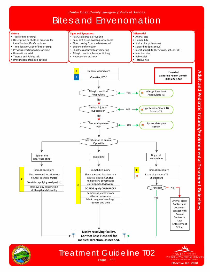
Effective Jan. 2016
Contra Costa County Emergency Medical Services
Bites and Envenomation
Treatment Guideline T02Page 1 of 2
History• Type of bite or sting• Description or photo of creature for identification, if safe to do so• Time, location, size of bite or sting• Previous reaction to bite or sting• Domestic vs. wild• Tetanus and Rabies risk• Immunocompromised patient
Differential• Animal bite• Human bite • Snake bite (poisonous)• Spider bite (poisonous)• Insect sting/bite (bee, wasp, ant, or tick)• Infection risk• Rabies risk• Tetanus risk
Signs and Symptoms• Rash, skin break, or wound• Pain, soft tissue swelling, or redness• Blood oozing from the bite wound• Evidence of infection• Shortness of breath or wheezing• Allergic reaction, hives, or itching• Hypotension or shock
If neededCalifornia Poison Control
(800) 222‐1222
General wound care E
P Consider, IV/IO
Serious injury orhypotension
Allergic reaction/Anaphylaxis
Moderate/severePain
No
No
Hypotension/Shock TGTrauma TG
Allergic Reaction/Anaphylaxis TG
Yes
Yes
Appropriate pain control
Yes
Identification of animal,if possible
Spider biteBee/wasp sting
Snake biteDog / cat
Human bite
Immobilize injury
E
Elevate wound location to a neutral position, if able
Consider, applying cold pack(s)
Remove any constricting clothing/bands/jewelry
Immobilize injury
E
Elevate wound location to a neutral position, if able
DO NOT apply COLD PACKS
Remove any constricting clothing/bands/jewelry
Remove all jewelry from affected extremity
Mark margin of swelling/ redness and time
Immobilize injuryE
Extremity trauma TGif indicated
Transport
Animal bites: Contact and document contact with
Animal Control or
Law Enforcement
Officer
No
Yes
Notify receiving facility. Contact Base Hospital for
medical direction, as needed.
Effective Jan. 2020
Effective Jan. 2016
Contra Costa County Emergency Medical Services
Bites and Envenomation
Treatment Guideline T02Page 2 of 2
Pearls• Human bites have higher infection rates than animal bites due to normally occurring mouth bacteria.• Carnivore bites are much more likely to become infected and may have risk of Rabies exposure.• Cat bites may progress to infection rapidly due to a specific bacteria (Pasteurella multicoda).• Poisonous snakes in our region are generally of the pit viper family: six rattlesnake species.• If no pain or swelling is present, envenomation is unlikely. About 25% of snake bites are dry bites.• Black Widow spider bites tend to be minimally painful initially, but over a few hours, muscular and severe abdominal pain may develop (black spider with a red hourglass on belly).• Brown Recluse spider bites are minimally painful to painless. Little reaction is noted initially but tissue necrosis
at the site of the bite develops over the next few days (brown spider with fiddle shape on back).• Evidence of infection includes: swelling, redness, drainage, fever, and red streaks proximal to wound.• Immunocompromised patients are at an increased risk for infection.• Consider contacting the California Poison Control Center for identification (800) 222‐1222.
Effective Jan. 2020
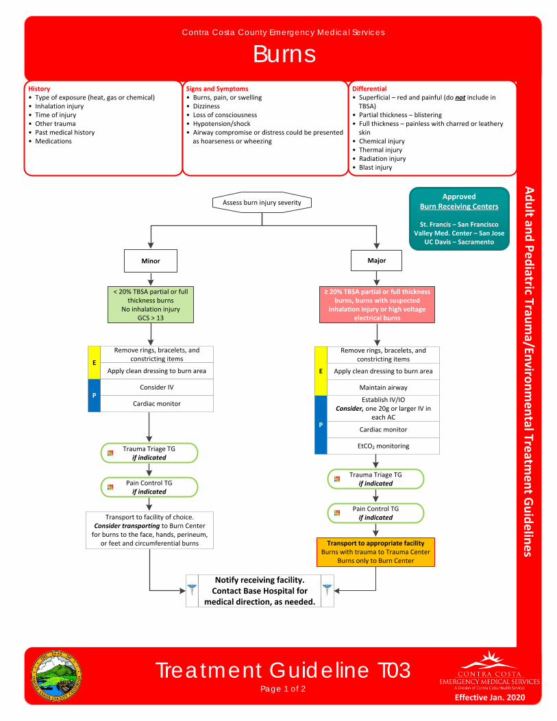
Effective Jan. 2016
Contra Costa County Emergency Medical Services
Burns
Treatment Guideline T03Page 1 of 2
History• Type of exposure (heat, gas or chemical)• Inhalation injury• Time of injury• Other trauma• Past medical history• Medications
Differential• Superficial – red and painful (do not include in TBSA)• Partial thickness – blistering • Full thickness – painless with charred or leathery skin• Chemical injury• Thermal injury• Radiation injury• Blast injury
Signs and Symptoms• Burns, pain, or swelling• Dizziness• Loss of consciousness• Hypotension/shock• Airway compromise or distress could be presented as hoarseness or wheezing
P
Assess burn injury severity
Pain Control TGif indicated
Minor Major
Remove rings, bracelets, and constricting items
EApply clean dressing to burn area
Consider IV
< 20% TBSA partial or full thickness burns
No inhalation injuryGCS > 13
≥ 20% TBSA partial or full thickness burns, burns with suspected
inhalation injury or high voltage electrical burns
Trauma Triage TGif indicated
Transport to facility of choice.Consider transporting to Burn Center for burns to the face, hands, perineum,
or feet and circumferential burns
P
Pain Control TGif indicated
Remove rings, bracelets, and constricting items
E Apply clean dressing to burn area
Establish IV/IOConsider, one 20g or larger IV in
each AC
Trauma Triage TGif indicated
Transport to appropriate facilityBurns with trauma to Trauma Center
Burns only to Burn Center
ApprovedBurn Receiving Centers
St. Francis – San FranciscoValley Med. Center – San Jose
UC Davis – Sacramento
Cardiac monitor
Cardiac monitor
EtCO2 monitoring
Maintain airway
Notify receiving facility. Contact Base Hospital for
medical direction, as needed.
Effective Jan. 2020
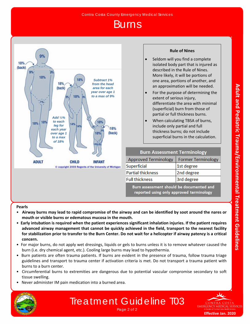
Effective Jan. 2016
Contra Costa County Emergency Medical Services
Burns
Treatment Guideline T03Page 2 of 2
Pearls• Airway burns may lead to rapid compromise of the airway and can be identified by soot around the nares or
mouth or visible burns or edematous mucosa in the mouth.• Early intubation is required when the patient experiences significant inhalation injuries. If the patient requires
advanced airway management that cannot be quickly achieved in the field, transport to the nearest facility for stabilization prior to transfer to the Burn Center. Do not wait for a helicopter if airway patency is a critical concern.
• For major burns, do not apply wet dressings, liquids or gels to burns unless it is to remove whatever caused the burn (i.e. dry chemical agent, etc.). Cooling large burns may lead to hypothermia.
• Burn patients are often trauma patients. If burns are evident in the presence of trauma, follow trauma triage guidelines and transport to trauma center if activation criteria is met. Do not transport a trauma patient with burns to a burn center.
• Circumferential burns to extremities are dangerous due to potential vascular compromise secondary to soft tissue swelling.
• Never administer IM pain medication into a burned area.
Add ½% to each leg for
each year over age 1 to a max of 18%
Rule of Nines
Seldom will you find a complete isolated body part that is injured as described in the Rule of Nines. More likely, it will be portions of one area, portions of another, and an approximation will be needed.
For the purpose of determining the extent of serious injury, differentiate the area with minimal (superficial) burn from those of partial or full thickness burns.
When calculating TBSA of burns, include only partial and full thickness burns; do not include superficial burns in the calculation.
Subtract 1% from the head area for each
year over age 1 to a max of 9%
Effective Jan. 2020
Effective Jan. 2016
Contra Costa County Emergency Medical Services
Extremity Trauma
Treatment Guideline T04Page 1 of 2
History• Type and time of injury• Mechanism (crush, penetrating, blunt, or amputation)• Open vs. closed wound/fracture• Past medical history• Medications
Differential• Abrasion• Contusion• Laceration• Sprain• Dislocation• Fracture• Amputation
Signs and Symptoms• Evidence of trauma• Pain, swelling, deformity, or bleeding• Altered sensation or motor function• Diminished pulse or capillary refill• Decreased extremity temperature
P
E
Place splints and cold packs to stabilize fractures as necessary
Exit to Airway TGif indicated
Cardiac monitor
Establish IV/IO
If SBP < 90 in adultsNormal Saline bolus 500ml IV/IOReassess patient for criteria aboveMay repeat to a Maximum 1L as long as criteria above exists
If poor perfusion or shock in pedsNormal Saline bolus IV/IO
Use PEDIATAPE and refer to dosing guide
Repeat to age dependent goal SBPMay repeat to a Maximum 1L as long as criteria above exists
In the absence of head trauma, age‐specific hypotension, poor perfusion
or AMSConsider pain control
Control hemorrhagingApply tourniquet for hemorrhage or
shock
Exit to Pain Control TGif indicated
Crush injury?
P
E
Secure airwayand support respiratory rate
Place splints and cold packs to stabilize fractures as necessary
Cardiac monitor
EtCO2 monitoring
Establish IV/IO
If SBP < 90 in adultsNormal Saline bolus 500ml IV/IOReassess patient for criteria above
May repeat as long as criteria above exists
If poor perfusion or shock in pedsNormal Saline bolus IV/IO
Use PEDIATAPE and refer to dosing guideRepeat to age dependent goal SBP
May repeat as long as criteria above exists
In the absence of head trauma, age‐specific hypotension, poor perfusion or AMS
Consider pain control
Early transport after releaseLimit scene time to 10 minutes
Control hemorrhagingApply tourniquet for hemorrhage or shock
For suspected hyperkalemia:• Peaked T‐waves; or
• QRS > 0.12 seconds; or• Loss of P‐waves
Albuterol nebulizer 5mg in 6ml Normal SalineCalcium Chloride 1gm over 60 seconds
20ml flush IV/IO prior to pushing next med
Sodium Bicarbonate 1mEq/kg
YesNo
Prior to release from entanglementAlbuterol nebulizer 5mg in 6ml Normal Saline
Tourniquet use should not be delayed until a patient is in shock or is clearly exsanguinating. It should be applied early and can be used safely without risk of patient injury. Do not wait; apply often and tighten if needed.
Notify receiving facility. Contact Base Hospital for
medical direction, as needed.
Consider Hemostatic Gauze
Consider Hemostatic Gauze
Effective Jan. 2021
Effective Jan. 2016
Contra Costa County Emergency Medical Services
Extremity Trauma
Treatment Guideline T04Page 2 of 2
Pearls
For partial amputations, splint affected extremity in anatomic location and elevate extremity.
For complete amputations, place amputated part in a dry container or bag and place on ice. Seal or tie off bag and place in second container or bag. DO NOT place amputated extremity directly on ice or in water. Elevate extremity and dress with dry gauze.
Penetrating trauma to an extremity may hide significant vascular injury and hemorrhage. Early application of a tourniquet should be considered.
In cases of clear‐cut traumatic arrest, epinephrine is not indicated in PEA or asystole. Epinephrine will not correct arrest caused by a tension pneumothorax, cardiac tamponade, or hemorrhagic shock. If there is any doubt as to the cause of arrest, treat as a non‐traumatic arrest.
Hypotension is age dependent. This is not always reliable and should be interpreted in context with the patient’s typical BP, if known. Shock may be present with a seemingly normal blood pressure initially.
Neonate: < 60mmHg or weak pulsesInfant: < 70mmHg or weak pulses1‐10 years: < 70mmHg + (age in years x2)Over 10 years: <90mmHgOver 65 years: <110mmHg
If vigorous hemorrhage is not controlled with elevation and direct pressure on wound, apply a tourniquet. Tourniquets may be used in pediatric patients.
Tourniquets and hemostatic gauze may also be appropriate for hemorrhage control in multi‐casualty incidents.
Consider the use of hemostatic gauze to pack the wound. More than hemostatic agent may be needed. Secure hemostatic gauze in place with a compression bandage.
Crush Injury Syndrome is caused by muscle crush injury and cell death. Most patients have an extensive area of involvement such as a large muscle mass in a lower extremity or the pelvis. May develop after one (1) hour in the presence of a severe crush, but usually requires at least four (4) hours of compression. Hypovolemia and hyperkalemia may occur, particularly in extended entrapments.
Avoid hyperventilation. Maintain an EtCO2 of 35 or greater, which may be unreliable if the patient was subject to multisystem trauma or poor perfusion.
Hypotension usually indicates injury or shock and should be treated aggressively.
An important item to monitor and document is a change in the level of consciousness by repeat examination.
Do not overlook the possibility of associated domestic violence or abuse.
Effective Jan. 2021
Effective Jan. 2016
Contra Costa County Emergency Medical Services
Head Trauma
Treatment Guideline T05Page 1 of 2
History• Time of injury• Mechanism (blunt vs. penetrating)• Loss of consciousness• Bleeding• Past medical history• Medications (anticoagulants)
Differential• Skull fracture• Spinal injury• Abuse
Signs and Symptoms• Evidence of trauma• Pain, swelling, or bleeding• AMS• Unconscious• Respiratory distress or failure• Vomiting• Seizure
P
Spinal Motion Restriction if indicated
Elevate head 30 degrees unless contraindicated. Position patient on left side if needed for vomiting
Cardiac monitor
EtCO2 monitoring
Establish IV/IO
If SBP approaching 90 or rapidly droppingin adults
Normal Saline bolus 1000ml IV/IOReassess patient for criteria above May give additional 500ml IV/IOas long as criteria above exists
If poor perfusion or shock in pedsNormal Saline bolus IV/IO
Use PEDIATAPE and refer to PEARLSRepeat to age dependent goal SBPMay repeat to a Maximum 1L as long as criteria above exists
For Nausea/Vomiting Consider Adults ‐ Ondansetron 4mg IV/IO/IM/ODT
(May repeat every 10 minutes to a Maximum 12mg)
Pediatric ≥ 4 years ‐ Ondansetron IV/IO/IM/ODTUse PEDIATAPE and refer to dosing guide
(May repeat x1 for peds > 40kg)
Hemorrhage Control Direct Pressure Pressure Bandage Consider Hemostatic Gauze
Limit scene time – Transport early
E
Exit to Airway TGif indicated
POTENTIAL SEVEREHEAD INJURY?
Any one of these: LOC at any point GCS ≤ 14 Any post‐traumatic seizure Multisystem trauma
requiring intubation?
Age Dependent Signs of Shock
Neonate: < 60mmHg or weak pulses
Infant: < 70mmHg or weak pulses
1‐10 years: < 70mmHg + (age in years x2)
Over 10 years: <90mmHg
Over 65 years: <110mmHg
P
P
Manage ABCsE
NO
Secure airway and support respiratory rate
High flow O2 via NRBMaintain SPO2 as close to 100% as possibleIf unable to maintain SPO2 with NRB & BLS
maneuvers – Proceed with BVMAVOID HYPERVENTILATION
EYES
Notify receiving facility. Contact Base Hospital for medical direction, as needed.
Effective Jan. 2021
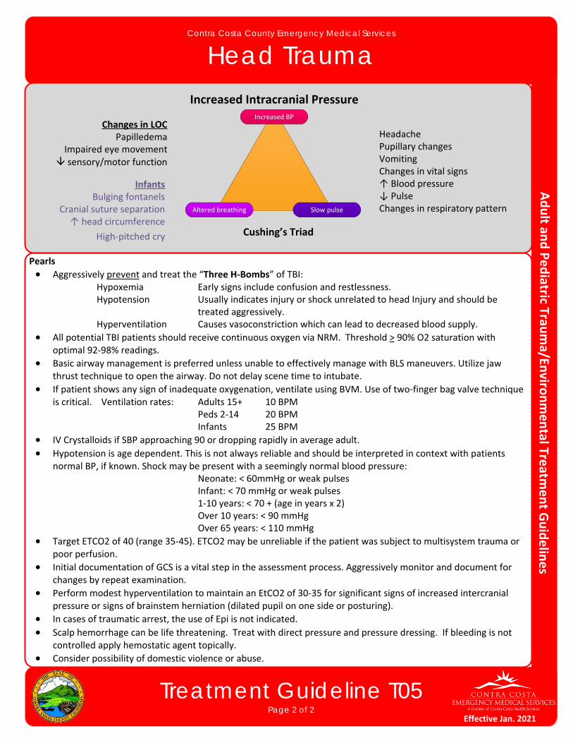
Effective Jan. 2016
Contra Costa County Emergency Medical Services
Head Trauma
Treatment Guideline T05Page 2 of 2
Pearls
Aggressively prevent and treat the “Three H‐Bombs” of TBI:Hypoxemia Early signs include confusion and restlessness.Hypotension Usually indicates injury or shock unrelated to head Injury and should be
treated aggressively.Hyperventilation Causes vasoconstriction which can lead to decreased blood supply.
All potential TBI patients should receive continuous oxygen via NRM. Threshold > 90% O2 saturation with optimal 92‐98% readings.
Basic airway management is preferred unless unable to effectively manage with BLS maneuvers. Utilize jaw thrust technique to open the airway. Do not delay scene time to intubate.
If patient shows any sign of inadequate oxygenation, ventilate using BVM. Use of two‐finger bag valve technique is critical. Ventilation rates: Adults 15+ 10 BPM
Peds 2‐14 20 BPMInfants 25 BPM
IV Crystalloids if SBP approaching 90 or dropping rapidly in average adult.
Hypotension is age dependent. This is not always reliable and should be interpreted in context with patients normal BP, if known. Shock may be present with a seemingly normal blood pressure:
Neonate: < 60mmHg or weak pulsesInfant: < 70 mmHg or weak pulses1‐10 years: < 70 + (age in years x 2)Over 10 years: < 90 mmHgOver 65 years: < 110 mmHg
Target ETCO2 of 40 (range 35‐45). ETCO2 may be unreliable if the patient was subject to multisystem trauma or poor perfusion.
Initial documentation of GCS is a vital step in the assessment process. Aggressively monitor and document for changes by repeat examination.
Perform modest hyperventilation to maintain an EtCO2 of 30‐35 for significant signs of increased intercranial pressure or signs of brainstem herniation (dilated pupil on one side or posturing).
In cases of traumatic arrest, the use of Epi is not indicated.
Scalp hemorrhage can be life threatening. Treat with direct pressure and pressure dressing. If bleeding is not controlled apply hemostatic agent topically.
Consider possibility of domestic violence or abuse.
Increased Intracranial Pressure
Changes in LOCPapilledema
Impaired eye movement sensory/motor function
HeadachePupillary changesVomitingChanges in vital signs↑ Blood pressure↓ PulseChanges in respiratory pattern
InfantsBulging fontanels
Cranial suture separation↑ head circumference
High‐pitched cry
Increased BP
Altered breathing Slow pulse
Cushing’s Triad
Effective Jan. 2021
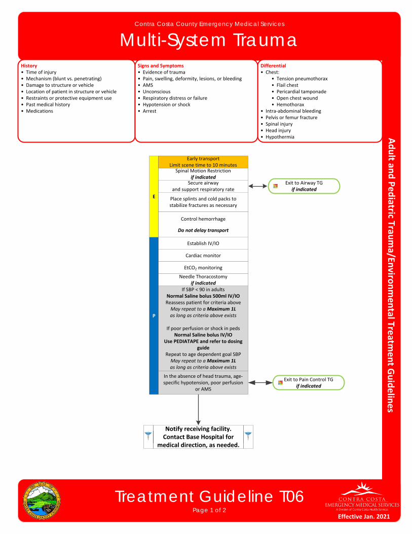
Effective Jan. 2016
Contra Costa County Emergency Medical Services
Multi-System Trauma
Treatment Guideline T06Page 1 of 2
History• Time of injury• Mechanism (blunt vs. penetrating)• Damage to structure or vehicle• Location of patient in structure or vehicle• Restraints or protective equipment use• Past medical history• Medications
Differential• Chest: • Tension pneumothorax • Flail chest • Pericardial tamponade • Open chest wound • Hemothorax• Intra‐abdominal bleeding• Pelvis or femur fracture• Spinal injury• Head injury• Hypothermia
Signs and Symptoms• Evidence of trauma• Pain, swelling, deformity, lesions, or bleeding• AMS• Unconscious• Respiratory distress or failure• Hypotension or shock• Arrest
P
Spinal Motion Restrictionif indicated
E
Secure airwayand support respiratory rate
Place splints and cold packs to stabilize fractures as necessary
Exit to Airway TGif indicated
Cardiac monitor
EtCO2 monitoring
Establish IV/IO
If SBP < 90 in adultsNormal Saline bolus 500ml IV/IOReassess patient for criteria aboveMay repeat to a Maximum 1L as long as criteria above exists
If poor perfusion or shock in pedsNormal Saline bolus IV/IO
Use PEDIATAPE and refer to dosing guide
Repeat to age dependent goal SBPMay repeat to a Maximum 1L as long as criteria above exists
In the absence of head trauma, age‐specific hypotension, poor perfusion
or AMS
Control hemorrhage
Do not delay transport
Early transportLimit scene time to 10 minutes
Needle Thoracostomyif indicated
Notify receiving facility. Contact Base Hospital for
medical direction, as needed.
Exit to Pain Control TGif indicated
Effective Jan. 2021

Effective Jan. 2016
Contra Costa County Emergency Medical Services
Multi-System Trauma
Pearls• ALS procedures in the field do not significantly improve patient outcome in critical trauma patients.• Basic airway management is preferred unless unable to effectively manage with BLS maneuvers. Utilize jaw
thrust technique to open the airway.• Intubation of head injury patients is best addressed at the hospital. Advanced Airways should not be used in
traumatic arrest. • In cases of clear‐cut traumatic arrest, epinephrine is not indicated in PEA or asystole. Epinephrine will not
correct arrest caused by a tension pneumothorax, cardiac tamponade, or hemorrhagic shock. If there is any doubt as to the cause of arrest, treat as a non‐traumatic arrest.
• Hypotension is age dependent. This is not always reliable and should be interpreted in context with the patient’s typical BP, if known. Shock may be present with a seemingly normal blood pressure initially.
▫ Neonate: < 60mmHg or weak pulses ▫ Infant: < 70mmHg or weak pulses
▫ 1‐10 years: < 70mmHg + (age in years x2)▫ Over 10 years: <90mmHg▫ Over 65 years: <110mmHg
• Avoid hyperventilation. Maintain an EtCO2 of 35 or greater, which may be unreliable if the patient was subject to multisystem trauma or poor perfusion.
• Hypotension usually indicates injury or shock and should be treated aggressively.• An important item to monitor and document is a change in the level of consciousness by repeat examination.• Do not overlook the possibility of associated domestic violence or abuse.
Treatment Guideline T06Page 2 of 2
Effective Jan. 2021
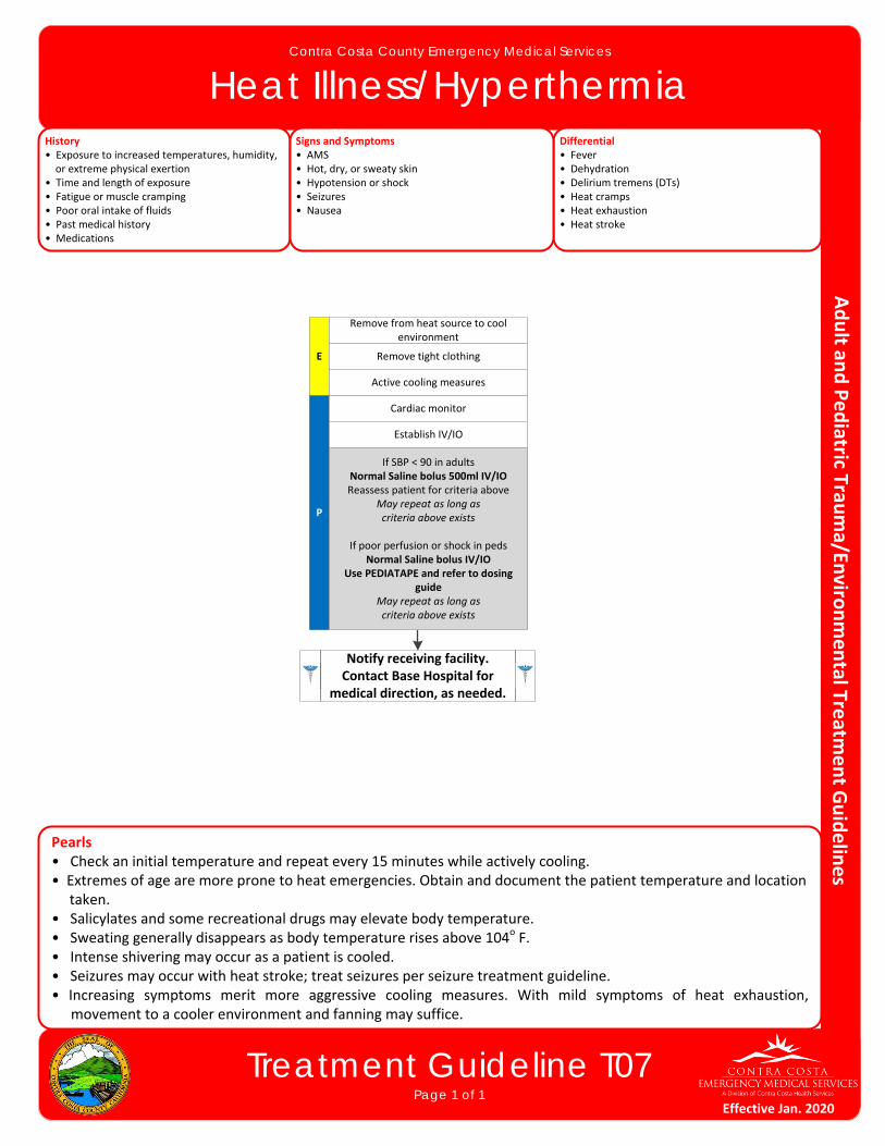
Effective Jan. 2016
Contra Costa County Emergency Medical Services
Heat Illness/Hyperthermia
Treatment Guideline T07Page 1 of 1
History• Exposure to increased temperatures, humidity, or extreme physical exertion• Time and length of exposure• Fatigue or muscle cramping• Poor oral intake of fluids• Past medical history• Medications
Differential• Fever• Dehydration• Delirium tremens (DTs)• Heat cramps• Heat exhaustion• Heat stroke
Signs and Symptoms• AMS• Hot, dry, or sweaty skin• Hypotension or shock• Seizures• Nausea
P
E Remove tight clothing
Cardiac monitor
Establish IV/IO
If SBP < 90 in adultsNormal Saline bolus 500ml IV/IOReassess patient for criteria above
May repeat as long as criteria above exists
If poor perfusion or shock in pedsNormal Saline bolus IV/IO
Use PEDIATAPE and refer to dosing guide
May repeat as long as criteria above exists
Remove from heat source to cool environment
Active cooling measures
Pearls• Check an initial temperature and repeat every 15 minutes while actively cooling.• Extremes of age are more prone to heat emergencies. Obtain and document the patient temperature and location taken.• Salicylates and some recreational drugs may elevate body temperature.• Sweating generally disappears as body temperature rises above 104o F.• Intense shivering may occur as a patient is cooled.• Seizures may occur with heat stroke; treat seizures per seizure treatment guideline.• Increasing symptoms merit more aggressive cooling measures. With mild symptoms of heat exhaustion,
movement to a cooler environment and fanning may suffice.
Notify receiving facility. Contact Base Hospital for
medical direction, as needed.
Effective Jan. 2020

This page intentionally left blank

Treatment Guideline T08Page 1 of 1
Contra Costa County Emergency Medical Services
HypothermiaHistory• Age• Exposure to decreased temperatures, but may occur in normal atmospheric temperatures• Time and length of exposure• Drug or alcohol use• Infection or sepsis• Past medical history• Medications
Differential• Sepsis• Environmental exposure• Hypoglycemia• CNS dysfunction • Stroke • Head injury • Spinal cord injury
Signs and Symptoms• AMS• Cold or clammy skins• Shivering• Extremity pain or sensory abnormality• Bradycardia• Hypotension or shock
P
E
Cardiac monitor
Establish IV/IO
If SBP < 90 in adultsNormal Saline bolus 500ml IV/IOReassess patient for criteria above
May repeat as long as criteria above exists
If poor perfusion or shock in pedsNormal Saline bolus IV/IO
Use PEDIATAPE and refer to dosing guide
Repeat to age dependent goal SBPMay repeat as long as criteria above exists
Blood glucose analysisExit to Diabetic TG
if indicated
Pearls• Severe hypothermia may cause cardiac instability. Avoidance of excess stimuli is important in severe hypothermia
as the heart is sensitive and interventions may induce arrhythmias. Necessary interventions should be done as gently as possible.
• Check for pulselessness for 30‐45 seconds to avoid unnecessary chest compressions.• Defer ACLS medications until patient is warmed.• If V‐Fib or pulseless V‐Tach is present, shock x1, and defer further shocks.• Extremes of age, malnutrition, alcohol, and other drug use are contributing factors to hypothermia. • Patients with prolonged hypoglycemia often become hypothermic; blood glucose analysis is essential.• If a temperature is unable to be measured, treat the patient based on the suspected temperature.• Warm packs can be placed in the armpit and groin areas. Care should be taken not to place directly on skin.
Spinal motion restrictionif indicated
Secure airway and support respiratory rate
if indicated
Exit to Airway TGif indicated
Notify receiving facility. Contact Base Hospital for
medical direction, as needed.
Remove wet clothing and cover with warm dry sheets or blankets
Gently move to a warm environment
Effective Jan. 2020

This page intentionally left blank
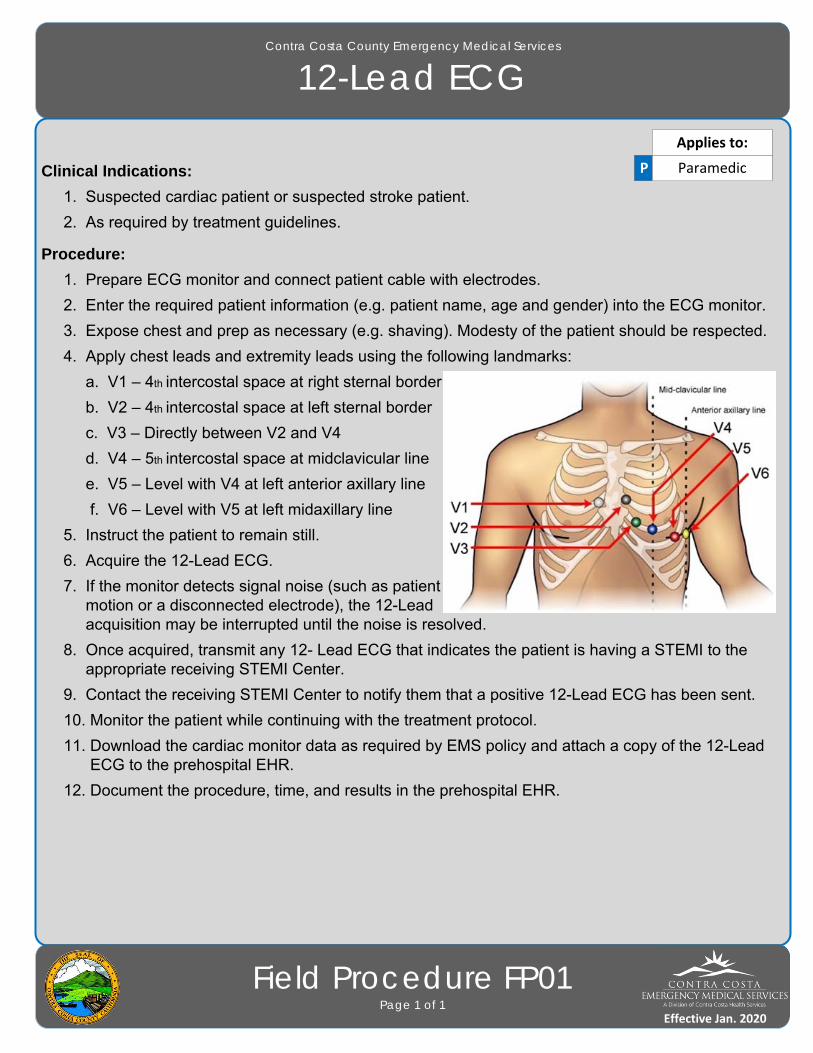
Clinical Indications:
1. Suspected cardiac patient or suspected stroke patient.
2. As required by treatment guidelines.
Procedure:
1. Prepare ECG monitor and connect patient cable with electrodes.
2. Enter the required patient information (e.g. patient name, age and gender) into the ECG monitor.
3. Expose chest and prep as necessary (e.g. shaving). Modesty of the patient should be respected.
4. Apply chest leads and extremity leads using the following landmarks:
a. V1 – 4th intercostal space at right sternal border
b. V2 – 4th intercostal space at left sternal border
c. V3 – Directly between V2 and V4
d. V4 – 5th intercostal space at midclavicular line
e. V5 – Level with V4 at left anterior axillary line
f. V6 – Level with V5 at left midaxillary line
5. Instruct the patient to remain still.
6. Acquire the 12-Lead ECG.
7. If the monitor detects signal noise (such as patient motion or a disconnected electrode), the 12-Lead acquisition may be interrupted until the noise is resolved.
8. Once acquired, transmit any 12- Lead ECG that indicates the patient is having a STEMI to the appropriate receiving STEMI Center.
9. Contact the receiving STEMI Center to notify them that a positive 12-Lead ECG has been sent.
10. Monitor the patient while continuing with the treatment protocol.
11. Download the cardiac monitor data as required by EMS policy and attach a copy of the 12-Lead ECG to the prehospital EHR.
12. Document the procedure, time, and results in the prehospital EHR.
Effective Jan. 2016
Contra Costa County Emergency Medical Services
12-Lead ECG
Field Procedure FP01Page 1 of 1
P Paramedic
Applies to:
Effective Jan. 2020
Effective Jan. 2016
Contra Costa County Emergency Medical Services
Airway: BLS Management
Field Procedure FP02Page 1 of 1
Clinical Indications:
1. A patient who is unable to maintain their airway without assistance.
Procedure:
1. Position the patient to optimize airway opening and facilitate ventilations.
a. Use the sniffing position with head extended (A) and neck flexed forward (B) unless there is a suspected spinal injury.
b. Position with head/shoulders elevated; anterior ear should be at the same horizontal level as the sternal notch (C). This is especially advantageous in larger or morbidly obese patients.
2. Using a two-person technique is the preferred method to ventilate patients with a Bag-Valve- Mask (BVM).
a. J – jaw thrust maneuver to open the airway.
b. A – use a nasal or oral airway. Appropriately measure before placing airway adjunct.
c. W – work together to ventilate the patient using a BVM. This should be done using two rescuers – one holding the mask to achieve an optimal seal and the other to deliver ventilations.
d. S – slow and small ventilations to produce visible chest rise.
3. It is required that the airway be monitored continuously through waveform capnography (ALS providers) and pulse oximetry.
Notes:
1. The goal of airway management is to ensure adequate ventilation and oxygenation. Initial airway management should always begin with BLS maneuvers.
2. Avoid excessive ventilation. In non-arrest patients, ventilation rates should be:
a. Adults – 10/minute
b. Children – 20/minute
c. Infants – 30/minute
EMT
A
B
C
E EMT
P Paramedic
Applies to:
Effective Jan. 2020
Effective Jan. 2016
Contra Costa County Emergency Medical Services
Airway: Bougie Device
Field Procedure FP03Page 1 of 1
Clinical Indications:
1. Patients meet clinical indications for oral intubation.
Contraindications:
1 Two attempts at intubation.
2. Age less than eight (8) or ETT size less than 6.5 mm.
Procedure:
1. Prepare, position, and oxygenate the patient with 100% oxygen.
2. Select the proper ET tube and remove stylette; test cuff and prepare suction.
3. Lubricate the distal end and cuff of the endotracheal tube (ETT) with a water-based lubricant and the distal 1/2 of the Bougie device. (Note: Failure to lubricate the Bougie and the ETT may result in being unable to pass the ETT).
4. Using the laryngoscope, visualize the vocal cords, if possible, using the BURP maneuver as needed.
5. Introduce the Bougie with the curved tip anteriorly and visualize the tip passing the vocal cords or above the arytenoids if the cords cannot be visualized.
6. Once inserted, gently advance the Bougie until you meet resistance; feel for the tracheal rings. If you do not meet resistance, you have a probable esophageal intubation and insertion should be reattempted or use a i-Gel.
7. Withdraw the Bougie ONLY to a depth sufficient to allow loading of the ETT while maintaining proximal control of the Bougie.
8. Gently advance the Bougie and loaded ETT until you have feel resistance again, thereby assuring tracheal placement and minimizing the risk of accidental displacement of the Bougie.
9. While maintaining a firm grasp on the proximal Bougie, introduce the ETT over the Bougie passing the tube to its appropriate depth.
10. If you are unable to advance the ETT into the trachea and the Bougie and ETT are adequately lubricated, withdraw the ETT slightly and rotate the ETT 90 degrees COUNTER clockwise to turn the bevel of the ETT posteriorly. If this technique fails to facilitate passing of the ETT you may attempt direct laryngoscopy while advancing the ETT (this will require an assistant to maintain the position of the Bougie and, if so desired, advance the ETT).
11. Once the ETT is correctly placed, hold the ETT securely and remove the Bougie.
12. Confirm tracheal placement according to the intubation protocol, inflate the cuff with 3 – 10cc of air, auscultate for equal breath sounds, and reposition accordingly.
13. When final position is determined secure the ETT, reassess breath sounds, apply EtCO2 monitoring, and record and monitor readings to assure continued tracheal intubation.
P Paramedic
Applies to:
Effective Jan. 2021

Field Treatment GuidelinesPage 3 of 3
Effective Jan. 2016
Contra Costa County Emergency Medical Services
Airway: Endotracheal Intubation
Field Procedure FP04Page 1 of 1
Clinical Indications:
1. Inability to adequately ventilate a patient with a Bag Valve Mask (BVM) and basic airway adjunct.
2. An unconscious patient without a gag reflex who is apneic or is demonstrating inadequate respiratory effort.
Procedure:
1. Using the Cormack Lehane difficult airway assessment tool to determine if the patient’s airway will be difficult to intubate. If it is determined to be difficult, use an i-Gel.
2. Prepare, position, and oxygenate the patient with 100% Oxygen.
3. Select proper ET tube and stylette; have suction ready.
4. The use of a Bougie device is strongly encouraged with all ET intubation attempts.
5. Using laryngoscope, visualize vocal cords. (Use the BURP maneuver to assist).
6. Limit each intubation attempt to 30 seconds with BVM between attempts.
7. Visualize tube passing through vocal cords.
8. Confirm and document tube placement using an EtCO2 monitoring.
9. Inflate the cuff with 3 – 10cc of air; secure the tube using a commercial tube holder.
10. Auscultate for bilaterally equal breath sounds and absence of sounds over the epigastrium. If you are unsure of placement, remove tube and ventilate patient with a BVM.
11. Apply waveform capnography. After 3 ventilations, EtCO2 should be >10 or comparable to pre-intubation values. If < 10, check for adequate circulation, equipment, and ventilatory rate. If EtCO2 remains < 10 without physiologic explanation, remove the ETT and ventilate using an airway adjunct and BVM.
12. Consider using an i-Gel if intubation efforts are unsuccessful.
13. Monitor EtCO2 and record readings on scene, en route to the hospital, and at the hospital.
14. Document ETT size, time, result (success) and placement location by the centimeter marks either at the patient’s teeth or lips in the EHR. Document all devices used to confirm initial tube placement. Also document positive or negative breath sounds before and after each movement of the patient.
15. It is required that the airway be monitored continuously through waveform capnography (ALS providers) and pulse oximetry.
P Paramedic
Applies to:
Effective Jan. 2020

Effective Jan. 2016
Contra Costa County Emergency Medical Services
Airway: Foreign Body Removal
Field Procedure FP05Page 1 of 1
Clinical Indications:
1. Sudden onset of respiratory distress often with coughing, wheezing, gagging, or stridor due to a foreign-body obstruction of the upper airway.
Procedure:
1. Assess the degree of foreign body obstruction.
a. Do not interfere with a mild obstruction; allow the patient to clear their airway by coughing.
b. In severe foreign-body obstructions, the patient may not be able to make a sound. The patient may clutch his/her neck demonstrating the universal choking sign.
2. For an infant, deliver 5 back blows (slaps) followed by 5 chest thrusts repeatedly until the object is expelled or the patient becomes unresponsive.
3. For a child, perform a subdiaphragmatic abdominal thrust (Heimlich Maneuver) until the object is expelled or the patient becomes unresponsive.
4. For adults, a combination of maneuvers may be required.
a. First, subdiaphragmatic abdominal thrusts (Heimlich Maneuver) should be used in rapid sequence until the obstruction is relieved.
b. If abdominal thrusts are ineffective, chest thrusts should be used. Chest thrusts should be used primarily in morbidly obese patients and in patients who are in the late stages of pregnancy.
5. If the patient becomes unresponsive, begin CPR immediately but look in the mouth before administering any ventilations. If a foreign-body is visible, remove it.
6. Do not perform blind finger sweeps in the mouth and posterior pharynx. This may push the object farther into the airway.
7. In unresponsive patients, Paramedics should visualize the posterior pharynx with a laryngoscope to potentially identify and remove the foreign-body using Magill forceps.
8. Document the methods used and result of these procedures in the EHR.
E EMT
P Paramedic
Applies to:
Effective Jan. 2020
Effective Jan. 2016
Contra Costa County Emergency Medical Services
Airway: Supraglottic Airway Devices
Field Procedure FP06Page 1 of 2
Definitions:1. Supraglottic Airway Device (SAD): A device that is placed into the oral pharynx and subsequently
placed over the glottic opening. This is done in a ‘blind’ maneuver without the aid of a laryngoscope. This will aid in the oxygenation and ventilation of the patient.
2. The I-Gel is the approved supraglottic airway device for Contra Costa County EMS.
Indications:1. Cardiac Arrest2. Respiratory arrest with no immediate reversible cause (i.e. hypoglycemia or opioid overdose)
3. Inability to adequately ventilate a patient with a Bag Valve Mask (BVM) and basic airway adjunct. 4. An unconscious patient without a gag reflex who is apneic or is demonstrating inadequate respiratory effort
5. For EMT's with approved optional scope: Patients greater than or equal to 15 years of age in need of airway protection or unable to be adequately ventilated with BVM.
Contraindications:1. Gag reflex
2. Caustic ingestion – Esophageal burns3. Known esophageal disease (e.g. cancer, varices or stricture)4. Laryngectomy with stoma – if present, place ETT in stoma5. Severe airway trauma6. Trismus
Complications:1. Airway and/or esophageal trauma2. Regurgitation3. Aspiration
Procedure:1. Prepare, position and oxygenate the patient with 100% Oxygen.2. Document the pre-intubation EtCO2 reading.3. Select proper I-Gel using weight based chart.4. Lubricate the I-Gel with water-based lubricant, get suction ready.5. If present, remove dentures or plates from the mouth prior to insertion.6. Remove the I-Gel cradle and gently press the chin downwards and introduce the iGel into the
mouth with the opening towards the chin, along the hard palatte until a definitive resistance is felt. Do not apply excessive force on the I-Gel upon insertion.
7. Attach BVM, ETCO2 and ventilate the patient.
O EMT Optional
P Paramedic
Applies to:
Effective Jan. 2020

Effective Jan. 2016
Contra Costa County Emergency Medical Services
Airway: Supraglottic Airway Devices
Field Procedure FP06Page 2 of 2
8. Auscultate for breath sounds and sounds over the epigastrium and lookfor the chest to rise and fall.
9. Confirm tube placement using EtCO2 and waveform capnography. It is required that the airway be monitored continuously through waveform capnography (ALS providers) and pulse oximetry.
10. Secure I-Gel airway to patient with an approved method.
O EMT Optional
P Paramedic
Applies to:
Effective Jan. 2020
Field Treatment GuidelinesPage 3 of 3
Effective Jan. 2016
Contra Costa County Emergency Medical Services
Airway: Stomal Intubation
Field Procedure FP07Page 1 of 1
Clinical Indications:
1. Patient requiring intubation who has a mature stoma and does not have a replacement tracheostomy tube available.
Procedure:
1. Select the largest endotracheal tube (ETT) that will fit through the stoma without force; check the cuff and remove the stylette.
2. Pre-oxygenate the patient with 100% oxygen using a BVM.
3. Wear sterile gloves. It is not necessary to lubricate the ETT.
4. Suction if necessary.
5. Pass the ETT and inflate the cuff. The pharynx has been bypassed, so the ETT will protrude from the neck by several inches.
6. Hold the tube in place and attach the BVM.
7. While ventilating the patient, watch for equal rise and fall of the chest.
8. Secure the tube and ventilate with 100% oxygen.
9. Auscultate for bilaterally equal breath sounds. Examine the neck for subcutaneous emphysema indicating false passage.
10. Do not take longer than 30 seconds to perform this procedure.
11. Document ETT size, time, result (success) and placement location by the centimeter marks either at the stomal opening in the EHR. Document all devices used to confirm initial tube placement. Also document positive or negative breath sounds before and after each movement of the patient.
12. It is required that the airway be monitored continuously through waveform capnography (ALS providers) and pulse oximetry.
P Paramedic
Applies to:
Effective Jan. 2020

Effective Jan. 2016
Contra Costa County Emergency Medical Services
Airway: Tracheostomy Tube Replacement
Field Procedure FP08Page 1 of 1
Clinical Indications:
1. Presence of Tracheostomy site.
2. Urgent or emergent indication to change the tube, such as obstruction that will not clear with suction, dislodgement, or inability to oxygenate/ventilate the patient without other obvious explanation.
Procedure:
1. Have all airway equipment prepared for standard airway management, including equipment for endotracheal intubation.
2. Have an airway device (endotracheal tube or tracheostomy tube) of the same size as the tracheostomy tube currently in place as well as 0.5 size smaller available (e.g., if the patient has a #6.0 Shiley, then have a 6.0 and a 5.5 tube).
3. Lubricate the replacement tube(s) with water-based lubricant and check the cuff.
4. Remove the tracheostomy tube from mechanical ventilation device and use a bag-valve mask to pre-oxygenate the patient as much as possible.
5. Once all equipment is in place, remove the device securing the tracheostomy tube.
6. If applicable, deflate the cuff on the tube. If unable to aspirate air with a syringe, cut the balloon off to allow the cuff to deflate.
7. Remove the tracheostomy tube.
8. Insert the replacement tube. Confirm placement via auscultation of the lungs.
9. If there is any difficulty placing the tube, re-attempt procedure with the smaller tube.
10. If difficulty is still encountered, use standard airway procedures such as oral bag-valve mask or endotracheal intubation. More difficulty with tube changing can be anticipated for tracheostomy sites that are immature (i.e. less than two weeks old). Great caution should be exercised in attempts to change immature tracheotomy sites.
11. Document the procedure, confirmation, patient response, and any complications in the EHR.
P Paramedic
Applies to:
Effective Jan. 2020
Effective Jan. 2016
Contra Costa County Emergency Medical Services
Cardiac Arrest Management
Field Procedure FP09Page 1 of 4
Clinical Indications:
Each and every out-of-hospital adult cardiac arrest (OHCA) which results in the activation of the EMS System shall be managed using CPR-Highly Defined (CPR-HD). CPR-HD is the expected standard of care for cardiac arrest in Contra Costa County.
Purpose: The purpose of CPR-HD is to provide a structured, standardized and choreographed approach to cardiac arrest management. The CPR-HD ‘Script’ is time driven and serves as the ‘CODE’ leader.
Principles:
1. Resuscitation is based on proper planning and organized execution. Procedures require space and patient access. Make room to work. Utilize a team focused approach assigning responders to predetermined tasks.
2. The unit first on scene shall establish and follow the CPR-HD Script. Efforts should be dispensed to ensure adequate timekeeping occurs throughout the resuscitation.
3. Cardiac arrest management efforts should be directed at high quality and continuous chest compressions with limited interruptions. Our goal is to provide two (2) minutes of straight continuous compressions with a less than ten (<10) second pause.
4. In cardiac arrest, drugs are of limited usefulness. High quality compressions and defibrillation are far more important.
5. Conduct resuscitation with goal of preserving cerebral function through meticulous attention to procedure.
6. Passive ventilation for the first three cycles (6 minutes) of CPR. After that time, the patient should be ventilated using a BLS airway and BVM at a rate of 6 ventilations/minute (1:10 seconds) with continuous CPR. Placement of an advanced airway should be deferred unless a provider is unable to ventilate the patient with a BLS airway and BVM.
7. Once transport is deemed appropriate and the patient has experienced a return of spontaneous circulation (ROSC) at any time throughout the resuscitation; transport to a STEMI receiving Center.
E EMT
Applies to:
P Paramedic
Effective Jan. 2021
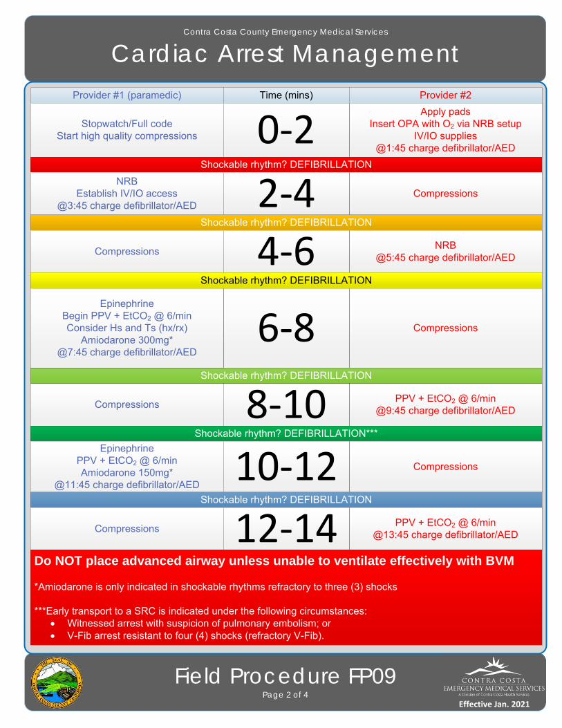
Effective Jan. 2016
Contra Costa County Emergency Medical Services
Cardiac Arrest Management
Field Procedure FP09Page 2 of 4
Provider #1 (paramedic) Time (mins) Provider #2
Stopwatch/Full codeStart high quality compressions
Apply padsInsert OPA with O2 via NRB setup
IV/IO supplies@1:45 charge defibrillator/AED
0‐2Shockable rhythm? DEFIBRILLATION
NRBEstablish IV/IO access
@3:45 charge defibrillator/AEDCompressions2‐4
Shockable rhythm? DEFIBRILLATION
CompressionsNRB
@5:45 charge defibrillator/AED4‐6Shockable rhythm? DEFIBRILLATION
Compressions6‐8Shockable rhythm? DEFIBRILLATION
CompressionsPPV + EtCO2 @ 6/min
@9:45 charge defibrillator/AED8‐10Shockable rhythm? DEFIBRILLATION***
EpinephrinePPV + EtCO2 @ 6/minAmiodarone 150mg*
@11:45 charge defibrillator/AED
Compressions10‐12Shockable rhythm? DEFIBRILLATION
CompressionsPPV + EtCO2 @ 6/min
@13:45 charge defibrillator/AED12‐14Do NOT place advanced airway unless unable to ventilate effectively with BVM
*Amiodarone is only indicated in shockable rhythms refractory to three (3) shocks
***Early transport to a SRC is indicated under the following circumstances: Witnessed arrest with suspicion of pulmonary embolism; or V-Fib arrest resistant to four (4) shocks (refractory V-Fib).
EpinephrineBegin PPV + EtCO2 @ 6/minConsider Hs and Ts (hx/rx)
Amiodarone 300mg*@7:45 charge defibrillator/AED
Effective Jan. 2021
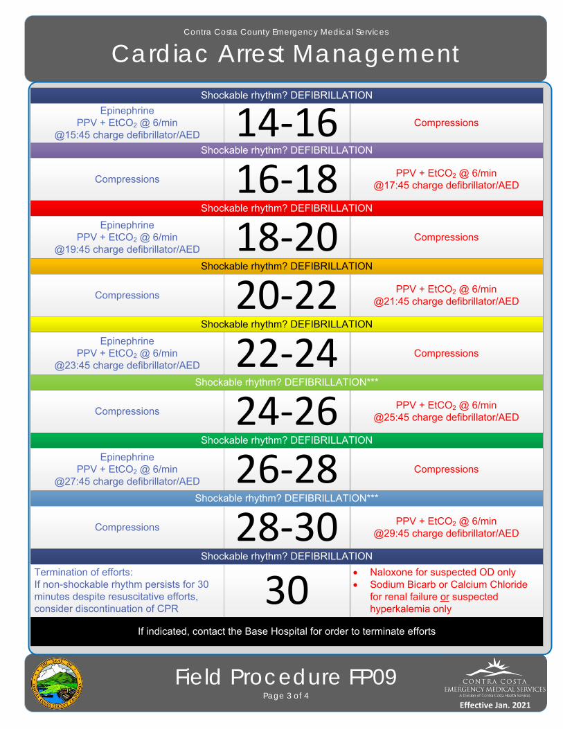
Effective Jan. 2016
Contra Costa County Emergency Medical Services
Cardiac Arrest Management
Field Procedure FP09Page 3 of 4
PPV + EtCO2 @ 6/min
EpinephrinePPV + EtCO2 @ 6/min
@15:45 charge defibrillator/AEDCompressions14‐16
Shockable rhythm? DEFIBRILLATION
CompressionsPPV + EtCO2 @ 6/min
@17:45 charge defibrillator/AED16‐18Shockable rhythm? DEFIBRILLATION
EpinephrinePPV + EtCO2 @ 6/min
@19:45 charge defibrillator/AEDCompressions18‐20
Shockable rhythm? DEFIBRILLATION
CompressionsPPV + EtCO2 @ 6/min
@21:45 charge defibrillator/AED20‐22Shockable rhythm? DEFIBRILLATION
EpinephrinePPV + EtCO2 @ 6/min
@23:45 charge defibrillator/AEDCompressions22‐24
Shockable rhythm? DEFIBRILLATION***
CompressionsPPV + EtCO2 @ 6/min
@25:45 charge defibrillator/AED24‐26Shockable rhythm? DEFIBRILLATION
EpinephrinePPV + EtCO2 @ 6/min
@27:45 charge defibrillator/AEDCompressions26‐28
Shockable rhythm? DEFIBRILLATION
Shockable rhythm? DEFIBRILLATION***
CompressionsPPV + EtCO2 @ 6/min
@29:45 charge defibrillator/AED28‐30Shockable rhythm? DEFIBRILLATION
Termination of efforts:If non-shockable rhythm persists for 30 minutes despite resuscitative efforts, consider discontinuation of CPR
Naloxone for suspected OD only Sodium Bicarb or Calcium Chloride
for renal failure or suspected hyperkalemia only
30If indicated, contact the Base Hospital for order to terminate efforts
Effective Jan. 2021
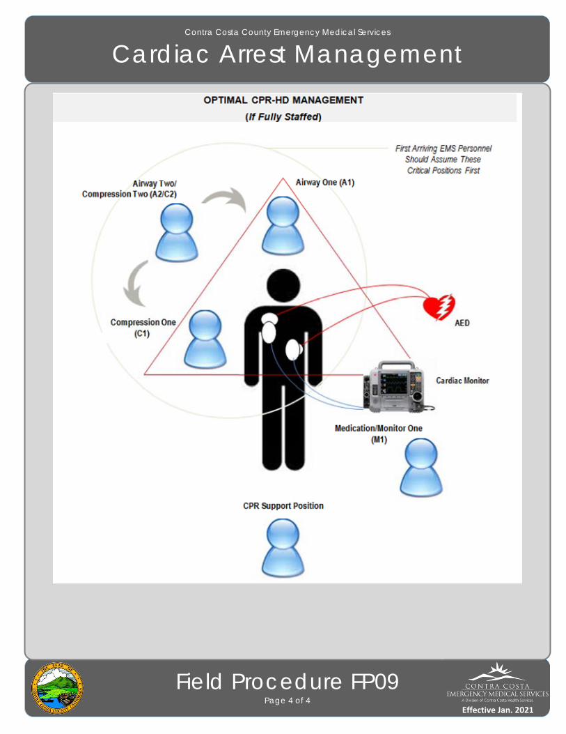
Effective Jan. 2016
Contra Costa County Emergency Medical Services
Cardiac Arrest Management
Field Procedure FP09Page 4 of 4
Effective Jan. 2021
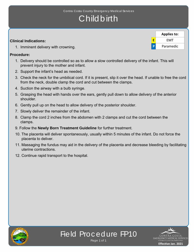
Effective Jan. 2016
Contra Costa County Emergency Medical Services
Childbirth
Field Procedure FP10Page 1 of 1
Clinical Indications:
1. Imminent delivery with crowning.
Procedure:
1. Delivery should be controlled so as to allow a slow controlled delivery of the infant. This will prevent injury to the mother and infant.
2. Support the infant’s head as needed.
3. Check the neck for the umbilical cord. If it is present, slip it over the head. If unable to free the cord from the neck, double clamp the cord and cut between the clamps.
4. Suction the airway with a bulb syringe.
5. Grasping the head with hands over the ears, gently pull down to allow delivery of the anterior shoulder.
6. Gently pull up on the head to allow delivery of the posterior shoulder.
7. Slowly deliver the remainder of the infant.
8. Clamp the cord 2 inches from the abdomen with 2 clamps and cut the cord between the clamps.
9. Follow the Newly Born Treatment Guideline for further treatment.
10. The placenta will deliver spontaneously, usually within 5 minutes of the infant. Do not force the placenta to deliver.
11. Massaging the fundus may aid in the delivery of the placenta and decrease bleeding by facilitating uterine contractions.
12. Continue rapid transport to the hospital.
E EMT
P Paramedic
Applies to:
Effective Jan. 2021
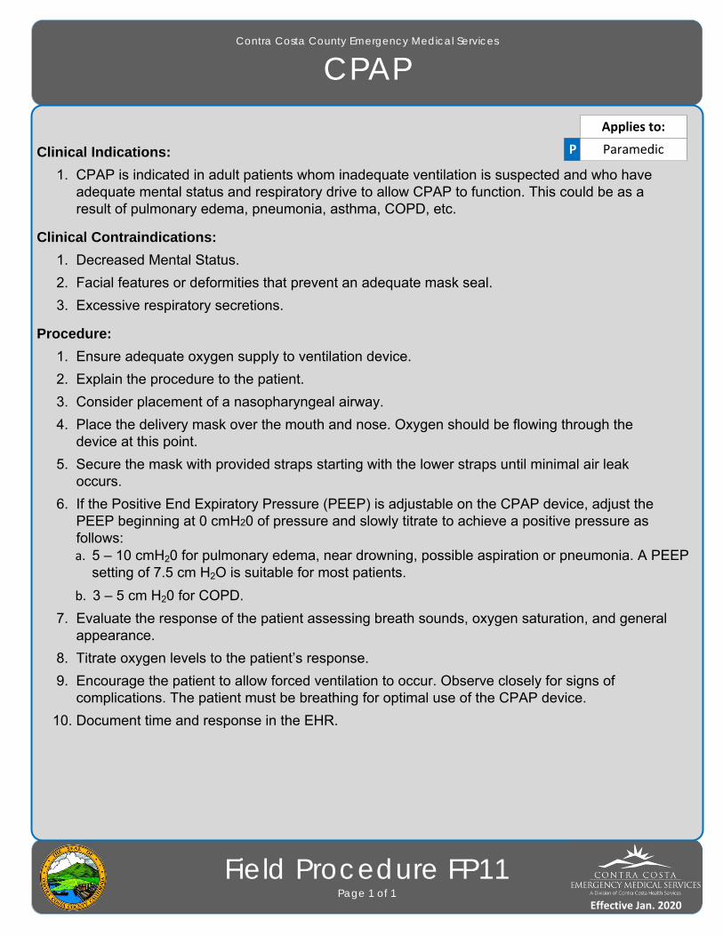
Effective Jan. 2016
Contra Costa County Emergency Medical Services
CPAP
Field Procedure FP11Page 1 of 1
Clinical Indications:
1. CPAP is indicated in adult patients whom inadequate ventilation is suspected and who have adequate mental status and respiratory drive to allow CPAP to function. This could be as a result of pulmonary edema, pneumonia, asthma, COPD, etc.
Clinical Contraindications:
1. Decreased Mental Status.
2. Facial features or deformities that prevent an adequate mask seal.
3. Excessive respiratory secretions.
Procedure:
1. Ensure adequate oxygen supply to ventilation device.
2. Explain the procedure to the patient.
3. Consider placement of a nasopharyngeal airway.
4. Place the delivery mask over the mouth and nose. Oxygen should be flowing through the device at this point.
5. Secure the mask with provided straps starting with the lower straps until minimal air leak occurs.
6. If the Positive End Expiratory Pressure (PEEP) is adjustable on the CPAP device, adjust the PEEP beginning at 0 cmH20 of pressure and slowly titrate to achieve a positive pressure as follows: a. 5 – 10 cmH20 for pulmonary edema, near drowning, possible aspiration or pneumonia. A PEEP setting of 7.5 cm H2O is suitable for most patients.
b. 3 – 5 cm H20 for COPD.
7. Evaluate the response of the patient assessing breath sounds, oxygen saturation, and general appearance.
8. Titrate oxygen levels to the patient’s response.
9. Encourage the patient to allow forced ventilation to occur. Observe closely for signs of complications. The patient must be breathing for optimal use of the CPAP device.
10. Document time and response in the EHR.
P Paramedic
Applies to:
Effective Jan. 2020
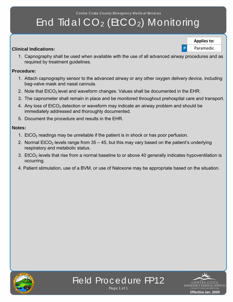
Effective Jan. 2016
Contra Costa County Emergency Medical Services
End Tidal CO2 (EtCO2) Monitoring
Field Procedure FP12Page 1 of 1
Clinical Indications:
1. Capnography shall be used when available with the use of all advanced airway procedures and as required by treatment guidelines.
Procedure:
1. Attach capnography sensor to the advanced airway or any other oxygen delivery device, including bag-valve mask and nasal cannula.
2. Note that EtCO2 level and waveform changes. Values shall be documented in the EHR.
3. The capnometer shall remain in place and be monitored throughout prehospital care and transport.
4. Any loss of EtCO2 detection or waveform may indicate an airway problem and should be immediately addressed and thoroughly documented.
5. Document the procedure and results in the EHR.
Notes:
1. EtCO2 readings may be unreliable if the patient is in shock or has poor perfusion.
2. Normal EtCO2 levels range from 35 – 45, but this may vary based on the patient’s underlying respiratory and metabolic status.
3. EtCO2 levels that rise from a normal baseline to or above 40 generally indicates hypoventilation is occurring.
4. Patient stimulation, use of a BVM, or use of Naloxone may be appropriate based on the situation.
P Paramedic
Applies to:
Effective Jan. 2020
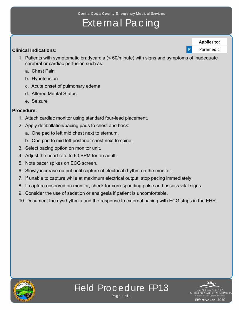
Effective Jan. 2016
Contra Costa County Emergency Medical Services
External Pacing
Field Procedure FP13Page 1 of 1
Clinical Indications:
1. Patients with symptomatic bradycardia (< 60/minute) with signs and symptoms of inadequate cerebral or cardiac perfusion such as:
a. Chest Pain
b. Hypotension
c. Acute onset of pulmonary edema
d. Altered Mental Status
e. Seizure
Procedure:
1. Attach cardiac monitor using standard four-lead placement.
2. Apply defibrillation/pacing pads to chest and back:
a. One pad to left mid chest next to sternum.
b. One pad to mid left posterior chest next to spine.
3. Select pacing option on monitor unit.
4. Adjust the heart rate to 60 BPM for an adult.
5. Note pacer spikes on ECG screen.
6. Slowly increase output until capture of electrical rhythm on the monitor.
7. If unable to capture while at maximum electrical output, stop pacing immediately.
8. If capture observed on monitor, check for corresponding pulse and assess vital signs.
9. Consider the use of sedation or analgesia if patient is uncomfortable.
10. Document the dysrhythmia and the response to external pacing with ECG strips in the EHR.
P Paramedic
Applies to:
Effective Jan. 2020

Effective Jan. 2016
Contra Costa County Emergency Medical Services
Helmet Removal
Field Procedure FP14Page 1 of 1
Clinical Indications:
1. Helmet interferes with airway management or spinal motion restriction.
2. Improper fit, allowing head to move within helmet.
3. Patient in cardiac arrest.
Contraindications:
1. Airway and spinal motion restriction can be addressed without helmet removal.
Procedure:
1. High Impact Helmets (e.g. motorcycle, car racing) - Whether the helmet is a closed or open-faced style helmet, the helmet must always be removed while providing spinal precautions.
2. Low Impact Helmets with Shoulder Pads (e.g. football, ice hockey, etc.) - In those patients wearing a well-fitted helmet which conforms closely to the patient's head, it may be preferable to leave the helmet and shoulder pads in place after removing the face mask. If the helmet is removed, the shoulder pads must also be removed to maintain neutral spinal alignment.
3. Low Impact Helmets without Shoulder Pads (e.g. baseball, bicycle, rollerblade, etc.) - Whether the helmet is a closed or open faced style helmet, the helmet must always be removed while providing spinal precautions.
4. Assess and document PMS.
5. While gently removing the helmet, maintain stabilization of the cervical spine. If indicated, place the patient in spinal motion restriction.
6. Reassess and document PMS.
E EMT
P Paramedic
Applies to:
Effective Jan. 2020

Effective Jan. 2016
Contra Costa County Emergency Medical Services
Intraosseous Access
Field Procedure FP15Page 1 of 2
Clinical Indications:
1. Patients where rapid, regular IV access is unavailable with any of the following:
a. Cardiac arrest
b. When IV access is unsuccessful or, after evaluation of potential sites, it is determined that an IV attempt would not be successful in the setting of:
i. Shock or evolving shock, regardless of the cause.
ii. Impending arrest or unstable dysrhythmia.
Contraindications:
1. Fracture of the targeted bone.
2. IO within the past 48 hours in the targeted bone.
3. Infection at the insertion site.
4. Burns that disrupt actual bone integrity at the insertion site.
5. Inability to locate landmarks or excessive tissue over the insertion site.
6. Previous orthopedic procedure near the insertion site (e.g. prosthetic limb or joint).
Procedure:
1. Proximal humerus (preferred site in patients with perfusing rhythm)
2. Proximal tibia
3. Distal tibia (if proximal humerus or proximal tibia are unsuitable)
Procedure:
1. Locate the insertion site:
a. The proximal humerus site is the greater tubercle, identifiable as a prominence on the humerus when the arm is rotated inward and the patient’s hand is on the abdomen.
b. The proximal tibia site is on the flat medial aspect of the tibia, 2 finger-breadths below the lower edge of the patella and medial to the tibial tuberosity.
c. The distal tibia site is 2 finger-breadths above the most prominent aspect of the medial malleolus (inside aspect of ankle) in the midline of the shaft of the tibia.
2. Prep the selected site with chlorhexidine and allow to air dry.
P Paramedic
Applies to:
Effective Jan. 2020
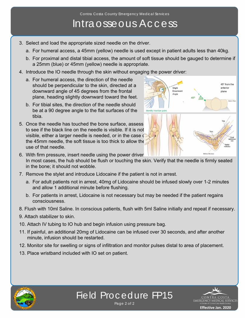
Effective Jan. 2016
Contra Costa County Emergency Medical Services
Intraosseous Access
Field Procedure FP15Page 2 of 2
3. Select and load the appropriate sized needle on the driver.
a. For humeral access, a 45mm (yellow) needle is used except in patient adults less than 40kg.
b. For proximal and distal tibial access, the amount of soft tissue should be gauged to determine if a 25mm (blue) or 45mm (yellow) needle is appropriate.
4. Introduce the IO needle through the skin without engaging the power driver:
a. For humeral access, the direction of the needle should be perpendicular to the skin, directed at a downward angle of 45 degrees from the frontal plane, heading slightly downward toward the feet.
b. For tibial sites, the direction of the needle should be at a 90 degree angle to the flat surfaces of the tibia.
5. Once the needle has touched the bone surface, assess to see if the black line on the needle is visible. If it is not visible, either a larger needle is needed, or in the case of the 45mm needle, the soft tissue is too thick to allow the use of that needle.
6. With firm pressure, insert needle using the power driver. In most cases, the hub should be flush or touching the skin. Verify that the needle is firmly seated in the bone; it should not wobble.
7. Remove the stylet and introduce Lidocaine if the patient is not in arrest.
a. For adult patients not in arrest, 40mg of Lidocaine should be infused slowly over 1-2 minutes and allow 1 additional minute before flushing.
b. For patients in arrest, Lidocaine is not necessary but may be needed if the patient regains consciousness.
8. Flush with 10ml Saline. In conscious patients, flush with 5ml Saline initially and repeat if necessary.
9. Attach stabilizer to skin.
10. Attach IV tubing to IO hub and begin infusion using pressure bag.
11. If painful, an additional 20mg of Lidocaine can be infused over 30 seconds, and after another minute, infusion should be restarted.
12. Monitor site for swelling or signs of infiltration and monitor pulses distal to area of placement.
13. Place wristband included with IO set on patient.
Effective Jan. 2020
Effective Jan. 2016
Contra Costa County Emergency Medical Services
Needle Decompression
Field Procedure FP16Page 1 of 1
Clinical Indications:
1. Patients who are exhibiting signs of a tension pneumothorax and have at least one of the following signs:
a. AMS
b. Hypotension
c. Increased pulse and respirations
d. Absent breath sounds or hyperresonance to percussion on affected side
e. Jugular vein distension
f. Difficulty ventilating
g. Tracheal shift
h. In patients with penetrating trauma to the chest or upper back, or gunshot wound to the neck or torso who are in respiratory distress, a weak or absent radial pulse may be substituted for blood pressure measurement as listed above; signs of tension pneumothorax listed above may also be present.
i. Patients in traumatic arrest with chest or abdominal trauma for whom resuscitation is indicated. These patients may require bilateral chest decompression even in the absence of the signs above.
Procedure:
1. Administer high flow oxygen.
2. Identify and prep the site:
a. Locate the second intercostal space in the mid-clavicular line on the same side as the pneumothorax.
b. Prepare the site with chlorhexidine and allow to air dry.
3. Insert a 14g catheter into the skin over the third rib and direct it just over the top of the rib (superior border) into the interspace.
4. Advance the catheter through the parietal pleura until a “pop” is felt and air or blood exits under pressure through the catheter, then advance catheter only to chest wall.
5. Remove the needle, leaving the plastic catheter in place.
6. Secure the catheter hub to the chest wall with an Asherman chest seal. The catheter should be stabilized within the flutter valve mechanism.
P Paramedic
Applies to:
Effective Jan. 2020
Effective Jan. 2016
Contra Costa County Emergency Medical Services
Pediatric Assessment
Field Procedure FP17Page 1 of 2
Clinical Indications:
1. Any child that can be measured with the Peditape.
Pediatric Assessment Triangle:
Primary Assessment:
E EMT
P Paramedic
Applies to:
Effective Jan. 2020
Effective Jan. 2016
Contra Costa County Emergency Medical Services
Pediatric Assessment
Field Procedure FP17Page 2 of 2
Pediatric GCS:
No response
Obeys commands
Effective Jan. 2020
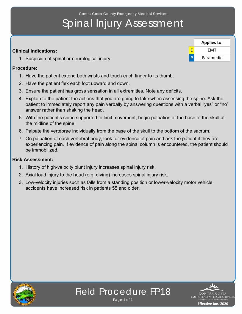
Effective Jan. 2016
Contra Costa County Emergency Medical Services
Spinal Injury Assessment
Field Procedure FP18Page 1 of 1
Clinical Indications:
1. Suspicion of spinal or neurological injury
Procedure:
1. Have the patient extend both wrists and touch each finger to its thumb.
2. Have the patient flex each foot upward and down.
3. Ensure the patient has gross sensation in all extremities. Note any deficits.
4. Explain to the patient the actions that you are going to take when assessing the spine. Ask the patient to immediately report any pain verbally by answering questions with a verbal “yes” or “no” answer rather than shaking the head.
5. With the patient’s spine supported to limit movement, begin palpation at the base of the skull at the midline of the spine.
6. Palpate the vertebrae individually from the base of the skull to the bottom of the sacrum.
7. On palpation of each vertebral body, look for evidence of pain and ask the patient if they are experiencing pain. If evidence of pain along the spinal column is encountered, the patient should be immobilized.
Risk Assessment:
1. History of high-velocity blunt injury increases spinal injury risk.
2. Axial load injury to the head (e.g. diving) increases spinal injury risk.
3. Low-velocity injuries such as falls from a standing position or lower-velocity motor vehicle accidents have increased risk in patients 55 and older.
E EMT
P Paramedic
Applies to:
Effective Jan. 2020

Effective Jan. 2016
Contra Costa County Emergency Medical Services
Spinal Motion Restriction
Field Procedure FP19Page 1 of 1
Clinical Indications: 1. Spinal motion restriction (SMR) as determined by spinal injury assessment.
a. Blunt trauma – full SMR (C-collar and full-length vacuum splint):
i. Major blunt trauma meeting trauma activation criteria
ii. Presence of neurological deficit, priapism or suspected spinal shock
iii. Obvious anatomic deformity of the spine
iv. Significant tenderness on palpation of vertebral column
v. Significant blunt trauma mechanism when a patient assessment is unreliable
b. Blunt trauma – modified SMR (C-collar only):
i. Blunt trauma not meeting above criteria but with pain complaints or concerns based on mechanism or patient risk
ii. Examples of patients may include those ambulatory after self-extrication, low-velocity mechanisms and those with no neurologic findings.
c. Penetrating trauma – full SMR:
i. Neurologic deficit or an obvious deformity of the spine
ii. Patients who have both penetrating and a significant blunt injury should be evaluated using blunt trauma criteria
Procedure: 1. Explain the procedure to the patient; assess and record extremity neuro status & distal pulses.
2. Place the patient in an appropriately sized C-collar while maintaining in-line stabilization of the cervical spine by a second provider.
3. If indicated, place the patient on a full-length vacuum splint.
4. Stabilize the patient with straps and head rolls or other similar device. Once the head is secured, the second provider may release manual in-line stabilization.
5. Assess and record extremity neuro status and distal pulses post-procedure. If worse, remove any immobilization devices and reassess.
Note: 1. SMR should reduce, rather than increase, patient discomfort. SMR that increases pain should be avoided. The cervical spine should never be moved if movement increases pain or in the presence of neurological deficits or neck spasms.
2. Suspected spinal injuries should be maintained in a neutral position; position will vary by patient.
3. Routine use of full spinal motion restriction should be reserved for patients with confirmatory physical findings or high suspicion of spinal injury.
4. AMS or presence of an entry/exit wound in proximity of spine are no longer indications for SMR.
E EMT
P Paramedic
Applies to:
Effective Jan. 2020
Effective Jan. 2016
Contra Costa County Emergency Medical Services
Taser Dart Removal
Field Procedure FP20Page 1 of 1
Clinical Indications: 1. Taser darts incurred from law enforcement intervention that are impeding patient care or are causing profound discomfort for the patient may be removed.
2. Do NOT remove a taser dart that is located in the face, neck, or groin.
Procedure: 1. Assess the patient for any potential injury after the taser was deployed. Note that taser deployment will cause a patient to fall to the ground.
2. When safe to do so, verify that all wires of the taser dart have been disconnected from the weapon.
3. Place one hand on the patient in the area where the taser dart is embedded and stabilize the skin surrounding the puncture site between two fingers. Keep your hand away from the taser dart.
4. With your other hand, in one fluid motion, pull the taser dart straight out from the puncture site.
5. Immediately dispose of the taser dart in an approved sharps container.
6. Apply direct pressure for bleeding and apply a sterile dressing to the wound.
Note: 1. Patients who have had taser darts removed shall not be medically cleared by prehospital personnel.
2. The following must be documented in the EHR: a. The patient’s presenting behavior or signs/symptoms which lead to law enforcement to tase the patient.
b. Time of taser dart removal.
c. Anatomic location of taser dart.
d. Whether or not the taser dart was intact following removal.
E EMT
P Paramedic
Applies to:
Effective Jan. 2020
Effective Jan. 2016
Contra Costa County Emergency Medical Services
Tourniquet
Field Procedure FP21Page 1 of 1
Clinical Indications:
1. Life threatening extremity hemorrhage that can not be controlled by other means.
2. May be appropriate for use to control hemorrhage in multi-casualty incidents.
Contraindications:
1. Non-extremity hemorrhage.
2. Hemorrhage that can be controlled with pressure or dressings.
Procedure:
1. Place tourniquet proximal to wound.
2. Tighten until hemorrhage stops or distal pulses in affected extremity disappear.
3. Secure the tourniquet and mark the time of application on extremity.
4. Note the time of tourniquet application in the electronic medical record and communicate this to the receiving facility.
5. Dress wounds as necessary.
6. If one tourniquet is not sufficient or not functional to control hemorrhage, consider the application of a second tourniquet more proximal to the first.
E EMT
P Paramedic
Applies to:
Effective Jan. 2020
Effective Jan. 2016
Contra Costa County Emergency Medical Services
Valsalva (Modified) Maneuver
Field Procedure FP22Page 1 of 1
Clinical Indications:
1. Clinically stable patient with narrow complex tachycardia. Do not attempt this procedure on a patient with serious signs or symptoms, which include: Hypotension; acutely altered mental status; signs of shock/poor perfusion; chest pain with evidence of ischemia (e.g. STEMI, T-wave inversions, or depressions); and acute CHF.
Procedure:
1. Place the patient on a cardiac monitor; ensure continuous ECG monitoring throughout procedure.
2. If not already completed, establish intravenous access.
3. Describe the procedure to the patient.
4. Place and position the patient on the gurney so that the patient is sitting in a semi-recumbent (45o) position.
5. Have the patient inhale and hold his/her breath while bearing down as if to have a bowel movement, or have the patient blow into a 10ml syringe while pinching their nose closed. Instruct the patient to continue bearing down or blowing into the syringe until told to stop; time the event for 15 seconds.
6. Immediately lie the patient supine and elevate the patient’s legs to 45o for 15 seconds.
7. Lower the patient’s legs such that the patient is in a supine position and reassess the cardiac rhythm after 45 seconds.
8. Continue to monitor the heart rhythm during the procedure. Stop the procedure if the patient becomes confused, the heart rate drops below 100 or asystole occurs.
9. If the patient remains in a narrow complex tachycardia, repeat the procedure one time.
10. Document the initial and all subsequent ECG rhythms and any dysrhythmia in the prehospital care record.
P Paramedic
Applies to:
Effective Jan. 2020
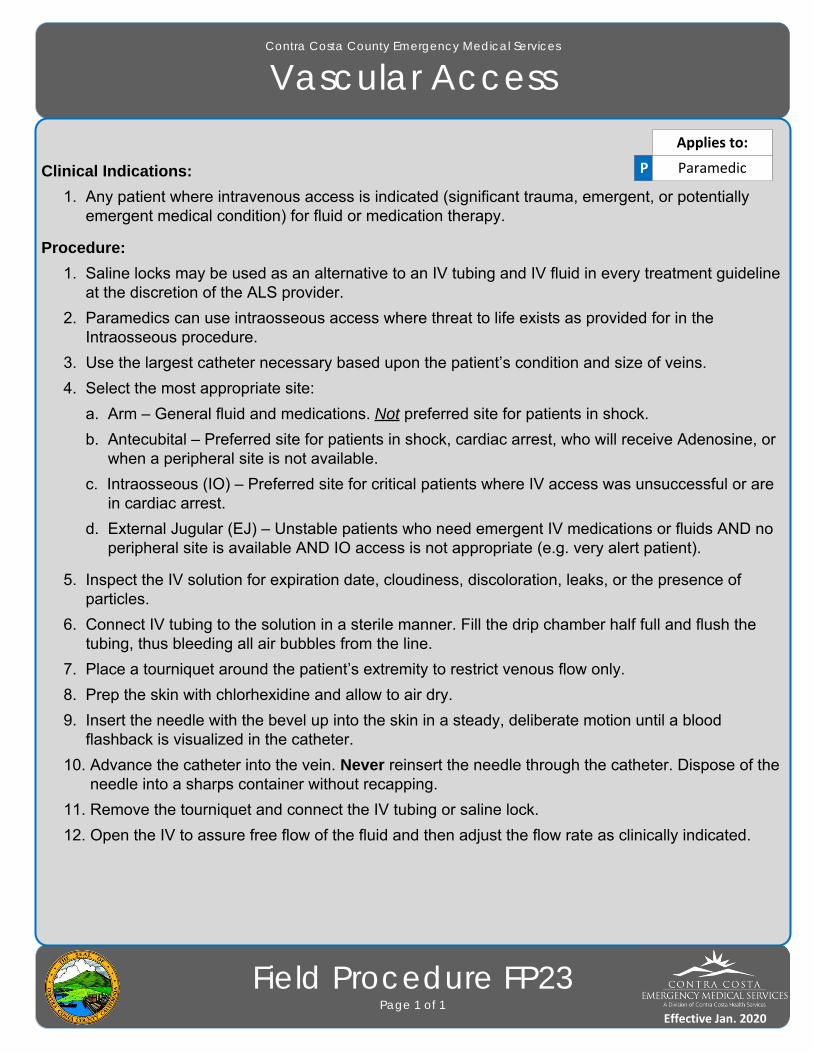
Effective Jan. 2016
Contra Costa County Emergency Medical Services
Vascular Access
Field Procedure FP23Page 1 of 1
Clinical Indications:
1. Any patient where intravenous access is indicated (significant trauma, emergent, or potentially emergent medical condition) for fluid or medication therapy.
Procedure:
1. Saline locks may be used as an alternative to an IV tubing and IV fluid in every treatment guideline at the discretion of the ALS provider.
2. Paramedics can use intraosseous access where threat to life exists as provided for in the Intraosseous procedure.
3. Use the largest catheter necessary based upon the patient’s condition and size of veins.
4. Select the most appropriate site:
a. Arm – General fluid and medications. Not preferred site for patients in shock.
b. Antecubital – Preferred site for patients in shock, cardiac arrest, who will receive Adenosine, or when a peripheral site is not available.
c. Intraosseous (IO) – Preferred site for critical patients where IV access was unsuccessful or are in cardiac arrest.
d. External Jugular (EJ) – Unstable patients who need emergent IV medications or fluids AND no peripheral site is available AND IO access is not appropriate (e.g. very alert patient).
5. Inspect the IV solution for expiration date, cloudiness, discoloration, leaks, or the presence of particles.
6. Connect IV tubing to the solution in a sterile manner. Fill the drip chamber half full and flush the tubing, thus bleeding all air bubbles from the line.
7. Place a tourniquet around the patient’s extremity to restrict venous flow only.
8. Prep the skin with chlorhexidine and allow to air dry.
9. Insert the needle with the bevel up into the skin in a steady, deliberate motion until a blood flashback is visualized in the catheter.
10. Advance the catheter into the vein. Never reinsert the needle through the catheter. Dispose of the needle into a sharps container without recapping.
11. Remove the tourniquet and connect the IV tubing or saline lock.
12. Open the IV to assure free flow of the fluid and then adjust the flow rate as clinically indicated.
P Paramedic
Applies to:
Effective Jan. 2020
Effective Jan. 2016
Contra Costa County Emergency Medical Services
Pediatric Medication Administration
Field Procedure FP24Page 1 of 1
Patient Safety is medication administration is paramount. Accurate administration of pediatricmedication requires multiple steps. Follow each of the steps below in every case.
Clinical Indications:1. Any patient under 15 years of age that requires a medication intervention.
Procedure:1. Assess Patient
Use the 6 RIGHTS – Right patient, Right drug (and indication), Right dose, Right route ofadministration, Right timing and frequency, and Right documentation.
2. Obtain weight estimate in KilogramsA. Use the Pediatape measurement device in every child of appropriate height to determine color
range of weight. Pediatape applies to patients less than 147 cm tall (4 foot, 10 inches)B. If taller than the Pediatape tape, estimate weight by patient/parent history or paramedic estimate
and ALWAYS convert to kilograms using conversion table.
3. Determine volume on Drug ChartA. Consult drug chart based on medication name to determine volume in ml.B. If patient is 50 Kg or greater, utilize adult dosages.
4. Draw up medicationA. Verify drug being administered.B. Utilize smallest syringe for volume (e.g. 1 ml or less, use tuberculin syringe).C. When giving IM or intranasal medication, load syringe ONLY with amount to be administered.
5. Double check to confirm volumeA. Double-check volume and dose with drug chart in hand – verbalize name of medication,
volume, dosage and route to another paramedic or trained EMT on scene. If possible, have another provider visually confirm dose in syringe and as called for on drug card.
6. Administer MedicationA. Administer medication by appropriate route.B. Observe the patient for signs of an adverse reaction.
7. DocumentationA. Always document dosages in your patient care report by milligrams (or if Dextrose, in grams).B. Document response to medication and any observed adverse reaction.
P Paramedic
Applies to:
Effective Jan. 2020

Effective Jan. 2016
Contra Costa County Emergency Medical Services
Finger Stick Blood Glucose
Field Procedure FP25Page 1 of 1
Clinical Indications: 1. Suspected diabetic patient
a. Signs and symptoms may include, altered mental status, combative orirritable, diaphoresis, seizure, abdominal pain, nausea or vomiting, weakness, dehydration, deep or rapid breathing
b. Medical bracelet found, insulin or other diabetic medications found, information received from patient and bystanders
2. Decreased LOC with unknown origin
Contraindications: 1. None
Equipment: 1. PPE 2. Glucometer 3. Alcohol prep pads 4. Blood glucose test strips 5. Lancet & band-aid 6. Sharps container
Procedure: 1. Activate 9-1-1 if not already activated 2. Assess patient to confirm indications are met 3. Insert test strip into glucometer 4. Cleanse finger with alcohol prep 5. Gently massage finger to promote blood flow and pierce with lancet 6. Apply blood sample to blood strip and document results 7. If BGL glucometer reading is < 60 follow treatment guideline for administration of oral glucose. 8. Continue to monitor, reassess and treat patient
Special Considerations: 1. Glucometer manufacturer instructions should be included in all Agency specific training
Treatment Guidelines: 1. Adult: G01, A03, A05, A07, A08, A12, A13, A16, A17, A18, A19, A20, T08 2. Pediatric: PC01, PC04, P03, P04, P05, P06, P07, P09, P12, P13
E EMT
P Paramedic
Applies to:
Effective Jan. 2020
Effective Jan. 2016
Contra Costa County Emergency Medical Services
EMT Epinephrine Administration
Field Procedure FP26Page 1 of 1
Clinical Indications: 1. Patient exposed to allergen and displays systemic signs and
symptoms such as:a. Mental status: Anxiety, sense of impending doom, confusion, unconscious b. Onset: Sudden, but can be more than an hour after exposure c. Skin/vascular system: Severe flushing, rash or hives, edema to the face and neck d. Respiration: Severe bronchoconstriction (wheezing), laryngospasm (stridor), difficulty breathing e. GI system: Severe cramps. Abdominal rumbling, diarrhea, vomiting f. Vital Signs: Early signs include increased heart rate and respiratory rate. Late signs included
falling respiratory rate and blood pressure, shock
Contraindications: 1. Use caution with patients who are >50 years of age and patients with cardiac history
a. Patients >50 years of age should receive pediatric dose (0.15 mg)
Equipment: 1. PPE 2. Epinephrine auto-injector (adult and pediatric) 3. Alcohol prep pads 4. Sharps container
Procedure: 1. Activate 9-1-1 if not already activated 2. Assess patient to confirm indications are met
a. Remove allergens b. Manage life threats, airway, breathing and circulation
3. Consider aseptic technique if appropriate 4. Administer appropriate epinephrine auto-injector (record time of administration)
a. Adult auto-injector: 15 through 49 years of age b. Pediatric auto-injector: 0 through 15 years of age and > 50 years of age
5. Continue to monitor, reassess and treat patient a. Be ready to support ventilations b. Secondary administration should be via IV route by a Paramedic
Special Considerations: 1. Manufacturer instructions should be included in all Agency specific training
Treatment Guidelines: 1. A04, P02
E EMT
Applies to:
Effective Jan. 2020
Effective Jan. 2016
Contra Costa County Emergency Medical Services
EMT Naloxone Administration
Field Procedure FP27Page 1 of 1
Clinical Indications: 1. Patient >22 kg. (50 lbs.) 2. Patient with suspected narcotic overdose and inadequate breathing
(respiratory rate <8). Signs and symptoms include: a. Deep snoring or gurgling b. Pale and or clammy skin c. Depressed LOC, no response to stimulation, heavy nod d. Pinpoint pupils
Contraindications: 1. Patient is breathing adequately and able to maintain own airway 2. Relative contraindication for allergy to naloxone 3. Not advised when patient has exercised their right under the California End of Life Act, are on
hospice or who are newly born
Equipment: 1. PPE 2. Naloxone prefilled syringe (pre-load or auto-injector) 3. Nasal atomizer (pre-load) 4. Alcohol prep pads 5. Sharps container
Procedure: 1. Activate 9-1-1 if not already activated 2. Assess patient to confirm indications are met
a. Manage life threats, airway, breathing and circulation 3. Administration of naloxone should be titrated to effect adequate breathing not to restore
consciousness. 4. Consider aseptic technique if appropriate 5. Administer naloxone intranasal (Record time of administration)
a. Attach nasal atomizer b. Administer 1 mg / 1mL into each nostril
6. Prepare for patient to vomit; utilize recovery position and suction 7. Continue to monitor, reassess and treat patient
Special Considerations: 1. Manufacturer instructions should be included in all Agency specific training
Treatment and Field Procedure Guidelines: 1. G02, G03, A13, A14, P08, P09, P10, FP09
E EMT
Applies to:
Effective Jan. 2020
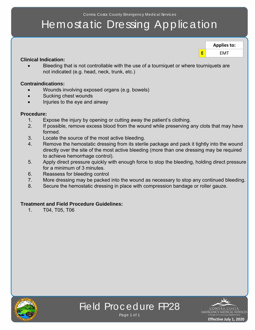
Effective Jan. 2016
Contra Costa County Emergency Medical Services
Hemostatic Dressing Application
Field Procedure FP28Page 1 of 1
Clinical Indication: Bleeding that is not controllable with the use of a tourniquet or where tourniquets are
not indicated (e.g. head, neck, trunk, etc.)
Contraindications: Wounds involving exposed organs (e.g. bowels) Sucking chest wounds Injuries to the eye and airway
Procedure:1. Expose the injury by opening or cutting away the patient’s clothing.2. If possible, remove excess blood from the wound while preserving any clots that may have
formed.3. Locate the source of the most active bleeding.4. Remove the hemostatic dressing from its sterile package and pack it tightly into the wound
directly over the site of the most active bleeding (more than one dressing may be required to achieve hemorrhage control).
5. Apply direct pressure quickly with enough force to stop the bleeding, holding direct pressure for a minimum of 3 minutes.
6. Reassess for bleeding control7. More dressing may be packed into the wound as necessary to stop any continued bleeding.8. Secure the hemostatic dressing in place with compression bandage or roller gauze.
Treatment and Field Procedure Guidelines: 1. T04, T05, T06
E EMT
Applies to:
Effective July 1, 2020

Effective Jan. 2016
Contra Costa County Emergency Medical Services
Influenza and COVID-19 Vaccination
Field Procedure FP29Page 1 of 2
Equipment required1. Vaccine2. 23-25 g, 1-inch needle
a. For larger patients, 1.5-inch needle length may be more appropriate.b. See “Needle Gauge/Length and Injection Site Guidance” below for additional information.c. COVID-19 vaccine may come as prefilled/ready to administer or require other injection supplies
or sizes.
Vaccine Administration Procedure1. Assess the need for the vaccine in question utilizing the current guidance on that vaccination.2. Screen for contraindications and precautions of inactivated vaccine (listed below).3. Collect and review Vaccine Consent/Record of Administration sheet
a. Confirm that the consent has been signed.4. To prevent syncope, vaccinate patients while they are seated or lying down and consider
observing them for 15 minutes after receipt of the vaccine. 5. Paramedics must maintain aseptic technique when administering the influenza or COVID
vaccines.6. The screening questionnaire must be completed prior to administration of the influenza or COVID vaccine.7. Wash hands and don gloves.8. Check expiration date of vaccine.9.Cleanse the area of the deltoid muscle with the alcohol prep.
a. Deltoid landmarks: 2-3 finger widths down from the acromion process; bottom edge isimaginary line drawn from axilla
10. Insert the needle at a 90-degree angle into the muscle.a. Pulling back on the plunger prior to injection is not necessary.
11. Inject the vaccine into the muscle.12. Withdraw the needle, and using the alcohol prep, apply slight pressure to the injection site.13. Do not recap or detach needle from syringe. All used syringes/needles should be placed in
puncture - proof containers.14. Monitor the patient for any symptoms of allergic reaction.15. Document the following information:
a. Date of vaccinationb. Name of patientc. Injection sited. Vaccine lot numbere. Vaccine manufacturer
P Paramedic
Applies to:
Effective during declared disaster
O EMT Optional
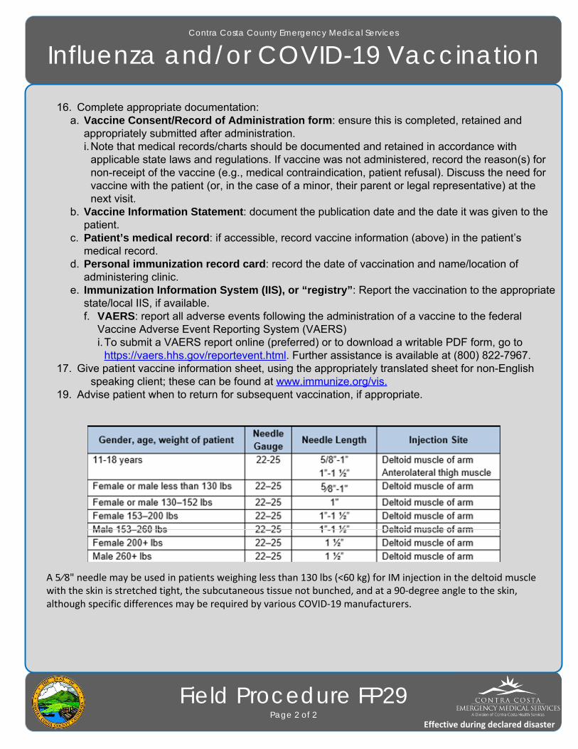
Effective Jan. 2016
Contra Costa County Emergency Medical Services
Influenza and/or COVID-19 Vaccination
Field Procedure FP29Page 2 of 2
16. Complete appropriate documentation:a. Vaccine Consent/Record of Administration form: ensure this is completed, retained and
appropriately submitted after administration.i.Note that medical records/charts should be documented and retained in accordance with
applicable state laws and regulations. If vaccine was not administered, record the reason(s) fornon-receipt of the vaccine (e.g., medical contraindication, patient refusal). Discuss the need forvaccine with the patient (or, in the case of a minor, their parent or legal representative) at the next visit.
b. Vaccine Information Statement: document the publication date and the date it was given to thepatient.
c. Patient’s medical record: if accessible, record vaccine information (above) in the patient’s medical record.
d. Personal immunization record card: record the date of vaccination and name/location of administering clinic.e. Immunization Information System (IIS), or “registry”: Report the vaccination to the appropriate
state/local IIS, if available.f. VAERS: report all adverse events following the administration of a vaccine to the federal
Vaccine Adverse Event Reporting System (VAERS)i.To submit a VAERS report online (preferred) or to download a writable PDF form, go to
https://vaers.hhs.gov/reportevent.html. Further assistance is available at (800) 822-7967. 17. Give patient vaccine information sheet, using the appropriately translated sheet for non-English
speaking client; these can be found at www.immunize.org/vis.19. Advise patient when to return for subsequent vaccination, if appropriate.
A 5⁄8" needle may be used in patients weighing less than 130 lbs (<60 kg) for IM injection in the deltoid muscle with the skin is stretched tight, the subcutaneous tissue not bunched, and at a 90‐degree angle to the skin, although specific differences may be required by various COVID‐19 manufacturers.
Effective during declared disaster

Effective Jan. 2016
Contra Costa County Emergency Medical Services
Pediatric Cardioversion
Pediatric Dosing GuidePage 1 of 1
Energy SelectionPhysio-Control LifePak 12 and LifePak 15
Note: Cardioversion in pediatric patients requires a Base Hospital order.
Gray
COLOR First Second
8 J 15 J
Pink 15 J 30 J
Red 15 J 30 J
Purple 20 J 30 J
Yellow 30 J 50 J
White 30 J 70 J
Blue 30 J 70 J
Orange 50 J 100 J
Green 70 J 125 J
40kg 70 J 150 J
45kg 100 J 175 J
Effective Jan. 2020
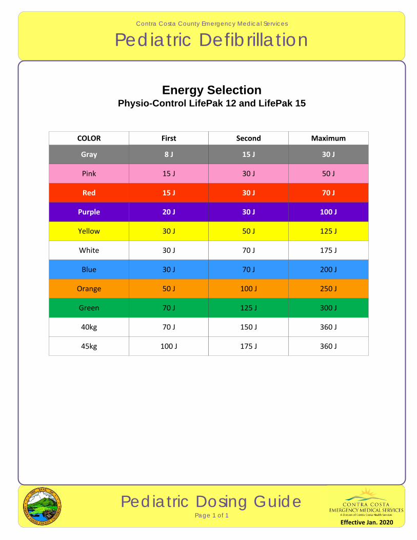
Effective Jan. 2016
Contra Costa County Emergency Medical Services
Pediatric Defibrillation
Pediatric Dosing GuidePage 1 of 1
Energy SelectionPhysio-Control LifePak 12 and LifePak 15
Gray
COLOR First Second Maximum
8 J 15 J 30 J
Pink 15 J 30 J 50 J
Red 15 J 30 J 70 J
Purple 20 J 30 J 100 J
Yellow 30 J 50 J 125 J
White 30 J 70 J 175 J
Blue 30 J 70 J 200 J
Orange 50 J 100 J 250 J
Green 70 J 125 J 300 J
40kg 70 J 150 J 360 J
45kg 100 J 175 J 360 J
Effective Jan. 2020
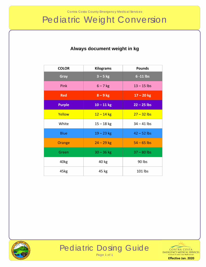
Effective Jan. 2016
Contra Costa County Emergency Medical Services
Pediatric Weight Conversion
Pediatric Dosing GuidePage 1 of 1
Always document weight in kg
Gray
COLOR Kilograms Pounds
3 – 5 kg 6 ‐11 lbs
Pink 6 – 7 kg 13 – 15 lbs
Red 8 – 9 kg 17 – 20 kg
Purple 10 – 11 kg 22 – 25 lbs
Yellow 12 – 14 kg 27 – 32 lbs
White 15 – 18 kg 34 – 41 lbs
Blue 19 – 23 kg 42 – 52 lbs
Orange 24 – 29 kg 54 – 65 lbs
Green 30 – 36 kg 37 – 80 lbs
40kg 40 kg 90 lbs
45kg 45 kg 101 lbs
Effective Jan. 2020

Effective Jan. 2016
Contra Costa County Emergency Medical Services
Adenosine
Pediatric Dosing GuidePage 1 of 1
Indication: SVTConcentration = 3 mg/ml
Note: Immediately follow with a rapid 10-20ml NS bolus.
Gray
COLOR Doses (mg) Give (ml)
0.45 mg
Pink
Red
Purple
Yellow
White
Blue
Orange
Green
40kg
45kg
0.9 mg 1st 0.15 ml 2nd 0.3 ml
0.66 mg1.35 mg
1st 0.22 ml 2nd 0.45 ml
0.9 mg1.8 mg
1st 0.3 ml 2nd 0.6 ml
1 mg2mg
1st 0.33 ml 2nd 0.67 ml
1.35 mg2.7 mg
1st 0.45 ml 2nd 0.9 ml
1.7 mg3.4 mg
1st 0.6 ml 2nd 1.2 ml
2.1 mg4.2 mg
1st 0.7 ml 2nd 1.4 ml
2.7 mg5.4 mg
1st 0.9 ml 2nd 1.8 ml
3.3 mg6.6 mg
1st 1.1 ml 2nd 2.2 ml
4mg8mg
1st 1.3 ml 2nd 2.7 ml
4.5 mg9 mg
1st 1.5 ml 2nd 3 ml
Effective Jan. 2020
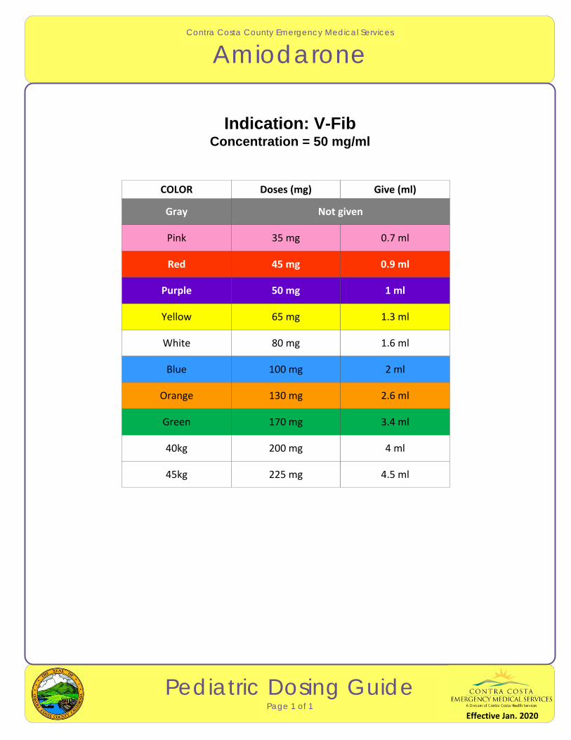
Effective Jan. 2016
Contra Costa County Emergency Medical Services
Amiodarone
Pediatric Dosing GuidePage 1 of 1
Indication: V-FibConcentration = 50 mg/ml
Gray
COLOR Doses (mg) Give (ml)
Pink
Red
Purple
Yellow
White
Blue
Orange
Green
40kg
45kg
Not given
35 mg 0.7 ml
45 mg 0.9 ml
50 mg 1 ml
65 mg 1.3 ml
80 mg 1.6 ml
100 mg 2 ml
130 mg 2.6 ml
170 mg 3.4 ml
200 mg 4 ml
225 mg 4.5 ml
Effective Jan. 2020

Effective Jan. 2016
Contra Costa County Emergency Medical Services
Atropine
Pediatric Dosing Guide Page 1 of 1
Indication: Symptomatic Bradycardia/OverdoseConcentration = 0.1 mg/ml
Minimum Dose = 0.1 mgMaximum Dose – 0.5 mg
Note: Ensure adequate ventilation before considering Atropine. NOT indicated for asystole.
Gray
COLOR Doses (mg) Give (ml)
Pink
Red
Purple
Yellow
White
Blue
Orange
Green
40kg
45kg
0.13 mg 1.3 ml
0.17 mg 1.7 ml
0.2 mg 2 ml
0.25 mg 2.5 ml
0.35 mg 3.5 ml
0.42 mg 4.2 ml
0.5 mg 5 ml
0.5 mg 5 ml
0.5 mg 5 ml
0.5 mg 5 ml
0.1 mg 1 ml
Effective Jan. 2020
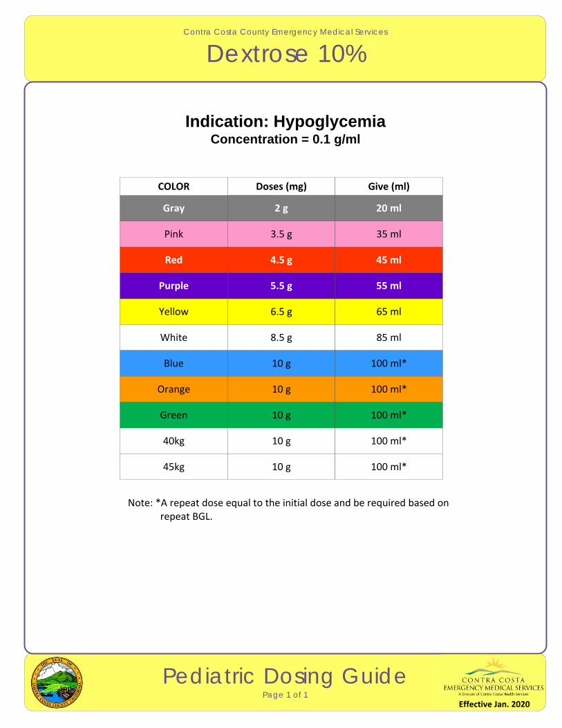
Effective Jan. 2016
Contra Costa County Emergency Medical Services
Dextrose 10%
Pediatric Dosing GuidePage 1 of 1
Indication: HypoglycemiaConcentration = 0.1 g/ml
Note: *A repeat dose equal to the initial dose and be required based on repeat BGL.
Gray
COLOR Doses (mg) Give (ml)
Pink
Red
Purple
Yellow
White
Blue
Orange
Green
40kg
45kg
3.5 g 35 ml
4.5 g 45 ml
5.5 g 55 ml
6.5 g 65 ml
8.5 g 85 ml
10 g 100 ml*
10 g 100 ml*
10 g 100 ml*
10 g 100 ml*
10 g 100 ml*
2 g 20 ml
Effective Jan. 2020
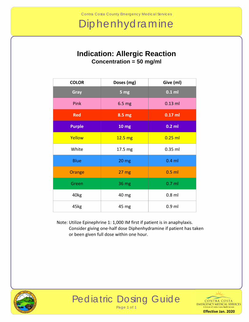
Effective Jan. 2016
Contra Costa County Emergency Medical Services
Diphenhydramine
Pediatric Dosing GuidePage 1 of 1
Indication: Allergic ReactionConcentration = 50 mg/ml
Note: Utilize Epinephrine 1: 1,000 IM first if patient is in anaphylaxis. Consider giving one‐half dose Diphenhydramine if patient has taken or been given full dose within one hour.
Gray
COLOR Doses (mg) Give (ml)
Pink
Red
Purple
Yellow
White
Blue
Orange
Green
40kg
45kg
6.5 mg 0.13 ml
8.5 mg 0.17 ml
10 mg 0.2 ml
12.5 mg 0.25 ml
17.5 mg 0.35 ml
20 mg 0.4 ml
27 mg 0.5 ml
36 mg 0.7 ml
40 mg 0.8 ml
45 mg 0.9 ml
5 mg 0.1 ml
Effective Jan. 2020
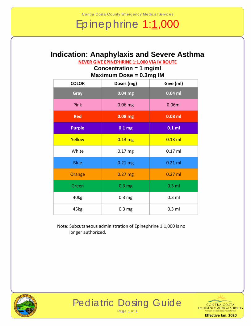
Effective Jan. 2016
Contra Costa County Emergency Medical Services
Epinephrine 1:1,000
Pediatric Dosing GuidePage 1 of 1
Indication: Anaphylaxis and Severe AsthmaNEVER GIVE EPINEPHRINE 1:1,000 VIA IV ROUTE
Concentration = 1 mg/mlMaximum Dose = 0.3mg IM
Note: Subcutaneous administration of Epinephrine 1:1,000 is no longer authorized.
Gray
COLOR Doses (mg) Give (ml)
Pink
Red
Purple
Yellow
White
Blue
Orange
Green
40kg
45kg
0.06 mg 0.06ml
0.08 mg 0.08 ml
0.1 mg 0.1 ml
0.13 mg 0.13 ml
0.17 mg 0.17 ml
0.21 mg 0.21 ml
0.27 mg 0.27 ml
0.3 mg 0.3 ml
0.3 mg 0.3 ml
0.3 mg 0.3 ml
0.04 mg 0.04 ml
Effective Jan. 2020
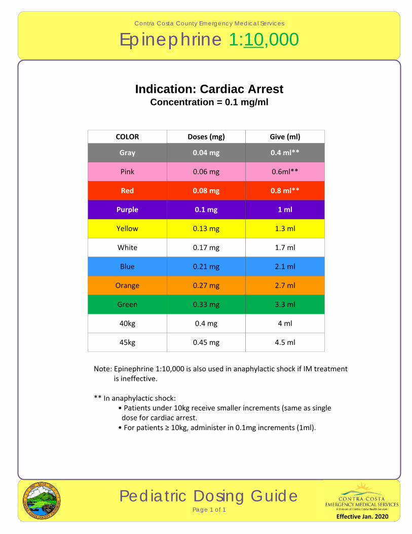
Effective Jan. 2016
Contra Costa County Emergency Medical Services
Epinephrine 1:10,000
Pediatric Dosing GuidePage 1 of 1
Indication: Cardiac ArrestConcentration = 0.1 mg/ml
Note: Epinephrine 1:10,000 is also used in anaphylactic shock if IM treatment is ineffective.
** In anaphylactic shock:• Patients under 10kg receive smaller increments (same as single dose for cardiac arrest.• For patients ≥ 10kg, administer in 0.1mg increments (1ml).
Gray
COLOR Doses (mg) Give (ml)
Pink
Red
Purple
Yellow
White
Blue
Orange
Green
40kg
45kg
0.06 mg 0.6ml**
0.08 mg 0.8 ml**
0.1 mg 1 ml
0.13 mg 1.3 ml
0.17 mg 1.7 ml
0.21 mg 2.1 ml
0.27 mg 2.7 ml
0.33 mg 3.3 ml
0.4 mg 4 ml
0.45 mg 4.5 ml
0.04 mg 0.4 ml**
Effective Jan. 2020
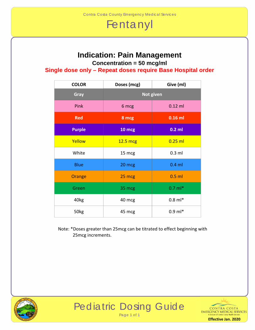
Effective Jan. 2016
Contra Costa County Emergency Medical Services
Fentanyl
Pediatric Dosing GuidePage 1 of 1
Indication: Pain ManagementConcentration = 50 mcg/ml
Single dose only – Repeat doses require Base Hospital order
Note: *Doses greater than 25mcg can be titrated to effect beginning with 25mcg increments.
Gray
COLOR Doses (mcg) Give (ml)
Pink
Red
Purple
Yellow
White
Blue
Orange
Green
40kg
50kg
6 mcg 0.12 ml
8 mcg 0.16 ml
10 mcg 0.2 ml
12.5 mcg 0.25 ml
15 mcg 0.3 ml
20 mcg 0.4 ml
25 mcg 0.5 ml
35 mcg 0.7 ml*
40 mcg 0.8 ml*
45 mcg 0.9 ml*
Not given
Effective Jan. 2020
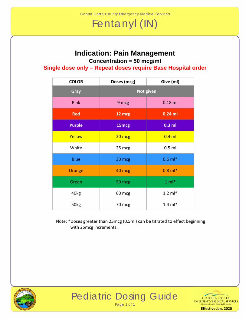
Effective Jan. 2016
Contra Costa County Emergency Medical Services
Fentanyl (IN)
Pediatric Dosing GuidePage 1 of 1
Indication: Pain ManagementConcentration = 50 mcg/ml
Single dose only – Repeat doses require Base Hospital order
Note: *Doses greater than 25mcg (0.5ml) can be titrated to effect beginning with 25mcg increments.
Gray
COLOR Doses (mcg) Give (ml)
Pink
Red
Purple
Yellow
White
Blue
Orange
Green
40kg
50kg
9 mcg 0.18 ml
12 mcg 0.24 ml
15mcg 0.3 ml
20 mcg 0.4 ml
25 mcg 0.5 ml
30 mcg 0.6 ml*
40 mcg 0.8 ml*
50 mcg 1 ml*
60 mcg 1.2 ml*
70 mcg 1.4 ml*
Not given
Effective Jan. 2020
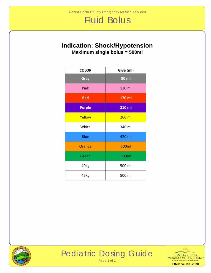
Effective Jan. 2016
Contra Costa County Emergency Medical Services
Fluid Bolus
Pediatric Dosing GuidePage 1 of 1
Indication: Shock/HypotensionMaximum single bolus = 500ml
Gray
COLOR Give (ml)
Pink
Red
Purple
Yellow
White
Blue
Orange
Green
40kg
45kg
130 ml
170 ml
210 ml
260 ml
340 ml
420 ml
500ml
500ml
500 ml
500 ml
80 ml
Effective Jan. 2020
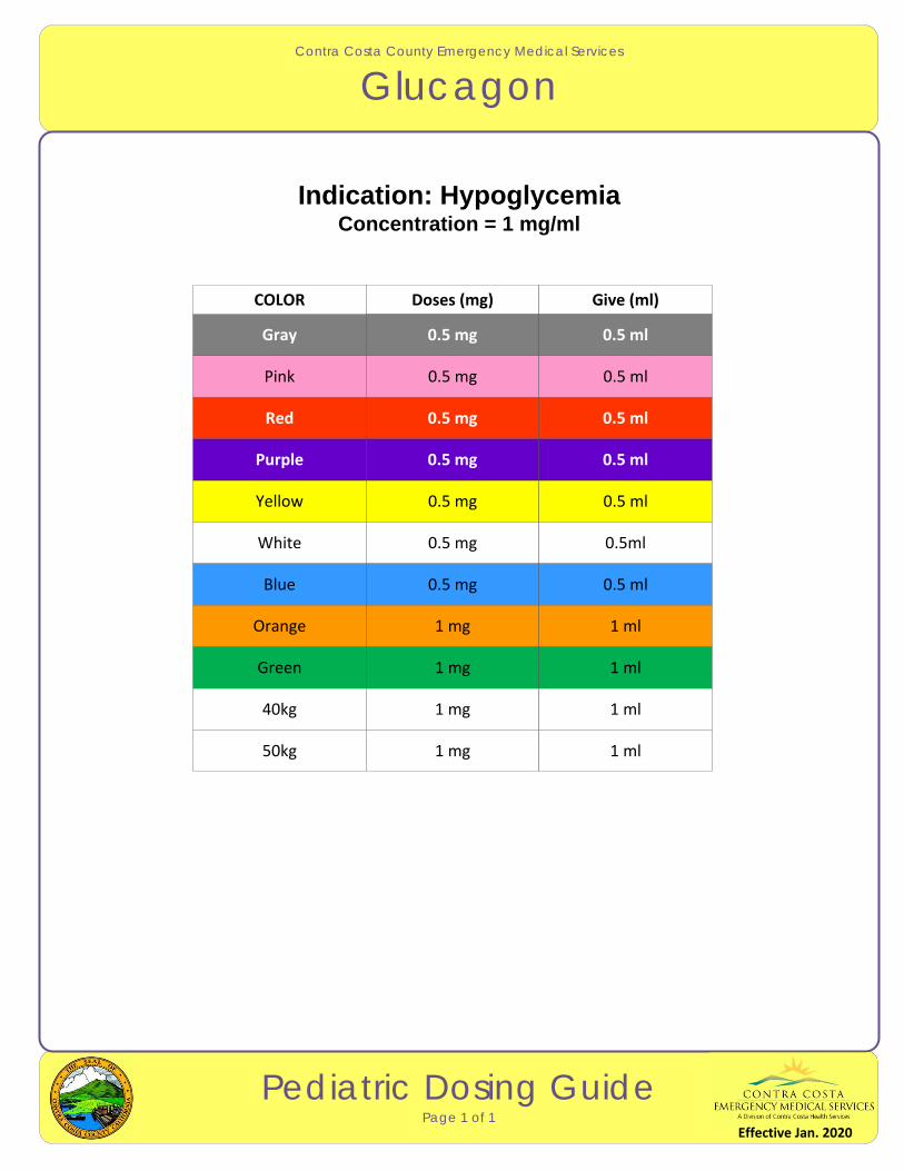
Effective Jan. 2016
Contra Costa County Emergency Medical Services
Glucagon
Pediatric Dosing GuidePage 1 of 1
Indication: HypoglycemiaConcentration = 1 mg/ml
Gray
COLOR Doses (mg) Give (ml)
Pink
Red
Purple
Yellow
White
Blue
Orange
Green
40kg
50kg
0.5 mg 0.5 ml
0.5 mg 0.5 ml
0.5 mg 0.5 ml
0.5 mg 0.5 ml
0.5 mg 0.5ml
0.5 mg 0.5 ml
1 mg 1 ml
1 mg 1 ml
1 mg 1 ml
1 mg 1 ml
0.5 mg 0.5 ml
Effective Jan. 2020
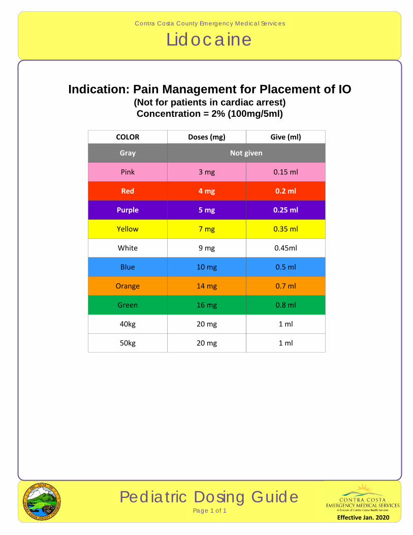
Effective Jan. 2016
Contra Costa County Emergency Medical Services
Lidocaine
Pediatric Dosing GuidePage 1 of 1
Indication: Pain Management for Placement of IO(Not for patients in cardiac arrest)Concentration = 2% (100mg/5ml)
Gray
COLOR Doses (mg) Give (ml)
Pink
Red
Purple
Yellow
White
Blue
Orange
Green
40kg
50kg
3 mg 0.15 ml
4 mg 0.2 ml
5 mg 0.25 ml
7 mg 0.35 ml
9 mg 0.45ml
10 mg 0.5 ml
14 mg 0.7 ml
16 mg 0.8 ml
20 mg 1 ml
20 mg 1 ml
Not given
Effective Jan. 2020

Effective Jan. 2016
Contra Costa County Emergency Medical Services
Midazolam – Status Epilepticus
Pediatric Dosing GuidePage 1 of 1
Indication: Status EpilepticusConcentration = 5 mg/ml
Note: Additional doses may be given every 3‐5 minutes until seizure cessation OR up to a maximum dose of 5mg (≤ 40 kg) OR 10mg (> 40 kg)
Gray
COLOR Doses (mg) Give (ml)
Pink
Red
Purple
Yellow
White
Blue
Orange
Green
≥ 40kg
1.4 mg 0.26 ml
1.8 mg 0.34 ml
2.2 mg 0.42 ml
2.8 mg 0.52 ml
3.6 mg 0.66 ml
4.6 mg 0.84 ml
5 mg 1 ml
5 mg 1 ml
10 mg 2 ml
1 mg 0.2 ml
IN (preferred)/IM
Effective Jan. 2021
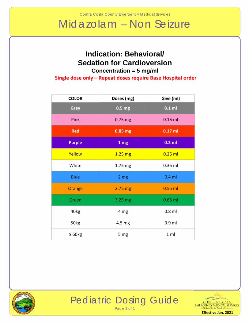
Effective Jan. 2016
Contra Costa County Emergency Medical Services
Midazolam – Non Seizure
Pediatric Dosing GuidePage 1 of 1
Indication: Behavioral/ Sedation for Cardioversion
Concentration = 5 mg/ml Single dose only – Repeat doses require Base Hospital order
Gray
COLOR Doses (mg) Give (ml)
Pink
Red
Purple
Yellow
White
Blue
Orange
Green
40kg
50kg
0.75 mg 0.15 ml
0.85 mg 0.17 ml
1 mg 0.2 ml
1.25 mg 0.25 ml
1.75 mg 0.35 ml
2 mg 0.4 ml
2.75 mg 0.55 ml
3.25 mg 0.65 ml
4 mg 0.8 ml
4.5 mg 0.9 ml
0.5 mg 0.1 ml
≥ 60kg 5 mg 1 ml
Effective Jan. 2021
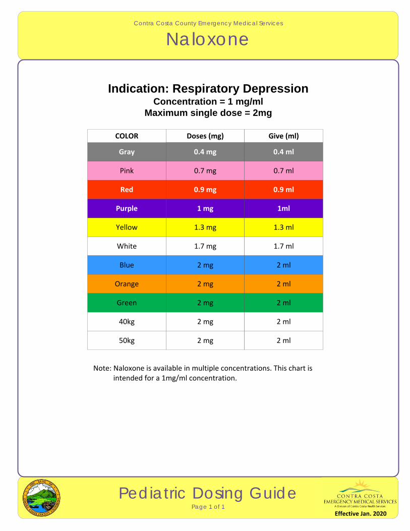
Effective Jan. 2016
Contra Costa County Emergency Medical Services
Naloxone
Pediatric Dosing GuidePage 1 of 1
Indication: Respiratory Depression Concentration = 1 mg/ml
Maximum single dose = 2mg
Note: Naloxone is available in multiple concentrations. This chart is intended for a 1mg/ml concentration.
Gray
COLOR Doses (mg) Give (ml)
Pink
Red
Purple
Yellow
White
Blue
Orange
Green
40kg
50kg
0.7 mg 0.7 ml
0.9 mg 0.9 ml
1 mg 1ml
1.3 mg 1.3 ml
1.7 mg 1.7 ml
2 mg 2 ml
2 mg 2 ml
2 mg 2 ml
2 mg 2 ml
2 mg 2 ml
0.4 mg 0.4 ml
Effective Jan. 2020
Effective Jan. 2016
Contra Costa County Emergency Medical Services
Ondansetron ODT
Pediatric Dosing GuidePage 1 of 1
Indication: Nausea/Vomiting Concentration = 4mg tablet
Maximum single dose = 4mg
Note: Ondansetron is not indicated for motion sickness.
Gray
COLOR Doses (mg) Give (ml)
Pink
Red
Purple
Yellow
White
Blue
Orange
Green
40kg
50kg
Not indicated Not indicated
Not indicated Not indicated
Not indicated Not indicated
Not indicated Not indicated
4 mg 1 tablet
4 mg 1 tablet
4 mg 1 tablet
4 mg 1 tablet
4 mg 1 tablet
4 mg 1 tablet
Not indicated Not indicated
Effective Jan. 2020
Effective Jan. 2016
Contra Costa County Emergency Medical Services
Ondansetron
Pediatric Dosing GuidePage 1 of 1
Indication: Nausea/vomiting Concentration = 2 mg/ml
Maximum single dose = 4mg
Note: Ondansetron is not indicated for motion sickness. May be repeated x1 for patients ≥ 40kg.
Gray
COLOR Doses (mg) Give (ml)
Pink
Red
Purple
Yellow
White
Blue
Orange
Green
40kg
50kg
Not indicated Not indicated
Not indicated Not indicated
Not indicated Not indicated
Not indicated Not indicated
1 mg 0.5 ml
2 mg 1 ml
2 mg 1 ml
3 mg 1.5 ml
4 mg 2 ml
4 mg 2 ml
Not indicated Not indicated
Effective Jan. 2020
Effective Jan. 2016
Contra Costa County Emergency Medical Services
Approved Abbreviations
ReferencePage 1 of 3
ABBREVIATION DEFINITION> Is greater than
< Is less than
≥ Is greater than or equal to
≤ Is less than or equal to
ACLS Advanced Cardiopulmonary Life Support
AED Automated External Defibrillator
ALS Advanced Life Support
AMA Against Medical Advice
AMI Acute Myocardial Infarction
AMS Altered Mental Status
BHP Base Hospital Physician
BLS Basic Life Support
BP Blood Pressure
BPM Beats Per Minute
BSA Body Surface Area
BSI Body Substance Isolation
BVM Bag Valve Mask
CAB Circulation, Airway, Breathing
CAD Computer Aided Dispatch
CHF Congestive Heart Failure
CHO UCSF Benioff Children’s Hospital of Oakland
CHP California Highway Patrol
COPD Chronic Obstructive Pulmonary Disease
CPAP Continuous Positive Airway Pressure
CPR Cardiopulmonary Resuscitation
CPS Child Protective Services
APS Adult Protective Services
CT Computed Tomography
CVA Cerebral Vascular Accident
DNR Do Not Resuscitate
Effective Jan. 2020
Effective Jan. 2016
Contra Costa County Emergency Medical Services
Approved Abbreviations
ReferencePage 2 of 3
ABBREVIATION DEFINITIONECG Electrocardiogram
ED Emergency Department
EMS Emergency Medical Services
EMT Emergency Medical Technician
EMTALA Emergency Medical Treatment and Active Labor Act
EOA Exclusive Operating Area
ETA Estimated Time of Arrival
EtCO2 End Tidal Carbon Dioxide
ETT Endotracheal Tube
GCS Glasgow Coma Score
HIPAA
HR Heart Rate (as depicted on an ECG) – not to be confused with pulse rate
IC Incident Commander
ICS Incident Command System
IFT Inter‐facility Transfer
IM Intramuscular
IN Intranasal
IO Intraosseous
IV Intravenous
IVP Intravenous Piggyback
LOC Level of Consciousness
MCI Multi Casualty Incident
MHOAC Medical/Health Operational Area Coordinator
NIMS National Incident Management System
NREMT National Registry of Emergency Medical Technicians
NS Normal Saline
H20 Water
NTG Nitroglycerin
MOI Mechanism of injury
EHR Electronic Health Record
Health Insurance Portability and Accountability Act
Effective Jan. 2020
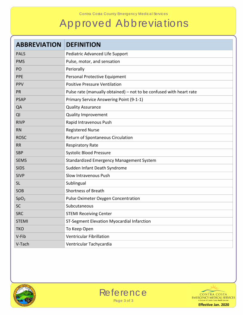
Effective Jan. 2016
Contra Costa County Emergency Medical Services
Approved Abbreviations
ReferencePage 3 of 3
ABBREVIATION DEFINITION
PO Periorally
PPE Personal Protective Equipment
PPV Positive Pressure Ventilation
PR Pulse rate (manually obtained) – not to be confused with heart rate
PSAP Primary Service Answering Point (9‐1‐1)
QA Quality Assurance
QI Quality Improvement
RIVP Rapid Intravenous Push
RN Registered Nurse
ROSC Return of Spontaneous Circulation
SBP
SEMS Standardized Emergency Management System
SIDS Sudden Infant Death Syndrome
SIVP Slow Intravenous Push
SL Sublingual
SOB Shortness of Breath
SpO2 Pulse Oximeter Oxygen Concentration
SC Subcutaneous
SRC STEMI Receiving Center
STEMI ST‐Segment Elevation Myocardial Infarction
TKO To Keep Open
V‐Fib Ventricular Fibrillation
V‐Tach Ventricular Tachycardia
RR Respiratory Rate
Systolic Blood Pressure
PALS Pediatric Advanced Life Support
PMS Pulse, motor, and sensation
Effective Jan. 2020

Effective Jan. 2016
Contra Costa County Emergency Medical Services
Drug Reference
ReferencePage 1 of 6
Drug
AdenosineNarrow complex tachycardia
Indication Dosing Cautions Comments
Initial – 6mg rapid IV
Repeat – 12mg rapid IV
Follow each dose with 20ml NS rapid IV
May cause transient heart block or asystole. Use caution when patient is taking carmbamazepine, dipyramidole, or methylxanthines. Do not administer if patient is experiencing acute asthma exacerbation.
Side effects include: chest pressure/pain, palpitations, hypotension, dyspnea, or feeling of impending doom.
Refer to pediatric dosing guide
Albuterol
Bronchospasm
5mg nebulized
Repeat as needed Use caution in patients taking MAOIs (antidepressants Nardil and Parnate)
None5mg nebulized
Repeat as needed
Crush injury ‐ hyperkalemia
5mg nebulized continuously
Amiodarone
V‐FibPulseless V‐Tach
Initial – 300mg IV/IO
Repeat – 150mg IV/IO if rhythm persists In patients with pulses,
may cause hypotension. Do not administer if patient is hypotensive. Do not use filter needle.
When creating infusion, careful mixing is needed to avoid foaming of medication.
Refer to pediatric dosing guide
Symptomatic stable V‐Tach
Initial – 150mg IV/IO drip over 10 minutes
Repeat – 150mg IV/IO if needed
AcetaminophenModerate to severe pain
1 gm over 15 minutes for patients greater than 50 kg
Active liver disease, history of transplant, patients currently taking Acetaminophen containing products such as cold/cough medicine, percocet or vicodin
Should not be directly administered into IV site. Set up as piggyback.
Not for use in patients with chest pain of cardiac origin.
Effective Jan. 2021
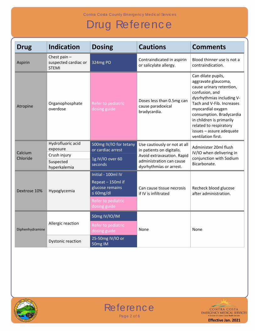
Effective Jan. 2016
Contra Costa County Emergency Medical Services
Drug Reference
ReferencePage 2 of 6
Drug
AtropineOrganophosphate overdose
Indication Dosing Cautions Comments
Doses less than 0.5mg can cause paradoxical bradycardia.
Can dilate pupils, aggravate glaucoma, cause urinary retention, confusion, and dysrhythmias including V‐Tach and V‐Fib. Increases myocardial oxygen consumption. Bradycardia in children is primarily related to respiratory issues – assure adequate ventilation first.
Refer to pediatric dosing guide
Calcium Chloride
500mg IV/IO for tetany or cardiac arrest
Use cautiously or not at all in patients on digitalis. Avoid extravasation. Rapid administration can cause dysrhythmias or arrest.
Administer 20ml flush IV/IO when delivering in conjunction with Sodium Bicarbonate.
1g IV/IO over 60 seconds
Dextrose 10% Hypoglycemia
Initial ‐ 100ml IV
Repeat – 150ml if glucose remains ≤ 60mg/dl
Can cause tissue necrosis if IV is infiltrated
Recheck blood glucose after administration.
Refer to pediatric dosing guide
Diphenhydramine
Allergic reaction
50mg IV/IO/IM
None NoneRefer to pediatric dosing guide
Dystonic reaction25‐50mg IV/IO or 50mg IM
Hydrofluoric acid exposure
Crush injury
Suspected hyperkalemia
AspirinChest pain – suspected cardiac or STEMI
324mg POContraindicated in aspirin or salicylate allergy.
Blood thinner use is not a contraindication.
Effective Jan. 2021
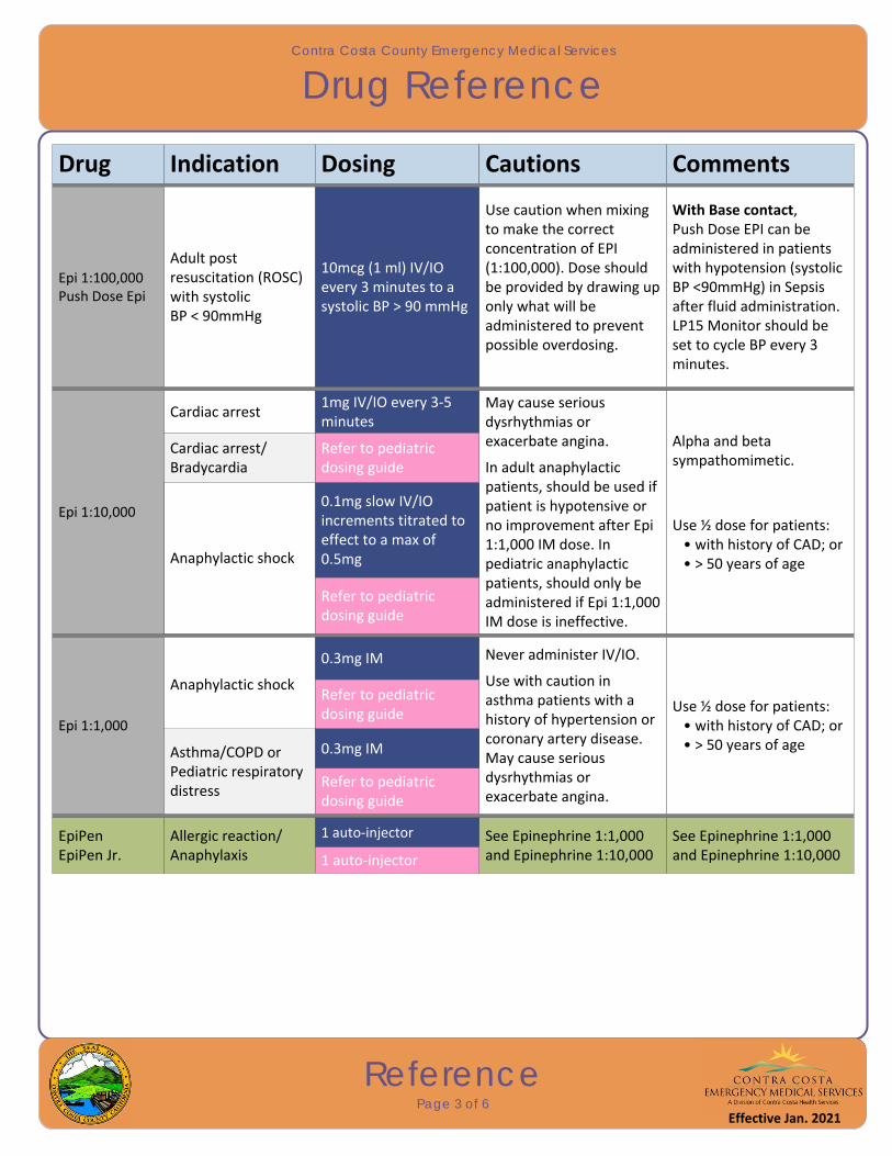
Effective Jan. 2016
Contra Costa County Emergency Medical Services
Drug Reference
ReferencePage 3 of 6
Drug Indication Dosing Cautions Comments
Epi 1:1,000
Anaphylactic shock
0.3mg IM Never administer IV/IO.
Use with caution in asthma patients with a history of hypertension or coronary artery disease. May cause serious dysrhythmias or exacerbate angina.
Use ½ dose for patients: • with history of CAD; or • > 50 years of age
Refer to pediatric dosing guide
Asthma/COPD orPediatric respiratory distress
0.3mg IM
Refer to pediatric dosing guide
EpiPenEpiPen Jr.
Allergic reaction/Anaphylaxis
1 auto‐injector See Epinephrine 1:1,000 and Epinephrine 1:10,000
See Epinephrine 1:1,000 and Epinephrine 1:10,0001 auto‐injector
Epi 1:10,000
Cardiac arrest1mg IV/IO every 3‐5 minutes
May cause serious dysrhythmias or exacerbate angina.
In adult anaphylactic patients, should be used if patient is hypotensive or no improvement after Epi 1:1,000 IM dose. In pediatric anaphylactic patients, should only be administered if Epi 1:1,000 IM dose is ineffective.
Alpha and beta sympathomimetic.
Use ½ dose for patients: • with history of CAD; or • > 50 years of age
Refer to pediatric dosing guide
Anaphylactic shock
0.1mg slow IV/IO increments titrated to effect to a max of 0.5mg
Refer to pediatric dosing guide
Cardiac arrest/Bradycardia
Epi 1:100,000Push Dose Epi
Adult postresuscitation (ROSC)with systolic BP < 90mmHg
10mcg (1 ml) IV/IO every 3 minutes to a systolic BP > 90 mmHg
Use caution when mixing to make the correct concentration of EPI (1:100,000). Dose should be provided by drawing up only what will be administered to prevent possible overdosing.
With Base contact, Push Dose EPI can be administered in patients with hypotension (systolic BP <90mmHg) in Sepsis after fluid administration.LP15 Monitor should be set to cycle BP every 3 minutes.
Effective Jan. 2021
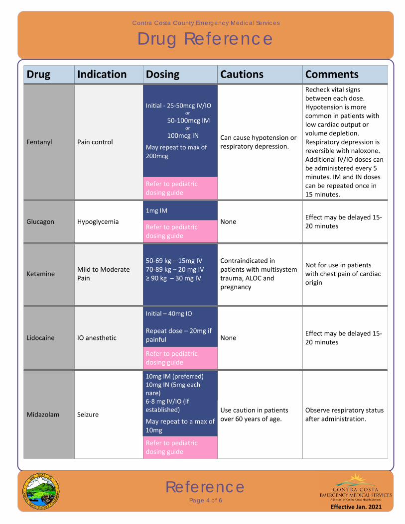
Effective Jan. 2016
Contra Costa County Emergency Medical Services
Drug Reference
ReferencePage 4 of 6
Drug Indication Dosing Cautions Comments
Midazolam Seizure
5mg IV/IO/IM/IN
May repeat to a max of 10mg
Use caution in patients over 60 years of age.
Observe respiratory status after administration.
Refer to pediatric dosing guide
Lidocaine NoneEffect may be delayed 15‐20 minutes
Refer to pediatric dosing guide
10mg IM (preferred)10mg IN (5mg each nare)6‐8 mg IV/IO (if established)
May repeat to a max of 10mg
IO anesthetic
Initial – 40mg IO
Repeat dose – 20mg if painful
Glucagon Hypoglycemia
1mg IM
NoneEffect may be delayed 15‐20 minutesRefer to pediatric
dosing guide
KetamineMild to Moderate Pain
50‐69 kg – 15mg IV70‐89 kg – 20 mg IV≥ 90 kg – 30 mg IV
Contraindicated in patients with multisystem trauma, ALOC and pregnancy
Not for use in patients with chest pain of cardiac origin
Fentanyl Pain control
Initial ‐ 25‐50mcg IV/IO or
50‐100mcg IM or
100mcg IN
May repeat to max of 200mcg
Can cause hypotension or respiratory depression.
Recheck vital signs between each dose. Hypotension is more common in patients with low cardiac output or volume depletion. Respiratory depression is reversible with naloxone. Additional IV/IO doses can be administered every 5 minutes. IM and IN doses can be repeated once in 15 minutes.
Refer to pediatric dosing guide
Effective Jan. 2021

Effective Jan. 2016
Contra Costa County Emergency Medical Services
Drug Reference
ReferencePage 5 of 6
Drug Indication Dosing Cautions Comments
NaloxoneRespiratory depression or apnea
2mg IN or
1‐2mg IV/IM Abrupt withdrawal symptoms and combative behavior may occur.
IN administration preferred unless patient is in shock or has copious secretions/blood in nares. Shorter duration of action than that of narcotics. Titrate to effect of normal respirations; it is not necessary to fully wake the patient.
Refer to pediatric dosing guide
Naloxone Overdose 1 preload syringe See Naloxone See Naloxone
Midazolam
Behavioral emergency
Initial ‐ 5mg IM/IN or
1‐3mg IV in 1mg
increments
May repeat to a max of
5mg
For excited delirium
Initial – 5mg IM/IN
May repeat to a max of
10mg
Use caution in patients over 60 years of age.
Observe respiratory status after administration.
For pediatric patients, repeat orders require Base Hospital orders.
Sedation for pacing or cardioversion
1mg IV/IO
Titrate in 1‐2mg increments to a max of 5mg
Refer to pediatric dosing guide
Sedation of patient with an advanced airway
2‐5mg IV/IO
May repeat to a max of 5mg
Refer to pediatric dosing guide
For patients ≥ 12 years of age only. Refer to pediatric dosing guide
Effective Jan. 2021
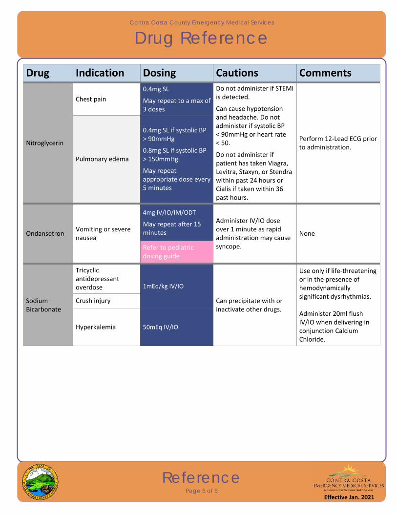
Effective Jan. 2016
Contra Costa County Emergency Medical Services
Drug Reference
ReferencePage 6 of 6
Drug Indication Dosing Cautions Comments
OndansetronVomiting or severe nausea
4mg IV/IO/IM/ODT
May repeat after 15 minutes
Administer IV/IO dose over 1 minute as rapid administration may cause syncope.
None
Refer to pediatric dosing guide
Sodium Bicarbonate
Tricyclic antidepressant overdose 1mEq/kg IV/IO
Can precipitate with or inactivate other drugs.
Use only if life‐threatening or in the presence of hemodynamically significant dysrhythmias.
Administer 20ml flush IV/IO when delivering in conjunction Calcium Chloride.
Crush injury
Hyperkalemia 50mEq IV/IO
Nitroglycerin
Pulmonary edema
0.4mg SL
May repeat to a max of 3 doses
Do not administer if STEMI is detected.
Can cause hypotension and headache. Do not administer if systolic BP < 90mmHg or heart rate< 50.
Do not administer if patient has taken Viagra, Levitra, Staxyn, or Stendra within past 24 hours or Cialis if taken within 36 past hours.
Perform 12‐Lead ECG prior to administration.
Chest pain
0.4mg SL if systolic BP > 90mmHg
0.8mg SL if systolic BP > 150mmHg
May repeat appropriate dose every 5 minutes
Effective Jan. 2021