Embed Size (px)
Citation preview
REVIEW Open Access
Preliminary guideline- andpathophysiology-based protocols forneurocritical careYasuhiro Norisue1,2* , Yoshihisa Fujimoto1 and Kazuma Nakagawa3
Abstract
Background: Because of the complex pathophysiological processes involved, neurocritical care has been driven byanecdotal experience and physician preferences, which has led to care variation worldwide. Standardization ofpractice has improved outcomes for many of the critical conditions encountered in the intensive care unit.
Main body: In this review article, we introduce preliminary guideline- and pathophysiology-based protocols for (1)prompt shivering management, (2) traumatic brain injury and intracranial pressure management, (3) neurologicalprognostication after cardiac arrest, (4) delayed cerebral ischemia after subarachnoid hemorrhage, (5) nonconvulsivestatus epilepticus, and (6) acute or subacute psychosis and seizure.
Conclusion: These tentative protocols may be useful tools for bedside clinicians who need to provide consistent,standardized care in a dynamic clinical environment. Because most of the contents of presented protocol are notsupported by evidence, they should be validated in a prospective controlled study in future. We suggest that theseprotocols should be regarded as drafts to be tailored to the systems, environments, and clinician preferences ineach institution.
Keywords: Neurocritical care, Protocols, Guidelines, Pathophysiology, Shivering, Neurological prognostication,Delayed cerebral ischemia, Nonconvulsive status epilepticus, Psychosis, Seizure
BackgroundThe art of neurocritical care requires an understandingof the pathophysiology of the highly complex centralnervous system. Because of its complexity and the lackof evidence, the approach to neurocritical care is oftenclinician-dependent, i.e., driven by anecdotal experienceand physician preferences, which leads to care variation.Overall, standardization of practice has improved out-comes for many critical conditions in the intensive careunit; thus, greater emphasis should be placed on redu-cing variation in neurocritical care practice.Guideline- and pathophysiology-based protocols are
concise yet comprehensive and are useful for bedsideclinicians who need to provide consistent, standardized
practice in a dynamic clinical environment. We intro-duce five preliminary protocols in this article. Becausemost of the text of the protocols addresses managementin neurocritical care fields that lack firm evidence, andbecause of the varied availability of medical resourcesamong institutions, we recommend that these protocolsbe used as drafts to be customized for the systems, envi-ronments, and clinical preferences of each institution.
ProtocolsPrompt shivering management (Fig. 1)Shivering is a physiological homeostatic response tomaintain or raise temperature in hypothermia or feverwhen the set point temperature is elevated. However,shivering counteracts the effort of fever managementand targeted temperature management (TTM)/thera-peutic hypothermia, which are critical interventions tomitigate secondary brain injury. With inadequate shiver-ing management, target temperature is difficult toachieve in a timely manner and may potentially worsen
* Correspondence: [email protected] of Emergency and Critical Care Medicine, Tokyo Bay UrayasuIchikawa Medical Center, 3-4-32 Todaijima, Urayasu, Chiba 2790001, Japan2Department of Emergency and Critical Care Medicine, St. MariannaUniversity Hospital, 2-16-1, Sugao, Kawasaki, Kanagawa 2168511, JapanFull list of author information is available at the end of the article
© The Author(s). 2018 Open Access This article is distributed under the terms of the Creative Commons Attribution 4.0International License (http://creativecommons.org/licenses/by/4.0/), which permits unrestricted use, distribution, andreproduction in any medium, provided you give appropriate credit to the original author(s) and the source, provide a link tothe Creative Commons license, and indicate if changes were made. The Creative Commons Public Domain Dedication waiver(http://creativecommons.org/publicdomain/zero/1.0/) applies to the data made available in this article, unless otherwise stated.
Norisue et al. Journal of Intensive Care (2018) 6:45 https://doi.org/10.1186/s40560-018-0316-6

outcome. Furthermore, shivering increases the cerebralmetabolic rate and may result in increased intracranialpressure (ICP) and brain oxygen consumption [1, 2].Lastly, shivering increases the total body metabolic rateand total CO2 production, which may raise the partialpressure of CO2 and raise ICP. Therefore, shiveringshould be regarded as a neurological emergency requir-ing immediate control in patients with acute brain in-jury, and any protocol for shivering management shouldencourage clinicians to expedite treatment. The presentdraft proposal for shivering management refers to theBedside Shivering Assessment Scale and shivering proto-col proposed by Badjatia and Brophy, respectively, [1, 2]and was refined, based on our practice, to achieveprompt shivering control (Fig. 1).
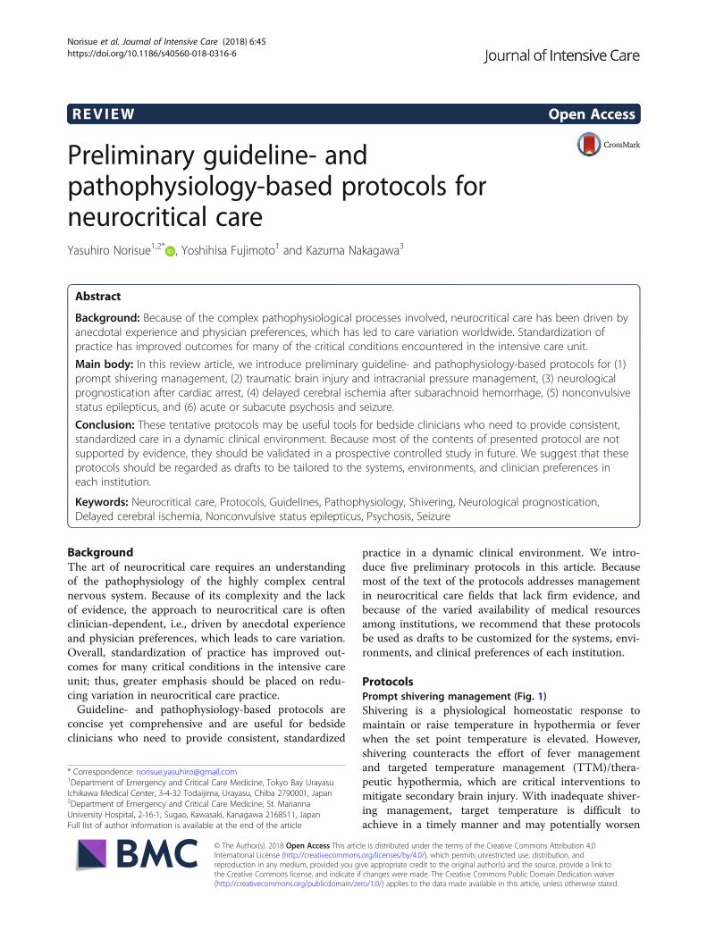
Traumatic brain injury and ICP management (Fig. 2)Intracranial hypertension, commonly defined as persist-ent elevation of ICP above 20–22 mmHg, is a relatively
common neurologic complication seen after traumaticbrain injury (TBI). If untreated, it can lead to cerebralischemia, brain herniation, and possibly brain death.Therefore, care providers must promptly recognize theearly clinical and radiographic features of elevated ICPand aggressively treat with a goal of reducing mortalityand morbidity. The adult brain is a nearly incompress-ible substance enclosed in a fixed cranium. Therefore,ICP will inevitably be affected by a volume change inany of the three main intracranial components—cere-brospinal fluid (CSF), brain parenchyma, and blood [3].In addition, if there is a new space-occupying lesionwithin the fixed cranium (i.e., hematoma), it will inevitablyincrease ICP. When assessing a patient with elevated ICP,it is important to determine whether the contributing fac-tor is a focal, global, or mixed process since the treatmentstrategy may be different for each type of mass effect. Ifthere is a focal, new mass-occupying lesion that is causinga regional mass effect and brain tissue compression, the
Fig. 1 Preliminary protocol for prompt shivering management [1, 2]. Modified from Brophy [2] with permission. Abbreviations: BSAS BedsideShivering Assessment Scale, ECG electrocardiogram, IV intravenously, PO per oral, H hour, min minute
Norisue et al. Journal of Intensive Care (2018) 6:45 Page 2 of 9

Fig. 2 (See legend on next page.)
Norisue et al. Journal of Intensive Care (2018) 6:45 Page 3 of 9
first step is to consider surgical evacuation. Once the focalmass effect is ruled out or treated, global elevation of ICPmust be addressed. The overarching strategy to controlglobally elevated ICP is to (1) optimize cerebral perfu-sion, oxygenation, and venous drainage; (2) preventfever, hypercapnia, hyponatremia, hypo/hyperglycemia,and seizure; (3) provide adequate cerebral metabolicsuppression with sedation; and (4) reduce cerebraledema with osmotic therapy. For refractory intracra-nial hypertension, treatments to be considered includepentobarbital-induced coma, therapeutic hypothermia,ventriculostomy placement for CSF diversion, anddecompressive craniectomy. Ideally, a protocol for TBImanagement should include not only ICP control butalso indications of initial surgical intervention for
intracranial hematoma and basic management to pre-vent secondary brain injury. Step 1 (indications forsurgical intervention) of the preliminary protocol isbased on the recommendations by Bullock et al. [4–7],and step 2 (indications for ICP monitoring) and step 3(basic management of TBI and ICP control) were devel-oped in accordance with the guidelines for managementof severe traumatic brain injury of the Brain TraumaFoundation and our practice (Fig. 2) [8].
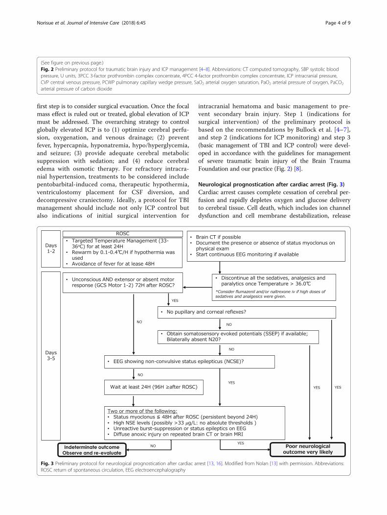
Neurological prognostication after cardiac arrest (Fig. 3)Cardiac arrest causes complete cessation of cerebral per-fusion and rapidly depletes oxygen and glucose deliveryto cerebral tissue. Cell death, which includes ion channeldysfunction and cell membrane destabilization, release
(See figure on previous page.)Fig. 2 Preliminary protocol for traumatic brain injury and ICP management [4–8]. Abbreviations: CT computed tomography, SBP systolic bloodpressure, U units, 3PCC 3-factor prothrombin complex concentrate, 4PCC 4-factor prothrombin complex concentrate, ICP intracranial pressure,CVP central venous pressure, PCWP pulmonary capillary wedge pressure, SaO2 arterial oxygen saturation, PaO2 arterial pressure of oxygen, PaCO2
arterial pressure of carbon dioxide
Fig. 3 Preliminary protocol for neurological prognostication after cardiac arrest [13, 16]. Modified from Nolan [13] with permission. Abbreviations:ROSC return of spontaneous circulation, EEG electroencephalography
Norisue et al. Journal of Intensive Care (2018) 6:45 Page 4 of 9

Fig. 4 Preliminary protocol for monitoring and diagnosis of delayed cerebral ischemia after subarachnoid hemorrhage [23, 24]. Abbreviations:SAH subarachnoid hemorrhage, DVT deep vein thrombosis, IVH intraventricular hemorrhage
Norisue et al. Journal of Intensive Care (2018) 6:45 Page 5 of 9

of destructive enzymes, cell swelling, and, eventually,apoptosis, can begin within 5 min after complete cessationof cerebral perfusion [9–11]. After return of spontaneouscirculation, neurological prognostication is essential be-cause it enables clinicians to provide information to familymembers or surrogates who must consider decisions tolimit care for patients with little hope of meaningfulneurological recovery [11]. To date, there is no definitivediagnostic test to accurately predict functional outcomeafter cardiac arrest. Moreover, clinical findings shortlyafter cardiac arrest have little relationship to patient out-comes [12]. However, the use of a systematic approachallows for reliable prediction of a very poor neurologicaloutcome (persistent vegetative state) and provides familymembers and surrogates with the information necessaryto make decisions [13–16]. A protocol that clearly ad-dresses “what to do next” would be helpful for clinicians.The present preliminary protocol is based on the 2015European Society of Intensive Care Medicine Guidelinesfor Post-resuscitation Care [13], but includes additionaldetailed stepwise instructions (Fig. 3).
Delayed cerebral ischemia after subarachnoidhemorrhage (Fig. 4)After subarachnoid hemorrhage, notably from a rupturedcerebral aneurysm, cerebral vasospasm can develop, thusleading to delayed cerebral ischemia (DCI) and possiblyinfarction. The mechanism of DCI is complex and is notsolely attributed to large-vessel narrowing and associatedlow blood flow distally [17, 18]. Other postulated mecha-nisms are early brain injury, microcirculatory dysfunctionwith loss of cerebral autoregulation, cortical spreadingdepolarization, and microthrombosis [19, 20]. A DCI diag-nosis is made clinically on the basis of symptoms such asnew changes in mental status and neurologic deficits.Additional relevant information includes findings of CTor MRI angiography, digital subtraction angiography, andtranscranial Doppler (TCD) ultrasound. In addition, otherreversible causes of neurological changes must be ruledout, such as delayed hydrocephalus, nonconvulsive seiz-ure, rebleeding, toxic-metabolic encephalopathy frominfection, and medication side effects. To date, nimodi-pine, a calcium-channel blocker with cerebral vasodilatory
Fig. 5 Preliminary protocol for diagnosis of nonconvulsive status epilepticus [32]. Abbreviations: AED anti-epileptic drug
Norisue et al. Journal of Intensive Care (2018) 6:45 Page 6 of 9

Fig. 6 Preliminary protocol for acute or subacute psychosis and newly onset seizure [33, 35]. Modified from Dubey [33] with permission.Abbreviations: VDRL venereal disease research laboratory, HSV herpes simplex virus, VZV varicella zoster virus, HIV human immunodeficiency virus,CMV cytomegalovirus, Anti-TPO anti-thyroid peroxidase, ANA anti-nuclear antibody, ANCA antineutrophil cytoplasmic antibody
Norisue et al. Journal of Intensive Care (2018) 6:45 Page 7 of 9
effect, is the only drug that has been shown to improveneurological outcomes in patients with subarachnoidhemorrhage [21]. Other calcium-channel blockers, such asnicardipine, have been used in countries where nimodipineis unavailable [22] but have not been shown to improveoutcomes. Hemodynamic augmentation to increase oxygendelivery to the brain, including volume optimization andinduced hypertension, is the mainstay for management ofnew-onset DCI. For refractory cases where medical man-agement is ineffective, intra-arterial interventions such asballoon angioplasty and intra-arterial administration ofcalcium-channel blockers are a second-line treatment [23].A protocol should include risk stratification and step-wise treatment for DCI in individual patients. Thepresent preliminary protocol describes basic manage-ment of subarachnoid hemorrhage and risk stratifica-tion and monitoring, based on our practice and theexisting literature [24–26]. Diagnosis and managementof DCI are based on recommendations from the neu-rocritical care society and our practice [23] (Fig. 4).
Diagnosis of nonconvulsive status epilepticus (Fig. 5)Nonconvulsive status epilepticus (NCSE) is characterizedby electrographic seizure activity without clinical convul-sions in patients who do not fully recover consciousnessbetween attacks [27]. Although the impact of treatingNCSE on clinical outcomes has not been investigated ina randomized controlled trial, the prognosis of NCSE isbelieved to be poor if not treated since untreated seizureis associated with secondary brain injury [28–30]. Adiagnosis of NCSE should be considered in any patientwith discrepancies between his/her neurological findingsand clinical history or imaging findings such as CT orMRI. The typical example is a patient who developssudden unexpected neurological deterioration after suc-cessful management of a structural brain injury withoutnew findings on CT or MRI. Although the condition isreferred to as nonconvulsive, patients with NCSE mayhave subtle motor symptoms such as sustained eye devi-ation, nystagmus, lip smacking, and twitching in the faceor extremities [31]. Definitive diagnosis requires electro-encephalography (EEG), and continuous EEG monitoringincreases the sensitivity and specificity of NCSE diagnosis.We attempted to develop an algorithmic protocol to inter-pret EEG and diagnose NCSE for bedside clinical use.Based on the current guidelines [27, 32], this draft proto-col for diagnosis of NCSE is designed to simplify NCSEdiagnosis and management (Fig. 5).
Acute or subacute psychosis and seizure (Fig. 6)Many medical conditions and pharmacologic side effectscan cause unexpected psychosis or seizure. However, sometreatable and reversible conditions, such as viral encephal-itis and autoimmune encephalitis, are frequently missed.
Delays in diagnosis and treatment of encephalitis may re-sult in poor neurological outcomes. It is essential to reviewall possible causes of unexpected psychosis and seizure andto start empirical treatment for a potentially treatable con-dition before obtaining all examination results. The presentdraft protocol for diagnosis and management of auto-immune encephalitis includes comprehensive differentialdiagnoses and algorithms for diagnostic evaluation andwas developed on the basis of a comprehensive literaturereview by Francesc et al., antibody prevalence in epilepsy(APE) score [33], and empirical treatment for autoimmuneencephalitis in cases of unexpected psychosis or seizure, asreflected by the expert opinions of the European Feder-ation of the Neurological Societies (EFNS) task force [34]and our practice (Fig. 6).
ConclusionThe present guideline- and pathophysiology-based pro-tocols that can be customized for particular clinical envi-ronments may help providing consistent, standardizedcare in neurocritical care. Because most of the contentsof presented protocol are not supported by evidence,they should be validated in a prospective controlledstudy in future.
AbbreviationsCSF: Cerebrospinal fluid; DCI: Delayed cerebral ischemia;EEG: Electroencephalography; ICP: Intracranial pressure; NCSE: Nonconvulsivestatus epilepticus; TBI: Traumatic brain injury; TCD: Transcranial Doppler;TTM: Targeted temperature management
Author contributionsYN is the guarantor of the manuscript content and protocols. YF and KNsubstantially contributed to the manuscript and protocols. All authors readand approved the final manuscript.
Ethics approval and consent to participateNot applicable
Consent for publicationNot required
Competing interestsThe authors declare that they have no competing interests.
Publisher’s NoteSpringer Nature remains neutral with regard to jurisdictional claims inpublished maps and institutional affiliations.
Author details1Department of Emergency and Critical Care Medicine, Tokyo Bay UrayasuIchikawa Medical Center, 3-4-32 Todaijima, Urayasu, Chiba 2790001, Japan.2Department of Emergency and Critical Care Medicine, St. MariannaUniversity Hospital, 2-16-1, Sugao, Kawasaki, Kanagawa 2168511, Japan.3Department of Medicine, Division of Neurology, John A. Burns School ofMedicine University of Hawai`i, 1301 Punchbowl Street, Honolulu, HI 96813,USA.
Norisue et al. Journal of Intensive Care (2018) 6:45 Page 8 of 9

Received: 12 April 2018 Accepted: 27 July 2018
References1. Badjatia N, Strongilis E, Gordon E, Prescutti M, Fernandez L, Fernandez A,
Buitrago M, Schmidt JM, Ostapkovich ND, Mayer SA. Metabolic impact ofshivering during therapeutic temperature modulation: the BedsideShivering Assessment Scale. Stroke. 2008;39(12):3242–7.
2. Brophy GM, Human T. Pharmacotherapy pearls for emergency neurologicallife support. Neurocrit Care. 2017;27(Suppl 1):51–73.
3. Fishman RA. Cerebrospinal fluid in diseases of the nervous system.Philadelphia: London: Saunders; 1980.
4. Bullock MR, Chesnut R, Ghajar J, Gordon D, Hartl R, Newell DW, Servadei F,Walters BC, Wilberger J, Surgical Management of Traumatic Brain InjuryAuthor G. Surgical management of posterior fossa mass lesions.Neurosurgery. 2006;58(3 Suppl):S47–55. discussion Si-iv
5. Bullock MR, Chesnut R, Ghajar J, Gordon D, Hartl R, Newell DW, Servadei F,Walters BC, Wilberger J, Surgical Management of Traumatic Brain InjuryAuthor G. Surgical management of depressed cranial fractures.Neurosurgery. 2006;58(3 Suppl):S56–60. discussion Si-iv
6. Bullock MR, Chesnut R, Ghajar J, Gordon D, Hartl R, Newell DW, Servadei F,Walters BC, Wilberger JE, Surgical Management of Traumatic Brain InjuryAuthor G. Surgical management of acute epidural hematomas.Neurosurgery. 2006;58(3 Suppl):S7–15. discussion Si-iv
7. Bullock MR, Chesnut R, Ghajar J, Gordon D, Hartl R, Newell DW, Servadei F,Walters BC, Wilberger JE, Surgical Management of Traumatic Brain InjuryAuthor G. Surgical management of acute subdural hematomas.Neurosurgery. 2006;58(3 Suppl):S16–24. discussion Si-iv
8. Carney N, Totten AM, O'Reilly C, Ullman JS, Hawryluk GW, Bell MJ, Bratton SL,Chesnut R, Harris OA, Kissoon N, et al. Guidelines for the management ofsevere traumatic brain injury, fourth edition. Neurosurgery. 2017;80(1):6–15.
9. Neumar RW. Molecular mechanisms of ischemic neuronal injury. Ann EmergMed. 2000;36(5):483–506.
10. Pulsinelli WA, Brierley JB, Plum F. Temporal profile of neuronal damage in amodel of transient forebrain ischemia. Ann Neurol. 1982;11(5):491–8.
11. Lee K. The neuroICU book. 2nd ed. New York: McGraw-Hill Medical; 2017.12. Rittenberger JC, Sangl J, Wheeler M, Guyette FX, Callaway CW. Association
between clinical examination and outcome after cardiac arrest.Resuscitation. 2010;81(9):1128–32.
13. Nolan JP, Soar J, Cariou A, Cronberg T, Moulaert VR, Deakin CD, Bottiger BW,Friberg H, Sunde K, Sandroni C. European Resuscitation Council andEuropean Society of Intensive Care Medicine guidelines for post-resuscitation care 2015: section 5 of the European Resuscitation Councilguidelines for resuscitation 2015. Resuscitation. 2015;95:202–22.
14. Wijdicks EF, Hijdra A, Young GB, Bassetti CL, Wiebe S, Quality StandardsSubcommittee of the American Academy of N. Practice parameter:prediction of outcome in comatose survivors after cardiopulmonaryresuscitation (an evidence-based review): report of the Quality StandardsSubcommittee of the American Academy of Neurology. Neurology 2006;67(2):203–210.
15. Zandbergen EG, de Haan RJ, Stoutenbeek CP, Koelman JH, Hijdra A.Systematic review of early prediction of poor outcome in anoxic-ischaemiccoma. Lancet. 1998;352(9143):1808–12.
16. Rossetti AO, Rabinstein AA, Oddo M. Neurological prognostication ofoutcome in patients in coma after cardiac arrest. Lancet Neurol. 2016;15(6):597–609.
17. Millikan CH. Cerebral vasospasm and ruptured intracranial aneurysm. ArchNeurol. 1975;32(7):433–49.
18. Etminan N, Vergouwen MD, Ilodigwe D, Macdonald RL. Effect ofpharmaceutical treatment on vasospasm, delayed cerebral ischemia, andclinical outcome in patients with aneurysmal subarachnoid hemorrhage: asystematic review and meta-analysis. J Cereb Blood Flow Metab. 2011;31(6):1443–51.
19. Budohoski KP, Guilfoyle M, Helmy A, Huuskonen T, Czosnyka M, Kirollos R,Menon DK, Pickard JD, Kirkpatrick PJ. The pathophysiology and treatment ofdelayed cerebral ischaemia following subarachnoid haemorrhage. J NeurolNeurosurg Psychiatry. 2014;85(12):1343–53.
20. Francoeur CL, Mayer SA. Management of delayed cerebral ischemia aftersubarachnoid hemorrhage. Crit Care. 2016;20(1):277.
21. Petruk KC, West M, Mohr G, Weir BK, Benoit BG, Gentili F, Disney LB, KhanMI, Grace M, Holness RO, et al. Nimodipine treatment in poor-grade
aneurysm patients. Results of a multicenter double-blind placebo-controlledtrial. J Neurosurg. 1988;68(4):505–17.
22. Tejada JG, Taylor RA, Ugurel MS, Hayakawa M, Lee SK, Chaloupka JC. Safetyand feasibility of intra-arterial nicardipine for the treatment of subarachnoidhemorrhage-associated vasospasm: initial clinical experience with high-doseinfusions. AJNR Am J Neuroradiol. 2007;28(5):844–8.
23. Diringer MN, Bleck TP, Claude Hemphill J 3rd, Menon D, Shutter L, Vespa P,Bruder N, Connolly ES Jr, Citerio G, Gress D, et al. Critical care managementof patients following aneurysmal subarachnoid hemorrhage:recommendations from the Neurocritical Care Society’s MultidisciplinaryConsensus Conference. Neurocrit Care. 2011;15(2):211–40.
24. Macdonald RL. Delayed neurological deterioration after subarachnoidhaemorrhage. Nat Rev Neurol. 2014;10(1):44–58.
25. Frontera JA, Claassen J, Schmidt JM, Wartenberg KE, Temes R, Connolly ESJr, MacDonald RL, Mayer SA. Prediction of symptomatic vasospasm aftersubarachnoid hemorrhage: the modified fisher scale. Neurosurgery. 2006;59(1):21–7. discussion 21-27
26. de Rooij NK, Greving JP, Rinkel GJ, Frijns CJ. Early prediction of delayedcerebral ischemia after subarachnoid hemorrhage: development andvalidation of a practical risk chart. Stroke. 2013;44(5):1288–94.
27. Brophy GM, Bell R, Claassen J, Alldredge B, Bleck TP, Glauser T, Laroche SM,Riviello JJ Jr, Shutter L, Sperling MR, et al. Guidelines for the evaluation andmanagement of status epilepticus. Neurocrit Care. 2012;17(1):3–23.
28. Towne AR, Pellock JM, Ko D, DeLorenzo RJ. Determinants of mortality instatus epilepticus. Epilepsia. 1994;35(1):27–34.
29. DeLorenzo RJ, Garnett LK, Towne AR, Waterhouse EJ, Boggs JG, Morton L,Choudhry MA, Barnes T, Ko D. Comparison of status epilepticus withprolonged seizure episodes lasting from 10 to 29 minutes. Epilepsia. 1999;40(2):164–9.
30. Waterhouse EJ, Garnett LK, Towne AR, Morton LD, Barnes T, Ko D,DeLorenzo RJ. Prospective population-based study of intermittent andcontinuous convulsive status epilepticus in Richmond, Virginia. Epilepsia.1999;40(6):752–8.
31. Husain AM, Horn GJ, Jacobson MP. Non-convulsive status epilepticus:usefulness of clinical features in selecting patients for urgent EEG. J NeurolNeurosurg Psychiatry. 2003;74(2):189–91.
32. Leitinger M, Beniczky S, Rohracher A, Gardella E, Kalss G, Qerama E, Hofler J,Hess Lindberg-Larsen A, Kuchukhidze G, Dobesberger J, et al. Salzburgconsensus criteria for non-convulsive status Epilepticus––approach toclinical application. Epilepsy Behav. 2015;49:158–63.
33. Dubey D, Alqallaf A, Hays R, Freeman M, Chen K, Ding K, Agostini M,Vernino S. Neurological autoantibody prevalence in epilepsy of unknownetiology. JAMA Neurol. 2017;74(4):397–402.
34. Vedeler CA, Antoine JC, Giometto B, Graus F, Grisold W, Hart IK, Honnorat J,Sillevis Smitt PA, Verschuuren JJ, Voltz R, et al. Management ofparaneoplastic neurological syndromes: report of an EFNS Task Force. Eur JNeurol. 2006;13(7):682–90.
35. Graus F, Delattre JY, Antoine JC, Dalmau J, Giometto B, Grisold W, HonnoratJ, Smitt PS, Vedeler C, Verschuuren JJ, et al. Recommended diagnosticcriteria for paraneoplastic neurological syndromes. J Neurol NeurosurgPsychiatry. 2004;75(8):1135–40.
Norisue et al. Journal of Intensive Care (2018) 6:45 Page 9 of 9