Embed Size (px)
Citation preview
PRENATAL DIAGNOSISPrenat Diagn 2002; 22: 802–805.Published online in Wiley InterScience (www.interscience.wiley.com). DOI: 10.1002/pd.415
SHORT COMMUNICATION
Prenatal and postnatal characterization of Y chromosomestructural anomalies by molecular cytogenetic analysis
C. Hernando1, M. Carrera2, I. Ribas2, N. Parear3, R. Baraibar4, J. Egocue1 and C. Fuster1*1Unitat de Biologia, Facultat de Medicina, Universitat Autonoma de Barcelona, E-08193 Bellaterra, Barcelona, Spain2Centro de Patologıa Celular y Diagnostico Prenatal, Barcelona, Spain3Servei de Ginecologia, Institut Universitari Dexeus, Barcelona, Spain4Servei de Pediatria, Institut Universitari Dexeus, Barcelona, Spain
We describe three cases in which we used fluorescence in situ hybridization (FISH), polymerase chainreaction (PCR) and comparative genomic hybridization (CGH) to characterize Y chromosome structuralanomalies, unidentifiable by conventional G-banding. Case 1 was a 46,X,+mar karyotype; FISH analysisrevealed an entire marker chromosome highlighted after hybridization with the Y chromosome paintingprobe. The PCR study showed the presence of Y chromosome markers AMG and SY620 and theabsence of SY143, SY254 and SY147. CGH results confirmed the loss of Yq11.2-qter. These resultsindicated the presence of a deletion: del(Y)(q11.2). Case 2 was a 45,X [14]/46,XY[86] karyotype witha very small Y chromosome. The PCR study showed the presence of Y chromosome markers SY620and AMG, and the absence of SY143, SY254 and SY147. CGH results showed gain of Yq11.2-pter andloss of Yq11.2-q12. These results show the presence of a Yp isodicentric: idic(Y)(q11.2). Case 3 was a45,X,inv(9)(p11q12)[30]/46,X,idic(Y)(p11.3?),inv(9)(p11q12)[70] karyotype. The FISH signal covered all theabnormal Y chromosome using a Y chromosome paint. The PCR study showed the presence of Y chromosomemarkers AMG, SY620, SY143, SY254 and SY147. CGH only showed gain of Yq11.2-qter. These resultssupport the presence of an unbalanced (Y;Y) translocation. Our results show that the combined use of molecularand classical cytogenetic methods in clinical diagnosis may allow a better delineation of the chromosomeregions implicated in specific clinical disorders. Copyright 2002 John Wiley & Sons, Ltd.
KEY WORDS: isochromosomes; FISH; PCR; CGH; prenatal diagnosis
INTRODUCTION
Many individuals with sex chromosome abnormalitiesremain undiagnosed because usually their phenotypefalls within the limits of ‘normality,’ although thekaryotypic change has an effect on most non-mosaiccases. Even now, when many abnormalities of thesex chromosomes have been defined and their clinicalconsequences reported in detail, the precise role ofthe sex chromosomes in sex differentiation and in thegenetic control of gametogenesis still remain obscure(Robinson et al., 1999; Kleiman et al., 2001).
In recent years, fluorescence in situ hybridization(FISH and multi-colour-FISH) and comparative genomichybridization (CGH) have allowed the correct identifi-cation of constitutional chromosomal aberrations. Thesetechniques, combined with conventional G-banding,have a clear diagnostic and prognostic value, and con-tribute to genetic counselling (Lichter et al., 1991; Bryn-dorf et al., 1995; Speicher et al., 1996; Daniely et al.,1999; Stumm et al., 1999; Rigola et al., 2001).
Here, we describe three cases where de novo Y chro-mosome structural anomalies were detected by pre and
*Correspondence to: C. Fuster, Unitat de Biologia, Departamentde Biologia Cellular, Fisiologia i Immunologia, Facultat de Medic-ina, Universitat Autonoma de Barcelona, E-08193 Bellaterra,Barcelona, Spain. E-mail: [email protected]
postnatal diagnosis and characterized by molecular cyto-genetic analysis. The present report illustrates the use-fulness of FISH, polymerase chain reaction (PCR) andCGH for the accurate identification of Y chromosomestructural anomalies, as well establishing a better corre-lation with the clinical manifestations.
CASE REPORTS
Case 1
Case 1 was a fetus with 46,X,+mar karyotype. A 38-year-old woman requested a prenatal diagnosis when shewas in the 18th week of pregnancy. Previously, she hadtwo spontaneous abortions and a phenotypically normalgirl. Cytogenetic analysis of cultured amniotic fluid cellsafter G-banding showed an abnormal karyotype with 46chromosomes, including a normal X chromosome anda small marker chromosome in 14 of the clones anal-ysed. To characterize the marker chromosome in thisfetus, FISH, PCR and CGH were carried out. Analysis ofperipheral blood lymphocytes from the parents showednormal karyotypes. Ecography revealed male genitalia,and no fetal malformations were observed. The coupledecided to continue with the pregnancy. An apparently
Copyright 2002 John Wiley & Sons, Ltd. Received: 21 November 2001Revised: 12 April 2002
Accepted: 21 April 2002
Y-CHROMOSOME STRUCTURAL ABNORMALITIES 803
normal boy was born at 39.3 weeks without complica-tions. He weighed 4.410 g, his length was 48 cm andthe head circumference was 37 cm. Physical examina-tion detected megasomia and a fronto-palpebral vascularangioma. Apparently normal male genitalia (no cryp-tochidism or hypospadias) were observed.
Case 2
Case 2 was a fetus with a unilateral renal agenesia.For rapid prenatal detection of common chromosomeaneuploidies, QF-PCR (quantitative fluorescent PCR)was carried out in amniocytes. No autosomal aneuploi-dies were found using the highly polymorphic STRmarkers S1313, S1818 and S2121; X22, HPRT andthe modified AMXY showed characteristic monoallelicpatterns and were uninformative for sex chromosomecopy number. Cytogenetic study of amniocytes revealeda mosaic 45,X[11]/46,X,der(Y)[16] karyotype. Analy-sis of peripheral blood lymphocytes from the parentsshowed normal karyotypes. The couple decided to con-tinue with the pregnancy. No evidence of intrauterinegrowth retardation was observed. An apparently pheno-typically normal boy was born at term. Birth weight,length and head circumference were 2910 g, 49 cm and36 cm, respectively. Apparently normal male genitalia(no cryptochidism or hypospadias) were observed. Toverify the fetal karyotype in the neonate and to charac-terize the derivative Y chromosome, conventional cyto-genetic and molecular cytogenetic analyses (PCR andCGH) were carried out in peripheral blood.
Case 3
Case 3 was a 15-year-old girl with primary amenorrheaand a 45,X,inv(9)(p11q12)[30]/46X,idic(Y)(p11.3?),inv(9)(p11q12)[70] karyotype. A clinical examinationdetected the presence of growth retardation (length149 cm and weight 44 kg) and bone age retardation.No other phenotypic signs of Turner syndrome werefound. She had apparently normal external female gen-italia. The internal genitalia had no testicular elements;she had a hyposplastic uterus and a streak right gonad.The secondary sex characteristics were apparently nor-mal. It is interesting to note the presence of a notablebreast asymmetry (the left breast was larger). Simpleichthyosis and epiphyseal closing were observed. Norenal or cardiac congenital malformations were detected.Analysis of peripheral blood lymphocytes from the par-ents showed normal karyotypes. FISH, PCR and CGHanalyses were carried out in peripheral blood to verifythe chromosome anomaly.
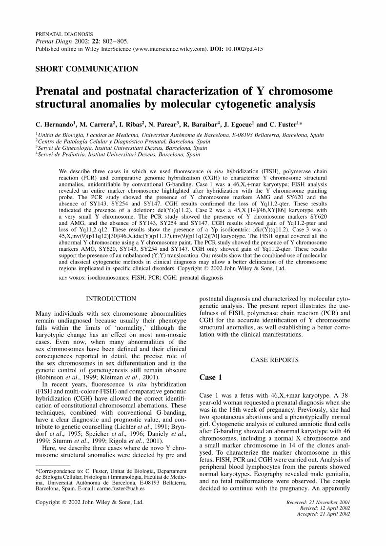
Figure 1 illustrates the partial karyotypes showing thesex chromosomes of all three cases.
MATERIAL AND METHODS
Cytogenetic and FISH analyses
Amniotic fluid cells or peripheral blood samples fromthese cases were cultured in Chang and RPMI-1640
Case 1 Case 2
X Y YX YX
Case 3
Figure 1—G-banded partial karyotypes showing the sex chromosomesfrom the three patients
media according to standard procedures. Metaphasechromosomes were analysed by Wright G-banding.FISH was performed with X and Y chromosome specificpaint probes (Chromoprobe-M Multiprobe, Cytocell Ltd,UK). Hybridization to metaphase spreads was performedaccording to the instructions of the supplier.
PCR and CGH analyses
DNA was extracted from amniotic fluid cells or periph-eral blood samples according to standard procedures.
PCR analyses for SY620 (AZFa), SY143 (AZFb),SY254 (DAZ-AZFc), SY147 (AZFc) used sequencetagged site markers from four different regions of thelong arm of the Y chromosome. Following amplifica-tion, the fluorescent PCR products were mixed with areference molecular standard (GeneScan-500 TAMRA,Applied Byosistems, USA) and electrophoresis was per-formed employing an automated DNA sequencer (ABI310 Genetic Analyser, Applied Byosistems, USA).
CGH was carried out to identify the abnormal Ychromosomes. For CGH, the red-labelled patient DNAand the green-labelled control DNA were hybridized onnormal male metaphases, following the instructions ofthe supplier (Vysis, Downer Grove, IL, USA). Standardnick-translation was performed using a commercial kit.The 1:1 probe mixture (700 ng) was hybridized incombination with unlabeled human Cot-1 DNA onnormal male metaphase spreads. Slides were analysedusing a Cytovision Ultra Workstation (Applied Imaging,Sunderland, UK). The software performed a calculationof the patient DNA to normal DNA fluorescence ratiosalong the length of each chromosome. Ratio values ofCGH above 1.25 and below 0.75 were considered torepresent chromosomal gains and losses respectively.
RESULTS
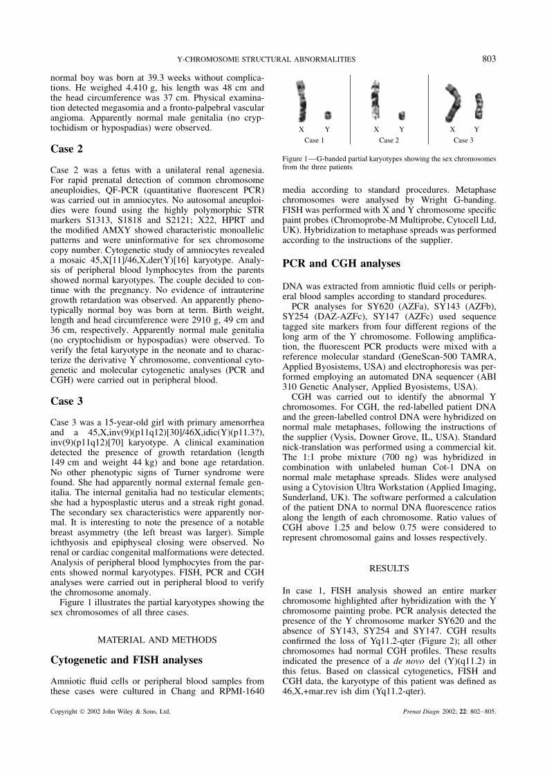
In case 1, FISH analysis showed an entire markerchromosome highlighted after hybridization with the Ychromosome painting probe. PCR analysis detected thepresence of the Y chromosome marker SY620 and theabsence of SY143, SY254 and SY147. CGH resultsconfirmed the loss of Yq11.2-qter (Figure 2); all otherchromosomes had normal CGH profiles. These resultsindicated the presence of a de novo del (Y)(q11.2) inthis fetus. Based on classical cytogenetics, FISH andCGH data, the karyotype of this patient was defined as46,X,+mar.rev ish dim (Yq11.2-qter).
Copyright 2002 John Wiley & Sons, Ltd. Prenat Diagn 2002; 22: 802–805.
804 C. HERNANDO ET AL.
Case 1
Y n=9
0.5 1.0 1.5
Y n=8
0.5 1.0 1.5
Y n=10
0.5 1.0 1.5
Case 2 Case 3
Figure 2—Y chromosomes and profiles obtained by CGH showing: loss of Yq11.2-qter in case 1, gain of Yq11.2-pter and loss of Yq11.2-q12in case 2, and gain of Yq11.2-qter in case 3
Evaluation of G-banded chromosomes of case2 at birth confirmed the presence of mosaicism45,X[14]/46,X,der(Y)[86] with a very small Y chro-mosome. The PCR study showed the presence of Ychromosome marker SY620 and the absence of SY143,SY254 and SY147. These findings and those obtainedby QF-PCR (the presence of AMGY located at Yp) arein agreement with the presence of an isodicentric Yp.CGH results confirmed the gain of Yq11.2-pter and theloss of Yq11.2-q12 (Figure 2); all other chromosomeshad normal CGH profiles. These analyses revealed thepresence of mosaicism for an isodicentric (Y)(q11.2) anda 45,X cell line in this newborn. Based on classical cyto-genetics and CGH data, the karyotype of this patientwas defined as 45,X[14]/46,X,der(Y)[86]. rev ish enh(Yq11.2-pter).rev ish dim (Yq11.2-q12).
In patient 3, FISH analysis showed a brightsignal on the entire chromosome after hybridiza-tion with the Y chromosome paint. PCR studyshowed the presence of Y chromosome markersSY620, SY143, SY254 and SY147. CGH results onlydetected the gain of Yq11.2-qter (Figure 2); all otherchromosomes had normal CGH profiles. These resultsindicate the presence of mosaicism for an unbalancedtranslocation between two Y chromosomes and a45,X cell line in this woman. Based on classicalcytogenetics, FISH, and CGH data, the karyotypeof this patient was defined as 45,X,inv(9)(p11q12)[30]/46X,der(Y),inv(9)(p11q12)[70]. rev ish enh(Yq11.2-qter).
DISCUSSION
The precise characterization of the chromosome frag-ments implicated in constitutive unbalanced karyotypeshas a considerable prognostic value and may contributeto the discovery of genes implicated in specific disor-ders. The prenatal detection of Y chromosome struc-tural anomalies poses a diagnostic dilemma because theireffect on fetal and postnatal development is difficult topredict. In 1989, Hsu published the first evidence of theexistence of a drastic difference in terms of the pheno-typic outcome between postnatal diagnosis and prenataldiagnosis of 45,X/46,XY mosaicism. Due to the biasedascertainment of postnatally diagnosed cases, all caseswere phenotypically abnormal while over 92% of pre-natal cases were born as phenotypically normal maleinfants with normal genitalia (Hsu, 1989). Similar find-ings have been described more recently by Telvi et al.(1999).
FISH and PCR analyses have proved useful forthe accurate identification of Y chromosome structuralalterations. Moreover, CGH has been shown to behelpful for the identification of marker chromosomes (Yuet al., 1993; Wang et al., 1995). In the present work wehave used these three methods (FISH, PCR and CGH)to characterize three de novo structural Y chromosomealterations in pre and postnatal diagnostic situations.
The cytogenetic study of case 1 revealed a dele-tion Yq that was characterized as del(Y)(q11.2) byCGH and PCR analyses. A comparison with previouspublished cases confirms that almost all patients with46,X,del(Y)(q11) were phenotypic males who, as adults,usually show short stature and infertility (Hsu, 1989; Maet al., 2000; Kirsch et al., 2000; Kleiman et al., 2001).The presence of AZFa in our patient does not discardthe presence of infertility, since recently, Martinez et al.,(2000) reported that no deletions of the AZFa regionwere detected in a screening for AZF deletions in a largeseries of patients with severely impaired spermatogen-esis. According to this information, the prognosis forthis newborn, with normal clinical features, is the prob-able presence of short stature and infertility on reachingadulthood.
Previously reported patients with Yp isodicentricswere 45,X mosaics due to the unstable nature of thedicentric chromosome. Predicting the phenotypic conse-quences of different duplications and deletions in dicen-tric chromosomes is complicated due to the variabilityresulting from the different degrees of mosaicism (Hsu,1989; Stuppia et al., 1996). Our case 2, as most reportedcases, was a de novo idic(Yp) and had a breakpoint atYq11.2. He was phenotypically male, as were 27% of thereported cases with the same isodicentric chromosome(Hsu, 1989). The only clinical abnormality observed incase 2 was the presence of a unilateral renal agenesia; toour knowledge, this phenotype has not been previouslydescribed associated with this chromosome alteration.Due to the loss of AZFb, AZFc-DAZ and AZFc, it isvery probable that our patient will be an infertile adult.
Translocation between two Y chromosomes isunusual. The presence of a 45,X cell line in mostY/Y translocation may suggest that some of these Y/Ytranslocations could be dicentric. A clear distinctionbetween a Y/Y translocation, a Yp isochromosomeand a Yq isodicentric chromosome is difficult usingcytogenetic techniques. The use of CGH identifiedthe chromosome alteration in our patient 3 as a Y/Ytranslocation. The high proportion of the 45,X cell line(30%) determined the presence of Turner syndromestigmata (Hsu et al., 1989; Bergendi et al., 1997).
Copyright 2002 John Wiley & Sons, Ltd. Prenat Diagn 2002; 22: 802–805.
Y-CHROMOSOME STRUCTURAL ABNORMALITIES 805
Moreover, the presence of spermatogenesis controllinggenes (AZF) in this patient could play an important rolein gonadal development and in the differentiation into aphenotypic female with some Turner stigmata (Bergendiet al., 1997; Jenderny et al., 1998; Robinson et al., 1999,Giltay et al., 2001).
The present report illustrates the importance of FISH,PCR and CGH techniques in complementing conven-tional cytogenetic methods for the accurate identificationand characterization of Y chromosome structural alter-ations in pre and postnatal diagnosis, and to correlatethem with clinical manifestations.
ACKNOWLEDGMENTS
Financial support was given by DGESIC (PB 98-0891)and CIRIT (2001, SGR-00201).
REFERENCES
Bergendi E, Plochl E, Vlasak I, Rittinger O, Muss W. 1997. ATurner-like phenotype in a girl with an isodicentric fluorescent Ychromosome mosaicism. Klin Padiatr 209: 133–136.
Bryndorf T, Kirchchoftt M, Rose H, et al. 1995. Comparativegenomic hybridization in clinical cytogenetics. Am J Hum Genet57: 1211–1220.
Daniely M, Barkai G, Godman B, Avidam-Goldring A. 1999. Struc-tural unbalanced chromosome rearrangements resolved by compar-ative genomic hybridization. Cytogenet Cell Genet 86: 51–55.
Giltay JC, Ausems MG, van Seumeren I, et al. 2001. Short stature asthe only presenting feature in a patient with isodicentric (Y)(q11.23)and gonadoblastoma: a clinical and molecular cytogenetic study.Eur J Pediatr 160: 154–158.
Hsu LYF. 1989. Prenatal diagnosis of 45,X/46,XY mosaicism—areview and update. Prenat Diagn 9: 31–48.
Jenderny J, Schmidt W, Held KR. 1998. Presence of the AZF regionin a female with an idic(Y)(q11). Clin Genet 54: 341–344.
Kirsch S, Weiss B, De Rosa M, Ogata T, Lombardi G, Rap-pold GA. 2000. FISH deletion mapping defines a single locationfor the Y chromosome stature gene, CGY. J Med Genet 37:593–599.
Kleiman SE, Maymon BBS, Yogev L, Paz G, Yavetz H. 2001.Prognostic value of Y deletion analysis. The prognostic role ofthe extent of Y microdeletion on spermatogenesis and maturity ofSertoli cells. Hum Reprod 16: 399–409.
Lichter P, Boyle AL, Cremer T, Ward DC. 1991. Analysis of genesand chromosomes by nonisotopic in situ hybridization. Genet AnalytTech Appl 8: 24–35.
Ma K, Mallidis C, Bhasin S. 2000. The role of Y chromosomedeletions in male infertility. Eur J Endocrinol 142: 418–430.
Martınez MC, Bernabe MJ, Gomez E, et al. 2000. Screening for AZFdeletion in a large series of severely impaired spermatogenesispatients. J Androl 21: 651–655.
Rigola MA, Carrera M, Ribas I, et al. 2001. Identification of two denovo partial trisomies by comparative genomic hybridization. ClinGenet 59: 106–110.
Robinson DO, Dalton P, Jacobs PA, et al. 1999. A molecular andFISH analysis of structurally abnormal Y-chromosomes in patientswith Turner syndrome. J Med Genet 36: 279–284.
Speicher MR, Ballard SG, Ward DC. 1996. Karyotyping humanchromosomes by combinatorial multi-fluor- FISH. Nature Genet 12:368–375.
Stumm M, Tonnies H, Wieacker PF. 1999. Molecular cytogenetictechniques for the diagnosis of chromosomal abnormalities inchildhood disease. Eur J Pediatr 158: 531–536.
Stuppia L, Calabarese G, Franchi PG, et al. 1996. Molecular studiesin three patients with isodicentric Y chromosome. Hum Genet 98:691–695.
Telvi L, Lebbar A, Del Pino O, Barber JP, Chaussain JL. 1999.45,X/46,XY Mosaicism: report of 27 Cases. Pediatrics 104:304–308.
Wang BBT, Yu LCh, Peng W, Falk RE, Williams J. 1995. Prenatalidentification of i(Yp) by molecular cytogenetic analysis. PrenatDiagn 15: 1115–1119.
Yu LC, Williams J Wang BT, et al. 1993. Characterization of thei(18p) in prenatal diagnosis by fluorescence in situ hybridization.Prenat Diagn 13: 355–361.
Copyright 2002 John Wiley & Sons, Ltd. Prenat Diagn 2002; 22: 802–805.