Embed Size (px)
Citation preview
Med. J. Malaysia Vol. 47 No. 3 September 1992
Prenatal diagnosis of intestinal obstruction due to ileal atresia
*S Raman, MRCOG
**L L Chan, MRCP
***K W Chang, FRCS
* S P Rachagan, MRCOG
*Department of Obstetrics and Gynaecology **Department of Paediatrics ***Department of Surgery
University Hospital, 59100 Kuala Lumpur, Malaysia
Summary
A case of intestinal obstruction due to ileal atresia where the diagnosis was made prenatally by ultrasound is presented. Close monitoring of the fetus was done ultrasonographically to look for any evidence of meconium peritonitis. The baby was delivered preterm but weighed 3.3 kg. Laparotomy and enterostomy was done and the baby is currently well.
Introduction
Small bowel atresia and stenosis occur in about 1 in 5000 live births and this presents as bowel obstruction. In the West meconium ileus is the commonest presenting symptom and occurs in 10 to 15% of cases. This problem is uncommon in the Asian population. The cause of sman bowel atresia or stenosis is mostofthe time due to vascular impairment, volvulus or intussusception. With the advent of high resolution ultrasound many of these abnormalities can be correctly diagnosed in utero. Associated extragastrointestinal abnormalities are uncommon but associated bowel complications like malrotation and intestinal duplications occur not infrequently. We present a case of ileal atresia correctly diagnosed in utero with successful surgical correction performed soon after birth.
Case Report
HHA was a 30 year old Gravida 2 Para 1 who was referred at 33 weeks gestation for 'cystic abdominal masses' in the fetus by a private obstetrician. This abnormality was detected on routine screening of the fetus by ultrasound. She had an uneventful antenatal course at the time of presentation. There was no family history of fetal anomalies. On examination her general condition was satisfactory. The blood pressure was 120/80 mmHg and there were no other abnormalities noted on general and systemic examination. Abdominal examination revealed a 34 weeks size gravid uterus. There was a single fetus in longitudinal lie and cephalic presentation. The fetal heart was heard and it was regular.
Ultrasound examination showed a single fetus in longitudinal lie and cephalic presentation. The biparietal diameter and femur length corresponded to about 32 weeks gestation. The only abnormality noted was in the fetal abdomen where multiple loops of intestines were noted (Fig 1). There was vigorous peristalsis and speckles of particles were noted in the bowel. The rest of the abdominal
228
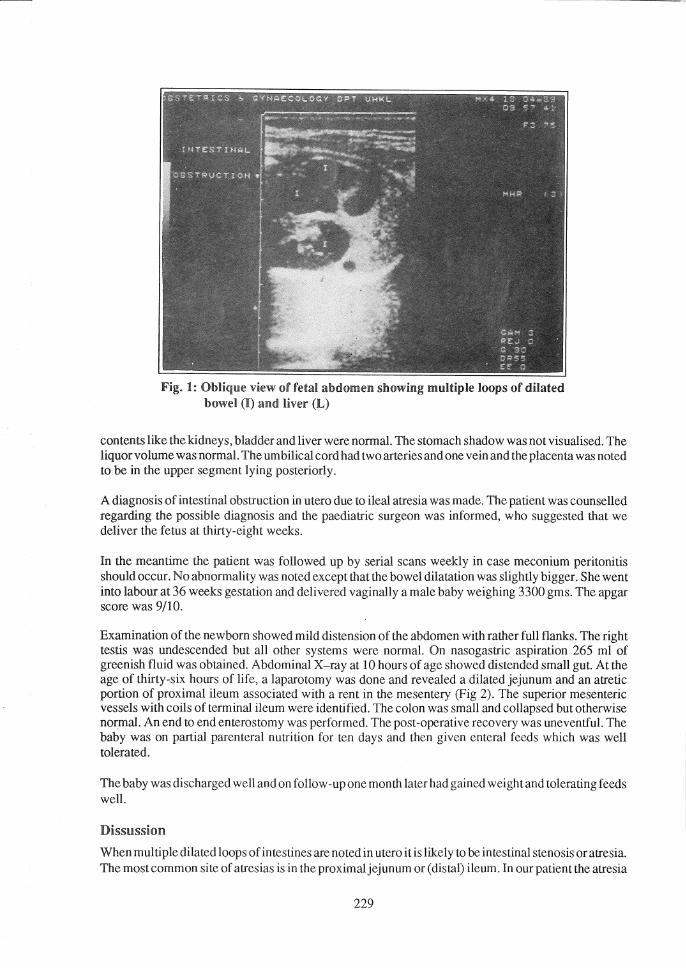
Fig. 1: Oblique view of fetal abdomen showing multiple loops of dilated bowel (I) and liver (L)
contents like the kidneys, bladder and liver were normal. The stomach shadow was not visualised. The liquor volume was normal. The urn bilical cord had two arteries and one vein and the placenta was noted to be in the upper segment lying posterioriy.
A diagnosis of intestinal obstruction in utero due to ileal atresia was made. The patient was counselled regarding the possible diagnosis and the paediatric surgeon was informed, who suggested that we deliver the fetus at thirty-eight weeks.
In the meantime the patient was followed up by serial scans weekly in case meconium peritonitis should occur. No abnormality was noted except that the bowel dilatation was slightly bigger. She went into labour at 36 weeks gestation and delivered vaginally a male baby weighing 3300 gms. The apgar score was 9/10.
Examination of the newborn showed mild distension of the abdomen with rather full flanks. The right testis was undescended but all other systems were normal. On nasogastric aspiration 265 ml of greenish fluid was obtained. Abdominal X-ray at 10 hours of age showed distended small gut. At the age of thirty-six hours of life, a laparotomy was done and revealed a dilated jejunum and an atretic portion of proximal ileum associated with a rent in the mesentery (Fig 2). The superior mesenteric vessels with coils of terminal ileum were identified. The colon was small and collapsed but otherwise normal. An end to end enterostomy was performed. The post-operative recovery was uneventful. The baby was on partial parenteral nutrition for ten days and then given enteral feeds which was well tolerated.
The baby was discharged well andon follow-up one month later had gained weight and tolerating feeds well.
Dissussion
When multiple dilated loops of intestines are noted in utero it is likely to be intestinal stenosis or atresia. The most common site of atresias is in the proximal jejunum or (distal) ileum. In our patient the atresia
229
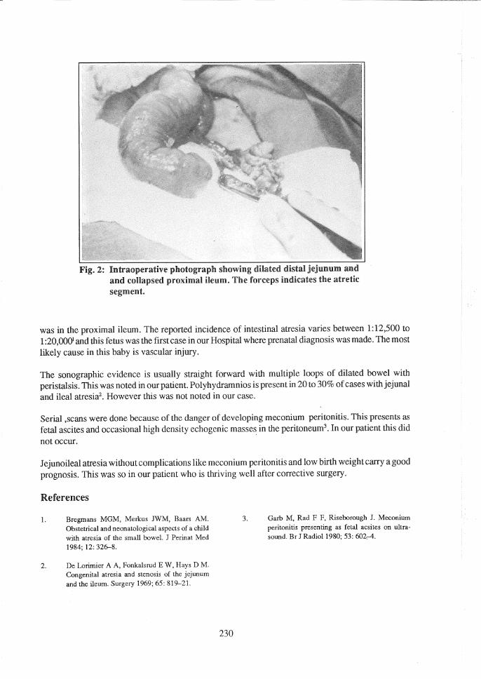
Fig. 2: Intraoperative photograph showing dilated distal jejunum and and collapsed proximal ileum. The forceps indicates the atretic segment.
was in the proximal ileum. The reported incidence of intestinal atresia varies between 1:12,500 to 1 :20,OO()l and this fetus was the first case in our Hospital where prenatal diagnosis was made. The most likely cause in this baby is vascular injury.
The sonographic evidence is usually straight forward with multiple loops of dilated bowel with peristalsis. This was noted in our patient. Polyhydramnios is present in 20 to 30% of cases with jejunal and ileal atresia2• However this was not noted in our case.
Serial ,scans were done because of the danger of developing meconium peritonitis. This presents as fetal ascites and occasional high density echogenic masses in the peritoneum3 . In our patient this did
not occur.
Jejunoileal atresia without complications like meconium peritonitis and low birth weight carry a good prognosis. This was so in our patient who is thriving well after corrective surgery.
References
1. Bregmans MGM, Merkus JWM, Baars AM. Obstetrical and neonatological aspects of a child with atresia of the small bowel. J Perinat Med 1984; 12: 326-8.
2. De Lorimier A A, Fonkalsrud E W, Hays D M. Congenital atresia and stenosis of the jejunum and the ileum. Surgery 1969; 65: 819-21.
3.
230
Garb M, Rad F F, Riseborough J. Meconium peritonitis presenting as fetal acsites on ultrasound. BrJ Radiol1980; 53: 602;-4.