Embed Size (px)
Citation preview
219
Charles Coutelle and Simon N. Waddington (eds.), Prenatal Gene Therapy: Concepts, Methods, and Protocols, Methods in Molecular Biology, vol. 891, DOI 10.1007/978-1-61779-873-3_11, © Springer Science+Business Media, LLC 2012
Chapter 11
Animal Models for Prenatal Gene Therapy: The Sheep Model
Khalil N. Abi-Nader , Michael Boyd , Alan W. Flake , Vedanta Mehta , Donald Peebles , and Anna L. David
Abstract
Large animal experiments are vital in the fi eld of prenatal gene therapy, to allow translation from small animals into man. Sheep provide many advantages for such experiments. They have been widely used in research into fetal physiology and pregnancy and the sheep fetus is a similar size to that in the human. Sheep are tolerant to in utero manipulations such as fetoscopy or even hysterotomy, and they are cheaper and easier to maintain than non-human primates. In this chapter, we describe the animal husbandry involved in generating time-mated sheep pregnancies, the large number of injection routes in the fetus that can be achieved using ultrasound or fetoscopic-guided injection, and laparotomy when these more mini-mally invasive routes of injection are not feasible.
Key words: Sheep , Ultrasound , Fetoscopy , Amniocentesis , Fetal blood sampling
If fetal gene therapy is to become clinically applicable, develop-ments in vector technology must be accompanied by improve-ments in minimally invasive methods of delivering vectors to the fetus. Traditionally, invasive surgical techniques such as maternal laparotomy or hysterotomy have been performed to access the fetus in small- and even large-animal models. However, in clinical practice, minimally invasive techniques such as ultrasound-guided injection, or even fetoscopy, could be used to deliver gene therapy to the fetus with less morbidity and mortality. It is likely that non-human primates will be the ultimate animal model that will be used for safety studies in the immediate preparation for a clinical trial of fetal gene therapy. However, the high maintenance costs and breeding conditions prohibit their use in the routine development of novel injection techniques.
1. Introduction
220 K.N. Abi-Nader et al.
Sheep are much easier to breed and maintain and are a well-established animal model of human fetal physiology. Sheep have a consistent gestation period of 145 days; the development of the fetus and of the immune system is very similar to humans. Using the pregnant sheep, we have adapted ultrasound-guided injection techniques from fetal medicine practice and developed new methods to deliver gene therapy to the fetal sheep including ultrasound-guided intratracheal injection to target the distal respi-ratory epithelium ( 1, 2 ) and ultrasound-guided intragastric injec-tion to target the intestinal mucosa ( 3 ) . Fetoscopic techniques can be utilized when improved visualization is needed beyond the resolu-tion of ultrasound guidance or when surgical interventions are required to facilitate optimal delivery of vector. Examples of feto-scopic techniques include performing intravascular injections prior to 70 days gestation in the sheep, targeting the umbilical veins at their convergence at the cord (Flake AW, unpublished observa-tion), or placement of an intratracheal balloon at the time of vector installation to enhance pulmonary epithelial transduction ( 4 ) . The combination of ultrasound guidance and fetoscopy is often utilized when small fetoscopes are employed. Ultrasound provides the big picture allowing guidance of the scope with its relatively limited fi eld of view to the point of intervention.
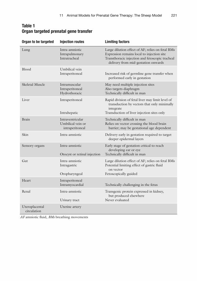
These techniques are described in detail in this chapter and the fetal organs that can be targeted via different routes of injection are listed in Table 1 . It is likely that many if not all of these methods could be adapted for use in other large animals such as the macaque, baboon, goat, or dog, and are likely to target similar organs to those in the fetal sheep.
Maternal mortality in the pregnant sheep is negligible and fetal mortality was between 3 and 15%, depending on the route of injec-tion. Over 90% of the fetal mortality is due to iatrogenic infection, usually with known fl eece commensals ( 5 ) . Thus, it is vital that any invasive procedure is performed with stringent aseptic technique. In clinical practice, cleaning by wiping the maternal abdominal skin with an antiseptic solution is suf fi cient. In the sheep, the fl eece needs to be scrubbed as an additional step, as described below.
Ultrasound-guided invasive procedures in sheep such as tra-cheal injection had a complication rate of 6%, which was related to blood vessel damage within the thorax ( 6 ) . Intracardiac and umbil-ical vein injection had an unacceptably high procedure-related fetal mortality in fi rst-trimester fetal sheep ( 5 ) and umbilical vein injec-tion was only reliably achieved from 70 days of gestation, equiva-lent to 20 weeks of gestation in humans. The relevant time windows for the different application routes in humans still need to be estab-lished with respect to technical feasibility, fetal physiology, and the development of the fetal immune system.
22111 Animal Models for Prenatal Gene Therapy: The Sheep Model
Table 1 Organ targeted prenatal gene transfer
Organ to be targeted Injection routes Limiting factors
Lung Intra-amniotic Large dilution effect of AF; relies on fetal BMs Intrapulmonary Expression remains local to injection site Intratracheal Transthoracic injection and fetoscopic tracheal
delivery from mid-gestation onwards
Blood Umbilical vein Increased risk of germline gene transfer when
performed early in gestation Intraperitoneal
Skeletal Muscle Intramuscular May need multiple injection sites Intraperitoneal Also targets diaphragm Hydrothoracic Technically dif fi cult in man
Liver Intraperitoneal
Intrahepatic
Rapid division of fetal liver may limit level of transduction by vectors that only minimally integrate
Transduction of liver injection sites only
Brain Intraventricular Technically dif fi cult in man Umbilical vein or
intraperitoneal Relies on vector crossing the blood brain
barrier; may be gestational age dependent
Skin Intra-amniotic Delivery early in gestation required to target deeper epidermal layers
Sensory organs Intra-amniotic
Otocyst or retinal injection
Early stage of gestation critical to reach developing ear or eye
Technically dif fi cult in man
Gut Intra-amniotic Large dilution effect of AF; relies on fetal BMs Intragastric
Oropharyngeal
Potential limiting effect of gastric fl uid on vector
Fetoscopically guided
Heart Intraperitoneal Technically challenging in the fetus Intramyocardial
Renal Intra-amniotic
Urinary tract
Transgenic protein expressed in kidney, but produced elsewhere
Never evaluated
Uteroplacental circulation
Uterine artery
AF amniotic fl uid, BMs breathing movements
222 K.N. Abi-Nader et al.
1. Ewes and rams in a ratio of 10:1. 2. Vaccination to Chlamydia abortus (Enzovax ® , Intervet UK)
and toxoplasmosis (Toxovax ® , Intervet UK). 3. Chronogest ® CR sponges (Intervet UK Limited, Milton
Keynes, UK) containing 20 mg Flugestone acetate. 4. Sponge applicator. 5. Ram Mating Raddle (Agrimark Limited, Isle of Man, UK). 6. Super Ewe & Lamb UFAS compound feed (J & W Atlee Ltd,
Dorking, Surrey, U K).
1. Ultrasound coupling gel (Electro Medical Supplies Ltd, Oxon, UK).
2. Clippers for fl eece (Masterclip Duo, Outlandish Items Ltd, Leicestershire, UK).
3. Ultrasound scanner with a curved-array 3.0–5.0 MHz abdom-inal probe with real-time image display, cine loop facility, B-mode, M-mode pulse wave Doppler fl ow mode and color fl ow mode, e.g., Logiq 400 (GE Medical Systems, Milwaukee, USA), or Acuson 128 XP10 ultrasound scanner (Siemens, Bracknell, UK).
4. Ultrasound charts in use for sheep ( 7, 8 ) .
1. Wood chips for bedding (GPM Shavings, East Angus, Canada). 2. A large sling with poles. 3. A balance to weigh the sheep (needs to measure up to 150 kg). 4. Clippers for fl eece (Masterclip Duo, Outlandish Items Ltd,
Leicestershire, UK). 5. 19-Gauge butter fl y winged perfusion set (Terumo Europe NV,
Leuven, Belgium). 6. BD Vacutainer EDTA, 9NC, and SST tubes (BD Vacutainer
systems, Plymouth, UK). 7. 15 ml of 10% w/v thiopental sodium i.v. (Thiovet, Novartis
Animal Health UK Ltd, Hertfordshire, UK). 8. 2–2.5% Iso fl urane (Iso fl urane-Vet, Merial Animal Health Ltd,
Essex, UK). 9. 100% Oxygen. 10. An oral gastric tube (Arnold Veterinary Products, Harlescott,
Shrewsbury, UK). 11. A laryngoscope with a 30-cm blade (Penlon, UK). 12. A 9.0-mm cuffed endotracheal tube (Portex, UK).
2. Materials
2.1. Generation of Time-Mated Sheep
2.2. Con fi rmation of Pregnancy and Gestational Age by Ultrasound
2.3. Sheep Anesthesia
22311 Animal Models for Prenatal Gene Therapy: The Sheep Model
13. A semiclosed anesthetic system that includes a capnograph (Ohmeda Anethesia system, BOC Healthcare).
14. A stethoscope (Littman, 3 M, Bracknell, UK). 15. A pulse oximeter (5250 RGM, Ohmeda). 16. A surgical theater table that can be raised. 17. A ventilator (Manley MP5, Blease Medical Equipment Ltd,
Chesham, Bucks, UK).
1. Povidone iodine antiseptic cleaning solution (Grampian Pharmaceuticals Ltd, UK).
2. Chlorhexidine gluconate cleaning scrub (Hibiscrub, AstraZeneca Ltd, UK).
3. Povidone iodine antiseptic solution (Grampian Pharmaceuticals Ltd, UK).
4. Sterile drapes. 5. A 3.0–5.0-MHz curved-array abdominal probe on an ultra-
sound scanner with real-time image display, cine loop facility, B-mode, M-mode pulse wave Doppler fl ow mode, and color fl ow mode, e.g., Logiq 400 (GE Medical Systems, Milwaukee USA), or Acuson 128 XP10 ultrasound scanner (Siemens, Bracknell, UK).
6. Sterile ultrasound coupling gel (Electro Medical Supplies Ltd, Oxon, UK).
7. A no. 11 blade (Swann-Morton, Shef fi eld, UK). 8. 18-G, 20-G, 15-cm long and 22-G, 9-cm long echotip needles
with a lancet tip (Cook Medical, UK). 9. Sterile physiological saline (0.9% NaCl). 10. 1-, 2-, and 5-ml syringes. 11. Viricide agent, e.g., Virkon (Antec International Limited).
1. Sterile preparation as described for ultrasound-guided and lap-arotomy procedures.
2. If ultrasound guidance is required, ultrasound equipment as described for ultrasound-guided procedures above.
3. If laparotomy is required, equipment as described for laparo-tomy procedures below.
4. Storz (Storz Endoskope, Tuttlingen, Germany) compatible videocart with minimum requirements of: (1) high de fi nition monitor, ideally con fi gured for picture in picture with the ultrasound image displayed on the same screen, (2) camera control unit, (3) camera head preferably 3-chip, (4) suction (optional) and irrigation system, (5) cold light source, and (6) video recording unit.
2.4. Ultrasound-Guided Procedures
2.5. Fetoscopic Procedures

224 K.N. Abi-Nader et al.
5. Storz fetoscopy set appropriate for planned procedure. We recommend the use of a remote eyepiece for maximum range of motion. Each set consists of a telescope and an operating sheath with various working channels, and accessories for speci fi c procedures. Telescopes range from the straight for-ward 0° 1 mm, 20 cm working length scope to the 0°–30° 2 mm 26 cm working length scopes (Storz endoscopy, USA). Corresponding operating sheaths are the 1.3-mm maximum diameter (6.5 Fr), pointed tip, sheath with laser fi ber or punc-ture needle working channel, and the 9 Fr (Storz endoscopy, USA). Operating sheath with laser fi ber working channel or more versatile 11, 11.5, or 14.5 Fr. operating sheaths (Storz endoscopy, USA) that include larger working channels or chan-nels for working inserts.
6. For intravascular injections, the straight forward 0° 1 mm, 20 cm working length scope (Storz Endoscopy, USA), the 1.3-mm maximum diameter (6.5 Fr), pointed tip, sheath with laser fi ber or puncture needle working channel, and the 0.6-mm 26-cm long puncture needle are used (Storz endos-copy, USA).
7. For fetal balloon tracheal occlusion, the 1.3-mm diameter, 30.6 cm working length, fetal tracheoscope and the 3.3-mm tracheoscopic sheath with three side ports (30.6 cm working length, precurved 30°) are ideal (Storz endoscopy, USA).
8. 10 Fr Flexor Check-Flo cannula (Check-Flo Introducer set). 9. BALTACCI—BDPE—detachable balloon catheter (outer
diameter max. 0.9 mm, Balt, Montmorency, France). 10. 3.0 Fr Slip-cath infusion catheter (Cook, USA). 11. Warm Lactated Ringers solution. 12. 3-0 Vicryl.
1. Povidone iodine antiseptic cleaning solution (Grampian Pharmaceuticals Ltd, UK).
2. Chlorhexidine gluconate cleaning scrub (Hibiscrub, AstraZeneca Ltd, UK).
3. Povidone iodine antiseptic solution (Grampian Pharmaceuticals Ltd, UK).
4. Sterile drapes. 5. An electrosurgical unit (ERBE VIO 300D Electrosurgical
Generator, Tubingen, Germany). 6. A no. 21 blade and handle (Swann-Morton, Shef fi eld, UK). 7. Surgical instruments: Mayo and Metzenbaum scissors, surgical
forceps set (atraumatic, Debackey, and grasping forceps), large and small needle holders (Fine Science Tools, InterFocus Ltd, Cambridge, UK).
2.6. Laparotomy Procedures
22511 Animal Models for Prenatal Gene Therapy: The Sheep Model
8. Surgical sutures: 5-mm Mersiline tape (Ethicon, St. Stevens-Woluwe, Belgium), 2–0 PDS (Ethicon, St. Stevens-Woluwe, Belgium), 2-0 Monocryl (Ethicon, St. Stevens-Woluwe, Belgium) and 2-0 and 3-0 prolene (Ethicon, Norderstedt, Germany).
9. Surgical vessel loops (Bard Ltd, Crawley, West Sussex, UK). 10. 23- and 25-G butter fl y winged perfusion sets (Terumo Europe
NV, Leuven, Belgium). 11. 2-, 5-, and 10-ml syringes. 12. Blood collection tubes: EDTA, 9NC, and SST tubes (BD,
Plymouth, UK). 13. Injectable antibiotics: Penstrep (200 mg/ml procaine penicil-
lin and 250 mg/ml dihydrostreptomycin; Norbrook Laboratories Ltd, County Down, UK), sodium benzylpenicil-lin G (Crystapen, Schering-Plough, Uxbridge, UK), and Gentamicin (Genticin Injectable, Roche Products Ltd, Hertfordshire, UK).
14. Injectable analgesics: Buprenorphine (Vetergesic, Alstoe Animal Health, York, UK).
1. Once the sheep for experiments have been selected, a vaccina-tion schedule against Chlamydia abortus and toxoplasmosis is drawn up and given. See Note 1.
2. To time sheep ovulation, Chronogest ® sponges (Flugestone acetate) containing 30 mg progesterone is placed in the vagina of ewes for 2 weeks to induce ovulation.
3. Two days after removal of the progesterone sponges, ewes are put in a pen with the ram overnight. The ram is marked on its belly with Ram Raddle ® , a colored powder mixed with liquid paraf fi n which is transferred and marks the back of the ewe once she has been served. Marked ewes are presumed to have been tupped.
4. Ewes that are unmarked or not marked clearly are put back for a “return service” with the ram 2 weeks later.
5. A spreadsheet is used to check on regular progress with tup-ping and to schedule experiments at speci fi c gestational ages, with close liaison with the farmer.
6. When the sheep breeding season is advanced (March onwards) superovulation may help. Chronogest ® sponges are placed vag-inally for 12 days followed by an intramuscular injection of 1,000 IU Folligon ® (Pregnant Mares Serum Gonadotrophin).
3. Methods
3.1. Generating Time-Mated Sheep
226 K.N. Abi-Nader et al.
The ewe is placed in with the ram 2 days later and returned as before 14 days later. Superovulation often results in higher order multiple pregnancies such as triplets and quadruplets, and the conception rate is not as high as that achieved using progesterone alone.
7. Sheep should be moved from the farm to the experimental animal facility (if they are separate), at least 1 week before the scheduled procedure. See Note 2.
1. All ewes that have been tupped are scanned to con fi rm preg-nancy, fetal number, and gestation age at approximately 40 days of age to allow enough time to move them to the experimental facility. See Note 3.
2. Ewes are caught, turned up, and held in a sitting position for scanning using a standard B-mode gray-scale ultrasound scan-ner with an abdominal probe. This is the same position as is used by farmers to clip the toenails and for shearing sheep. Sheep are generally placid and will happily remain in this posi-tion for at least 15 min.
3. An area of the fl eece is clipped close to the skin just above the udders extending 10 cm up the abdomen, to allow a clear view of the whole uterus.
4. The sonographer applies ultrasound coupling medium to the clipped area and scans the uterus using a 3.0–5.0-MHz probe held just above the udders. It is important to scan across both horns of the uterus to examine for twins and triplet pregnancies.
5. Still images of the occipito-snout length and biparietal diame-ter are obtained and the dimensions measured on the machine. Gestational age is con fi rmed according to standard tables.
1. Before surgery ewes are starved for 12 h with free access to water, and bedded on wood chips. This is to prevent bloating which can occur when sheep under anesthesia are unable to belch (eructate) normally and release methane gas which is a by-product of fermentation of their food ( 9 ) .
2. Animals are weighed to allow calculation of the correct dose for drugs.
3. On the morning of surgery sheep are walked to theater. This should be a dedicated theater space if possible that is kept clean, has anesthetic gas extraction facilities and air-condition-ing to maintain the temperature at 37°C.
4. The sheep are turned up with an assistant sitting comfortably behind. The wool is clipped from their necks, the jugular vein is cannulated using a 19-Gauge butter fl y winged perfusion set, and the cannula is securely taped in.
3.2. Con fi rmation of Pregnancy and Gestational Age by Ultrasound
3.3. Sheep Anesthesia

22711 Animal Models for Prenatal Gene Therapy: The Sheep Model
5. Blood can be taken at this point for baseline investigations of antibodies, hematology, and biochemistry parameters. Blood is collected into BD Vacutainer tubes containing potassium EDTA for plasma and into plain tubes for serum (SST).
6. 15 ml of thiopental sodium (10% w/v) is given intravenously over 1 min for induction of anesthesia.
7. Once asleep the ewes are rolled onto a mat with poles or han-dles to attach to a hoist and they are then lifted mechanically onto the operating table and laid supine. With the sheep supine, the neck is extended and the jaw is lifted up using tape applied across the tongue and teeth. A laryngoscope with a 30-cm blade is passed down the trachea to enable visualization of the vocal cords. A 9.0-mm cuffed endotracheal tube containing a stylet is passed through the vocal cords. The stylet is removed and the cuff is in fl ated with the required volume of air to pre-vent inhalation of regurgitated ruminal contents. The tube is secured with tape tied around the lower jaw.
8. Alternatively, the ewe may also be intubated while she is held sitting up by an assistant behind her. Using a tape the upper jaw is lifted up to straighten the trachea, and while the tongue is gently pulled to one side with some gauze, the endotracheal tube is passed through the vocal cords.
9. The endotracheal tube is connected to the anesthetic gas machine and the correct placement of the endotracheal tube is checked by a variety of methods: Auscultate both lungs while the bag and valve mask is being
compressed to force air into the lungs. Observe the bag and valve mask in fl ating and de fl ating in the
closed system. Feel movement of air within the endotracheal tube as the ewe
breathes. 10. Once the sheep is supine on the operating table and anesthetized
the head is lowered to allow drainage of saliva and ruminal fl uid ( 10 ) . Because access to the whole abdomen is needed for proce-dures the animals are kept in dorsal recumbency. Over long peri-ods the rumen can compress the lungs causing dyspnoea and the vena cava reducing venous return to the heart. This is not felt to be a problem in the case of in utero ultrasound-guided proce-dures because they are usually of suf fi ciently short duration.
11. Anesthesia is maintained by inhalation on 2–2.5% iso fl urane in 100% oxygen run at 2–3 l/min for spontaneously breathing animals and at 6–8 l/min for animals on intermittent positive pressure ventilation (IPPV) to maintain an end-tidal CO 2 <5%, O 2 saturation >95%, and a respiratory rate of 12–14/min using a semiclosed anesthetic circuit.

228 K.N. Abi-Nader et al.
12. For lengthy experiments (over 20 min), it is advisable to pass an oral gastric tube into the sheep’s stomach to allow the release of methane gas. A capnograph should also be connected to the endotracheal tube to monitor carbon dioxide saturation. We aim to keep end-tidal CO 2 <5% if possible.
13. For experiments that are expected to last over 30 min (compli-cated ultrasound-guided procedures or laparotomy), the ewe should be ventilated to prevent CO 2 retention. If the venous return or oxygen saturation is poor, and the position of the animal is compromised, the animal’s head may be tilted too far down to rely on spontaneous breathing, in which case the ewe is then ventilated electively.
14. A pulse oximeter is placed on the ewe’s ear or tongue and is used to monitor oxygen saturation and pulse rate. This data is documented every 15 min together with respiration rate, car-bon dioxide concentration, anesthetic gas concentration, and level of anesthesia on an anesthetic sheet.
15. The anesthetic gas is turned off 1–2 min before the end of the experiment to allow recovery. Analgesia is not usually given for minimally invasive ultrasound-guided procedures. See Note 4.
16. The ewe is hoisted off the theater table to the fl oor and rolled onto her sternum. Her legs are placed underneath to ensure she can get upright as soon as awake. She is extubated once strong swallowing re fl exes return and she is then returned to the sheep pen.
17. The ewe is watched carefully to ensure she stands up within a few minutes of the procedure, and she is helped to stand if she does not do so spontaneously after 20 min. Standing allows recovery of ruminal function, which can be affected by a long general anesthetic.
18. The day after the procedure, the well-being of the ewe is assessed and fetal survival is determined on ultrasound scan by turning the ewe up for ultrasound assessment. Ultrasound-guided injection sites are examined carefully for infection.
19. For experiments where the fetus will be delivered, serial fetal ultrasound assessment should be made during gestation and a plan made for care of the ewe during labor (see Chapter 14 and Note 5).
1. Once anesthetized, the wool is clipped from the ewe’s abdomen from the udders up to the sternum and round to the sides.
2. The skin is scrubbed for 5 min using povidone iodine antisep-tic cleaning solution. See Note 6.
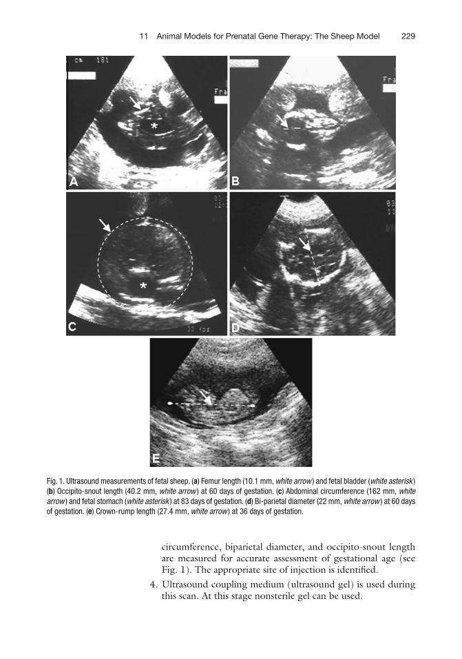
3. A detailed ultrasound examination of the uterus and its contents is performed using a 3–5-MHz probe on the ultrasound scan-ner. Fetal measurements such as fetal femur length, abdominal
3.4. Ultrasound-Guided Procedures
22911 Animal Models for Prenatal Gene Therapy: The Sheep Model
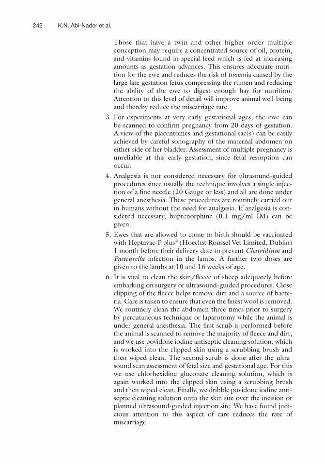
circumference, biparietal diameter, and occipito-snout length are measured for accurate assessment of gestational age (see Fig. 1 ). The appropriate site of injection is identi fi ed.
4. Ultrasound coupling medium (ultrasound gel) is used during this scan. At this stage nonsterile gel can be used.
Fig. 1. Ultrasound measurements of fetal sheep. ( a ) Femur length (10.1 mm, white arrow ) and fetal bladder ( white asterisk ) ( b ) Occipito-snout length (40.2 mm, white arrow ) at 60 days of gestation. ( c ) Abdominal circumference (162 mm, white arrow ) and fetal stomach ( white asterisk ) at 83 days of gestation. ( d ) Bi-parietal diameter (22 mm, white arrow ) at 60 days of gestation. ( e ) Crown-rump length (27.4 mm, white arrow ) at 36 days of gestation.
230 K.N. Abi-Nader et al.
5. The abdomen is then scrubbed again for a further 5 min using chlorhexidine gluconate cleaning solution and a fi nal clean with povidone iodine solution is made over the injection site.
6. The ultrasound probe is wiped over with 70% ethanol and then sprayed again and the ethanol is allowed to evaporate.
7. The operators (usually two) scrub up and don sterile gowns and gloves. All instruments (needles, syringes containing the vector, and saline) are laid out on a small operating table draped with a sterile drape. The vector is drawn up into syringes ready to be administered immediately to the fetus.
8. The abdomen is now draped with sterile drapes to reveal a window for the ultrasound guided injection sit. See Note 7.
9. Sterile ultrasound coupling medium (ultrasound gel) is now used for ultrasound scanning. See Note 8.
10. A small cut in the skin, using a no. 11 blade may facilitate needle insertion and avoid blunting the sharp needle tip. This is only necessary when a larger needle (18 Gauge and higher) is used.
11. Under ultrasound guidance the needle is inserted (with an angle of 45° relative to the ultrasound beam) through the maternal skin, the uterus, and into the amniotic cavity avoiding passage through the placentomes if possible. Care should be taken to keep the whole needle length and particularly the needle tip under ultrasound view as this view most accurately re fl ects the needle position, otherwise it is easy to be mislead about the true needle orientation.
12. For each injection route a freehand technique is used. Needle-guides can be used but reduce the fl exibility of the approach, especially if the fetal position moves relative to the needle as it is inserted. The fetus does not move around once the ewe has been under general anesthesia for at least 10 min.
13. The needle is then inserted through the fetal skin into the tar-get by fl icking it through the uterine wall and into the fetus. Once the needle tip is thought to be in the correct position, the stylet is removed and, the vector is injected followed by 40 μ l 0.9% saline to fl ush the vector from the dead space of the spinal needle. This is important because in many experiments, small volumes of vector are often applied.
14. If the position of the needle tip is not certain, a small volume (40 μ l 0.9% saline) can be introduced to con fi rm it. A white fl ash will be seen on the screen as turbulence was created by microbubbles.
15. After removal of the needle from the uterus, the fetus is observed for signs of fetal well-being such as a normal fetal heart rate, hematoma formation, or bleeding See Note 9.
23111 Animal Models for Prenatal Gene Therapy: The Sheep Model
16. Procedures are always video-taped so that after-injection review can be done to con fi rm the injection route.
17. The fetus should be scanned the day after injection to check on fetal well-being, by turning up the ewe. This allows examina-tion of the injection site on the ewe’s abdomen to be inspected.
1. A 22-G Quincke type spinal needle (Becton Dickinson, UK) is used for procedures performed at any gestational age.
2. A good-sized pocket of amniotic fl uid is identi fi ed by ultrasound.
3. The needle is aimed towards the center of the pocket of amni-otic fl uid while avoiding fetal parts, and passage through one of the placentomes.
4. The needle tip is advanced through the amniotic membrane using a rapid thrust since the amniotic membrane in sheep is relatively fl accid especially during early gestation.
5. The needle tip can be easily identi fi ed as a hyperechoic point inside fl uid- fi lled anechoic amniotic cavity (even when using needles without an echotip). The needle stylet is removed and a small volume of amniotic fl uid is aspirated for con fi rmation.
6. A small volume of 0.9% saline is injected and correct needle placement is recon fi rmed by observing microbubbles moving around the fetus in the amniotic sac.
7. The vector is injected.
1. A 20- or 22-Gauge Echotip Lancet needle is used, depending on gestational age. 20 Gauge is used after 80 days of gestation.
2. A midsagittal ultrasonographic plane of the fetal abdomen is obtained. The needle tip is targeted below the level and to the side of the cord insertion and just superior to the fetal bladder. Care should be taken to prevent damage to umbilical cord insertion or the umbilical arteries as they fl ow down medio-laterally along the posterior aspect of the anterior fetal abdomi-nal wall. Color or power Doppler is helpful in delineating the vascular anatomy if the operator is in doubt.
3. The position of the needle is viewed in two planes to con fi rm correct placement. Needle placement can also be veri fi ed by injecting 100 μ l saline and observing turbulence as microbub-bles spreading throughout the peritoneal cavity. In addition, an echodense area surrounding the fetal bowel usually devel-ops at the injection site.
4. The vector is delivered in volumes of 100–500 μ l before 60 days, up to 1 ml between 60 and 70 days and 1–2 ml after 70 days of gestation.
3.4.1. Intra-amniotic Injection ( see Note 10)
3.4.2. Intraperitoneal Injection
232 K.N. Abi-Nader et al.
1. A 22-Gauge Echotip Lancet needle is used for all procedures up to 85 days of gestation. A 20-Gauge Echotip Lancet needle is used for experiments at later gestational ages.
2. The intrahepatic portion of the umbilical vein is the preferred site for intravascular injection in the sheep fetus. This is best visualized using a cross-sectional plane of the abdomen with a slight tilt towards the fetal head. Attempts should be made to puncture the vessel as close as possible to 90° as more parallel angles are associated with failed punctures. Injection of the fetal intrahepatic umbilical vein can be achieved reliably from approximately 80 days of gestation in the fetal sheep (unpub-lished observation).
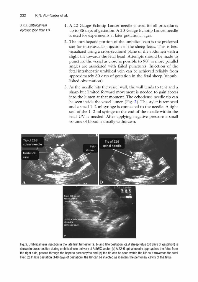
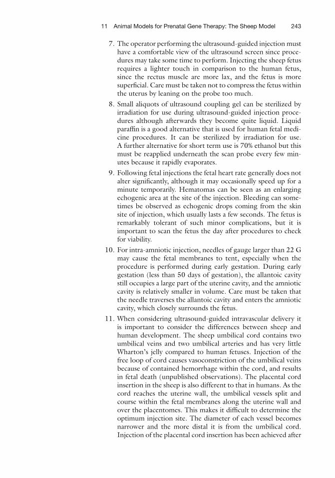
3. As the needle hits the vessel wall, the wall tends to tent and a sharp but limited forward movement is needed to gain access into the lumen at that moment. The echodense needle tip can be seen inside the vessel lumen (Fig. 2 ). The stylet is removed and a small 1–2 ml syringe is connected to the needle. A tight seal of the 1–2 ml syringe to the end of the needle within the fetal UV is needed. After applying negative pressure a small volume of blood is usually withdrawn.
3.4.3. Umbilical Vein Injection (See Note 11)
Fig. 2. Umbilical vein injection in the late fi rst trimester ( a , b ) and late gestation ( c ). A sheep fetus (60 days of gestation) is shown in cross-section during umbilical vein delivery of AdhFIX vector. ( a ) A 22-G spinal needle approaches the fetus from the right side, passes through the hepatic parenchyma and ( b ) the tip can be seen within the UV as it traverses the fetal liver. ( c ) In late gestation (140 days of gestation), the UV can be injected as it enters the peritoneal cavity of the fetus.
23311 Animal Models for Prenatal Gene Therapy: The Sheep Model
4. If the blood does not come immediately, the needle is carefully and very slightly withdrawn while negative pressure is applied with the syringe, since sometimes the needle is touching the opposite vessel wall. As the needle moves away from the vessel side wall opposite, the blood should be easily withdrawn.
5. Once correct needle placement is con fi rmed, the syringe is then swopped to that containing the vector, which is then injected in a volume of 100–1,000 μ l slowly over 1 min. Turbulence can be seen within the umbilical vein as microbub-bles passing along the vessel. Great care must be taken during this change to the syringe containing the vector to avoid dis-lodging the needle from the umbilical vein.
6. Inadvertent intrahepatic injection can be seen as echogenic white material in the vessel wall and surrounding hepatic parenchyma.
1. A 20-Gauge Echotip Lancet needle is used for this procedure to ensure the needle passes through the chest wall into the heart.
2. The left or right ventricle is the preferred site of injection. A four-chamber view of the fetal heart should be obtained if possible with the anterior chest wall facing anterior or laterally.
3. The heart should be approached from the inferior aspect so that the needle avoids the cardiac conduction system. This route has been found to reduce the mortality rate ( 11 ) .
4. The needle should be passed through the fetal chest wall, between the fetal ribs and aiming for the ventricle in the direction of the AV valves. Once the needle tip is within the ventricle, the stylet is removed and fetal blood is aspirated to check cor-rect needle position.
5. Once correct needle placement is con fi rmed, the syringe con-taining the vector is applied to the needle and a volume of 100–1,000 μ l is injected slowly over 1 min. Turbulence can be seen within the ventricle as microbubbles pass through the chamber.
6. After removal of the needle, the fetal heart should be observed under ultrasound to con fi rm normal heart rate and to check for pericardial hemorrhage.
1. A 22-Gauge Echotip Lancet needle is used for early gestation procedures. A 20-G needle may be used for procedures per-formed after midgestation.
2. Under ultrasound guidance the needle is aimed through the anterior fetal abdominal wall towards a point superior to the intrahepatic portion of the umbilical vein.
3. The position of the needle is viewed in two planes to con fi rm correct placement before delivery of the viral vector.
3.4.4. Intracardiac Injection ( see Note 12)
3.4.5. Intrahepatic Injection
234 K.N. Abi-Nader et al.
4. The vector is delivered in a volume of 100–200 μ l in early gestation. A higher volume may be delivered after midgestation.
5. During intrahepatic vector administration, an echodense area develops within the hepatic parenchyma at the injection site.
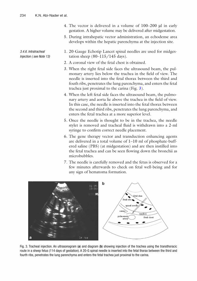
1. 20-Gauge Echotip Lancet spinal needles are used for midges-tation sheep (80–115/145 days).
2. A coronal view of the fetal chest is obtained. 3. When the right fetal side faces the ultrasound beam, the pul-
monary artery lies below the trachea in the fi eld of view. The needle is inserted into the fetal thorax between the third and fouth ribs, penetrates the lung parenchyma, and enters the fetal trachea just proximal to the carina (Fig. 3 ).
4. When the left fetal side faces the ultrasound beam, the pulmo-nary artery and aorta lie above the trachea in the fi eld of view. In this case, the needle is inserted into the fetal thorax between the second and third ribs, penetrates the lung parenchyma, and enters the fetal trachea at a more superior level.
5. Once the needle is thought to be in the trachea, the needle stylet is removed and tracheal fl uid is withdrawn into a 2-ml syringe to con fi rm correct needle placement.
6. The gene therapy vector and transduction enhancing agents are delivered in a total volume of 1–10 ml of phosphate-buff-ered saline (PBS) (at midgestation) and are then instilled into the fetal trachea and can be seen fl owing down the bronchii as microbubbles.
7. The needle is carefully removed and the fetus is observed for a few minutes afterwards to check on fetal well-being and for any sign of hematoma formation.
3.4.6. Intratracheal Injection ( see Note 13)
Fig. 3. Tracheal injection. An ultrasonogram ( a ) and diagram ( b ) showing injection of the trachea using the transthoracic route in a sheep fetus (114 days of gestation). A 20-G spinal needle is inserted into the fetal thorax between the third and fourth ribs, penetrates the lung parenchyma and enters the fetal trachea just proximal to the carina.
23511 Animal Models for Prenatal Gene Therapy: The Sheep Model
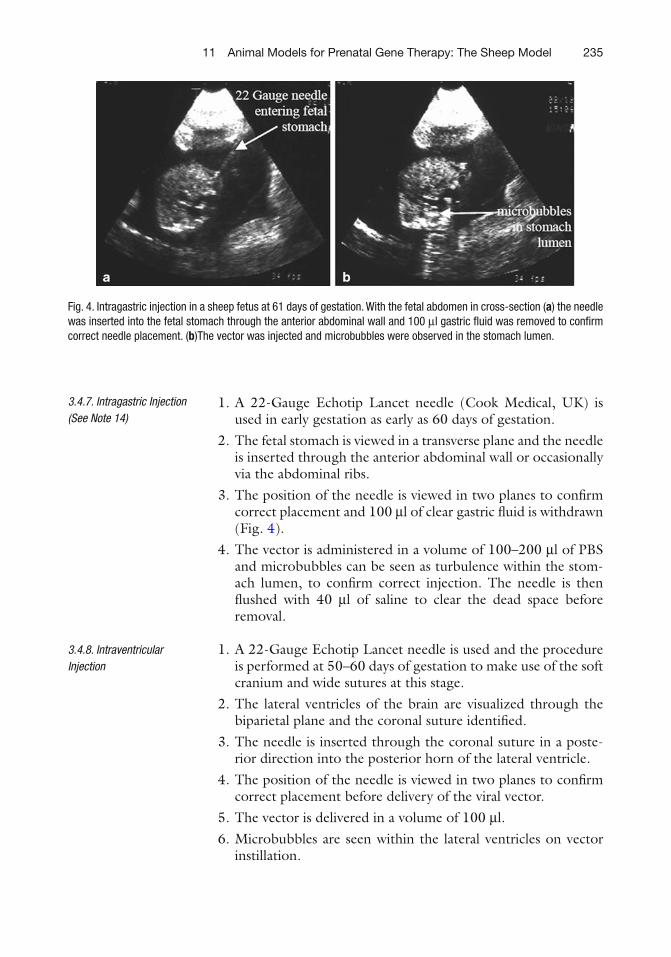
1. A 22-Gauge Echotip Lancet needle (Cook Medical, UK) is used in early gestation as early as 60 days of gestation.
2. The fetal stomach is viewed in a transverse plane and the needle is inserted through the anterior abdominal wall or occasionally via the abdominal ribs.
3. The position of the needle is viewed in two planes to con fi rm correct placement and 100 μ l of clear gastric fl uid is withdrawn (Fig. 4 ).
4. The vector is administered in a volume of 100–200 μ l of PBS and microbubbles can be seen as turbulence within the stom-ach lumen, to con fi rm correct injection. The needle is then fl ushed with 40 μ l of saline to clear the dead space before removal.
1. A 22-Gauge Echotip Lancet needle is used and the procedure is performed at 50–60 days of gestation to make use of the soft cranium and wide sutures at this stage.
2. The lateral ventricles of the brain are visualized through the biparietal plane and the coronal suture identi fi ed.
3. The needle is inserted through the coronal suture in a poste-rior direction into the posterior horn of the lateral ventricle.
4. The position of the needle is viewed in two planes to con fi rm correct placement before delivery of the viral vector.
5. The vector is delivered in a volume of 100 μ l. 6. Microbubbles are seen within the lateral ventricles on vector
instillation.
3.4.7. Intragastric Injection (See Note 14)
3.4.8. Intraventricular Injection
Fig. 4. Intragastric injection in a sheep fetus at 61 days of gestation. With the fetal abdomen in cross-section ( a ) the needle was inserted into the fetal stomach through the anterior abdominal wall and 100 μ l gastric fl uid was removed to con fi rm correct needle placement. ( b )The vector was injected and microbubbles were observed in the stomach lumen.

236 K.N. Abi-Nader et al.
1. A 20- or 22-Gauge Echotip Lancet needle is inserted along the length of the femur and/or the buttock. 22 Gauge is used before 70 days of gestation and 20 Gauge used after 70 days of gestation. Echotip needles are used because the tip is easier to see while next to the echogenic femur bone, when compared with other needles.
2. Prior to injection, a sagittal ultrasonographic plane of the fetal thigh is obtained. The fetal thigh is best observed by obtaining a cross-sectional view of the fetal bladder and the femora are seen extending either side.
3. The fetal thigh (quadriceps or hamstring) and buttock muscles are chosen for injection because these are the largest accessible muscle groups at this gestational age in the sheep fetus ( 12 )
4. The cross-sectional diameter of the fetal thigh or buttock in early gestation muscle is as wide as the bevel of the Echotip needle. Thus, insertion of the needle tip into the muscle in a transverse section may result in immediate loss of the vector into the amniotic fl uid from the needle tip. To avoid this and to ensure a longer needle track, the needle is placed deeply within the muscle parallel to the bone.
5. In later gestations (100 days of gestation and beyond), the muscle bulk is larger and a more angled approach to the fetal bone is available without risk of vector leakage.
6. The needle position is now checked in longitudinal and trans-verse sections.
7. Once the correct needle position is con fi rmed, the stylet is removed. With a longitudinal view of the needle within the fetal muscle, the syringe containing the vector is then attached to the needle and as the needle is slowly withdrawn along the length of the muscle, the vector is injected. Echogenic foci can be observed within the muscle parenchyma con fi rming vector placement.
8. If necessary to achieve the required vector dose, a number of intramuscular injections on the same fetus can be done, on the same leg or using the opposite leg, if accessible.
9. The vector is delivered in a total volume of 100–500 μ l by preferably 1 (up to 4) injection depending on the viral titre.
10. After needle removal, the fetal injection site is checked for bleeding.
11. Injection sites include the fetal thigh muscles (quadriceps), buttocks, and biceps/triceps.
1. A view of the fetal chest in longitudinal section is achieved. 2. A hydrothorax is created by inserting a 20-Gauge Echotip
Lancet needle through the thoracic musculature between the ribs and up to the margin of the lung parenchyma.
3.4.9. Intramuscular Injection ( see Note 15)
3.4.10. Intrapleural Injection
23711 Animal Models for Prenatal Gene Therapy: The Sheep Model
3. The needle position is checked in longitudinal and transverse sections of the fetal chest.
4. 500 μ l of PBS is then injected until a pool of fl uid is seen by ultra-sound. This not only con fi rms the correct needle position but also creates a hydrothorax into which the vector is delivered.
5. The vector is then injected into the pool of fl uid in a volume of 100 μ l.
6. After needle removal, the fetal injection site is checked for bleeding.
Fetoscopic procedures are required when higher resolution is required than can be provided by ultrasound guidance alone, or when the procedure requires real-time visualization that is beyond the capacity of ultrasound to provide. Fetoscopic procedures are often combined with ultrasound guidance to give the “big pic-ture” and guide the fetoscope to the area of the intervention. In addition, if fetal positioning is an issue, as it often is in sheep with multiple fetuses, laparotomy may be the simplest way to improve access to the various amniotic compartments, to position the fetus or fetuses, and accurately place the fetoscope. We have employed this technique to perform intravascular injections as early as 55 days in the sheep fetus.
1. Sheep anesthesia and abdominal skin preparation/scrubbing are performed according to that described earlier for ultra-sound-guided procedures.
2. Drape the sheep abdomen with sterile drapes. 3. If ultrasound guidance is utilized the ultrasound probe is pre-
pared as above and the sheep abdomen is scanned to assess the location, orientation, and number of amniotic cavities, and fetuses within the maternal abdomen.
4. If the convergence of umbilical veins from each cotyledon (i.e., the root of the umbilical cord) can be visualized and is tangen-tially accessible from the maternal abdominal wall, the proce-dure can be performed percutaneously without laparotomy. This is the exception, however, and in most cases obtaining the optimal orientation of the fetoscope to the vessels as they emerge from the intercotyledonary membranes requires a maternal laparotomy.
5. If a maternal laparotomy is required, perform a laparotomy as described below and expose the uterine horn of interest. With the uterus exposed, the root of the umbilical cord can be rela-tively easily identi fi ed by either ultrasound, or transillumina-tion of the uterine wall. See Note 16.
6. The insertion site of the fetoscope is chosen to achieve a 45° angle to the root of the cord in alignment with a major tribu-tary from multiple placentomes.
3.5. Fetoscopic Procedures
3.5.1. Fetoscopically Assisted Early Gestation Intravascular Injection

238 K.N. Abi-Nader et al.
7. Depending upon the sharpness of the fetoscope (1.3-mm pointed operating sheath) and the gestational age, the feto-scope may be directly inserted into the amniotic space, or a disposable cannula and fetoscopic trocar may be utilized for initial entry into the amniotic space and the fetoscope may then be directly introduced through the cannula. See Note 17.
8. The fetoscope is advanced under ultrasound guidance to the root of the umbilical cord.
9. The vessels are surveyed and a suitable vessel is chosen at the point of its emergence from the intercotyledenary membrane as it rises towards the con fl uence forming the cord. At this point the vessel is relatively tethered rather than freely fl oating which facilitates its puncture. See Note 18.
10. With the trocar in proper alignment to the chosen vessel and the vessel in view, the needle is advanced down the working channel of the fetoscope until it comes into view in the lower visual fi eld and extends a few millimeter from the end of the scope.
11. The entire fetoscope and needle are advanced until the tip of the needle just approximates the vessel. The obturator is with-drawn from the needle and the vector-containing syringe is loaded. As the needle contains minimal dead space and the manipulations, once the needle is in the vessel risks loss of placement, no attempt is made to clear the deadspace in the needle.
12. The fetoscope is then advanced to the point where the needle indents the center of the vessel with the bevel directed inferi-orly. A quick, very short thrust is made with the needle to penetrate the vein wall. If the needle penetrates the opposite wall it is slowly withdrawn until blood return is obtained. The vector is then injected and followed by a fl ush of 0.05 ml of saline. See Note 19.
13. The needle is withdrawn. A small amount of bleeding will occur but it will rapidly stop forming a clot on the vein.
14. The fetoscope is then withdrawn and the procedure repeated if a second fetus is targeted.
Injection of vector directly into the larynx or trachea can result in signi fi cant loss of vector through the epiglottis depending upon a number of factors. To prevent this loss, one approach is reversible tracheal occlusion using a deployable balloon device. In addition to preventing loss of vector containing solution, balloon tracheal occlusion has been shown to increase pulmonary epithelial prolif-eration, which might theoretically further enhance expression, either through transduction of dividing cells or expansion of trans-duced cells. The technique can be successfully employed after 80 days gestation in the lamb fetus.
3.5.2. Fetoscopic Balloon Tracheal Occlusion to Facilitate Gene Transfer to Pulmonary Epithelium

23911 Animal Models for Prenatal Gene Therapy: The Sheep Model
1. Sheep anesthesia, aseptic abdominal wall preparation and draping, and ultrasound guidance with or without laparotomy are as previously described.
2. Expose the uterine horn as previously described. 3. Position the fetus in the uterine horn upright with mouth facing
the operator. 4. Insert the 10 Fr. Flexor Check-Flo cannula using seldinger
technique through the uterine wall 5–10 cm from the fetal mouth.
5. Insert the tracheoscopic sheath with three side ports (3.3 mm outer diameter, 30.6 cm working length, and precurved 30°) containing the fetal tracheoscope (1.3 mm diameter, 30.6 cm working length) and the pre fi lled BALTACCI—BDPE—detachable balloon catheter (outer diameter max. 0.9 mm) in one of the side ports. See Note 20.
6. Maneuver the scope into the fetal mouth maintaining midline axial orientation with the curvature of the scope facing down-ward. Identify the tongue and follow it posteriorly to the epi-glottis and cords. Advance the sheath into the trachea (clearly identi fi ed by the tracheal rings). See Note 21.
7. Advance a 3.0-Fr Slip-cath infusion catheter into the distal tra-chea through the empty side port for vector injection.
8. Advance the Balt balloon catheter to a position in the proximal trachea below the cords. In fl ate the Balt balloon to the prede-termined volume to approximately 1.5× the diameter of the trachea and withdraw the mandrel of the catheter to detach the balloon. See Note 22.
9. Withdraw any excess lung fl uid distal to the balloon by aspira-tion through the Slip cath. Replace the syringe with the vector-containing syringe, and inject the vector into the trachea distal to the balloon, chasing the vector injection with an injection of saline equal to the catheter deadspace.
10. Withdraw the Slip-cath out of the trachea and withdraw the fetoscope and sheath.
11. Re fi ll the amniotic space with warm Lactated Ringers solution if signi fi cant amniotic fl uid leakage occurred.
12. Place a full thickness 3-0 Vicryl pursestring suture through the myometrium and membranes around the cannula and tighten and tie the pursestring as the cannula is removed.
Laparotomy is chosen over ultrasound-guided or fetoscopic deliv-ery techniques, when using minimally invasive routes of delivery into the fetus are not possible, for example, intraspinal delivery, or when the risk of ewe or fetal morbidity or mortality is high, for example, bleeding after injection of the uterine artery.
3.6. Laparotomy for Local Delivery of Gene Therapy into the Uterine Artery

240 K.N. Abi-Nader et al.
1. Sheep anesthesia and abdominal skin preparation/scrubbing are performed according to that described earlier for ultra-sound-guided delivery.
2. Drape the sheep abdomen with sterile drapes to expose the site of incision.
3. Make a midline lower abdominal incision in the skin from just above the pubic bone to just below the umbilicus using a no. 11 blade.
4. Carefully diathermy any blood vessels within the fat. 5. Incise the rectus sheath and open the peritoneal cavity
carefully. 6. Check the orientation of the uterus and identify the uterine
arteries bilaterally. The bowel is then tucked away using two or three wet lap sponges to expose one of the uterine arteries.
7. The visceral peritoneum overlying the main uterine artery is incised 2–3 cm proximal to its fi rst bifurcation and the under-lying vessel is exposed.
8. Dissect it bluntly from it fascial attachment to the uterine wall using a right angle Kelly clamp.
9. A vessel loop is passed around the artery and held by a small Kelly clamp to stabilize the vessel and elevate it slightly.
10. The vessel is occluded digitally and the vector is diluted in 10 ml PBS and injected over 1 min distal to the occlusion site using a 23-G butter fl y perfusion set connected to a 10-ml syringe.
11. The needle is removed and the injection site is digitally clamped while occlusion is kept on proximally as before for a further 4 min to minimize vector washout.
12. Hemostasis is secured and the visceral peritoneum overlying the artery is closed using 3-0 Prolene. See Note 23.
13. The procedure is repeated on the contralateral uterine artery. 14. Remove the lap sponges and instil Crystapen 3 g (sodium ben-
zylpenicillin G) + gentamicin 80 mg into the peritoneal cavity before closure for antibiotic prophylaxis.
15. Close the ewe’s abdomen in layers. The rectus sheath is closed with continuous 6-mm nylon tape to prevent herniation of the abdominal contents. In adolescent ewes where the rectus sheath can be fragile, an alternative suture material is inter-rupted mattress sutures with 0 Prolene. The subcutaneous tis-sue and the skin are closed with continuous 1-0 PDS and 1-0 Vicryl, respectively.
16. Intramuscular Penstrep 3 ml (200 mg/ml procaine penicillin and 250 mg/ml dihydrostreptomycin) is given for infection prophylaxis.
3.6.1. Laparotomy for Local Delivery of Gene Therapy into the Uterine Artery
24111 Animal Models for Prenatal Gene Therapy: The Sheep Model
1. After vector injection, the vein accompanying the main uterine artery is identi fi ed.
2. A 25-G butter fl y perfusion set connected to a 2-ml syringe is inserted into the vein through its covering visceral peritoneum and blood is collected at predetermined intervals from vector injection. See Note 24.
3. Once blood collection is complete, the needle is withdrawn and hand pressure is exerted using a gauze to secure hemostasis.
1. An adenoviral vector concentration of around 5 × 10 11 to 5 × 10 12 particles/ml is advised since higher doses may be toxic ( 5, 13 ) . AAV concentration of 1 × 10 12 vector genomes/kg is not toxic in early and late gestation, and gave up to 6 months of transgenic protein expression ( 14 ) . See Notes 25– 28 .
2. For local delivery, the adenoviral particles may be complexed with diethylamnioethyl (DEAE) dextran for better transfection ef fi ciency ( 15 ) . The adenovirus polycation complexes are pre-pared by addition of adenovirus particles to PBS containing DEAE dextran (5 mg/mL) and allowed to form for 30 min at room temperature before injection.
3. All vectors are aliquoted freshly using pipettes, diluted in the needed volume, and delivered to the fetus within 10–15 min.
4. After vector delivery the contaminated needle is immediately placed into a sharps bin to avoid sharps injury. The syringe is fl ushed with a viricide such as Virkon for 30 min to destroy the vector particles before being discarded.
1. Research in pregnant sheep requires considerable planning to ensure time-mated ewes that are presented to the researcher at the correct gestational age in the best of health. A dedicated animal research unit is required as well as good communica-tion between the animal technicians looking after the fl ock and the researcher themselves. Time-mating or tupping of sheep is usually begun in early autumn at the beginning of the sheep breeding season. A few breeds can be tupped all year round (e.g., Pol Dorset sheep) but they tend to be more expensive and have more multiple pregnancies.
2. Large animals need to be moved to the experimental facility at least 1 week before experiments to ensure that they acclima-tize. Care needs to be taken with animal welfare so as to reduce stress, for example, housing sheep in pairs, or close by so that they can see each other is recommended. The diet of pregnant sheep is tailored to whether they have a multiple pregnancy.
3.6.2. Uterine Vein Blood Sampling
3.7. Vector Dose
4. Notes
242 K.N. Abi-Nader et al.
Those that have a twin and other higher order multiple conception may require a concentrated source of oil, protein, and vitamins found in special feed which is fed at increasing amounts as gestation advances. This ensures adequate nutri-tion for the ewe and reduces the risk of toxemia caused by the large late gestation fetus compressing the rumen and reducing the ability of the ewe to digest enough hay for nutrition. Attention to this level of detail will improve animal well-being and thereby reduce the miscarriage rate.
3. For experiments at very early gestational ages, the ewe can be scanned to con fi rm pregnancy from 20 days of gestation. A view of the placentomes and gestational sac(s) can be easily achieved by careful sonography of the maternal abdomen on either side of her bladder. Assessment of multiple pregnancy is unreliable at this early gestation, since fetal resorption can occur.
4. Analgesia is not considered necessary for ultrasound-guided procedures since usually the technique involves a single injec-tion of a fi ne needle (20 Gauge or less) and all are done under general anesthesia. These procedures are routinely carried out in humans without the need for analgesia. If analgesia is con-sidered necessary, buprenorphine (0.1 mg/ml IM) can be given.
5. Ewes that are allowed to come to birth should be vaccinated with Heptavac-P plus ® (Hoechst Roussel Vet Limited, Dublin) 1 month before their delivery date to prevent Clostridium and Pasteurella infection in the lambs. A further two doses are given to the lambs at 10 and 16 weeks of age.
6. It is vital to clean the skin/ fl eece of sheep adequately before embarking on surgery or ultrasound-guided procedures. Close clipping of the fl eece helps remove dirt and a source of bacte-ria. Care is taken to ensure that even the fi nest wool is removed. We routinely clean the abdomen three times prior to surgery by percutaneous technique or laparotomy while the animal is under general anesthesia. The fi rst scrub is performed before the animal is scanned to remove the majority of fl eece and dirt, and we use povidone iodine antiseptic cleaning solution, which is worked into the clipped skin using a scrubbing brush and then wiped clean. The second scrub is done after the ultra-sound scan assessment of fetal size and gestational age. For this we use chlorhexidine gluconate cleaning solution, which is again worked into the clipped skin using a scrubbing brush and then wiped clean. Finally, we dribble povidone iodine anti-septic cleaning solution onto the skin site over the incision or planned ultrasound-guided injection site. We have found judi-cious attention to this aspect of care reduces the rate of miscarriage.
24311 Animal Models for Prenatal Gene Therapy: The Sheep Model
7. The operator performing the ultrasound-guided injection must have a comfortable view of the ultrasound screen since proce-dures may take some time to perform. Injecting the sheep fetus requires a lighter touch in comparison to the human fetus, since the rectus muscle are more lax, and the fetus is more super fi cial. Care must be taken not to compress the fetus within the uterus by leaning on the probe too much.
8. Small aliquots of ultrasound coupling gel can be sterilized by irradiation for use during ultrasound-guided injection proce-dures although afterwards they become quite liquid. Liquid paraf fi n is a good alternative that is used for human fetal medi-cine procedures. It can be sterilized by irradiation for use. A further alternative for short term use is 70% ethanol but this must be reapplied underneath the scan probe every few min-utes because it rapidly evaporates.
9. Following fetal injections the fetal heart rate generally does not alter signi fi cantly, although it may occasionally speed up for a minute temporarily. Hematomas can be seen as an enlarging echogenic area at the site of the injection. Bleeding can some-times be observed as echogenic drops coming from the skin site of injection, which usually lasts a few seconds. The fetus is remarkably tolerant of such minor complications, but it is important to scan the fetus the day after procedures to check for viability.
10. For intra-amniotic injection, needles of gauge larger than 22 G may cause the fetal membranes to tent, especially when the procedure is performed during early gestation. During early gestation (less than 50 days of gestation), the allantoic cavity still occupies a large part of the uterine cavity, and the amniotic cavity is relatively smaller in volume. Care must be taken that the needle traverses the allantoic cavity and enters the amniotic cavity, which closely surrounds the fetus.
11. When considering ultrasound-guided intravascular delivery it is important to consider the differences between sheep and human development. The sheep umbilical cord contains two umbilical veins and two umbilical arteries and has very little Wharton’s jelly compared to human fetuses. Injection of the free loop of cord causes vasoconstriction of the umbilical veins because of contained hemorrhage within the cord, and results in fetal death (unpublished observations). The placental cord insertion in the sheep is also different to that in humans. As the cord reaches the uterine wall, the umbilical vessels split and course within the fetal membranes along the uterine wall and over the placentomes. This makes it dif fi cult to determine the optimum injection site. The diameter of each vessel becomes narrower and the more distal it is from the umbilical cord. Injection of the placental cord insertion has been achieved after
244 K.N. Abi-Nader et al.
68 days of gestation (unpublished observations) but depends on good visualization, which can be improved using Doppler analysis of fetal blood fl ow within the vessel. Injection of the fetal cord insertion has not been tested but is likely to result in hemorrhage and fetal compromise as this is the position where the two umbilical veins join to become one as they enter the fetal liver.
12. The intracardiac injection route is limited by gestational age. Injection before 60 days of gestation led to miscarriage in all cases ( 5 ) due to pericardial hemorrhage. When the procedure was done on awake, on restrained ewes, the fetal loss rate from 100 days of gestation is reported to be 4.5% ( 16 ) . Pericardial hemorrhage was the main cause of death. Fetoscopic intracar-diac injection had an unacceptable 30% failure rate with 80% mortality ( 17 ) . For systemic delivery, the intrahepatic umbili-cal vein route of injection is probably more useful because vec-tor can be delivered at earlier gestational ages and with a lower miscarriage rate.
13. To improve transduction of the airways, NaCaprate (100 mM/ml, 5 ml) can be injected 5 min before delivery of the adenovi-rus vector. NaCaprate transiently opens the tight junctions between the airway epithelia, allowing the virus to reach the viral receptors on the basolateral side of the cell membrane. Complexing of the adenovirus with DEAE dextran (5 μ g/ml) up to 20 min before vector use can also improve gene transfer ( 2 ) . Injection of per fl ubron (5 ml) following vector injection can be used to fl ush the vector distally and enhance transduc-tion of the distal airways. The small airways and lung paren-chyma become very echogenic once per fl ubron is injected and this obscures the ultrasound view.
14. The fetal stomach can be injected in the sheep from 60 days of gestation. Earlier injection is associated with a very high mortal-ity rate, and there are dif fi culties visualizing it before 50 days of gestation ( 3 ) . To improve transduction of the fetal gut, NaCaprate (100–200 μ l, 100 mM/ml) can be injected into the stomach lumen 5 min before adenovirus vector injection. Complexing the adenovirus vector with DEAE dextran (5 μ g/ml) for 20 min before delivery improves gene transfer also ( 3 ) . Flushing the vector with per fl ubron in a volume of 1,000–1,500 μ l may be used to transduce distal bowel segments.
15. Gestational age at injection is an important consideration for intramuscular injection. Before approximately 70–80 days of gestation the fetal thigh musculature is relatively small com-pared to the size of the needle, making the injection technically more challenging. On the other hand, the relatively smaller muscle bulk means more is exposed to the vector, leading to better levels of gene transfer, and transduced muscle tissue is more easily identi fi ed on postmortem analysis.

24511 Animal Models for Prenatal Gene Therapy: The Sheep Model
16. The sheep placenta consists of between 70 and 100 placentomes each of which is made up of the uterine caruncle (maternal side) and fetal cotyledon. Each placentome has a vascular pedicle containing a single artery and vein. These converge in an arborized fashion from the two poles of the elongated chorionic sac to form the umbilical veins at the root of the umbilical vein. The area of the root of the umbilical vein can be imaged by ultrasound by following the umbilical vein to the uterine wall, or can be seen by transillumination of the uterus or by both techniques. It is important to approach the vessels at approxi-mately a 45° angle to the plane of the intercotyledonary mem-brane for successful venous puncture so trocar position is critical and generally requires the freedom afforded by mater-nal laparotomy.
17. The sharpness of the fetoscope or trocar is critical. The membranes are poorly fi xed and will tent and detach easily. The Storz pyra-midal tip trocars designed for fetoscopy work reasonably well but must be inserted with a short rapid thrust. An alternative is a 14-Gauge or larger angiocath that will accommodate the scope. At younger gestations the chorionic sac is prominent and must be traversed to enter the amnion. We have utilized amnioinfusion to distend the amniotic space and provide inter-nal counter-resistance for the amniotic membrane and reduce tenting of the membranes by the trocar or scope.
18. It can be exceedingly dif fi cult to puncture the freely fl oating umbilical vein. While sheep umbilical vessels have less Wharton’s jelly than human vessels, the vessel wall is nevertheless elastic and the vessel will roll unless tethered by membranes. We have found that it is easiest to puncture the vein at the point of emergence from the intercotyledonary membrane while it still has the orientation of the uterine wall rather than as it ascends into the umbilical cord. The other point of tethering, which we have not used but would be feasible is the fetal umbilical insertion of the cord.
19. The fetus is relatively tolerant of intravascular volume com-pared to intracavitary volume (intraperitoneal for instance) in early gestation likely due to the increased capacitance of the vascular system related to the placenta. Thus, volumes of up to 3 ml can be given to a 50-day lamb fetus without adverse con-sequence. In practice the vector dose is usually concentrated in 1 ml or less for administration.
20. The fetoscopic equipment described is optimal and is what is currently used in the clinical fetoscopic tracheal occlusion expe-rience for prenatal treatment of congenital diaphragmatic her-nia. Storz Endoscope currently makes state-of-the-art fetoscopy equipment and has little competition in the clinical market-place. Other scopes with similar speci fi cations can be used with the critical elements being three channels of adequate size, for
246 K.N. Abi-Nader et al.
a scope with at least 1,000 pixel resolution (generally 1.2 mm or greater), a working channel that can accommodate the Balt detachable balloon catheter, and a working channel that will admit a catheter or needle for vector injection. The Balt bal-loon must currently be pre fi lled with a predetermined volume of saline using a blunt tipped needle that is inserted through the valve. The balloon is then guided over the mandrel and the catheter tip, which is already inserted through the fetoscopic sheath. The balloon then empties into the catheter, fi lling the dead space. When in the correct position, the same volume is injected into the catheter lumen, re fi lling the balloon. Withdrawal of the catheter out of the valve using the scope tip for counter pressure deploys the balloon.
21. A familiarity with airway anatomy is helpful and tracheal intu-bation can be practiced on fetal or neonatal sheep that are being harvested for other purposes. The 30° curvature of the scope is helpful in cannulating the airway without needing marked hyperextension of the neck, but not critical. Care must be taken with straight scopes not to perforate the trachea posteriorly.
22. Tracheal diameter will depend on gestational age. The trachea is reasonably compliant and will accommodate balloon in fl ation up to approximately two times its diameter without tearing. Overdistension of the balloon keeps it in place for an adequate time for vector transduction and for the physiologic effect on lung growth. The balloon will generally subsequently dislodge with fetal growth and be coughed out. Alternatively, the bal-loon can be removed at birth, or can be de fl ated with an ultra-sound-guided needle when it will then be coughed out.
23. After uterine artery injection if bleeding persists from the puncture site despite continued pressure, a 6-0 Prolene suture can be used to close the defect in the vessel wall.
24. When sampling blood from the uterine vein, the parietal peri-toneum should not be removed since it reduces blood loss from the injection site. Any venous bleeding can be easily controlled using hand pressure with sterile gauze. A larger volume syringe (e.g., 5 ml) may be used, however, care should be taken not to exert extra negative pressure in order to prevent vein collapse.
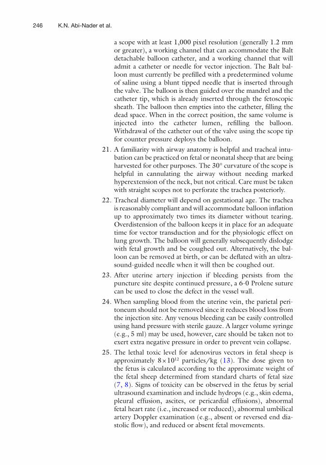
25. The lethal toxic level for adenovirus vectors in fetal sheep is approximately 8 × 10 12 particles/kg ( 13 ) . The dose given to the fetus is calculated according to the approximate weight of the fetal sheep determined from standard charts of fetal size ( 7, 8 ) . Signs of toxicity can be observed in the fetus by serial ultrasound examination and include hydrops (e.g., skin edema, pleural effusion, ascites, or pericardial effusions), abnormal fetal heart rate (i.e., increased or reduced), abnormal umbilical artery Doppler examination (e.g., absent or reversed end dia-stolic fl ow), and reduced or absent fetal movements.
24711 Animal Models for Prenatal Gene Therapy: The Sheep Model
26. We have found adenovirus vectors to provide short term and highly ef fi cient muscular gene transfer to fetal sheep. The vectors are tolerated to a high dose with minimal observed muscular damage on histological analysis ( 3, 5, 15 ) .
27. We observed that adeno-associated virus vectors gave long-term gene transfer (up to 6 months) to fetal sheep after intra-peritoneal delivery in early and late gestation ( 14 ) .
28. Other vector systems have not proved so useful. Gene transfer to fetal sheep muscle in vitro is achievable using retrovirus vec-tors ( 18 ) . We have found, however, that retrovirus vectors based on the Moloney Leukemia Virus (MLV) give poor gene transfer to the quadriceps of late gestation fetal sheep (120 days of gestation). This may re fl ect the relatively low dose that was applied (1 × 10 8 particles/kg fetus) to a relatively large muscle bulk at that gestation. Lentivirus vectors also seem to give poor gene transfer in the fetal sheep. Vectors based on human immunode fi ciency virus (HIV) or equine immune anemia virus (EIAV) and pseudotyped with VSV-G or Mokola gave no signi fi cant gene transfer to the fetal sheep muscle in early ges-tation when injected at a dose of 1 × 10 8 particles/kg fetus.
References
1. David AL, Peebles DM, Gregory L et al (2003) Percutaneous ultrasound-guided injection of the trachea in fetal sheep: a novel technique to target the fetal airways. Fetal Diagn Ther 18: 385–390
2. Peebles D, Gregory LG, David A et al (2004) Widespread and ef fi cient marker gene expres-sion in the airway epithelia of fetal sheep after minimally invasive tracheal application of recombinant adenovirus in utero . Gene Ther 11:70–78
3. David AL, Peebles DM, Gregory L et al (2006) Clinically applicable procedure for gene deliv-ery to fetal gut by ultrasound-guided gastric injection: toward prenatal prevention of early-onset intestinal diseases. Hum Gene Ther 17: 767–779
4. Davey MG, Hedrick HL, Bouchard S et al (2003) Temporary tracheal occlusion in fetal sheep with lung hypoplasia does not improve postnatal lung function. J Appl Physiol 94: 1054–1062
5. David AL, Cook T, Waddington S et al (2003) Ultrasound guided percutaneous delivery of ade-noviral vectors encoding the beta-galactosidase and human factor IX genes to early gestation fetal sheep in utero . Hum Gene Ther 14: 353–364
6. David AL, Weisz B, Gregory L et al (2006) Ultrasound-guided injection and occlusion of
the trachea in fetal sheep. Ultrasound Obstet Gynaecol 28:82–88
7. Barbera A, Jones OW, Zerbe GO et al (1995) Ultrasonographic assessment of fetal growth: comparison between human and ovine fetus. Am J Obstet Gynecol 173:1765–1769
8. Kelly RW, Newnham JP (1989) Estimation of gestational age in Merino ewes by ultrasound measurement of fetal head size. Aust J Agr Res 40:1293–1299
9. Wolfensohn S, Lloyd M (1998) Handbook of laboratory animal management and welfare. Blackwell, Oxford, pp 257–277
10. Taylor PM (1991) Anaesthesia in Sheep and Goats. In: Melling M, Alder M (eds) Sheep and goat practice. Saunders, London, pp 99–116
11. Newnham JP, Kelly RW, Boyne P, Reid SE (1989) Ultrasound guided blood sampling from fetal sheep. Aust J Agr Res 40:401–407
12. Joubert DM (1956) A study of prenatal growth and development in the sheep. J Agric Sci 47: 382–427
13. Themis M, Schneider H, Kiserud T et al (1999) Successful expression of galactosidase and factor IX transgenes in fetal and neonatal sheep after ultrasound-guided percutaneous adenovi-rus vector administration into the umbilical vein. Gene Ther 6:1239–1248

248 K.N. Abi-Nader et al.
14. David AL, McIntosh J, Peebles DM et al (2011) rAAV mediated in utero gene transfer gives therapeutic transgene expression in the sheep. Hum Gene Ther 22(4):419–426
15. Weisz B, David AL, Gregory LG et al (2005) Targeting the respiratory muscles of fetal sheep for prenatal gene therapy for Duchenne mus-cular dystrophy. Am J Obstet Gynecol 193: 1105–1109
16. Newnham JP, Kelly RW (1993) Ultrasound for research with foetal sheep. In: Neilson JP,
Chambers SE (eds) Obstetric Ultrasound I. Oxford Medical Publications, pp 203–222
17. Kohl T, Strumper D, Witteler R et al (2000) Fetoscopic direct fetal cardiac access in sheep: an important experimental milestone along the route to human fetal cardiac intervention. Circulation 102:1602–1604
18. John HA (1994) Variable ef fi ciency of retrovi-ral-mediated gene transfer into early-passage cultures of fetal lamb epithelial, mesenchymal, and neuroectodermal tissues. Hum Gene Ther 5:283–293