Embed Size (px)
Citation preview
From the Center for Brachial Plexus and Traumatic Nerve Injury,Surgery, New York, New York.
Received for publication July 28, 2013; accepted in revised form S
No benefits in any form have been received or will be receiveindirectly to the subject of this article.
Corresponding author: Scott W. Wolfe, MD, Center for Brachial PNerve Injury, Hospital for Special Surgery, 535 East 70th St., New [email protected].
0363-5023/13/-A--0001$36.00/0http://dx.doi.org/10.1016/j.jhsa.2013.09.042
SCIENTIFIC ARTICLE
Preoperative Donor Nerve Electromyography as a
Predictor of Nerve Transfer Outcomes
Joseph J. Schreiber, MD, Joseph H. Feinberg, MD, David J. Byun, BS,Steve K. Lee, MD, Scott W. Wolfe, MD
Purpose We hypothesized that health of the donor nerve and corresponding muscle, asassessed by electromyography (EMG), could predict the outcome of nerve transfer surgery.
Methods A retrospective review was performed to investigate outcomes of nerve transfers forelbow flexion and shoulder abduction. Motor strength was graded preoperatively and after aminimum 1-year follow-up. Preoperative EMG results were classified as functionally normalor affected based on motor unit recruitment pattern and correlated with follow-up motorstrength and range of motion.
Results Forty nerve transfers were identified: 27 were performed for elbow flexion and 13 forshoulder abduction. Overall, the 29 transfers in the normal EMG cohort showed significantlygreater postoperative improvement in motor strength (Medical Research Council grade0.2e4.1) than the 11 transfers in the affected EMG cohort (grade 0.0e3.0). In the shouldercohort, normal donor nerves resulted in greater strength (grade 4.0 vs. 2.4) and active motion(83� vs. 25�) compared with affected donor nerves. Double fascicular transfers with 2 normaldonor nerves demonstrated improved strength compared with double nerve transfers when 1donor nerve was affected (grade 4.5 vs. 3.2).
Conclusions Our findings demonstrate that a simple EMG classification that describes thequality of donor nerves can predict outcome as measured by postoperative motor strength andrange of motion. Preoperative EMG evaluation should be considered a valuable supple-mentary component of the donor nerve selection process when planning brachial plexusreconstruction. (J Hand Surg Am. 2013;-:-e-. Copyright � 2013 by the American Societyfor Surgery of the Hand. All rights reserved.)
Type of study/level of evidence Prognostic II.Keywords Nerve transfer, brachial plexus, electromyography.
B RACHIAL PLEXUS INJURIES ARE catastrophicevents that can produce debilitating limitationsof shoulder, elbow, and hand function.1,2 In
C5e6, or upper trunk injuries, restoration of elbow
Hospital for Special
eptember 26, 2013.
d related directly or
lexus and Traumaticrk, NY 10021; e-mail:
flexion is of primary importance, followed by theneed for shoulder stability, external rotation, andabduction.1,3e5 Whereas various interventions in-cluding nerve repair or nerve grafting have tradi-tionally played a central role in plexus reconstruction,nerve transfer has assumed increasing importancebecause of the relative short reinnervation distances,reliable and robust axon sources, and the steadilyexpanding variety of nerve transfer options availablefor these complex injuries.6e9
In 1994, Oberlin et al3 described a transfer usingfascicles of the ulnar nerve to the biceps motor branchto restore elbow flexion. This was followed by severalreports documenting restoration of biceps functionwithminimal ulnar nerve donor sitemorbidity.1,5,10e16
� 2013 ASSH r Published by Elsevier, Inc. All rights reserved. r 1
2 ELECTROMYOGRAPHY AND NERVE TRANSFER OUTCOMES
Besides addressing elbow flexion, restoring shoulderabduction and external rotation is critical to increasethe total functional sphere of the upper extremity.4
At a minimum, a stable glenohumeral joint is essen-tial to prevent dissipation of elbow flexion forceinto proximal humeral migration. A technique in-volving transfer of the long head triceps branch to thedeltoid branches of the axillary nerve gained popu-larity following early outstanding results.4 Whenperformed together, Oberlin and triceps-to-axillarynerve transfers are a reliable and effective techniquefor functional recovery of elbow flexion as well asshoulder abduction in C5e6 and upper trunk brachialplexus injuries.1,5,15,17
Several factors have been identified that maypredict success of nerve transfers including patientage,18e21 time from injury to surgery,14,18,21e23 bodymass index,18 and the number of active motor neu-rons in the donor fascicle.24,25
Electromyography (EMG) is often used beforesurgery to assess electrical conduction, muscledenervation, and the presence of functional motorunits in patients with brachial plexus injuries.26 Theuse of EMG during surgery has been explored tochoose the optimal donor fascicles, and motor out-comes have been correlated with evoked potentialpatterns.27
Preoperative EMG of potential donor nerves mayshow abnormalities in muscles that appear normalclinically. This presents a dilemma to the surgeon indetermining whether fascicles of the donor nervecould be transferred with expectation of success.Most authors recommend choosing donor nervesfrom muscles with clinically normal (British MedicalResearch Council [MRC] grade 5) or minimallyaffected (MRC grade 4 or 4þ). We hypothesized thathealth of the donor nerve and its correspondingmuscle, as assessed by EMG, could help predict theoutcome of nerve transfer surgery.
MATERIALS AND METHODSAn institutional review boardeapproved retrospec-tive review of our prospective Health InsurancePortability and Accountability Actecompliant trau-matic nerve injury registry was performed to inves-tigate the outcomes of 2 common nerve transfersperformed over the past decade: (1) ulnar or mediannerve fascicular branch transfer to the biceps or bra-chialis motor branches and (2) long or medial headtriceps branch transfer to motor branches of theaxillary nerve. A minimum of 1-year clinical follow-up after nerve transfer was required for inclusion in
J Hand Surg Am. r
the study. Exclusion criteria included abnormal clin-ical strength in the donor nerve distribution (<M4),incomplete preoperative EMG evaluation of both thedonor and the recipient nerves, or lack of 1-yearfollow-up. Forty such transfers were identified, with16 transfers to the biceps, 11 transfers to the bra-chialis, and 13 transfers to the deltoid.
All patients underwent detailed preoperative clin-ical examination including motor strength evaluationaccording to the modified MRC grading system,range of motion measurements, and sensory exami-nation. Throughout the study, MRC grading ofmuscles in the donor nerve distributions includedtriceps strength for radial nerve evaluation; flexorcarpi ulnaris muscle, ulnar portion of the flexor dig-itorum profundus, and first dorsal interosseous mus-cle for ulnar nerve evaluation; and flexor carpiradialis, pronator, flexor pollicis longus, and abductorpollicis brevis for median nerve evaluation. Preop-erative motor strength in the innervated muscles ofthe donor nerve distribution was required to be M4 orM5 for consideration of nerve transfer.
All donor nerves (ulnar, median, radial) wereevaluated before surgery by EMG. All EMG evalua-tions were performed and interpreted by a singleelectrodiagnostic expert. The donor nerve distributionEMG results were retrospectively classified as func-tionally normal or affected based on the recordedmotor unit recruitment patterns. Recruitment is anevaluation of functional motor units and is graded on a4-category scale: full, decreased, discrete, or none(Fig. 1). Full motor recruitment occurs when there iscomplete motor unit activation and the EMG screen isentirely filled. Decreased recruitment occurs whenthere is a large quantity of motor units firing, the entireEMG screen is not filled, but individual motor unitscannot be identified. Discrete recruitment patternshows a smaller quantity of motor units firing,resulting in individual motor units being identifiableduring maximum effort. We defined functionallynormal donor nerves as those that displayed either fullor decreased recruitment patterns in all of the muscleswithin the nerve’s innervation territory. Functionallyaffected donor nerves displayed a discrete pattern orcomplete lack of motor recruitment in some or all ofthe muscles within the nerve’s innervation territory.
The presence of abnormal spontaneous activityin the form of positive sharp waves and fibrillationswas also recorded on a scale of 0 to 4þ. Zero is theabsence of fibrillations or positive sharp waves, 1þ ispersistent/unsustained single trains in at least 2 muscleregions, 2þ is moderate numbers in 3 or more muscleareas, 3þ is many in all muscle regions, 4þ is an
Vol -, - 2013
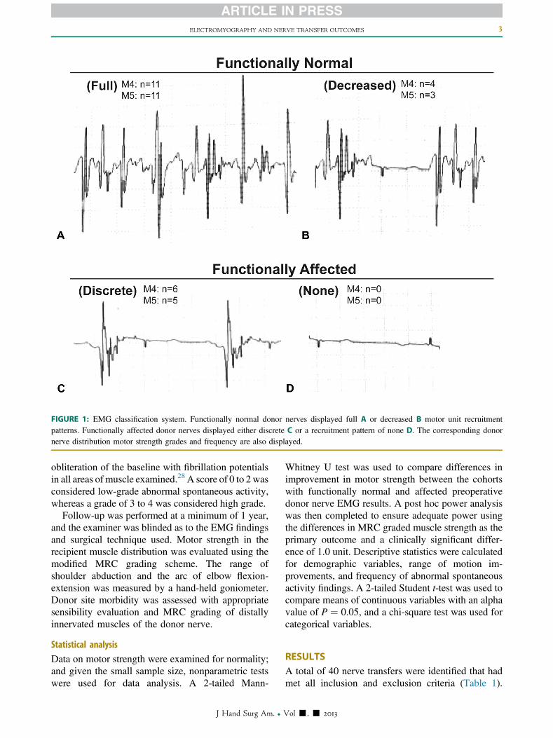
FIGURE 1: EMG classification system. Functionally normal donor nerves displayed full A or decreased B motor unit recruitmentpatterns. Functionally affected donor nerves displayed either discrete C or a recruitment pattern of none D. The corresponding donornerve distribution motor strength grades and frequency are also displayed.
ELECTROMYOGRAPHY AND NERVE TRANSFER OUTCOMES 3
obliteration of the baseline with fibrillation potentialsin all areas of muscle examined.28 A score of 0 to 2 wasconsidered low-grade abnormal spontaneous activity,whereas a grade of 3 to 4 was considered high grade.
Follow-up was performed at a minimum of 1 year,and the examiner was blinded as to the EMG findingsand surgical technique used. Motor strength in therecipient muscle distribution was evaluated using themodified MRC grading scheme. The range ofshoulder abduction and the arc of elbow flexion-extension was measured by a hand-held goniometer.Donor site morbidity was assessed with appropriatesensibility evaluation and MRC grading of distallyinnervated muscles of the donor nerve.
Statistical analysis
Data on motor strength were examined for normality;and given the small sample size, nonparametric testswere used for data analysis. A 2-tailed Mann-
J Hand Surg Am. r
Whitney U test was used to compare differences inimprovement in motor strength between the cohortswith functionally normal and affected preoperativedonor nerve EMG results. A post hoc power analysiswas then completed to ensure adequate power usingthe differences in MRC graded muscle strength as theprimary outcome and a clinically significant differ-ence of 1.0 unit. Descriptive statistics were calculatedfor demographic variables, range of motion im-provements, and frequency of abnormal spontaneousactivity findings. A 2-tailed Student t-test was used tocompare means of continuous variables with an alphavalue of P ¼ 0.05, and a chi-square test was used forcategorical variables.
RESULTSA total of 40 nerve transfers were identified that hadmet all inclusion and exclusion criteria (Table 1).
Vol -, - 2013
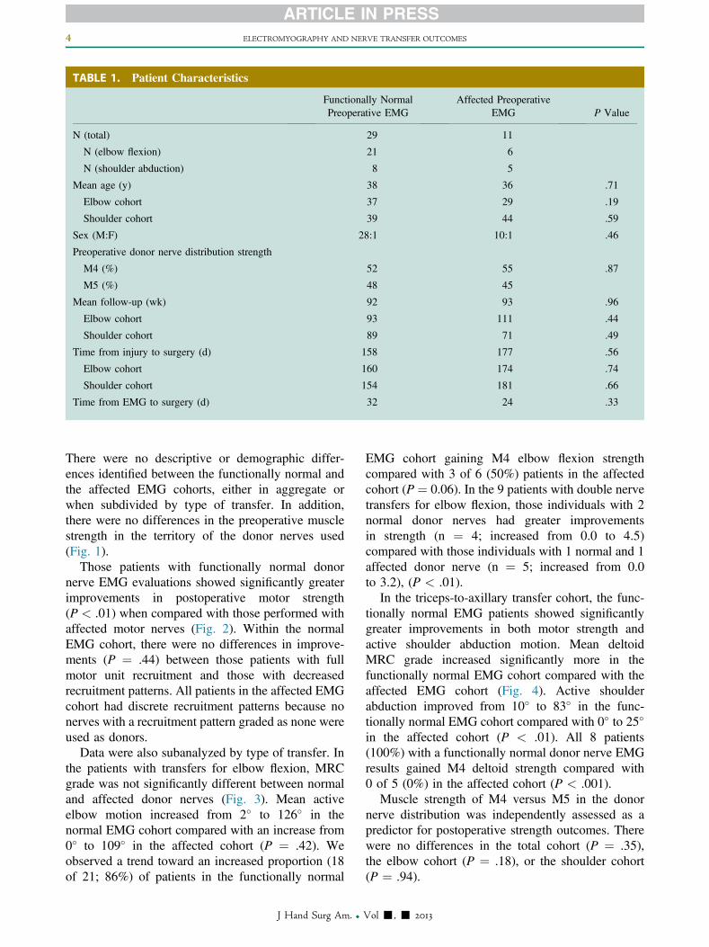
TABLE 1. Patient Characteristics
Functionally NormalPreoperative EMG
Affected PreoperativeEMG P Value
N (total) 29 11
N (elbow flexion) 21 6
N (shoulder abduction) 8 5
Mean age (y) 38 36 .71
Elbow cohort 37 29 .19
Shoulder cohort 39 44 .59
Sex (M:F) 28:1 10:1 .46
Preoperative donor nerve distribution strength
M4 (%) 52 55 .87
M5 (%) 48 45
Mean follow-up (wk) 92 93 .96
Elbow cohort 93 111 .44
Shoulder cohort 89 71 .49
Time from injury to surgery (d) 158 177 .56
Elbow cohort 160 174 .74
Shoulder cohort 154 181 .66
Time from EMG to surgery (d) 32 24 .33
4 ELECTROMYOGRAPHY AND NERVE TRANSFER OUTCOMES
There were no descriptive or demographic differ-ences identified between the functionally normal andthe affected EMG cohorts, either in aggregate orwhen subdivided by type of transfer. In addition,there were no differences in the preoperative musclestrength in the territory of the donor nerves used(Fig. 1).
Those patients with functionally normal donornerve EMG evaluations showed significantly greaterimprovements in postoperative motor strength(P < .01) when compared with those performed withaffected motor nerves (Fig. 2). Within the normalEMG cohort, there were no differences in improve-ments (P ¼ .44) between those patients with fullmotor unit recruitment and those with decreasedrecruitment patterns. All patients in the affected EMGcohort had discrete recruitment patterns because nonerves with a recruitment pattern graded as none wereused as donors.
Data were also subanalyzed by type of transfer. Inthe patients with transfers for elbow flexion, MRCgrade was not significantly different between normaland affected donor nerves (Fig. 3). Mean activeelbow motion increased from 2� to 126� in thenormal EMG cohort compared with an increase from0� to 109� in the affected cohort (P ¼ .42). Weobserved a trend toward an increased proportion (18of 21; 86%) of patients in the functionally normal
J Hand Surg Am. r
EMG cohort gaining M4 elbow flexion strengthcompared with 3 of 6 (50%) patients in the affectedcohort (P ¼ 0.06). In the 9 patients with double nervetransfers for elbow flexion, those individuals with 2normal donor nerves had greater improvementsin strength (n ¼ 4; increased from 0.0 to 4.5)compared with those individuals with 1 normal and 1affected donor nerve (n ¼ 5; increased from 0.0to 3.2), (P < .01).
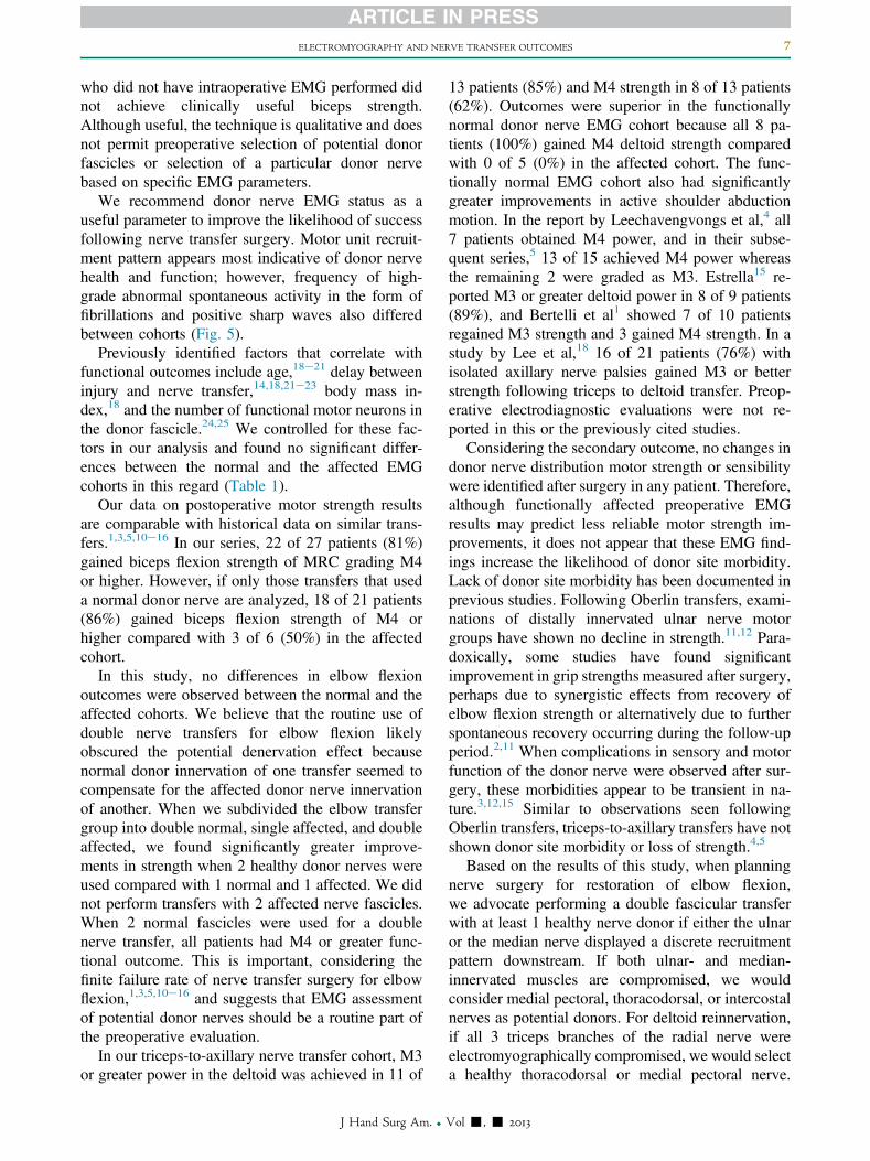
In the triceps-to-axillary transfer cohort, the func-tionally normal EMG patients showed significantlygreater improvements in both motor strength andactive shoulder abduction motion. Mean deltoidMRC grade increased significantly more in thefunctionally normal EMG cohort compared with theaffected EMG cohort (Fig. 4). Active shoulderabduction improved from 10� to 83� in the func-tionally normal EMG cohort compared with 0� to 25�
in the affected cohort (P < .01). All 8 patients(100%) with a functionally normal donor nerve EMGresults gained M4 deltoid strength compared with0 of 5 (0%) in the affected cohort (P < .001).
Muscle strength of M4 versus M5 in the donornerve distribution was independently assessed as apredictor for postoperative strength outcomes. Therewere no differences in the total cohort (P ¼ .35),the elbow cohort (P ¼ .18), or the shoulder cohort(P ¼ .94).
Vol -, - 2013
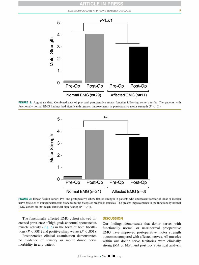
FIGURE 2: Aggregate data. Combined data of pre- and postoperative motor function following nerve transfer. The patients withfunctionally normal EMG findings had significantly greater improvements in postoperative motor strength (P < .01).
FIGURE 3: Elbow flexion cohort. Pre- and postoperative elbow flexion strength in patients who underwent transfer of ulnar or mediannerve fascicles to musculocutaneous branches to the biceps or brachialis muscles. The greater improvements in the functionally normalEMG cohort did not reach statistical significance (P ¼ .41).
ELECTROMYOGRAPHY AND NERVE TRANSFER OUTCOMES 5
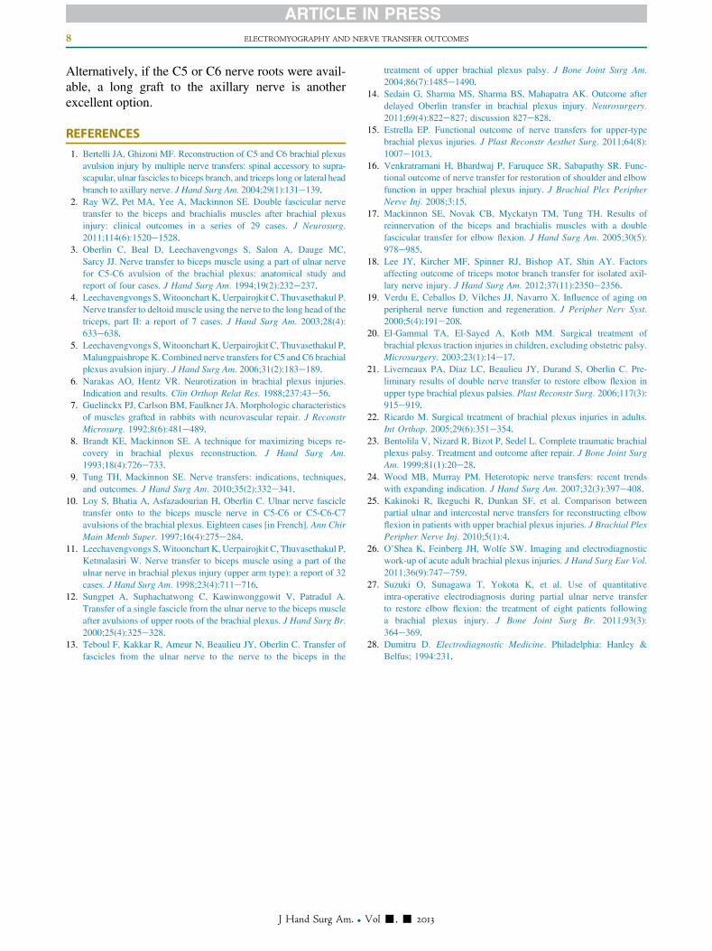
The functionally affected EMG cohort showed in-creased prevalence of high-grade abnormal spontaneousmuscle activity (Fig. 5) in the form of both fibrilla-tions (P < .001) and positive sharp waves (P < .001).
Postoperative clinical examination demonstratedno evidence of sensory or motor donor nervemorbidity in any patient.
J Hand Surg Am. r
DISCUSSIONOur findings demonstrate that donor nerves withfunctionally normal or near-normal preoperativeEMG have improved postoperative motor strengthoutcomes compared with affected nerves. All muscleswithin our donor nerve territories were clinicallystrong (M4 or M5), and post hoc statistical analysis
Vol -, - 2013
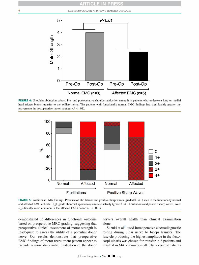
FIGURE 4: Shoulder abduction cohort. Pre- and postoperative shoulder abduction strength in patients who underwent long or medialhead triceps branch transfer to the axillary nerve. The patients with functionally normal EMG findings had significantly greater im-provements in postoperative motor strength (P < .01).
FIGURE 5: Additional EMG findings. Presence of fibrillations and positive sharp waves (graded 0e4þ) seen in the functionally normaland affected EMG cohorts. High-grade abnormal spontaneous muscle activity (grade 3e4þ fibrillations and positive sharp waves) weresignificantly more common in the affected EMG cohort (P < .001).
6 ELECTROMYOGRAPHY AND NERVE TRANSFER OUTCOMES
demonstrated no differences in functional outcomebased on preoperative MRC grading, suggesting thatpreoperative clinical assessment of motor strength isinadequate to assess the utility of a potential donornerve. Our results demonstrate that preoperativeEMG findings of motor recruitment pattern appear toprovide a more discernible evaluation of the donor
J Hand Surg Am. r
nerve’s overall health than clinical examinationalone.
Suzuki et al27 used intraoperative electrodiagnostictesting during ulnar nerve to biceps transfer. Thefascicle producing the highest amplitude in the flexorcarpi ulnaris was chosen for transfer in 6 patients andresulted in M4 outcomes in all. The 2 control patients
Vol -, - 2013
ELECTROMYOGRAPHY AND NERVE TRANSFER OUTCOMES 7
who did not have intraoperative EMG performed didnot achieve clinically useful biceps strength.Although useful, the technique is qualitative and doesnot permit preoperative selection of potential donorfascicles or selection of a particular donor nervebased on specific EMG parameters.
We recommend donor nerve EMG status as auseful parameter to improve the likelihood of successfollowing nerve transfer surgery. Motor unit recruit-ment pattern appears most indicative of donor nervehealth and function; however, frequency of high-grade abnormal spontaneous activity in the form offibrillations and positive sharp waves also differedbetween cohorts (Fig. 5).
Previously identified factors that correlate withfunctional outcomes include age,18e21 delay betweeninjury and nerve transfer,14,18,21e23 body mass in-dex,18 and the number of functional motor neurons inthe donor fascicle.24,25 We controlled for these fac-tors in our analysis and found no significant differ-ences between the normal and the affected EMGcohorts in this regard (Table 1).
Our data on postoperative motor strength resultsare comparable with historical data on similar trans-fers.1,3,5,10e16 In our series, 22 of 27 patients (81%)gained biceps flexion strength of MRC grading M4or higher. However, if only those transfers that useda normal donor nerve are analyzed, 18 of 21 patients(86%) gained biceps flexion strength of M4 orhigher compared with 3 of 6 (50%) in the affectedcohort.
In this study, no differences in elbow flexionoutcomes were observed between the normal and theaffected cohorts. We believe that the routine use ofdouble nerve transfers for elbow flexion likelyobscured the potential denervation effect becausenormal donor innervation of one transfer seemed tocompensate for the affected donor nerve innervationof another. When we subdivided the elbow transfergroup into double normal, single affected, and doubleaffected, we found significantly greater improve-ments in strength when 2 healthy donor nerves wereused compared with 1 normal and 1 affected. We didnot perform transfers with 2 affected nerve fascicles.When 2 normal fascicles were used for a doublenerve transfer, all patients had M4 or greater func-tional outcome. This is important, considering thefinite failure rate of nerve transfer surgery for elbowflexion,1,3,5,10e16 and suggests that EMG assessmentof potential donor nerves should be a routine part ofthe preoperative evaluation.
In our triceps-to-axillary nerve transfer cohort, M3or greater power in the deltoid was achieved in 11 of
J Hand Surg Am. r
13 patients (85%) and M4 strength in 8 of 13 patients(62%). Outcomes were superior in the functionallynormal donor nerve EMG cohort because all 8 pa-tients (100%) gained M4 deltoid strength comparedwith 0 of 5 (0%) in the affected cohort. The func-tionally normal EMG cohort also had significantlygreater improvements in active shoulder abductionmotion. In the report by Leechavengvongs et al,4 all7 patients obtained M4 power, and in their subse-quent series,5 13 of 15 achieved M4 power whereasthe remaining 2 were graded as M3. Estrella15 re-ported M3 or greater deltoid power in 8 of 9 patients(89%), and Bertelli et al1 showed 7 of 10 patientsregained M3 strength and 3 gained M4 strength. In astudy by Lee et al,18 16 of 21 patients (76%) withisolated axillary nerve palsies gained M3 or betterstrength following triceps to deltoid transfer. Preop-erative electrodiagnostic evaluations were not re-ported in this or the previously cited studies.
Considering the secondary outcome, no changes indonor nerve distribution motor strength or sensibilitywere identified after surgery in any patient. Therefore,although functionally affected preoperative EMGresults may predict less reliable motor strength im-provements, it does not appear that these EMG find-ings increase the likelihood of donor site morbidity.Lack of donor site morbidity has been documented inprevious studies. Following Oberlin transfers, exami-nations of distally innervated ulnar nerve motorgroups have shown no decline in strength.11,12 Para-doxically, some studies have found significantimprovement in grip strengths measured after surgery,perhaps due to synergistic effects from recovery ofelbow flexion strength or alternatively due to furtherspontaneous recovery occurring during the follow-upperiod.2,11 When complications in sensory and motorfunction of the donor nerve were observed after sur-gery, these morbidities appear to be transient in na-ture.3,12,15 Similar to observations seen followingOberlin transfers, triceps-to-axillary transfers have notshown donor site morbidity or loss of strength.4,5
Based on the results of this study, when planningnerve surgery for restoration of elbow flexion,we advocate performing a double fascicular transferwith at least 1 healthy nerve donor if either the ulnaror the median nerve displayed a discrete recruitmentpattern downstream. If both ulnar- and median-innervated muscles are compromised, we wouldconsider medial pectoral, thoracodorsal, or intercostalnerves as potential donors. For deltoid reinnervation,if all 3 triceps branches of the radial nerve wereelectromyographically compromised, we would selecta healthy thoracodorsal or medial pectoral nerve.
Vol -, - 2013
8 ELECTROMYOGRAPHY AND NERVE TRANSFER OUTCOMES
Alternatively, if the C5 or C6 nerve roots were avail-able, a long graft to the axillary nerve is anotherexcellent option.
REFERENCES
1. Bertelli JA, Ghizoni MF. Reconstruction of C5 and C6 brachial plexusavulsion injury by multiple nerve transfers: spinal accessory to supra-scapular, ulnar fascicles to biceps branch, and triceps long or lateral headbranch to axillary nerve. J Hand Surg Am. 2004;29(1):131e139.
2. Ray WZ, Pet MA, Yee A, Mackinnon SE. Double fascicular nervetransfer to the biceps and brachialis muscles after brachial plexusinjury: clinical outcomes in a series of 29 cases. J Neurosurg.2011;114(6):1520e1528.
3. Oberlin C, Beal D, Leechavengvongs S, Salon A, Dauge MC,Sarcy JJ. Nerve transfer to biceps muscle using a part of ulnar nervefor C5-C6 avulsion of the brachial plexus: anatomical study andreport of four cases. J Hand Surg Am. 1994;19(2):232e237.
4. Leechavengvongs S,Witoonchart K, Uerpairojkit C, Thuvasethakul P.Nerve transfer to deltoid muscle using the nerve to the long head of thetriceps, part II: a report of 7 cases. J Hand Surg Am. 2003;28(4):633e638.
5. Leechavengvongs S,Witoonchart K, Uerpairojkit C, Thuvasethakul P,Malungpaishrope K. Combined nerve transfers for C5 and C6 brachialplexus avulsion injury. J Hand Surg Am. 2006;31(2):183e189.
6. Narakas AO, Hentz VR. Neurotization in brachial plexus injuries.Indication and results. Clin Orthop Relat Res. 1988;237:43e56.
7. Guelinckx PJ, Carlson BM, Faulkner JA. Morphologic characteristicsof muscles grafted in rabbits with neurovascular repair. J ReconstrMicrosurg. 1992;8(6):481e489.
8. Brandt KE, Mackinnon SE. A technique for maximizing biceps re-covery in brachial plexus reconstruction. J Hand Surg Am.1993;18(4):726e733.
9. Tung TH, Mackinnon SE. Nerve transfers: indications, techniques,and outcomes. J Hand Surg Am. 2010;35(2):332e341.
10. Loy S, Bhatia A, Asfazadourian H, Oberlin C. Ulnar nerve fascicletransfer onto to the biceps muscle nerve in C5-C6 or C5-C6-C7avulsions of the brachial plexus. Eighteen cases [in French]. Ann ChirMain Memb Super. 1997;16(4):275e284.
11. Leechavengvongs S,Witoonchart K, Uerpairojkit C, Thuvasethakul P,Ketmalasiri W. Nerve transfer to biceps muscle using a part of theulnar nerve in brachial plexus injury (upper arm type): a report of 32cases. J Hand Surg Am. 1998;23(4):711e716.
12. Sungpet A, Suphachatwong C, Kawinwonggowit V, Patradul A.Transfer of a single fascicle from the ulnar nerve to the biceps muscleafter avulsions of upper roots of the brachial plexus. J Hand Surg Br.2000;25(4):325e328.
13. Teboul F, Kakkar R, Ameur N, Beaulieu JY, Oberlin C. Transfer offascicles from the ulnar nerve to the nerve to the biceps in the
J Hand Surg Am. r
treatment of upper brachial plexus palsy. J Bone Joint Surg Am.2004;86(7):1485e1490.
14. Sedain G, Sharma MS, Sharma BS, Mahapatra AK. Outcome afterdelayed Oberlin transfer in brachial plexus injury. Neurosurgery.2011;69(4):822e827; discussion 827e828.
15. Estrella EP. Functional outcome of nerve transfers for upper-typebrachial plexus injuries. J Plast Reconstr Aesthet Surg. 2011;64(8):1007e1013.
16. Venkratramani H, Bhardwaj P, Faruquee SR, Sabapathy SR. Func-tional outcome of nerve transfer for restoration of shoulder and elbowfunction in upper brachial plexus injury. J Brachial Plex PeripherNerve Inj. 2008;3:15.
17. Mackinnon SE, Novak CB, Myckatyn TM, Tung TH. Results ofreinnervation of the biceps and brachialis muscles with a doublefascicular transfer for elbow flexion. J Hand Surg Am. 2005;30(5):978e985.
18. Lee JY, Kircher MF, Spinner RJ, Bishop AT, Shin AY. Factorsaffecting outcome of triceps motor branch transfer for isolated axil-lary nerve injury. J Hand Surg Am. 2012;37(11):2350e2356.
19. Verdu E, Ceballos D, Vilches JJ, Navarro X. Influence of aging onperipheral nerve function and regeneration. J Peripher Nerv Syst.2000;5(4):191e208.
20. El-Gammal TA, El-Sayed A, Kotb MM. Surgical treatment ofbrachial plexus traction injuries in children, excluding obstetric palsy.Microsurgery. 2003;23(1):14e17.
21. Liverneaux PA, Diaz LC, Beaulieu JY, Durand S, Oberlin C. Pre-liminary results of double nerve transfer to restore elbow flexion inupper type brachial plexus palsies. Plast Reconstr Surg. 2006;117(3):915e919.
22. Ricardo M. Surgical treatment of brachial plexus injuries in adults.Int Orthop. 2005;29(6):351e354.
23. Bentolila V, Nizard R, Bizot P, Sedel L. Complete traumatic brachialplexus palsy. Treatment and outcome after repair. J Bone Joint SurgAm. 1999;81(1):20e28.
24. Wood MB, Murray PM. Heterotopic nerve transfers: recent trendswith expanding indication. J Hand Surg Am. 2007;32(3):397e408.
25. Kakinoki R, Ikeguchi R, Dunkan SF, et al. Comparison betweenpartial ulnar and intercostal nerve transfers for reconstructing elbowflexion in patients with upper brachial plexus injuries. J Brachial PlexPeripher Nerve Inj. 2010;5(1):4.
26. O’Shea K, Feinberg JH, Wolfe SW. Imaging and electrodiagnosticwork-up of acute adult brachial plexus injuries. J Hand Surg Eur Vol.2011;36(9):747e759.
27. Suzuki O, Sunagawa T, Yokota K, et al. Use of quantitativeintra-operative electrodiagnosis during partial ulnar nerve transferto restore elbow flexion: the treatment of eight patients followinga brachial plexus injury. J Bone Joint Surg Br. 2011;93(3):364e369.
28. Dumitru D. Electrodiagnostic Medicine. Philadelphia: Hanley &Belfus; 1994:231.
Vol -, - 2013