Embed Size (px)
Citation preview

Malaysian Journal of Analytical Sciences, Vol 21 No 2 (2017): 365 - 371
DOI: https://doi.org/10.17576/mjas-2017-2102-11
365
MALAYSIAN JOURNAL OF ANALYTICAL SCIENCES
Published by The Malaysian Analytical Sciences Society
PREPARATION AND CHARACTERIZATION OF DIFFERENT LOADING OF
ZINC OXIDE ON ACTIVATED CARBON NANOFIBERS
(Penyediaan dan Pencirian Gentian-Nano Karbon Teraktif Pada Kepekatan Zink Oksida Yang
Berbeza)
Faten Ermala Che Othman, Norhaniza Yusof*, Amirul Afiat Raffi, Hasrinah Hasbullah, Farhana Aziz,
Wan Norharyati Wan Salleh, Ahmad Fauzi Ismail
Advanced Membrane Technology Research Centre,
Faculty of Chemical & Energy Engineering,
Universiti Teknologi Malaysia, 81310 Skudai, Johor Bahru, Malaysia
*Corresponding author: [email protected]
Received: 26 August 2016; Accepted: 8 January 2017
Abstract
The study deals on the modified PAN-based activated carbon nanofibers (ACNFs) embedded with different amount of zinc
oxides (ZnO) (0, 5, 10, and 15% relative to PAN wt.) to be used as adsorbents for natural gas adsorption. The nanofibers (NFs)
were successfully fabricated via electrospinning process at optimize parameters. The resultant NFs underwent three steps of
pyrolysis process which are stabilization, carbonization and activation at optimum parameters. The morphological structure and
diameter of pure and modified ACNFs were characterized using SEM while the existences of chemical bonds were analyzed by
FTIR analysis. XRD analysis was done to identify the crystallinity of the ACNFs. BET method was used to identify the specific
surface area (SSA) and nitrogen adsorption isotherm of the samples. The results showed that the SSA of ACNF5 (163.04 m2/g) is
significantly higher compared to the pristine and other modified ACNFs, nevertheless the obtained results is much lower
compared to average theoretical value. SEM micrograph depicted that all ACNF samples possessed average diameter of 300 –
500 nm with smooth and aligned structure. The presence of white spots as ZnO alongside the NFs has been confirmed with FTIR
and XRD analysis. From these findings, it is believed that ACNFs/ZnO will become a new adsorbent with great potential for gas
adsorption and storage in the near future applications.
Keywords: activated carbon nanofibers, zinc oxide, polyacrylonitrile, specific surface area
Abstrak
Kajian ini membincangkan tentang gentian-nano karbon teraktif (ACNFs) terubah suai berasaskan polimer PAN yang
digabungkan dengan kandungan zink oksida (ZnO) yang berbeza (0, 5, 10, dan 15% berdasarkan berat PAN) untuk digunakan
sebagai penjerap dalam penjerapan gas asli. Gentian-nano (NFs) telah berjaya direka melalui proses putaran elektro
menggunakan parameter-paramter optimum daripada kajian terdahulu. Kemudian, NFs yang terbentuk akan melalui tiga
peringkat proses pirolisis iaitu penstabilan, karbonisasi, dan pengaktifan menggunakan parameter optimum daripada kajian
terdahulu. Struktur morfologi dan diameter ACNFs tulen dan yang telah diubahsuai dengan ZnO telah dicirikan menggunakan
SEM manakala kewujudan ikatan kimia telah dianalisis menggunakan FTIR. Selain itu, untuk mengenalpasti penghabluran
ACNFs yang terhasil, analisis yang dikenali sebagai XRD telah dijalankan. Kaedah BET pula dijalankan untuk mengenal pasti
luas permukaan tertentu (SSA) dan isoterma penjerapan nitrogen. Hasil kajian menunjukkan bahawa SSA sampel ACNF5
(163,04 m2/g) adalah lebih tinggi berbanding dengan ACNFs tulen atau ACNFs yang telah diubahsuai yang lain..Walau
bagaimanapun, keputusan yang diperoleh menunjukkan SSA yang jauh lebih rendah berbanding dengan nilai teori purata.
Melalui SEM mikrograf, semua sampel ACNFs yang dihasilkan melalui kajian ini memiliki diameter purata dari 300 hingga 500
nm dengan struktur licin dan sejajar. Kehadiran bintik putih sebagai ZnO telah disahkan melalui analisis FTIR dan XRD.
ISSN
1394 - 2506

Faten Ermala et al: PREPARATION AND CHARACTERIZATION OF DIFFERENT LOADING OF ZINC
OXIDE ON ACTIVATED CARBON NANOFIBERS
366
Penemuan ini membuktikan bahawa ACNFs/ZnO akan menjadi penjerap baru yang mempunyai potensi besar dalam aplikasi
penjerapan dan penyimpanan gas pada masa hadapan.
Kata kunci: gentian karbon teraktif, zink oksida, poliakrilonitril, luas permukaan spesifik
Introduction
Activated carbon (AC) has been widely utilized as adsorbent in various applications in recent years included in
water or gas adsorption due to their porous structure, high specific area, and cost-friendly [1]. Currently, AC can be
developed by using various types of precursors such as pitch, lignin, polymers, and also synthetic precursor,
however polyacrylonitrile (PAN)-based adsorbents have become an attractive alternative as PAN precursor could
produce high carbon yield up to 56% as compared to the other precursors [2]. However, a new enhanced material
known as activated carbon nanofibers (ACNFs) with highly microporous structure with finer structure and greater
specific surface area [3] have been developed in order to overcome the disadvantages of AC that possessed low SSA
and limited the gas adsorption capabilities. Ascribed by its low resistance to bulk flow, high adsorption-desorption
kinetics, heat stability, and high strength; PAN-based ACNFs have received major attention from researcher all over
the globe and were studied for their potential as excellent gas adsorbents [4].
Nanofibers (NFs) can be fabricated by various techniques included template synthesis, phase separation,
electrospinning, and self-assembly [5]. Out of those available techniques, electrospinning seems to produce NFs
with smaller diameter with uniform distribution structures. Moreover, electrospinning offers more advantages than
other techniques such as versatility, efficiency, and feasibility in large quantities production. Recently, in order to
improvised the NFs structures, the electrospun NFs were subjected to activation process to create new macropores
and micropores structures, with smaller fiber diameter and greater SSA. Furthermore, incorporation of additives
such as metal oxide into the NFs was proven can enhance the structure of the NFs. Metal oxide nanoparticles such
as magnesium oxide (MgO), manganese dioxide (MnO2), zinc oxide (ZnO), and nickel oxide (NiO) have been
profound to be as excellent candidate that could enhance the physicochemical properties of PAN-based ACNFs due
to their catalytic effects [6]. These nanoparticles themselves are high in SSA and the addition of right amount of
these nanoparticles into the NFs solution are believed could improve their structure as well as adsorption
capabilities.
In this study, the ACNFs embedded with ZnO were produced through suitable electrospinning and activation
method under optimum parameters obtained from previous studies [7]. The objective of this present study is to
prepare the ACNFs incorporated with ZnO and to study their microstructural properties and surface area for suitable
gas adsorbent materials.
Materials and Methods
Nanofibers fabrication
The polymer polyacrynitrile (PAN) and solvent N, N-dimethylformamide (DMF) were purchased from Sigma-
Aldrich while zinc oxide (ZnO) powder was purchased from Alfa Aesar. Homogenous DMF solutions of PAN
containing 0, 5, 10, and 15% (relative to PAN wt.) of ZnO were stirred gently for 24 hours at room temperature. The
nanofibers (NFs) were prepared by using a lab-scale electrospinning machine NFOM1000-1503 supplied from
Nfiber at optimum voltage of 12.4kV, the distance between the needle tip to collector of 20 cm, and flow rate of 1
ml/hour. The resultant NFs with 0, 5, 10, and 15% of ZnO were denoted as NF0, NF5, NF10, and NF15,
respectively.
Activation of nanofibers
The resultant NFs were underwent three stages of pyrolysis process. Start with stabilization with pure air until the
temperature of 275 °C, followed by carbonization under inert air condition until 600
°C, and finally activation with
activating gas such as carbon dioxide until 800 °C, in order to improve the porous structure of the NFs and directly
increasing the specific surface area with 30 minutes of resting time for each stage. After activation, the NFs were
known as activated carbon nanofibers (ACNFs). In this study, ACNFs with 0, 5, 10, and 15% of ZnO were denoted
as ACNF0, ACNF5, ACNF10, and ACNF15, individually.

Malaysian Journal of Analytical Sciences, Vol 21 No 2 (2017): 365 - 371
DOI: https://doi.org/10.17576/mjas-2017-2102-11
367
Characterization
In order to better understanding of the PAN-based ACNF/ZnO produced, there are several characterization analyses
have been conducted. The morphology and diameter of the NFs have been characterized using field emission
scanning electron microscopy (FESEM) while the elemental analysis and chemical studies of the NFs were
analyzed by using X-ray diffraction (XRD) analysis and Frontier Transform Infrared (FTIR) analysis, respectively.
Moreover, Brunauer–Emmett–Teller (BET) analysis was used in order to study specific surface area and pore size
distribution of all the resultant NFs. The surface area and microspore analysis of NFs and ACNFs were investigated
using nitrogen gas (N2) adsorption isotherm at 77 K (ASAP2020, Micromeritics, USA).
Results and Discussion
Morphological studies of nanofibers
Figure 1 shows the SEM images of PAN-based ACNF/ZnO with different loading of ZnO prior activation. It can be
seen that the all NFs possessed smooth, straight, and aligned structure with several white spots attached alongside
the fibers (Figure 1b, c, d) that represent the existence of ZnO. The fibers diameter was varied in all concentrations.
From the figure, it obviously can be seen that the pristine NFs possessed the largest fiber diameter as compared to
NFs embedded with ZnO. It was found that addition of ZnO has reduced the diameter of the NFs up to 45%, with
average diameter of NF0 is 576.8 ± 55.91 nm and was reduced to 325 ± 43.1 nm in all modified NFs (NFs
embedded with ZnO).
After activation, the NFs were transformed into activated carbon nanofibers (ACNFs) and the SEM photographs of
all pristine and modified ACNFs are shown in Figure 2. It can be seen that the ACNFs possessed rougher, irregular,
and undulated structure compared to NFs prior activation. The average diameter of the all NFs also decreased up to
40% from 450 nm to 270 nm due to the removal of PAN and other molecules during the heat treatment resulting in
weight loss and NFs’ diameter reduction [8, 9]. As shown in Figure 2(b), ACNF5 possessed the finest fiber structure
with well-dispersed and a few agglomeration of ZnO alongside/inside the ACNFs as compared to the others.
Figure 1. SEM micrograph of nanofibers with different loading of zinc oxide prior activation (a) NF0, (b) NF5, (c)
NF10 at 2500x magnification and (d) NF15 at 1500x magnification

Faten Ermala et al: PREPARATION AND CHARACTERIZATION OF DIFFERENT LOADING OF ZINC
OXIDE ON ACTIVATED CARBON NANOFIBERS
368
Figure 2. SEM micrograph of nanofibers with different loading of zinc oxide after activation (a) ACNF0, (b)
ACNF5, (c) ACNF10, and (d) ACNF15 at 2500x magnification
Although this result counterback the result of previous research conducted by Dadvar et al. [6], as they said the
increasing the amount of metal oxide loaded, the smaller the diameter of the NFs; this study has concluded that NFs
were only affected by the amount of metal oxide, which is up to its maximum loading depends on the type of the
metal oxide used in order to produce NFs with better structure. This phenomenon is believed could start as early as
during the dope preparation and fiber preparation stages as the amount and types of the metal oxide used can affect
the viscoelasticity of the solution and varies the ejection time of jet to arrive at the collector and these behaviors are
believed to give impact to the diameter of the NFs [10].
Chemical bond studies
Figure 3 shows the FTIR spectra for NFs embedded with 0 and 5% of ZnO prior and after activation. As can be
seen, there are various peaks appeared in the NFs prior activation (Figure 3a) and some of the peaks were removed
after activation due to heat treatment (Figure 3b), and it is believed only carbon and hydrogen peaks were left.
Theoretically, numerous new transition structures were formed during pyrolysis. As can be seen in Figure 3(a),
there are five major peaks can be detected in both NF0 and NF5 that represent different vibrations existed in the
NFs prior activation. However, there is one high peak was detected at wavenumber 445.7 cm-1
in NF5 that
represents Zn-O vibrations [11, 12]. Nevertheless, the appearance of unknown peak around wavenumber 400 – 700
cm-1
in Figure 3 is believed due to the existence of other vibrations that overlapped at the same wavenumber range
such as C-C, C-N, and C-O. After activation, there are only three major peaks were detected at 1285.5, 1433.1, and
1581.7 cm-1
represent C=O, C-H, and C=C vibrations, respectively [13].

Malaysian Journal of Analytical Sciences, Vol 21 No 2 (2017): 365 - 371
DOI: https://doi.org/10.17576/mjas-2017-2102-11
369
Figure 3. FTIR spectra of NF0 and NF5 (a) prior activation and (b) after activation
Elemental analysis
The crystallographical behaviors of both NF0 and NF5 prior and after activation have been analyzed by using X-ray
diffraction analysis as shown in Figure 4. It can be seen that prior activation, the structure of the NFs is amorphous
and after activation, the structure become more crystalline. As shown in Figure 4(a), the obvious peak detected at
16.7o in NF0 represents the polymer PAN peak [12] while the other two sharp peaks that only appeared in NF5 at
31.85 and 36.3o represents the ZnO peaks. However, in Figure 4(b), the polymer peak changed to carbon peak after
activation and it was detected at 13.2o [13]. As detected in Figure 4(a), the same ZnO peaks were also observed in
ACNF5 with addition of two other ZnO peaks at 34.6 and 47.55o [14 – 16]. It is believed these two peaks already
appeared in the NF5; however it cannot be seen due to amorphous structure of the NFs.
Figure 4. X-ray diffractogram of NF0 and NF5 (a) prior activation and (b) after activation
BET surface area
Brunauer–Emmett–Teller (BET) analysis was used to determine the specific surface area (SSA) of all resultant NFs
as depicted in Table 1. Prior activation, the addition of low or high concentration of ZnO bring no effect to the
enhancement of SSA of the NFs (see data in Table 1), in contrast it shows pristine NFs possessed the largest SSA.
Faten Ermala et al: PREPARATION AND CHARACTERIZATION OF DIFFERENT LOADING OF ZINC
OXIDE ON ACTIVATED CARBON NANOFIBERS
370
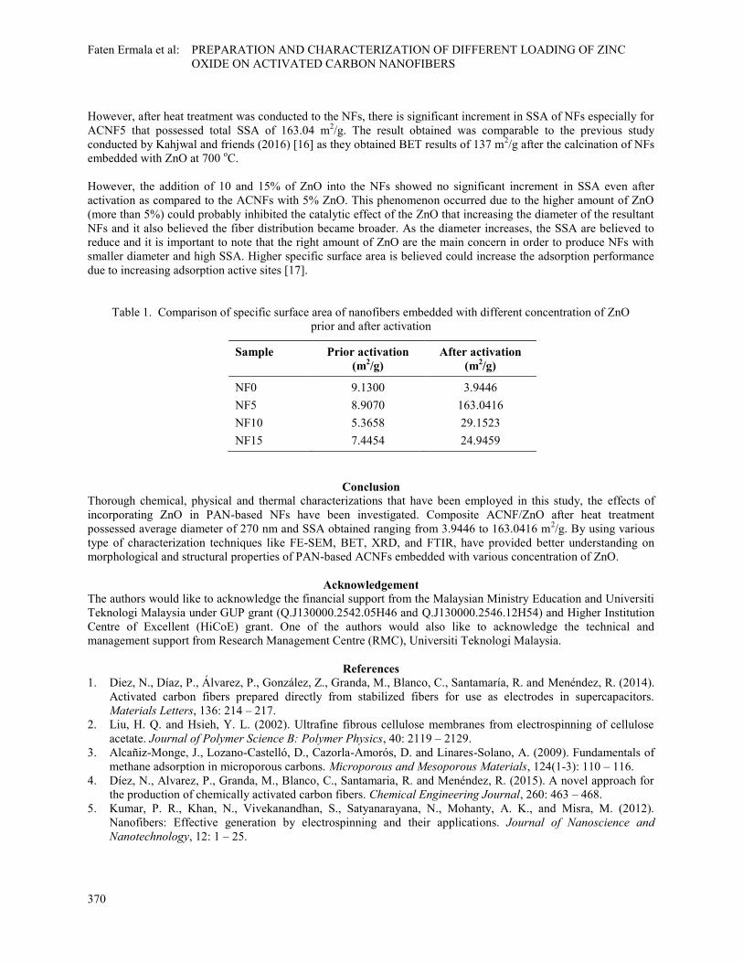
However, after heat treatment was conducted to the NFs, there is significant increment in SSA of NFs especially for
ACNF5 that possessed total SSA of 163.04 m2/g. The result obtained was comparable to the previous study
conducted by Kahjwal and friends (2016) [16] as they obtained BET results of 137 m2/g after the calcination of NFs
embedded with ZnO at 700 oC.
However, the addition of 10 and 15% of ZnO into the NFs showed no significant increment in SSA even after
activation as compared to the ACNFs with 5% ZnO. This phenomenon occurred due to the higher amount of ZnO
(more than 5%) could probably inhibited the catalytic effect of the ZnO that increasing the diameter of the resultant
NFs and it also believed the fiber distribution became broader. As the diameter increases, the SSA are believed to
reduce and it is important to note that the right amount of ZnO are the main concern in order to produce NFs with
smaller diameter and high SSA. Higher specific surface area is believed could increase the adsorption performance
due to increasing adsorption active sites [17].
Table 1. Comparison of specific surface area of nanofibers embedded with different concentration of ZnO
prior and after activation
Sample Prior activation
(m2/g)
After activation
(m2/g)
NF0 9.1300 3.9446
NF5 8.9070 163.0416
NF10 5.3658 29.1523
NF15 7.4454 24.9459
Conclusion
Thorough chemical, physical and thermal characterizations that have been employed in this study, the effects of
incorporating ZnO in PAN-based NFs have been investigated. Composite ACNF/ZnO after heat treatment
possessed average diameter of 270 nm and SSA obtained ranging from 3.9446 to 163.0416 m2/g. By using various
type of characterization techniques like FE-SEM, BET, XRD, and FTIR, have provided better understanding on
morphological and structural properties of PAN-based ACNFs embedded with various concentration of ZnO.
Acknowledgement
The authors would like to acknowledge the financial support from the Malaysian Ministry Education and Universiti
Teknologi Malaysia under GUP grant (Q.J130000.2542.05H46 and Q.J130000.2546.12H54) and Higher Institution
Centre of Excellent (HiCoE) grant. One of the authors would also like to acknowledge the technical and
management support from Research Management Centre (RMC), Universiti Teknologi Malaysia.
References
1. Diez, N., Díaz, P., Álvarez, P., González, Z., Granda, M., Blanco, C., Santamaría, R. and Menéndez, R. (2014).
Activated carbon fibers prepared directly from stabilized fibers for use as electrodes in supercapacitors.
Materials Letters, 136: 214 – 217.
2. Liu, H. Q. and Hsieh, Y. L. (2002). Ultrafine fibrous cellulose membranes from electrospinning of cellulose
acetate. Journal of Polymer Science B: Polymer Physics, 40: 2119 – 2129.
3. Alcañiz-Monge, J., Lozano-Castelló, D., Cazorla-Amorós, D. and Linares-Solano, A. (2009). Fundamentals of
methane adsorption in microporous carbons. Microporous and Mesoporous Materials, 124(1-3): 110 – 116.
4. Díez, N., Alvarez, P., Granda, M., Blanco, C., Santamaria, R. and Menéndez, R. (2015). A novel approach for
the production of chemically activated carbon fibers. Chemical Engineering Journal, 260: 463 – 468.
5. Kumar, P. R., Khan, N., Vivekanandhan, S., Satyanarayana, N., Mohanty, A. K., and Misra, M. (2012).
Nanofibers: Effective generation by electrospinning and their applications. Journal of Nanoscience and
Nanotechnology, 12: 1 – 25.

Malaysian Journal of Analytical Sciences, Vol 21 No 2 (2017): 365 - 371
DOI: https://doi.org/10.17576/mjas-2017-2102-11
371
6. Dadvar, S., Tavanai, H. and Morshed, M. (2012). Effect of embedding MgO and Al2O3 nanoparticles in the
precursor on the pore characteristics of PAN based activated carbon nanofibers. Journal of Analytical and
Applied Pyrolysis, 98: 98 – 105.
7. Bhardwaj, N. and Kundu, S. C. (2010). Electrospinning: A fascinating fiber fabrication technique.
Biotechnology Advances, 28: 325 – 347.
8. Venugopal, J., Zhang, Y. Z. and Ramakrishna, S. (2004). Electrospun nanofibres: biomedical applications.
Proceedings of the Institution of Mechanical Engineers, Part N: Journal of Nanomaterials, Nanoengineering
and Nanosystems, 218(1): 35 – 45.
9. Khalil, K. A., Sherif, E. M., Nabawy, A. M., Abdo, H. S., Marzouk, W. W. and Alharbi, H. F. (2016). Titanium
carbide nanofibers-reinforced aluminium compacts, a new strategy to enhance mechanical properties.
Materials, 9(5): 399 – 413.
10. Haroosh, H. J., Dong, Y., Chaudhary, D. S., Ingram, G. D. and Yusa, S. I. (2013). Electrospun PLA/PCL
composites embedded with unmodified and 3-aminopropyltriethoxysilane (ASP) modified halleysite nanotubes
(HNT). Applied Physics A: Materials Science and Processing, 110(2): 433 – 442.
11. Harish, K. and Renu, R. (2013). Structural and optical characterization of ZnO nanoparticles synthesized by
microemulsion route. International Letters of Chemistry, Physical and Astronomy, 19: 26 – 36.
12. Sun, M., Lan, B., Lin, T., Cheng, G., Ye, F., Yu, L., Cheng, X. and Zheng, X. (2013). Controlled synthesis of
nanostructured manganese oxide: crystalline evolution and catalytic activities. CrystEngComm, 15(35): 7010 –
7018.
13. Yusof, N. and Ismail, A. F. (2012). Post spinning and pyrolysis processes of polyacrylonitrile (PAN)-based
carbon fiber and activated carbon fiber: A review. Journal of Analytical and Applied Pyrolysis, 93: 1 – 13.
14. Babu, K. S., Reddy, A. R., Sujatha, C., Reddy, K. V. and Mallika, A. N. (2013). Synthesis and optical
characterization of porous ZnO. Journal of Advanced Ceramics, 2(3): 260 – 265.
15. Cipriani, E., Zanetti, M., Bracco, P., Brunella, V., Luda, M. P. and Costa, L. (2015) Crosslinking and
carbonization processes in PAN films and nanofibers. Polymer Degradation and Stability, 123: 178 –188.
16. Kanjwal, M. A., Barakat, N. A., Sheikh, F. A., Park, D. K. and Kim, H. Y. (2010). Physicochemical
characterizations of electrospun (ZnO-GeO2) nanofibers and their optical properties. Journal of Materials
Science, 45(14): 3833 – 3840.
17. Im, J. S., Park, S. J., Kim, T. J., Kim, Y. H. and Lee, Y. S. (2009). The study of controlling pore size on
electrospun carbon nanofibers for hydrogen adsorption. Journal of Colloid and Interface Science, 318 (1): 42 –
49.