Embed Size (px)
Citation preview

Iraqi J Pharm Sci, Vol.27(1) 2018 Domperidone nanoparticles DOI: http://dx.doi.org/10.31351/vol27iss1pp39-52
39
Preparation and Characterization of Domperidone Nanoparticles for
Dissolution Improvement Malath H. Oudah*, Firas A. Rahi** and Mohammed S. Al-lami***,1
*College of Pharmacy, University of Kufa , Al- Najaf, Iraq. ** Faculty of Pharmacy, Ibn Hayyan University College, Karbala, Iraq. *** College of Pharmacy, University of Basra, Al Basrah, Iraq
Abstract This study was carried out to prepare and characterize domperidone nanoparticles to enhance
solubility and the release rate. Domperidone is practically insoluble in water and has low erratic
bioavailability range from 13%-17%. The domperidone nanoparticles were prepared by solvent/antisolvent
precipitation method at different polymer:drug ratios of 1:1 and 2:1 using different polymers and grades of
poly vinyl pyrolidone, hydroxy propyl methyl cellulose and sodium carboxymethyl cellulose as stabilizers.
The effect of polymer type, ratio of polymer:drug, solvent:antisolvent ratio, stirring rate and stirring time on
the particle size, were investigated and found to have a significant (p≤ 0.05) effect on particle size. The best
formula was obtained with lowest average particle size of 84.05nm, which composed from 2:1 of PVP-
K15:drug and solvent/antisolvent volume ratio of 1:10. This formula was freeze dried and studied for
compatibility by FTIR and DSC, surface morphology by Field Emission Scanning Electron Microscope
(FESEM) and crystalline state by XRPD. Then domperidone nanoparticles were formulated into a simple
capsule dosage form in order to study of the in vitro release of drug from nanoparticles in comparison pure
drug and mixture of polymer:drug ratios of 2:1. The release of domperidone from best formula was highly
improved with a significant (p≤ 0.05) increase. it can conclude that nanoparticles showed better in vitro
dissolution profiles in comparison with pure drug Keywords: Domperidone, Solvent/antisolvent precipitation, Polymers, Polyvinyl pyrrolidone, Nanoparticles,
Dissolution rate, Release.
التذوب تحسينالجسيمات النانوية للدومبيريدون ل وصيفتحضير وت 1،***و محمد صبار اللامي **، فراس عزيز راهي *ملاذ هاتف عودة
كلية الصيدلة ، جامعة الكوفة ،النجف ، العراق .* كربلاء ، العراق .كلية ابن حيان الجامعة ،قسم الصيدلة ، **
، العراق . ، البصرة بصرةكلية الصيدلة ، جامعة ال***
ةالخلاص بريدونمسرعة إطلاق الدواء. الدوزيادة و يتهذوبانزيادة دومبيريدون للل الجسيمات النانوية وصيفلتحضير وتأجريت هذه الدراسة
.٪٣١ -٣١ن م الحيوينقص التوافر يعاني مبريدون غير قابل للذوبان في الماء وولغثيان والقيء، الدويستخدم مضاد لهو معاكس للدوبامين
و ٣:١ مر: نسببوليالباستخدام نسب مختلفه من المذيب دومبيريدون بواسطة طريقة ترسيب المذيب / مضاد للتم تحضير الجسيمات النانوية
كمثبتات. (HPMC-E50, HPMC-E15, CMC-30, PVP-K30, PVP-K15) باستخدام بوليميراتودواء بوليمر لمن ال ٣:٣
ياس الجسيمات النانويه من خلال قحجم على مضاد المذيب وسرعة ووقت التحريك الىو تركيزه ونسبة المذيب البوليمر نوعتم دراسة تاثير
ونسبة حجم مذيب ٣:١والتي تحتوي على نسبة بوليمرلدواء تساوي F8 للجسيمات . التوزيعحجم الجسيمات والمساحة السطحيه لها ومعامل
تم تجفيفها والتحقيق فيها لدراسات نانومتر و ٨:.٥٠جسيمات للتم اختيارها كأفضل الصيغ مع متوسط حجم فقد ٣::٣لمضاد مذيب تساوي
-X، الحاله البلورية للجسيمات النانويه باستخدامFESEM)) باستخدام ( ، شكل الجزيئات(FTIRمن خلال البوليمراتالتوافق بين الدواء و
RAYتأثر حسب يحجم الجسيمات النانوية انالنتائج إلى اضهرتكبسولة. النانويه فيت دومبيريدون ثم تمت صياغة جزيئا .لها ، والاستقرار
حرير الدواء . وجد ان تناتجووقت تحريك ال سرعةالمذيب: نسبة المضادة للمذيبات، نسبة البوليمر، الى نوع وتركيز البوليمر، ونسبة الدواء
من الدواء الخام ومزيجها مع البوليمرات . عاليهاسرع وبنسبه من الجسيمات النانوية كان ، الاطلاق. الاذابة الكلمات المفتاحية: الدومبيريدون ، الترسيب بالمذيب ومضاد المذيب ، البوليمر ، بولي فينايل بايروليدون ، الجسيمات النانوية ، معدل
Introduction The solubility, dissolution rate and
bioavailability of drugs are important factors for
achieving in vivo effectiveness. The
bioavailability of orally administered medications
depends on their capability to be absorbed via
gastrointestinal tract. It appears that enhancement
1Corresponding author E-mail: [email protected]
Received: 5/12/ 2017
Accepted: 3/2/2018 Iraqi Journal of Pharmaceutical Sciences

Iraqi J Pharm Sci, Vol.27(1) 2018 Domperidone nanoparticles
40
of the solubility of poorly water soluble APIs can
translate to an increase in their bioavailability.
Beside the coming of new technologies in drug
detection, combinatorial chemistry, and computer
helped drug design, there was growing in the
progress of new chemical entities with perfect
therapeutic potential. However due to of the
complicated chemistry, nearly 40% of the drug
applicants in the development pipeline and about
60% of new APIs produced by chemical synthesis
are introduced with poor aqueous solubility
causing in low and variable bioavailability (1,2). At
the present time nanotechnology offers various
methods in the area of dissolution enhancement of
low aqueous soluble drugs. nanoparticles
formulation technology has achieved a
considerable attention by the formulation
scientists. Pharmaceutical nanoparticles are
defined as solid, submicron-sized drug carrier that
may or may not be biodegradable. The advantages
of nanoparticles include lower drug toxicity,
reduce the dose needed, improved bioavailability,
increase drug targeting ability, decrease drug
resistance ,increase patient compliance and
reduced cost of treatment(3,4).The aim of this
research to formulate domperidone as
nanoparticles in capsule dosage form using
precipitation method of solvent/antisolvent to
improve dissolution rate. Domperidone is
antiemetic drug has chemical structure is 5-
chloro-1-[1-[3-(2,3-dihydro-2-oxo-1H-
benzimidazole-1-yl) propyl] -4piperidinyl1] -1,3
dihydro -2H –benzimidazole -2-one) (5),
domperidone is practically insoluble in water and
slightly soluble in ethanol and methanol(6), and
have low oral bioavailability (13-17%) is thought
to be due to hepatic first-pass and intestinal
metabolism and poor aqueous solubility (7).
Materials and method Materials
Domperidone (Science Lab–INDIA),
hydroxy propyl methyl cellulose of HPMC-
E50LV and HPMC-E15LV (Gromax Chemicals–
USA), polyvinyl pyrrolidone of PVP-K15, PVP-
K30 (ALPHA Chemika-INDIA) Na-CMC-30
(Calbiochem–USA), DMF (Sinopharm Chemical
Reagent–China), disodium hydrogen
orthophosphate, sodium Chloride, and
Hydrochloric acid 37% (BDH Laboratory-
England), potassium dihydrogen orthophosphate
(Fine Chem–India) and Lactose (Carlo Erba
Reagent-Italy) were used as received in this study.
Methods
Preparation of domperidone nanoparticles
The domperidone nanoparticles were
prepared using solvent/antisolvent of
precipitation technique (8). A certain amount of
pure domperidone was completely dissolved in
dimethyl formamide (DMF). The drug solution
with specific concentration was injected at
1mL/min using ordinary syringe into water
solution containing specific concentration of
stabilizer of each (PVP-K15, PVP-K30, HPMC-
E15, HPMC-E50, andCMC-30) with continuous
stirring. Precipitation of nanoparticles in form of
colloidal solution occurred gradually upon
mixing. The nanoparticles were then lyophilized
to obtain the nanoparticles powder. The
composition and variable conditions of
preparation of different formulas are listed in
table (1).
Table 1. Composition of domperidone
nanoparticles formulas
Formula Polymer polymer:
drug
ratio
Solvent:
anti
solvent
ratio
F1 HPMC-E50 1:1 1:10
F2 HPMC-E15 1:1 1:10
F3 PVP-K15 1:1 1:10
F4 PVP-K30 1:1 1:10
F5 CMC-30 1:1 1:10
F6 HPMC-E50 2:1 1:10
F7 HPMC-E15 2:1 1:10
F8 PVP-K15 2:1 1:10
F9 PVP-K30 2:1 1:10
F10 CMC-30 2:1 1:10
F11 HPMC-E50 2:1 0.5:10
F12 HPMC-E15 2:1 0.5:10
F13 PVP-K15 2:1 0.5:10
F14 PVP-K30 2:1 0.5:10
F15 CMC-30 2:1 0.5:10
F16 HPMC-E50 2:1 2:10
F17 HPMC-E15 2:1 2:10
F18 PVP-K15 2:1 2:10
F19 PVP-K30 2:1 2:10
F20 CMC-30 2:1 2:10
Particle size and poly dispersity index
measurement (PDI)
The ABT-9000 dynamic light scattering
nano laser (Angstrom-USA particle size analyzer
was used to measure the average particle size and
poly dispersity index (PDI), as measures for the

Iraqi J Pharm Sci, Vol.27(1) 2018 Domperidone nanoparticles
41
width of the size distribution, and the specific
surface area (SSA) for all prepared domperidone
nanoparticles formulas.
Study of variables affecting on size of
domperidone nanoparticles
Effect of type and concentration of stabilizer
Different stabilizer at two ratios of polymer
to drug concentration of 1:1 and 2:1 were used in
the preparation of domperidone nanoparticles.
Formulas F1-F10, were prepared and used to
illustrate the effect of polymer type and
concentration on the size of domperidone
nanoparticles.
Effect of time of stirring
The effect of time of stirring of 5 and
30min from finished addition of drug solution on
size of formed nanoprticles was studied using
polymers of PVP-K15, HPMC-E50, and CMC-30
at two ratios polymer to drug concentration of
1:1and 2:1.
Effect of rate of stirring
The effect of stirring rate was studied using
500, 700 and 1100rpm; this effect was examined
in all formulas.
Effect of solvent /anti solvent ratio
The effect of the ratio of volume of
solvent:antisolvent on the size of the formed
nanoparticles was studied in three ratios of 0.5:10,
1:10 and 2:10 in all polymers at polymer:drug
ratio of 2:1.
Characterization of lyophilized domperidone
nanoparticles
Determination of drug content and loading
efficiency. Assay was carried out by taking 6mg
powder of lyophilized nanoparticles and
dissolved in 20mL DMF in dry volumetric flask
and sonicated for 20min and then volume was
completed to 60mL with same solvent and filtered
on 0.45µm filter. The absorbance of filtrate was
then determined using UV-visible
spectrophotometer and the drug content was
calculated accordingly. The loading efficiency of
nanoparticles was determined from the theoretical
and actual drug contents (9). This experiment was
done in triplicate.
%𝐋𝐨𝐚𝐝𝐢𝐧𝐠 𝐄𝐟𝐟𝐢𝐜𝐢𝐞𝐧𝐜𝐲 (𝐋𝐄)
= 𝐀𝐜𝐭𝐮𝐚𝐥 𝐝𝐫𝐮𝐠 𝐜𝐨𝐧𝐭𝐞𝐧𝐭
𝐓𝐡𝐞𝐨𝐫𝐞𝐭𝐢𝐜𝐚𝐥 𝐝𝐫𝐮𝐠 𝐜𝐨𝐧𝐭𝐞𝐧𝐭 × 𝟏𝟎𝟎
Determination of saturated solubility
Solubility of pure domperidone and
domperidone nanoparticles was determined in
each medium of 0.1N HCl of pH 1.2, phosphate
buffer solution of pH 6.8, and distilled water using
the shacking-flask method (10). Plus, to ensure
increase in saturated solubility of domperidone
nanoparticles is due to reduction particle size, physical mixture of polymer of PVP-K15 and
pure domperidone in ratio of 2:1 was used and
studied. An excess amount of each powder was
added in test tube containing 10mL medium, the
tubes were sealed well and covered with
aluminum foil then incubated in a shacking water
bath at 37o C for 72h. Sample of solution was
drawn, filtered and the concentration of
domperidone was determined
spectrophometrically at the measured λmax using
corresponding calibration curve equation in each
media. The experiment was performed in
triplicate and the average value was calculated.
Field emission scanning electron microscope
Field emission scanning electron microscope
(FESEM) (Zeiss-Germany) of pure domperidone
powder, PVP-K15 powder were confirmed by
direct dusting of powder on carbon tape, while
FESEM for liquid formula (F8), sample was done
by the droplet evaporation technique. A droplet of
liquid was settled on carbon tape and dried at
room temperature. Images were taken by
secondary electrons using 1kV and different
magnification powers.
X-ray powder diffraction (XRPD)
X-ray powder diffraction was used to study
crystalline structure of drug, polymer and the
prepared nanoparticles. The X-ray diffraction
have the operating voltage and current were 60
(kV) and 80 (mA) respectively.
Fourier Transform Infrared
Spectroscopy(FTIR)
FTIR spectra were obtained using FTIR
spectroscope. Samples that studied were pure
domperidone, PVP-K15, physical mixture of
PVP-K15 and pure domperidone (at ratio of 2:1),
and selected nanoparticles formula. Sample was
milled, mixed with potassium bromide and
pressed in a form of disc of 13mm in diameter.
The disc was analyzed by FTIR spectroscopy at
4000-400cm-1.
Differential scanning calorimetry (DSC)
DSC (-60, Shimadzu-Japan) was used to
determine the crystalline state of drug particularly
when converted to nanoparticles and thermal
characteristics of samples were determined by an
automatic thermal analyzer system. Exactly
weighed samples of 5mg were placed in non-
hermetically aluminum pan and heated at the rate
of 20º C/min against an empty aluminum pan as a
reference covering a temperature range of 50 to
300º C (11).

Iraqi J Pharm Sci, Vol.27(1) 2018 Domperidone nanoparticles
42
In vitro dissolution study of domperidone
nanoparticles from capsule dosage form
Capsules prepared by simple manual
filling of hard gelatin capsule with fixed quantity
domperidone and lactose as filler. Then
dissolution study for capsule containing
domperidone nanoparticles were performed in a
paddle type dissolution apparatus according to BP
2009 monograph (12). Three types of capsule were
prepared, containing lyophilized domperidone
nanoparticles equivalent to 10mg of
domperidone, 10mg of pure domperidone and
physical mixture of polymer:pure domperidone
using PVP-K15 in ratio of 2:1 equivalent to 10mg
of domperidone. Each capsule was dispersed in
900 ml of 0.1NHCl of pH1.2 with aid of special
sinker of butter fly to prevent floating at 37±0.5°
C and rotated paddle 50rpm. A 5mL sample was
draw at specific time intervals from 5-120min for
analysis and replaced with same volume of fresh
media to maintain sink condition at 37±0.5° C.
Then sample was analyzed using UV-
spectrophotometer at wave length 284nm.
Afterward the accumulative percentage of release
was calculated and draw against time. The
experiment was performed in triplicate and the
average value were calculated.
Statistical analysis
The results of the experiments are given as
a mean samples ± standard deviation (SD) and
were analyzed for differences using one-way
analysis of variance (ANOVA) at p≤0.05 and the
dissolution profiles data were fitted to f1 and f2
difference and similarity factor to determine the
effect of nanoparticles formulation on the
dissolution patterns of domperidone from the
prepared dosage form (13).
Results and Discussion Evaluations of prepared domperidone
nanoparticle
Particle size and polydispersity index analysis
The particle size of all the prepared
formulas were characterized and found within a
range of nanometer sizes as shown in table (2).
The most essential parameters for the produced
suspended nanoparticles were the mean particle
size and poly dispersity index that in turn
determine and control the physicochemical
properties like saturated solubility and dissolution
profile (14). ABT-9000 nano laser particle size
analyzer is a particle size analyzer working on
basis of dynamic light scattering theory (DLS),
was used to measure the size of domperidone
nanoparticles and PDI "The common range of
PDI values are 0-0.05 (monodisperse standard),
0.05-0.08 (nearly monodisperse), 0.08-0.7 (mid-
range polydispersity), and >0.7 (very
polydisperse)'' (15). All formulas were showed
monodisperse PDI except, F10 which showed a
nearly monodisperse PDI while F7 and F9,
showed medium range of PDI. The specific
surface area (SSA) of the particles is the
summation of the areas of the exposed surfaces of
the particles per unit mass. Result of this study
showed a reduction in particle size and
consequently high surface area of domperidone
nanoparticles when compare with pure drug (16).
Particle size of formulas F1-F5 of different
polymer used at same polymer:drug ratio of 1:1
gave different particle size range of 334-667.5nm,
this indicates that, polymers have different
affinity to domperidone particle, even at same
ratio. The lowest particle size was achieved with
PVP-K15 polymer in F3, the measured particle
size of formula F6-F10 at polymer:drug ratio of
2:1 was significantly (p≤0.05) decreased that are
in range from 84.05-265.5nm, the smallest
particle size was attained with PVP-K15 polymer
(F8) at this ratio. The smallest size obtained in F8
as shown in figure (1) this might be due to high
affinity of PVP-K15 to domperidone and low
viscosity grade than other polymers of HPMC and
CMC. Also that, it is found that the increase in the
polymer concentration lead to a decrease in the
prepared particle size of domperidone
nanoparticles, as result of complete wrapping or
covering and stabilize of drug particle in small
size. Therefore, F8 was selected and subjected for
further studies. The polymers used in this study
were anionic and cationic, which might be play
an important role in stabilizing of the system by
steric effect, this is could be achieved by
adsorbing of polymer onto the surface of particle
through an anchor part that is strongly interacts
with the dispersed particles, while the other
solvated tail part extends into the bulk medium(17).
The effect of stirring time on domperidone
nanoparticles as shown in figure (2), it is found
that the increase in the time of stirring lead to
increase size of domperidone nanoparticles. This
finding agrees with that obtained by Chopra (18). The higher stirring rate induces rapid nucleation
toward smaller drug particles (19). The effect of
stirring rate on size of the prepared domperidone
nanoparticles and was found that, the increase in
stirring rate led to a significant (p≤0.05) decrease in the size of the prepared domperidone
nanoparticles as shown in figure (3). The effect of
ratio of volume of solution containing drug
(solvent) to the solution containing polymer
(antisolvent) on the size of the prepared
domperidone nanoparticles was studied and

Iraqi J Pharm Sci, Vol.27(1) 2018 Domperidone nanoparticles
43
results are concise in figure (4), and it was found
that the solvent: antisolvent volume ratio of 1:10
gave the significant (p≤0.05) lowest mean of
particle size in comparison to other ratios, that is
might be result of optimum molecular distribution
of drug and polymer for stabilization. The same
result has been observed by Dong and coworkers
in the preparation of spironolactone nanoparticles
using 1:10 ratio (20).
Figure 1. Effect of type and concentration of
polymers in on average size (n=3) of
domperidone nanoparticles
Figure 2. Effect of time of stirring on average
size (n=3) of domperidone nanoparticles
Figure 3. Effect of stirring rate on the average
size (n=3) of domperidone nanoparticles
Figure 4. Effect of solvent:anti solvent ratio on
the average size (n=3) of domperidone
nanoparticles
Iraqi J Pharm Sci, Vol.27(1) 2018 Domperidone nanoparticles
44
Table 2. Average of particle size ranges (n=3),
averages, poly dispersity index (PDI) and
specific surface area (SSA) of prepared
domperidone nanoparticles.
Formula Particle
size range
PDI SSA m2/g
F1 472.5 0.009 4.5
F2 421 0.009 5.01
F3 334 0.014 6.3
F4 375 0.012 6.01
F5 667.5 0.014 3.32
F6 188 0.05 11.34
F7 167.5 0.15 12.49
F8 84.05 0.011 28.07
F9 118 0.3 17.07
F10 236.5 0.064 9.22
F11 530.5 0.01 4.43
F12 530.5 0.009 4.38
F13 375 0.026 5.82
F14 472.5 0.018 4.8
F15 840.5 0.036 2.62
F16 426 0.009 5.06
F17 334 0.009 6.39
F18 375 0.009 6.2
F19 421 0.021 5.19
F20 749 0.045 2.89
Evaluation of selected formulas of domperidone
nanoparticles
Drug content and loading efficiency
The measured drug content result from
formula F8 was 1.94±0.01mg. The loading
efficiency of F8 was 97.3±0.5%, so that the
solvent:antisolvent method was effective in
preparing domperidone nanoparticles.
Saturated solubility of pure domperidone and
nanoparticles.
General statement of solubility increases
when particle size decreases, is due to the increase
of the surface area was considered. So on, the
saturated solubility of domperidone nanoparticles
of F8 was increased significantly (p≤0.05) in all
solvent media of 0.1N HCl of pH 1.2, phosphate
buffer of pH 6.8 and water as shown in table (3),
and to ensure increase in saturated solubility of
domperidone nanoparticles is due to reduction
particle size, physical mixture of PVP-K15 and
pure domperidone (at ratio of 2:1) was used and
studied.
Table 3. The average (±SEM, n=3) of
saturated solubility in mg/mL of domperidone
nanoparticles and pure domperidone at
37±0.5° C.
0.1N
HCl of
pH 1.2
Phosphate
buffer of
pH 6.8
Water
Pure
domperidone
0. 98±
0.0033
0.02±
0.001
0.016±
0.0004
F8 2.769±
0.0003
0.08±
0.001
0.08±
0.002
Physical
mixture of
PVP-
K15:pure
domperidone
(2:1)
1.4±
0.03
0.03±
0.001
0.035±
0.0005
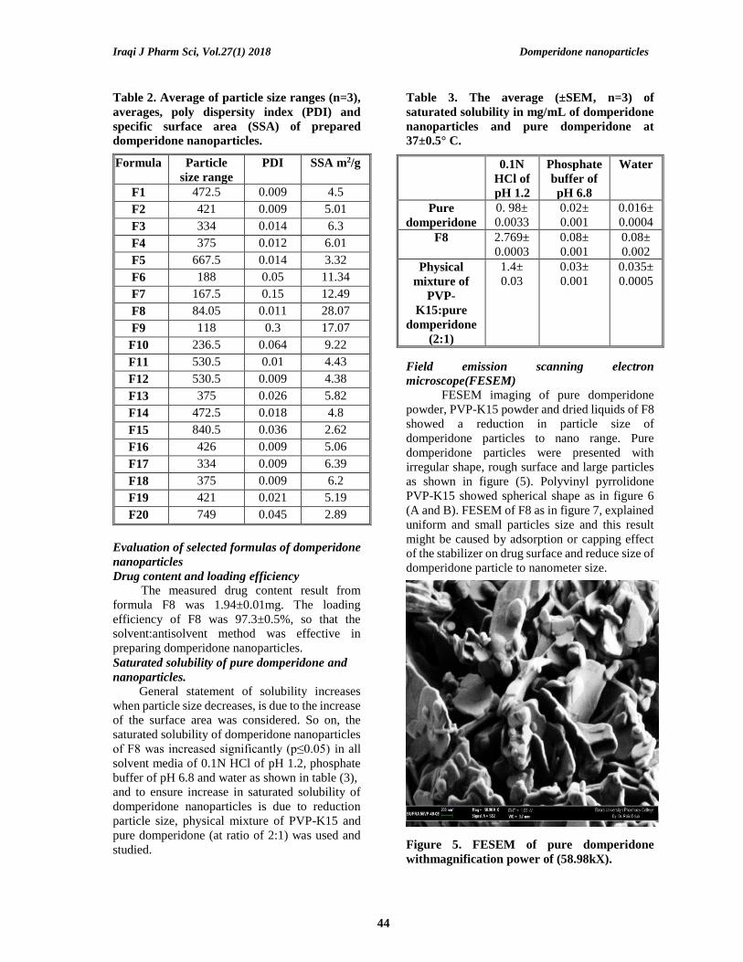
Field emission scanning electron
microscope(FESEM)
FESEM imaging of pure domperidone
powder, PVP-K15 powder and dried liquids of F8
showed a reduction in particle size of
domperidone particles to nano range. Pure
domperidone particles were presented with
irregular shape, rough surface and large particles
as shown in figure (5). Polyvinyl pyrrolidone
PVP-K15 showed spherical shape as in figure 6
(A and B). FESEM of F8 as in figure 7, explained
uniform and small particles size and this result
might be caused by adsorption or capping effect
of the stabilizer on drug surface and reduce size of
domperidone particle to nanometer size.
Figure 5. FESEM of pure domperidone
withmagnification power of (58.98kX).

Iraqi J Pharm Sci, Vol.27(1) 2018 Domperidone nanoparticles
45
Figure 6. FESEM of PVP-K15 particle with
magnification power of A-395X and B-1.84kX.
Figure 7. FESEM of F8 with
magnificationpower of 180.35 kX.
X –ray powder diffraction analysis
The obtained spectrum of X-ray
diffraction test of pure domperidone showed
several strong characteristic peaks at 2θ = 9.28º,
14.94º, 15.58º, 19.80º, 24.80º, and 32º as shown
in figure (8), which indicates the crystalline state
of pure drug. XRPD of PVP-K15 showed low
intense peaks due to the amorphous nature as
shown in figure (9). Some domperidone peaks
were still appeared with the physical mixture as in
figure (10). Domperidone nanoparticles of F8 as
shown in figure (11) showed less number and low
intense of diffraction peaks in comparison to that
of pure domperidone, which indicates that the
crystalline structure of domperidone was reduced
and part of it converted into amorphous state,
similar result was also found in candisartan
nanoparticles that prepared by Filipcsei’s research
group (21).
Figure 8. XRPD spectrum of pure domperidone

Iraqi J Pharm Sci, Vol.27(1) 2018 Domperidone nanoparticles
46
Figure 9. XRPD spectrum of polymer PVP-K15
Figure 10. XRPD spectrum of physical mixture of PVP-K15 and pure domperidone (at ratio of 2:1)).

Iraqi J Pharm Sci, Vol.27(1) 2018 Domperidone nanoparticles
47
Figure 11. XRPD spectrum of F8
Fourier Transform Infrared Spectroscopy
(FTIR)
FTIR was carried out for domperidone and
nanoparticles of F8. Domperidone exhibited a
strong characteristic absorbance band at 1714 cm-
1 due to C = O stretching vibrations of amide
functional group CONHR, and N ̶ H bending
characteristic band at 1693 cm-1, N ̶ H stretching
band of secondary amine appeared at 3126 cm-1
as a single band, symmetric and asymmetric C ̶ H
stretching bands appeared at 2810 and 2941 cm-1
respectively. As well as aromatic symmetric and
asymmetric C ̶ H stretching bands appeared at
3024 and 3074 cm-1 respectively, and the aromatic
C = C stretching band appeared at 1624 cm-1(22) as
shown in figure (12). The resulted FTIR of PVP-
K15, the physical mixtures of PVP-K15:pure
domperidone (2:1) and F8 as in figures (13, 14
and 15) showed the presence of main peaks of
domperidone which indicates there is no
interaction or complexation between drug and
polymer during preparation of nanoparticles.
Figure 12. FTIR spectrum of pure domperidone

Iraqi J Pharm Sci, Vol.27(1) 2018 Domperidone nanoparticles
48
Figure 13. FTIR spectrum of PVP-K15
Figure 14. FTIR spectrum of physical mixture of PVP-K15:pure domperidone (2:1)
Figure 15 . FTIR spectrum of F8
Differential scanning calorimetry (DSC)
DSC thermogram of domperidone as in
figure (16) showed a sharp endothermic peak at
249ºC, this melting point of pure domperidone as
referenced (23) and revealed that the drug has
crystalline nature with high purity. DSC of PVP-
K15 as in figure (17) showed broad peak of water
evaporation at 107º C this indicate amorphous
nature of this polymer. Physical mixture of PVP-
K15:pure domperidone (2:1) showed broad and
low intensity peak of domperidone which is
nearly at same position within the range of
melting point in figure (18). This indicates no
chemical reaction or complexation between drug
and polymer, DSC of F8 in figure (19) showed
remarkable reduction in peak intensity in

Iraqi J Pharm Sci, Vol.27(1) 2018 Domperidone nanoparticles
49
comparison with pure domperidone, that indicates
a reduction of crystalline state of domperidone
and conversion of part of it to amorphous state,
this result with that obtained X-ray diffraction
analysis of formula (F8) agrees with Younis
finding (24).
Figure 16. DSC thermogram of pure domperidone.
Figure 17. DSC thermogram of PVP-K15

Iraqi J Pharm Sci, Vol.27(1) 2018 Domperidone nanoparticles
50
Figure 18. DSC thermogram of physical mixture of PVP-K15:pure domperidone (2:1)
Figure 19. DSC thermogram of domperidone nanoparticles (F8)
In-vitro dissolution study of domperidone
nanoparticles form capsule dosage form
The percentage of drug release at15min
and the time required for released 100% of drug
were considered for the comparison of the
dissolution results between pure and
nanoparticles of domperidone. The release of
domperidone from F8 was faster in comparison
with pure domperidone, and reached to100% of
accumulative % of drug release at 15min.,
whereas the pure domperidone reached to 58%
and 90% at 15min at the end of the study,
respectively, as shown in figure (20). In addition,
physical mixture of PVP-K15:pure domperidone
(2:1) reached 60% and 90.9% at15 and 75min,
respectively. These results indicate that, the
increase in percentage of release of domperidone
from nanoparticles was due to the increase in the
surface area of these particles (25) and to the
reduction in crystalline state of domperidone.
These findings support that, the use of precipitation method was efficient and effective in
preparing nanoparticles.

Iraqi J Pharm Sci, Vol.27(1) 2018 Domperidone nanoparticles
51
Figure 20. The In-vitro release of the pure
domperidone, F8, physical mixture of PVP-
K15:pure domperidone (2:1) in 0.1N HCl of
pH 1.2 at 50rpm and 37° C (±SEM, n=3).
The comparison between the dissolution profiles
was done using difference and similarity test of f1
and f2 respectively. The data of accumulative
percentages of release of drug from selected
formula was fitted using a Microsoft Excel
program to calculate f1 and f2 and the obtained
results as illustrated in table (4). It was noticed
that the dissolution profiles of domperidone from
nanoparticles was not similar in comparison with
the pure drug as a reference, F8were not similar
as f1 values was higher than 15, whereas their f2
values were lower than 50(26). From these results,
it can conclude that nanoparticles showed better
in vitro dissolution profiles in comparison with
pure drug.
Table 4. Difference and similarity test (f1 and
f2) of selected formula and physical mixture of
PVP-K15:pure domperidone (2:1) compared
versus pure domperidone
f1 f2
F8 37.6 29.46
Physical mixture of
PVP-K15:pure
domperidone (2:1)
9.02 50.5
Conclusions Depending on obtained results, one can
concludes that, PVP-K15 gave best formula (F8),
the variables of type, concentration of stabilizer,
volume ratio of solvent: anti solvent, time and rate
of stirring showed considerable effect on
decreasing of the size of domperidone
nanoparticles. on increase of polymer: drug ratio, the size of produced domperidone nanoparticles
was decreased, and 1:10 volume ratio of solvent:
antisolvent was best than the other ratios.
Analysis by DSC, FESEM and XRPD of
nanoparticles of F8 indicated a reduction in
crystalline state of domperidone nanoparticles.
The dissolution of domperidone was highly
improved against pure drug, which give 100%
accumulative release at 15min.
References 1. Khadka, P., Ro, J., Kim, H., Kim, I., Kim, J.
T., Kim, H., Lee, J. Pharmaceutical particle
technologies: An approach to improve drug
solubility, dissolution and bioavailability.
Asian Journal of Pharmaceutical Sciences
2014; 9, 304–316.
2. Nekkanti, V. and Rueda, J. Nanoparticles for
Improved Delivery of Poorly Soluble Drugs.
Journal of Drug, USA 2016;1, 18-27.
3. Dhillon Balvinder, Narendra Kr. Goyal,
Rishabha Malviya and Pramod K. Sharma.
Poorly Water Soluble Drugs: Change in
Solubility for Improved Dissolution
Characteristics a Review.Global Journal of
Pharmacology 2014; 8, 26–35.
4. Bhatia, S. Natural polymer drug delivery
systems: Nanoparticles, plants, and algae.
Springer International Publishing 2016; 1-
225.
5. Alim, M., Karna, S., Chaturvedi, S., Agrawal,
V.K. Validated UV spectrophotometric
method for estimation of domperidone for
dissolution study. Der Pharmacia Lettre 2015;
7, 53–58.
6. Castle P (2005). European Pharmacopoeia
(EP), USDA and MAFF standards--will they
ever be harmonised under the VICH
umbrella? Dev Biol (Basel) .121, 227-234.
7. Guleria, R., Sharma, V., Kapoor, A., Kaith,
N.S., Singh, R. Polyethylene Glycol Enhances
Solubility of Domperidone through Solid
Dispersion. American Journal of Pharm Tech
Research 2012; 2, 630-638.
8. Mansouri,M. Preparation and characterization
of Ibuprofen nanoparticles by using
solvent/antisolvent precipitation. The Open
Conference Proceedings Journal 2011; 2, 88–
94.
9. Nakarani M., Misra A.K., Patel J.K., Vaghani
S.S. Itraconazole Nanosuspension Meant for
Oral Use : Development of Formulation,
Characterization and In Vitro Comparison
with marketed formulation. Daru 2010; 18,
84-90.
10. Baka, E., Comer, J. E. A. and Tak, K.Study of
equilibrium solubility measurement by

Iraqi J Pharm Sci, Vol.27(1) 2018 Domperidone nanoparticles
52
saturation shake-flask method using
hydrochlorothiazide as model compound.J
ournal of Pharmaceutical and Biomedical
Analysis 2008; 46, 335–341.
11. Amin, M. A., Osman, S. K. and Aly, U. F.
Preparation and characterization of
Ketoprofen nanosuspension for solubility and
dissolution velocity enhancement
.International Journal of Pharma and Bio
Sciences2013; 4, 768–780.
12. British Pharmacopoeia, (2009). Dissolution
monograph of domperidone.
13. FDA guidance for industry dissolution testing
of immediate evaluation, (1997); 4, 15–22.
14. Ginoya S, A. S. Nanosuspention: A novel
approach towards the drug delivery system.
Int. j. Pharm. Sci. 2013; 4, 100–122.
15. Gadad, P. Sharath Chandra, P.M. Dandagi and
V.S. Mastiholimath. Moxifloxacin Loaded
Polymeric Nanoparticles for Sustained Ocular
Drug Delivery. Journal of Pharmaceutical
Sciences 2012; 5, 1727–1734.
16. Akbari, B., Tavandashti, M. P. and
Zandrahimi, M. Particle Size Characterization
of Nanoparticles. Iranian Journal of Materials
Science & Engineering 2011; 8,48–56.
17. Wu, L., Zhang, J. and Watanabe, W. Physical
and chemical stability of drug nanoparticles.
Advanced Drug Delivery Reviews 2011; 63,
456–469.
18. Chopra, M., Kaur, P., Bernela, M., Thakur, R.
Synthesis and optimization of streptomycin
loaded Chitosan-Alginate nanoparticles.
International Journal of Scientific and
technology research 2012; 1, 31–34.
19. Lonare, A. A. and Patel, S. R. Antisolvent
Crystallization of Poorly Water Soluble
Drugs. International Journal of Chemical
Engineering and Applications 2013; 4, 337–
341.
20. Dong,Y., Ng, W.K., Shen, S., Kim, S. and
Tan, R. B. Preparation and characterization of
spironolactone nanoparticles by antisolvent
precipitation. International Journal of
Pharmaceutics 2009; 375,84-88.
21. Genovéva Filipcsei, Ötvös, Z., Pongrácz, K.
and D. Nanoparticulate candesartan cilexitile
compositions, process for the preparation
there of and pharmaceutical containing them.
U.S. Patent Application 2010; 255,379.
22. Ghodke D.S, G. M. Chaulang, K. S. Patil, P.
D. Nakhat, P. G. Yeole, N. S. Naikwade and
C.S. Magdum. Solid State Characterization of
Domperidone : Hydroxypropyl- β -
Cyclodextrin Inclusion Complex. Indian
Journal of Pharmaceutical Sciences 2010;
245–249.
23. Essa, E. A. and Balata, G. F. Preparation and
characterization of domperidone solid
dispersions. Pak. J. Pharm. Sci. 2009; 25:
783–791.
24. Younis, M. A. Enhancement of domperidone
dissolution rate via formulation of adsorbates
co-adsorbate A.E. International Journal of
Pharmaceutical Sciences and Research 2016;
7,951–960.
25. Junghanns, J. U. A. H. and Müller, R. H.
Nanocrystal technology, drug delivery and
clinical applications. International Journal of
Nanomedicine 2008; 3, 295–309.
26. Muhammad Qamar Khan, Nighat Razvi,
Fakhsheena Anjum, L. G. and Shoaib, S. A. S.
and M. H. Evaluation and comparison of
different brands of domperidone tablets
available in Evaluation and comparison of
different brands of domperidone. Pakistan
journal of pharmaceutical sciences 2014; 27,
935-938.