Embed Size (px)
Citation preview
ORIGINAL ARTICLE
Preparation and characterization of nanosized Ag/SLN compositeand its viability for improved occlusion
G. Cynthia Jemima Swarnavalli1 • S. Dinakaran2 • S. Divya1
Received: 5 December 2015 / Accepted: 25 January 2016 / Published online: 15 February 2016
� The Author(s) 2016. This article is published with open access at Springerlink.com
Abstract Nanocomposites consisting of silver and solid
lipid nanoparticles (SLN) elicit interest for their synergistic
effect based enhanced properties in skin hydration. The
nanocomposite preparation aims at combining the antimi-
crobial activity of silver with skin hydration performance
of SLN. The nanocomposites designated Ag/SAN (silver/
stearic acid nanoparticles), Ag/PAN (silver/palmitic acid
nanoparticles) were prepared by incorporating silver
nanoparticles into the dispersion of SLN and sonicating for
10 min followed by heating for 1 h at 50 �C in a thermo-
stat. The occlusive property of the two nanocomposites was
evaluated in comparison with the pure SLN by adopting de
Vringer-de Ronde in vitro occlusion test. The incorporation
of silver nanoparticles has improved occlusion factor by
10 % in the case of both composites at SLN concentration
of 0.14 mmol. Characterization studies include XRD, DSC,
HRSEM, DLS and zeta potential measurement. High res-
olution scanning electron microscopy (HRSEM) images
divulge that the nanoparticles of composite (Ag/SAN)
shows halo effect where the hydrophobic stearic acid is
oriented at the core and is surrounded by silver nanopar-
ticles while Ag/PAN shows cashew shaped SLN dispersed
in silver nanoparticles matrix.
Keywords Silver nanoparticles � SLN � Nanocomposite �Skin hydration � Zetapotential � HRSEM
Introduction
Since the last three decades, nanotechnology has been
impressed practically all the frontier areas of research
including cosmetics and pharmaceutics. It has the special
form of colloidal drug delivery system which includes
Solid Lipid Nanoparticles (SLNs), liposomes, den-
drimers, and polymeric nanoparticles. SLN are lipid
nanoparticles with solid matrix which has relatively low
melting points and some of them are present in food-
stuffs and in the human body. It has high specific surface
due to their small diameter, spherical shape and
favourable zeta potential (Gasco 2007). The stratum
corneum (SC) is made up of ceramides, cholesterol,
cholesterol esters and free fatty acids in which the per-
centage of stearic and palmitic acid is 15 % (Anantha-
padmanabhan et al. 2013). Fatty acids penetrate the skin
effectively and enhance delivery of certain co-applied
drugs and cosmetic actives (Golden et al. 1987). Fatty
acids are more susceptible to surfactant-induced removal
than other lipids necessitating replenishing of stratum
corneum lipids. Skin preservation largely depends on
incorporation of fatty acids such as stearic acid and
palmitic acid into creams and moisturizing body
cleansers to minimize their extraction by surfactants and
reload lost fatty acids to promote skin barrier preserva-
tion (Ananthapadmanabhan et al. 2013). One of the best
ways to integrate these fatty acids is in the form of solid
lipid nanoparticles since small size ensures a close
contact to the stratum corneum and preservation of
moisture on the surface. Interesting skin hydration effect
is observed when SLNs are incorporated into a mois-
turizing/day cream. It has been noted that the enhance-
ment of skin hydration effect is due to occlusive
property of SLNs (Muller et al. 2002; Wissing and
& G. Cynthia Jemima Swarnavalli
1 Department of Chemistry, Women’s Christian College,
Chennai 600006, India
2 School of Advanced Sciences, VIT University,
Vellore 632014, India
123
Appl Nanosci (2016) 6:1065–1072
DOI 10.1007/s13204-016-0522-2
Muller 2002a). Numerous in depth cosmetic and der-
matological studies with SLN targeting the skin and
incorporation of SLN into topical cosmetic and phar-
maceutical preparations, such as creams and gels have
been reported (Jenning et al. 2000; Wissing and Muller
2003a; Souto and Muller 2008). SLN possess higher
occlusive factor in comparison to nano structured lipid
carrier (NLC) of the same lipid content (Souto et al.
2004). It was also observed that an increase in oil con-
tent leads to a decrease in the occlusive factor (Teer-
anachaideekul et al. 2008) thus favouring SLN for skin
hydration. Wissing and Muller (2003b) reported that the
skin hydration effect of an o/w cream containing SLN
has superior skin hydration than a conventional o/w
cream. Pardeike et al. reported a significant increase in
skin hydration for an NLC incorporated cream compared
to conventional cream (Pardeike et al. 2009). The
occlusive character of SLN is due to film formation after
application on the skin leading to decreased water
evaporation (Wissing and Muller 2001a). De Vringer
(1997) has demonstrated that nanoparticles of lipids have
been found to be 15-folds more occlusive than micro
particles. Occlusion can be enhanced by low melting
lipids, highly crystalline SLN, decreasing particle size
and increase the concentration of the lipids (Wissing
et al. 2001; Wissing and Muller 2002b). Generally, the
mean particle size of SLN is in the rage of about 40 to
1000 nm (Pardeike et al. 2009). Since SLN combines the
advantages of various traditional carriers, such as lipo-
somes and polymeric nanoparticles, it finds application
in protecting chemically labile actives and giving the
ability to modulate cosmetic active or drug release.
Cosmetic actives, like coenzyme Q10 (Muller et al.
2007) ascorbyl palmitate (Uner et al. 2005) tocopherol
acetate (Wissing and Muller 2001b) and retinol (vitamin
A) (Volkhard Jenning SHG 2001) has proved enhance-
ment of chemical stability due to incorporation into lipid
nanocarriers.
In cosmetics and personal care, a new target is to use
compounds possessing dual or more properties and recent
research is focused on modifying the existing character-
istics of a compound by incorporating another ingredient.
A survey of literature reveals that SLN are incorporated
into creams and lotions owing to their occlusive effect
which is responsible for restoring moisture to the stratum
corneum. Apart from that SLN are good carriers of
chemically labile actives and possess sun blocking effi-
ciency (Lacatusu et al. 2010) due to its particulate nature.
In order to take advantage of the antimicrobial activity of
silver nanoparticles (Marambio-Jones and Hoek 2010)
they are integrated into SLN and the interesting obser-
vation of the present study is that it has enhanced the
occlusion property of SLN.
Materials and methods
Stearic acid, palmitic acid, ethanol, n-hexane, ethyl acetate
and Poly ethylene glycol (PEG) (M. wt 20,000 kDa) were
purchased from Merck. AgNO3 was obtained from Quali-
gen chemicals. TWEEN-80 (Polyoxyethylene derivative of
sorbitan mono-oleate) was purchased from Sigma-Aldrich
chemicals. The solvents were used after distillation and
confirming their melting point for purity. Stearic acid and
Palmitic acid were used as such and deionized water was
used in all preparations.
Synthesis of silver nanoparticles
Silver nitrate solution (20 mL, 1 mM) was taken in a
250 mL beaker and to this 5 mL of sodium hydroxide
was added with continuous stirring. About 3 mL of 3 %
solution of PEG (M.wt 20,000) was added drop wise to
the above solution and stirred for 5-10 min followed by
the drop wise addition of 10 mL of 0.01 M dextrose as
reducing agent. The stirring further continued for 5 min
and then the beaker containing the solution was heated
in a thermostat at 60 �C for 1 h. The solution turned
black and was cooled to room temperature and cen-
trifuged at 16,000 rpm (REMI cooling centrifuge) to
yield a black precipitate. The silver colloid was washed
repeatedly with deionized water and re-dispersed in
deionized water by sonication (Pci Ultrasonic bath
sonicator, 36 kHz) to give a yellow sol. The sample was
designated SN.
Preparation of solid lipid nanoparticles
The SLN were prepared by nano precipitation method
developed by Hatem Curt et al. (1992) The organic phase
contained various concentrations of stearic acid (0.08, 0.1,
0.12, 0.14 mmol) dissolved in a blend of 18 mL of ethyl
acetate and 2 mL of ethanol. The aqueous phase contained
specified amount of the emulsifier (TWEEN-80) dissolved
in 25 mL deionized water. The organic phase was added
drop wise to the aqueous phase with stirring (400 rpm) at
room temperature. The mixture immediately became
opalescent as a result of the formation of lipid nanoparti-
cles. The suspension was stirred during an additional time
of 5–10 min. The organic solvents were removed under
reduced pressure and the pH of the cooled dispersion was
adjusted to 1.2 by adding 0.1 M hydrochloric acid solution
to the precipitated SLN, and the precipitate was then col-
lected by centrifuging at 12,000 rpm. The fine white pre-
cipitate was re dispersed in deionized water by sonication.
SLN of Palmitic acid of different concentration (0.08, 0.1,
0.12, 0.14 mmol) was also prepared by the above
1066 Appl Nanosci (2016) 6:1065–1072
123

mentioned procedure with the organic phase containing a
blend of 18 mL of n-hexane and 2 mL of ethanol. The solid
lipid nanoparticles of stearic and palmitic acid are desig-
nated SAN and PAN respectively.
Preparation of the nanocomposite
The composites were prepared by mixing 20 mL of the
aqueous nanosuspension of the SLN and 2 mL of
nanosuspension of silver (10 Wt % of SLN) and sonicating
the mixture for 10 min followed by heating in a thermostat
at 50 �C for 1 h. The composite was obtained as a
nanosuspension after concentration. The composites of
silver and stearic acid and silver and palmitic acid were
designated as Ag/SAN and Ag/PAN respectively.
In vitro occlusion test
An in vitro occlusion test adapted by de Vringer was used
in order to test the skin hydration efficiency of SLN and the
composites (De Vringer and De Ronde 1995). Five beakers
(100 mL) were filled with 50 mL of water, covered with
filter paper (cellulose acetate filter) and sealed. One of the
beakers was left as such and 200 mg each of the four
samples was applied on the filter surface. The sample was
spread evenly with a spatula. The samples were stored at
32 �C and 50–55 % RH for 48 h. The samples were
weighed after 6, 24, and 48 h, giving the water loss due to
evaporation at each time (water flux through the filter
paper). Beaker covered with filter paper but without an
applied sample served as standard value. Every experiment
was performed in triplicate under constant conditions. The
occlusion factor F was calculated according to equation.
F ¼ 100� A� Bð Þ=Að Þ ð1Þ
where, A is the water loss without sample (standard) B is
the water loss with sample.
Characterization studies
Silver nanoparticles in the sol were characterized by
measuring the absorption wavelength using a UV–Visible
Spectrophotometer (Systronics DBS 2303). Powder X-Ray
diffraction (PXRD) analysis of the prepared samples were
carried out with a RICHSEIFER powder diffractometer,
using nickel filtered copper K-alpha radiations (k = 1.5461
A) with a scanning rate of 0.02�. Size and morphology of
the particles were studied using HRSEM (FEI quanta FEG
200—HRSEM). The average diameter, polydispersity
index (PI) and zeta potential measurements were measured
using dynamic light scattering (DLS) using a Malvern
Zetasizer nano-ZS (Malvern Instruments, UK). Prior to the
measurement, all samples were diluted using ultra-purified
water to yield a suitable scattering intensity. Samples were
analyzed at 25 �C using the general purpose mode. Thermo
grams were recorded with a differential scanning
calorimeter (NETZSCH DSC204). Samples were heated at
the scanning rate of 3 �C/min over a temperature range
between 30 and 200 �C.
Results
The SLN of palmitic and stearic acids were prepared by
nano precipitation method. Silver nanoparticles were pre-
pared by chemical reduction and the nanocomposites were
obtained by ultrasound assisted thermal method. The col-
loidal silver sol was characterized by UV–Visible spec-
trophotometer that exhibited a typical Surface Plasmon
resonance (SPR) peak at 420 nm (Fig. 1a). The
200 300 400 500 600 700 800
0.0
0.5
1.0
1.5
2.0
2.5A
bsor
banc
e (a
.u)
Wavelength (nm)
(b)
(a)
Fig. 1 a UV–Vis Absorption spectrum and b HRSEM micrographs
(scale bar 500 nm) of silver nanoparticles (SN)
Appl Nanosci (2016) 6:1065–1072 1067
123
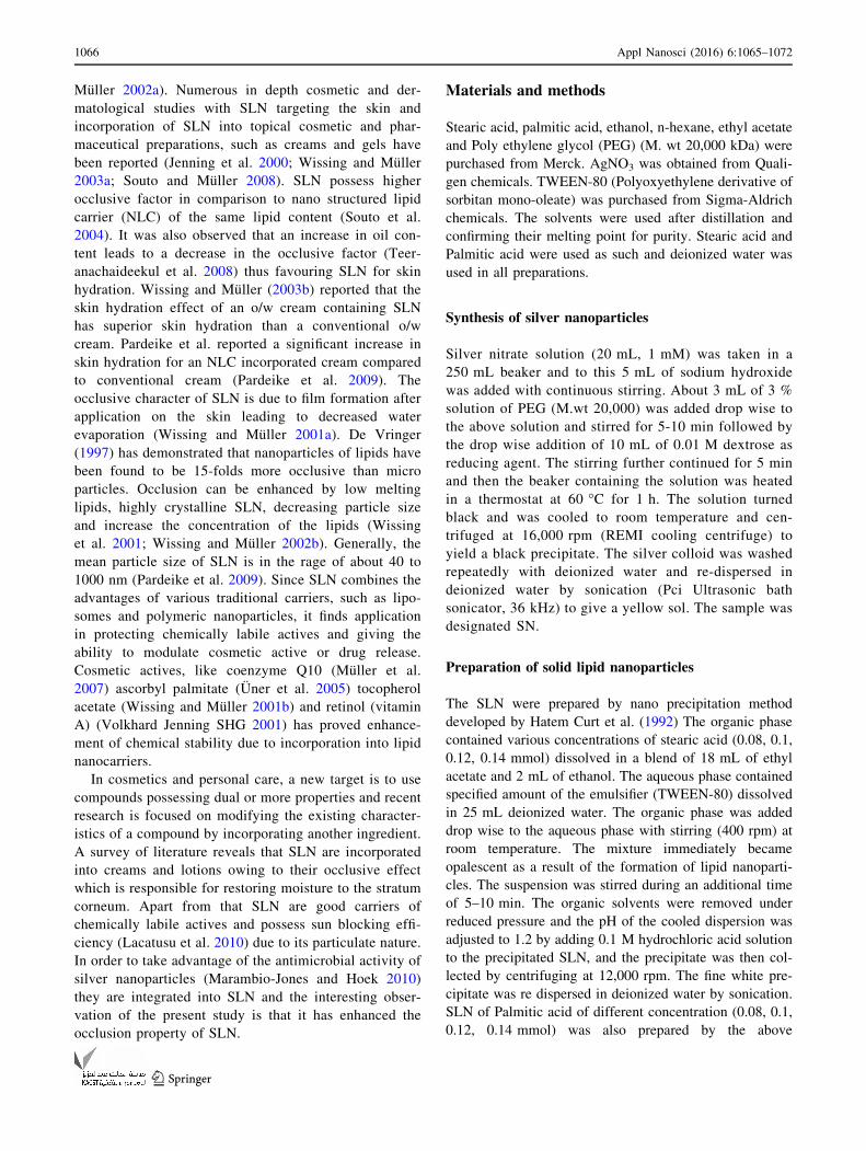
morphology of the obtained samples were investigated by
HRSEM images. Figure 1b shows the average size of silver
nanoparticle about 35 nm. Comparison of HRSEM images
of pure SLN and the Ag/SLN composites are as shown in
Fig. 2. SEM images of stearic and palmitic acid nanopar-
ticles (Fig. 2a, b) revealed that most of the particles were
spherical in shape with an average diameter of 100 and
80 nm respectively. The Ag/SAN sample exhibited a sur-
face morphology different from that of the pure SLN where
SLN appeared as a white domain surrounded by silver
nanoparticles (Fig. 2c). The hydrophobic SAN appeared to
be preferentially oriented to the core with the silver
nanoparticles arranged as a loosely organized layer at a
small distance from the surface of the nanosphere.
In the other composite Ag/PAN (Fig. 2d) the spherical
SLN of palmitic acid was found to be elongated in shape
resembling a cashew nut and firmly dispersed in the matrix
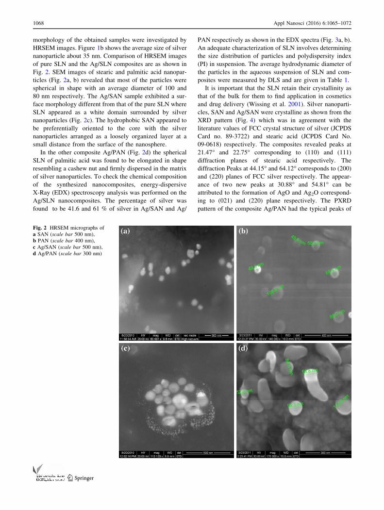
of silver nanoparticles. To check the chemical composition
of the synthesized nanocomposites, energy-dispersive
X-Ray (EDX) spectroscopy analysis was performed on the
Ag/SLN nanocomposites. The percentage of silver was
found to be 41.6 and 61 % of silver in Ag/SAN and Ag/
PAN respectively as shown in the EDX spectra (Fig. 3a, b).
An adequate characterization of SLN involves determining
the size distribution of particles and polydispersity index
(PI) in suspension. The average hydrodynamic diameter of
the particles in the aqueous suspension of SLN and com-
posites were measured by DLS and are given in Table 1.
It is important that the SLN retain their crystallinity as
that of the bulk for them to find application in cosmetics
and drug delivery (Wissing et al. 2001). Silver nanoparti-
cles, SAN and Ag/SAN were crystalline as shown from the
XRD pattern (Fig. 4) which was in agreement with the
literature values of FCC crystal structure of silver (JCPDS
Card no. 89-3722) and stearic acid (JCPDS Card No.
09-0618) respectively. The composites revealed peaks at
21.47� and 22.75� corresponding to (110) and (111)
diffraction planes of stearic acid respectively. The
diffraction Peaks at 44.15� and 64.12� corresponds to (200)
and (220) planes of FCC silver respectively. The appear-
ance of two new peaks at 30.88� and 54.81� can be
attributed to the formation of AgO and Ag2O correspond-
ing to (021) and (220) plane respectively. The PXRD
pattern of the composite Ag/PAN had the typical peaks of
Fig. 2 HRSEM micrographs of
a SAN (scale bar 500 nm),
b PAN (scale bar 400 nm),
c Ag/SAN (scale bar 500 nm),
d Ag/PAN (scale bar 300 nm)
1068 Appl Nanosci (2016) 6:1065–1072
123
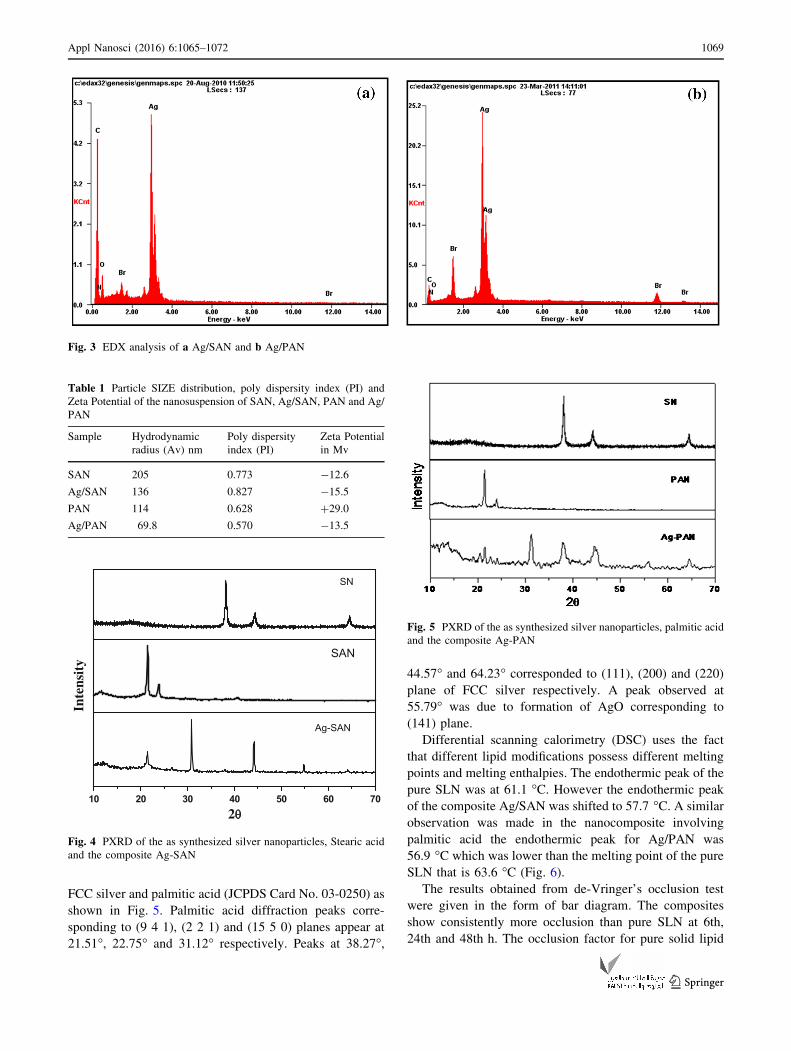
FCC silver and palmitic acid (JCPDS Card No. 03-0250) as
shown in Fig. 5. Palmitic acid diffraction peaks corre-
sponding to (9 4 1), (2 2 1) and (15 5 0) planes appear at
21.51�, 22.75� and 31.12� respectively. Peaks at 38.27�,
44.57� and 64.23� corresponded to (111), (200) and (220)
plane of FCC silver respectively. A peak observed at
55.79� was due to formation of AgO corresponding to
(141) plane.
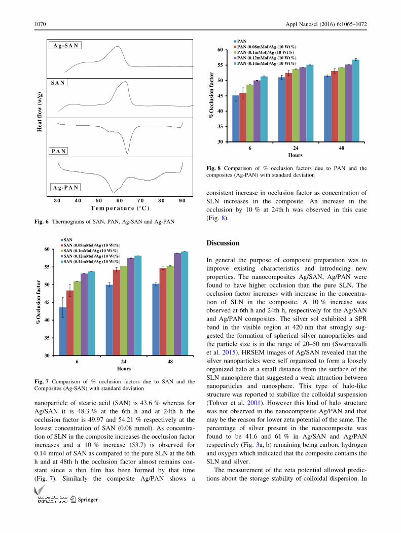
Differential scanning calorimetry (DSC) uses the fact
that different lipid modifications possess different melting
points and melting enthalpies. The endothermic peak of the
pure SLN was at 61.1 �C. However the endothermic peak
of the composite Ag/SAN was shifted to 57.7 �C. A similar
observation was made in the nanocomposite involving
palmitic acid the endothermic peak for Ag/PAN was
56.9 �C which was lower than the melting point of the pure
SLN that is 63.6 �C (Fig. 6).
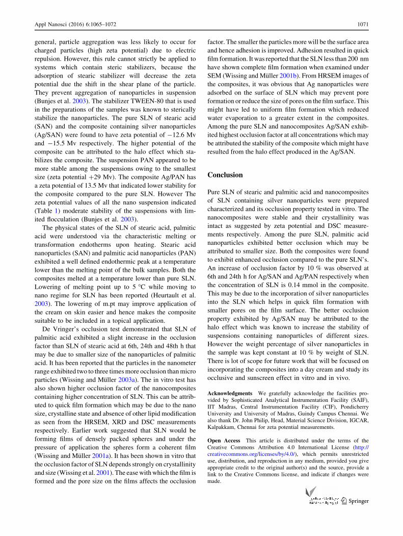
The results obtained from de-Vringer’s occlusion test
were given in the form of bar diagram. The composites
show consistently more occlusion than pure SLN at 6th,
24th and 48th h. The occlusion factor for pure solid lipid
Fig. 3 EDX analysis of a Ag/SAN and b Ag/PAN
Table 1 Particle SIZE distribution, poly dispersity index (PI) and
Zeta Potential of the nanosuspension of SAN, Ag/SAN, PAN and Ag/
PAN
Sample Hydrodynamic
radius (Av) nm
Poly dispersity
index (PI)
Zeta Potential
in Mv
SAN 205 0.773 -12.6
Ag/SAN 136 0.827 -15.5
PAN 114 0.628 ?29.0
Ag/PAN 69.8 0.570 -13.5
10 20 30 40 50 60 70
SN
Inte
nsit
y
SAN
2
Ag-SAN
Fig. 4 PXRD of the as synthesized silver nanoparticles, Stearic acid
and the composite Ag-SAN
Fig. 5 PXRD of the as synthesized silver nanoparticles, palmitic acid
and the composite Ag-PAN
Appl Nanosci (2016) 6:1065–1072 1069
123
nanoparticle of stearic acid (SAN) is 43.6 % whereas for
Ag/SAN it is 48.3 % at the 6th h and at 24th h the
occlusion factor is 49.97 and 54.21 % respectively at the
lowest concentration of SAN (0.08 mmol). As concentra-
tion of SLN in the composite increases the occlusion factor
increases and a 10 % increase (53.7) is observed for
0.14 mmol of SAN as compared to the pure SLN at the 6th
h and at 48th h the occlusion factor almost remains con-
stant since a thin film has been formed by that time
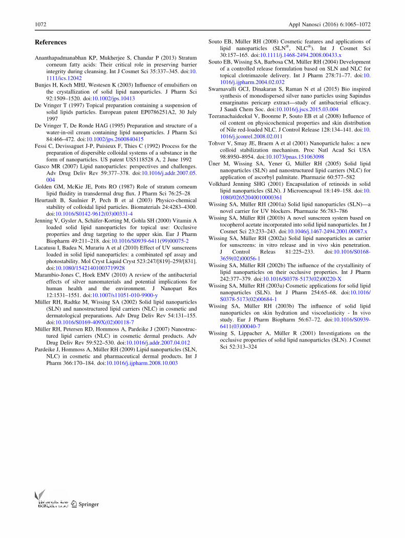
(Fig. 7). Similarly the composite Ag/PAN shows a
consistent increase in occlusion factor as concentration of
SLN increases in the composite. An increase in the
occlusion by 10 % at 24th h was observed in this case
(Fig. 8).
Discussion
In general the purpose of composite preparation was to
improve existing characteristics and introducing new
properties. The nanocomposites Ag/SAN, Ag/PAN were
found to have higher occlusion than the pure SLN. The
occlusion factor increases with increase in the concentra-
tion of SLN in the composite. A 10 % increase was
observed at 6th h and 24th h, respectively for the Ag/SAN
and Ag/PAN composites. The silver sol exhibited a SPR
band in the visible region at 420 nm that strongly sug-
gested the formation of spherical silver nanoparticles and
the particle size is in the range of 20–50 nm (Swarnavalli
et al. 2015). HRSEM images of Ag/SAN revealed that the
silver nanoparticles were self organized to form a loosely
organized halo at a small distance from the surface of the
SLN nanosphere that suggested a weak attraction between
nanoparticles and nanosphere. This type of halo-like
structure was reported to stabilize the colloidal suspension
(Tohver et al. 2001). However this kind of halo structure
was not observed in the nanocomposite Ag/PAN and that
may be the reason for lower zeta potential of the same. The
percentage of silver present in the nanocomposite was
found to be 41.6 and 61 % in Ag/SAN and Ag/PAN
respectively (Fig. 3a, b) remaining being carbon, hydrogen
and oxygen which indicated that the composite contains the
SLN and silver.
The measurement of the zeta potential allowed predic-
tions about the storage stability of colloidal dispersion. In
30 40 50 60 70 80 90
A g -P A N
T em p era tu re (°C )
Hea
t fl
ow (
w/g
)
P A N
S A N
A g -S A N
Fig. 6 Thermograms of SAN, PAN, Ag-SAN and Ag-PAN
30
35
40
45
50
55
60
6 24 48
%O
cclu
sion
fac
tor
Hours
SANSAN (0.08mMol)/Ag (10 Wt%)SAN (0.1mMol)/Ag (10 Wt%)SAN (0.12mMol)/Ag (10 Wt%)SAN (0.14mMol)/Ag (10 Wt%)
Fig. 7 Comparison of % occlusion factors due to SAN and the
Composites (Ag-SAN) with standard deviation
30
35
40
45
50
55
60
6 24 48
%O
cclu
sion
fac
tor
Hours
PANPAN (0.08mMol)/Ag (10 Wt%)PAN (0.1mMol)/Ag (10 Wt%)PAN (0.12mMol)/Ag (10 Wt%)PAN (0.14mMol)/Ag (10 Wt%)
Fig. 8 Comparison of % occlusion factors due to PAN and the
composites (Ag-PAN) with standard deviation
1070 Appl Nanosci (2016) 6:1065–1072
123
general, particle aggregation was less likely to occur for
charged particles (high zeta potential) due to electric
repulsion. However, this rule cannot strictly be applied to
systems which contain steric stabilizers, because the
adsorption of stearic stabilizer will decrease the zeta
potential due the shift in the shear plane of the particle.
They prevent aggregation of nanoparticles in suspension
(Bunjes et al. 2003). The stabilizer TWEEN-80 that is used
in the preparations of the samples was known to sterically
stabilize the nanoparticles. The pure SLN of stearic acid
(SAN) and the composite containing silver nanoparticles
(Ag/SAN) were found to have zeta potential of -12.6 Mv
and -15.5 Mv respectively. The higher potential of the
composite can be attributed to the halo effect which sta-
bilizes the composite. The suspension PAN appeared to be
more stable among the suspensions owing to the smallest
size (zeta potential ?29 Mv). The composite Ag/PAN has
a zeta potential of 13.5 Mv that indicated lower stability for
the composite compared to the pure SLN. However The
zeta potential values of all the nano suspension indicated
(Table 1) moderate stability of the suspensions with lim-
ited flocculation (Bunjes et al. 2003).
The physical states of the SLN of stearic acid, palmitic
acid were understood via the characteristic melting or
transformation endotherms upon heating. Stearic acid
nanoparticles (SAN) and palmitic acid nanoparticles (PAN)
exhibited a well defined endothermic peak at a temperature
lower than the melting point of the bulk samples. Both the
composites melted at a temperature lower than pure SLN.
Lowering of melting point up to 5 �C while moving to
nano regime for SLN has been reported (Heurtault et al.
2003). The lowering of m.pt may improve application of
the cream on skin easier and hence makes the composite
suitable to be included in a topical application.
De Vringer’s occlusion test demonstrated that SLN of
palmitic acid exhibited a slight increase in the occlusion
factor than SLN of stearic acid at 6th, 24th and 48th h that
may be due to smaller size of the nanoparticles of palmitic
acid. It has been reported that the particles in the nanometer
range exhibited two to three timesmore occlusion thanmicro
particles (Wissing and Muller 2003a). The in vitro test has
also shown higher occlusion factor of the nanocomposites
containing higher concentration of SLN. This can be attrib-
uted to quick film formation which may be due to the nano
size, crystalline state and absence of other lipid modification
as seen from the HRSEM, XRD and DSC measurements
respectively. Earlier work suggested that SLN would be
forming films of densely packed spheres and under the
pressure of application the spheres form a coherent film
(Wissing and Muller 2001a). It has been shown in vitro that
the occlusion factor of SLN depends strongly on crystallinity
and size (Wissing et al. 2001). The easewithwhich the film is
formed and the pore size on the films affects the occlusion
factor. The smaller the particlesmore will be the surface area
and hence adhesion is improved. Adhesion resulted in quick
film formation. It was reported that the SLN less than 200 nm
have shown complete film formation when examined under
SEM (Wissing and Muller 2001b). From HRSEM images of
the composites, it was obvious that Ag nanoparticles were
adsorbed on the surface of SLN which may prevent pore
formation or reduce the size of pores on the film surface. This
might have led to uniform film formation which reduced
water evaporation to a greater extent in the composites.
Among the pure SLN and nanocomposites Ag/SAN exhib-
ited highest occlusion factor at all concentrations which may
be attributed the stability of the composite which might have
resulted from the halo effect produced in the Ag/SAN.
Conclusion
Pure SLN of stearic and palmitic acid and nanocomposites
of SLN containing silver nanoparticles were prepared
characterized and its occlusion property tested in vitro. The
nanocomposites were stable and their crystallinity was
intact as suggested by zeta potential and DSC measure-
ments respectively. Among the pure SLN, palmitic acid
nanoparticles exhibited better occlusion which may be
attributed to smaller size. Both the composites were found
to exhibit enhanced occlusion compared to the pure SLN’s.
An increase of occlusion factor by 10 % was observed at
6th and 24th h for Ag/SAN and Ag/PAN respectively when
the concentration of SLN is 0.14 mmol in the composite.
This may be due to the incorporation of silver nanoparticles
into the SLN which helps in quick film formation with
smaller pores on the film surface. The better occlusion
property exhibited by Ag/SAN may be attributed to the
halo effect which was known to increase the stability of
suspensions containing nanoparticles of different sizes.
However the weight percentage of silver nanoparticles in
the sample was kept constant at 10 % by weight of SLN.
There is lot of scope for future work that will be focused on
incorporating the composites into a day cream and study its
occlusive and sunscreen effect in vitro and in vivo.
Acknowledgments We gratefully acknowledge the facilities pro-
vided by Sophisticated Analytical Instrumentation Facility (SAIF),
IIT Madras, Central Instrumentation Facility (CIF), Pondicherry
University and University of Madras, Guindy Campus Chennai. We
also thank Dr. John Philip, Head, Material Science Division, IGCAR,
Kalpakkam, Chennai for zeta potential measurements.
Open Access This article is distributed under the terms of the
Creative Commons Attribution 4.0 International License (http://
creativecommons.org/licenses/by/4.0/), which permits unrestricted
use, distribution, and reproduction in any medium, provided you give
appropriate credit to the original author(s) and the source, provide a
link to the Creative Commons license, and indicate if changes were
made.
Appl Nanosci (2016) 6:1065–1072 1071
123
References
Ananthapadmanabhan KP, Mukherjee S, Chandar P (2013) Stratum
corneum fatty acids: Their critical role in preserving barrier
integrity during cleansing. Int J Cosmet Sci 35:337–345. doi:10.
1111/ics.12042
Bunjes H, Koch MHJ, Westesen K (2003) Influence of emulsifiers on
the crystallization of solid lipid nanoparticles. J Pharm Sci
92:1509–1520. doi:10.1002/jps.10413
De Vringer T (1997) Topical preparation containing a suspension of
solid lipids particles. European patent EP0786251A2, 30 July
1997
De Vringer T, De Ronde HAG (1995) Preparation and structure of a
water-in-oil cream containing lipid nanoparticles. J Pharm Sci
84:466–472. doi:10.1002/jps.2600840415
Fessi C, Devissaguet J-P, Puisieux F, Thies C (1992) Process for the
preparation of dispersible colloidal systems of a substance in the
form of nanoparticles. US patent US5118528 A, 2 June 1992
Gasco MR (2007) Lipid nanoparticles: perspectives and challenges.
Adv Drug Deliv Rev 59:377–378. doi:10.1016/j.addr.2007.05.
004
Golden GM, McKie JE, Potts RO (1987) Role of stratum corneum
lipid fluidity in transdermal drug flux. J Pharm Sci 76:25–28
Heurtault B, Saulnier P, Pech B et al (2003) Physico-chemical
stability of colloidal lipid particles. Biomaterials 24:4283–4300.
doi:10.1016/S0142-9612(03)00331-4
Jenning V, Gysler A, Schafer-Korting M, Gohla SH (2000) Vitamin A
loaded solid lipid nanoparticles for topical use: Occlusive
properties and drug targeting to the upper skin. Eur J Pharm
Biopharm 49:211–218. doi:10.1016/S0939-6411(99)00075-2
Lacatusu I, Badea N, Murariu A et al (2010) Effect of UV sunscreens
loaded in solid lipid nanoparticles: a combinated spf assay and
photostability. Mol Cryst Liquid Cryst 523:247/[819]–259/[831].
doi:10.1080/15421401003719928
Marambio-Jones C, Hoek EMV (2010) A review of the antibacterial
effects of silver nanomaterials and potential implications for
human health and the environment. J Nanopart Res
12:1531–1551. doi:10.1007/s11051-010-9900-y
Muller RH, Radtke M, Wissing SA (2002) Solid lipid nanoparticles
(SLN) and nanostructured lipid carriers (NLC) in cosmetic and
dermatological preparations. Adv Drug Deliv Rev 54:131–155.
doi:10.1016/S0169-409X(02)00118-7
Muller RH, Petersen RD, Hommoss A, Pardeike J (2007) Nanostruc-
tured lipid carriers (NLC) in cosmetic dermal products. Adv
Drug Deliv Rev 59:522–530. doi:10.1016/j.addr.2007.04.012
Pardeike J, Hommoss A, Muller RH (2009) Lipid nanoparticles (SLN,
NLC) in cosmetic and pharmaceutical dermal products. Int J
Pharm 366:170–184. doi:10.1016/j.ijpharm.2008.10.003
Souto EB, Muller RH (2008) Cosmetic features and applications of
lipid nanoparticles (SLN�, NLC�). Int J Cosmet Sci
30:157–165. doi:10.1111/j.1468-2494.2008.00433.x
Souto EB, Wissing SA, Barbosa CM, Muller RH (2004) Development
of a controlled release formulation based on SLN and NLC for
topical clotrimazole delivery. Int J Pharm 278:71–77. doi:10.
1016/j.ijpharm.2004.02.032
Swarnavalli GCJ, Dinakaran S, Raman N et al (2015) Bio inspired
synthesis of monodispersed silver nano particles using Sapindus
emarginatus pericarp extract—study of antibacterial efficacy.
J Saudi Chem Soc. doi:10.1016/j.jscs.2015.03.004
Teeranachaideekul V, Boonme P, Souto EB et al (2008) Influence of
oil content on physicochemical properties and skin distribution
of Nile red-loaded NLC. J Control Release 128:134–141. doi:10.
1016/j.jconrel.2008.02.011
Tohver V, Smay JE, Braem A et al (2001) Nanoparticle halos: a new
colloid stabilization mechanism. Proc Natl Acad Sci USA
98:8950–8954. doi:10.1073/pnas.151063098
Uner M, Wissing SA, Yener G, Muller RH (2005) Solid lipid
nanoparticles (SLN) and nanostructured lipid carriers (NLC) for
application of ascorbyl palmitate. Pharmazie 60:577–582
Volkhard Jenning SHG (2001) Encapsulation of retinoids in solid
lipid nanoparticles (SLN). J Microencapsul 18:149–158. doi:10.
1080/02652040010000361
Wissing SA, Muller RH (2001a) Solid lipid nanoparticles (SLN)—a
novel carrier for UV blockers. Pharmazie 56:783–786
Wissing SA, Muller RH (2001b) A novel sunscreen system based on
tocopherol acetate incorporated into solid lipid nanoparticles. Int J
Cosmet Sci 23:233–243. doi:10.1046/j.1467-2494.2001.00087.x
Wissing SA, Muller RH (2002a) Solid lipid nanoparticles as carrier
for sunscreens: in vitro release and in vivo skin penetration.
J Control Releas 81:225–233. doi:10.1016/S0168-
3659(02)00056-1
Wissing SA, Muller RH (2002b) The influence of the crystallinity of
lipid nanoparticles on their occlusive properties. Int J Pharm
242:377–379. doi:10.1016/S0378-5173(02)00220-X
Wissing SA, Muller RH (2003a) Cosmetic applications for solid lipid
nanoparticles (SLN). Int J Pharm 254:65–68. doi:10.1016/
S0378-5173(02)00684-1
Wissing SA, Muller RH (2003b) The influence of solid lipid
nanoparticles on skin hydration and viscoelasticity - In vivo
study. Eur J Pharm Biopharm 56:67–72. doi:10.1016/S0939-
6411(03)00040-7
Wissing S, Lippacher A, Muller R (2001) Investigations on the
occlusive properties of solid lipid nanoparticles (SLN). J Cosmet
Sci 52:313–324
1072 Appl Nanosci (2016) 6:1065–1072
123