Embed Size (px)
Citation preview

© 2012 Pearson Education, Inc.
PowerPoint® Lecture Slides Prepared by Patty Bostwick-Taylor,Florence-Darlington Technical College
C H A P T E R 5
The Skeletal System

• Title• Classification of Bones
and Gross Anatomy of Long Bones
• Essential Question• Describe how bones are
classified. Describe the gross anatomy of long bones.

The Skeletal System
• Parts of the skeletal system• Bones (skeleton)
• Joints
• Cartilages
• Ligaments
• Two subdivisions of the skeleton• Axial skeleton
• Appendicular skeleton

Functions of Bones• Support the body
• Protect soft organs
• Skull and vertebrae for brain and spinal cord
• Rib cage for thoracic cavity organs
• Allow movement due to attached skeletal muscles• Store minerals and fats
• Calcium and phosphorus
• Fat in the internal marrow cavity
• Blood cell formation (hematopoiesis)
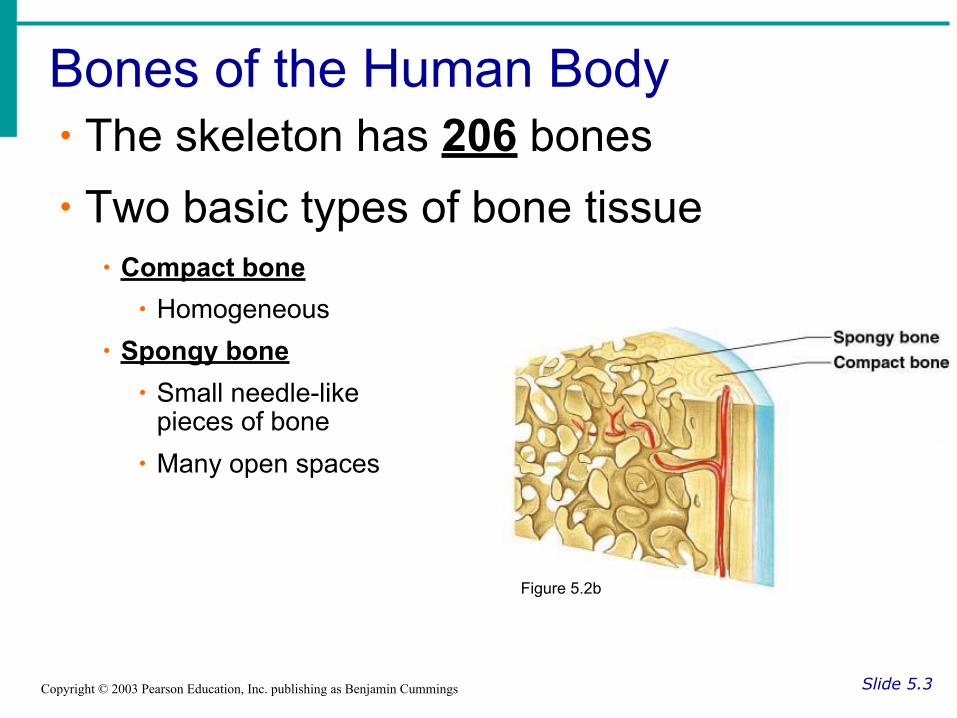
Bones of the Human Body
Slide 5.3Copyright © 2003 Pearson Education, Inc. publishing as Benjamin Cummings
∙ The skeleton has 206 bones∙ Two basic types of bone tissue∙ Compact bone∙ Homogeneous
∙ Spongy bone∙ Small needle-like
pieces of bone∙ Many open spaces
Figure 5.2b
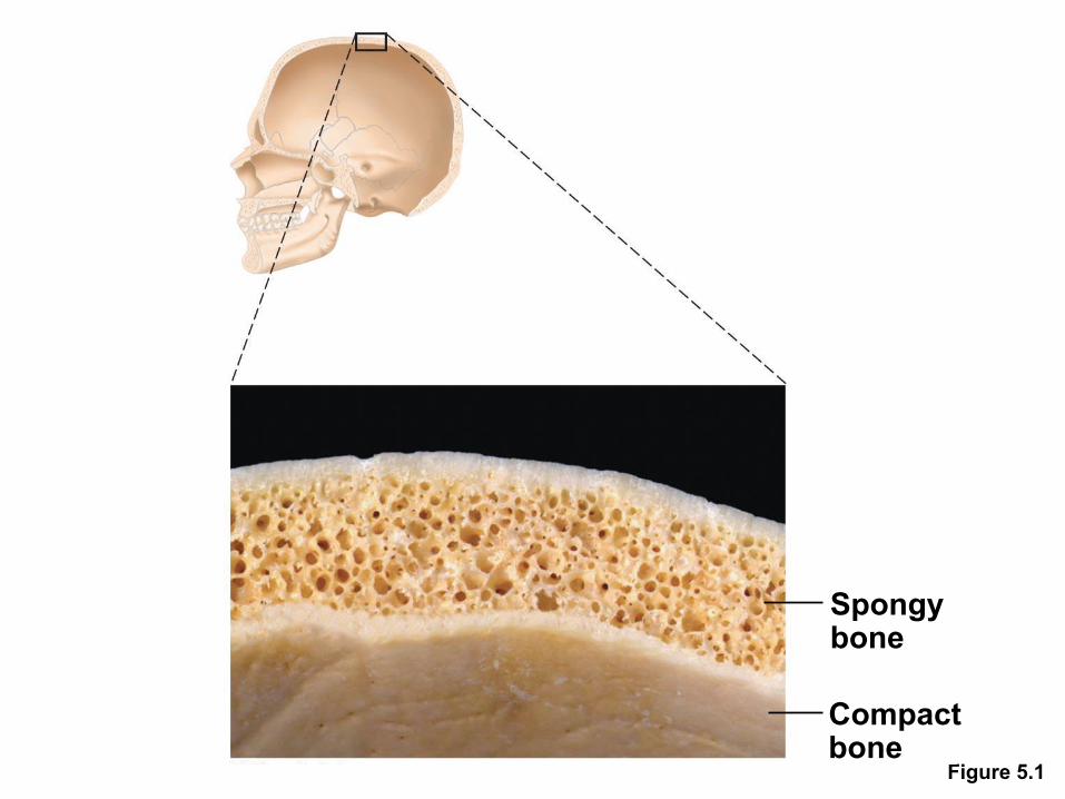
Figure 5.1
Spongybone
Compactbone

Figure 5.2

Classification of Bones
Slide 5.4aCopyright © 2003 Pearson Education, Inc. publishing as Benjamin Cummings
∙ Long bones∙ Typically longer than they are
wide
∙ Shaft with heads situated at both ends
∙ Contain mostly compact bone
∙ All of the bones of the limbs (except wrist, ankle, and kneecap bones)
∙ Examples: ■ Humerus, ulna, radius
■ Metacarpals, metatarsals
■ Phalanges (on both hands and feet)
■ Femur, tibia, fibula

Figure 5.2a

Classification of Bones
Slide 5.4bCopyright © 2003 Pearson Education, Inc. publishing as Benjamin Cummings
∙ Short bone∙ Generally cube-shaped
∙ Contain mostly spongy bone
∙ Includes bones of the wrist and ankle
∙ Sesamoid bones are a type of short bone which form within tendons (patella)
∙ Examples:
● Carpals, tarsals
● Talus, calcaneus
● patella

Figure 5.2d

Classification of Bones
Slide 5.5aCopyright © 2003 Pearson Education, Inc. publishing as Benjamin Cummings
∙ Flat bones∙ Thin and flattened
∙ curved
∙ Thin layers of compact bone around a layer of spongy bone
∙ Examples: ● All skull bones
● Sternum
● Clavicle
● Scapula
● All ribs

Figure 5.1
Spongybone
Compactbone

Figure 5.2c

Classification of Bones
Slide 5.5bCopyright © 2003 Pearson Education, Inc. publishing as Benjamin Cummings
∙ Irregular bones∙ Irregular shape
∙ Do not fit into other bone classification categories
∙ Example:
● All vertebrate
● All bones of the pelvic girdle

Figure 5.2b

Structures of a Long Bone
Slide 5.8bCopyright © 2003 Pearson Education, Inc. publishing as Benjamin Cummings
Figure 5.2a

Gross Anatomy of a Long Bone
Slide 5.6Copyright © 2003 Pearson Education, Inc. publishing as Benjamin Cummings
∙ Diaphysis∙Shaft∙Composed of compact bone
Figure 5.2a

Gross Anatomy of a Long Bone
Slide 5.6Copyright © 2003 Pearson Education, Inc. publishing as Benjamin Cummings
∙ Epiphysis ∙Ends of the bone∙Composed mostly of spongy bone
Figure 5.2a

Structures of a Long Bone
Slide 5.7Copyright © 2003 Pearson Education, Inc. publishing as Benjamin Cummings
∙ Periosteum∙Outside covering of the diaphysis∙ Fibrous connective tissue membrane
Figure 5.2c

Structures of a Long Bone
Slide 5.7Copyright © 2003 Pearson Education, Inc. publishing as Benjamin Cummings
∙ Sharpey’s fibers∙Secure periosteum to underlying bone
Figure 5.2c

Structures of a Long Bone
Slide 5.7Copyright © 2003 Pearson Education, Inc. publishing as Benjamin Cummings
∙ Arteries∙Supply bone cells with nutrients
Figure 5.2c

Structures of a Long Bone
Slide 5.8aCopyright © 2003 Pearson Education, Inc. publishing as Benjamin Cummings
∙ Articular cartilage∙Covers the epiphyses
∙Made of hyaline cartilage
∙Decreases friction at joint surfaces
Figure 5.2a

Figure 5.3b
Compact bone
Spongy bone
Articularcartilage
(b)

Structures of a Long Bone
Slide 5.8bCopyright © 2003 Pearson Education, Inc. publishing as Benjamin Cummings
∙ Medullary cavity∙Contains yellow marrow (mostly fat) in adults
∙Contains red marrow (for blood cell formation) in infants
Figure 5.2a

Bone Markings
Slide 5.9Copyright © 2003 Pearson Education, Inc. publishing as Benjamin Cummings
•Surface features of bones•Sites of attachments for muscles, tendons, and ligaments•Passages for nerves and blood vessels
•Categories of bone markings•Projections or processes—grow out from the bone surface
•Terms often begin with “T”•Depressions or cavities—indentations
•Terms often begin with “F”


• Title• Microscopic Anatomy
and Bone Growth
• Essential Question• How do the microscopic
structures of bone help in the process of bone growth and remodeling?
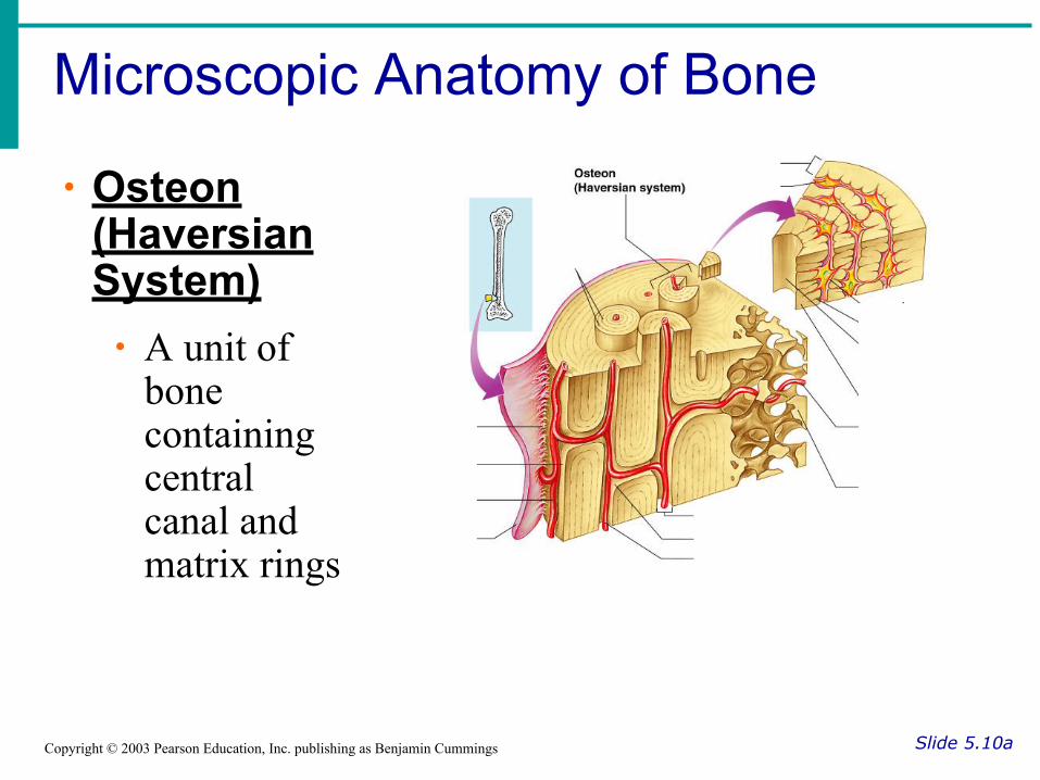
Microscopic Anatomy of Bone
Slide 5.10aCopyright © 2003 Pearson Education, Inc. publishing as Benjamin Cummings
∙ Osteon (Haversian System)∙ A unit of
bone containing central canal and matrix rings

Microscopic Anatomy of Bone
Slide 5.10aCopyright © 2003 Pearson Education, Inc. publishing as Benjamin Cummings
∙ Central (Haversian) canal∙ Opening in the center of an osteon∙ Carries blood vessels and nerves

Microscopic Anatomy of Bone
Slide 5.10aCopyright © 2003 Pearson Education, Inc. publishing as Benjamin Cummings
∙ Perforating (Volkman’s) canal∙ Canal perpendicular to the central canal∙ Carries blood vessels and nerves

Microscopic Anatomy of Bone
Slide 5.11aCopyright © 2003 Pearson Education, Inc. publishing as Benjamin Cummings
∙ Lacuna∙Cavities containing bone cells (osteocytes)∙Arranged in concentric rings
Figure 5.3

Figure 5.4c
Osteon
Lacuna
Centralcanal
Interstitiallamellae
(c)

Microscopic Anatomy of Bone
Slide 5.11aCopyright © 2003 Pearson Education, Inc. publishing as Benjamin Cummings
∙ Lamella∙Rings around the central canal∙Sites of lacunae
Figure 5.3

Microscopic Anatomy of Bone
Slide 5.11bCopyright © 2003 Pearson Education, Inc. publishing as Benjamin Cummings
∙ Canaliculus∙Tiny canals∙Radiate from the central canal to lacunae∙Form a transport system
Figure 5.3

Types of Bone Cells
Slide 5.15Copyright © 2003 Pearson Education, Inc. publishing as Benjamin Cummings
∙ Osteocytes∙Mature bone cells
∙ Osteoblasts∙Bone-forming cells

Types of Bone Cells
Slide 5.15Copyright © 2003 Pearson Education, Inc. publishing as Benjamin Cummings
∙ Osteoclasts∙Bone-destroying cells∙Break down bone matrix for remodeling and release of calcium in response to parathyroid hormone
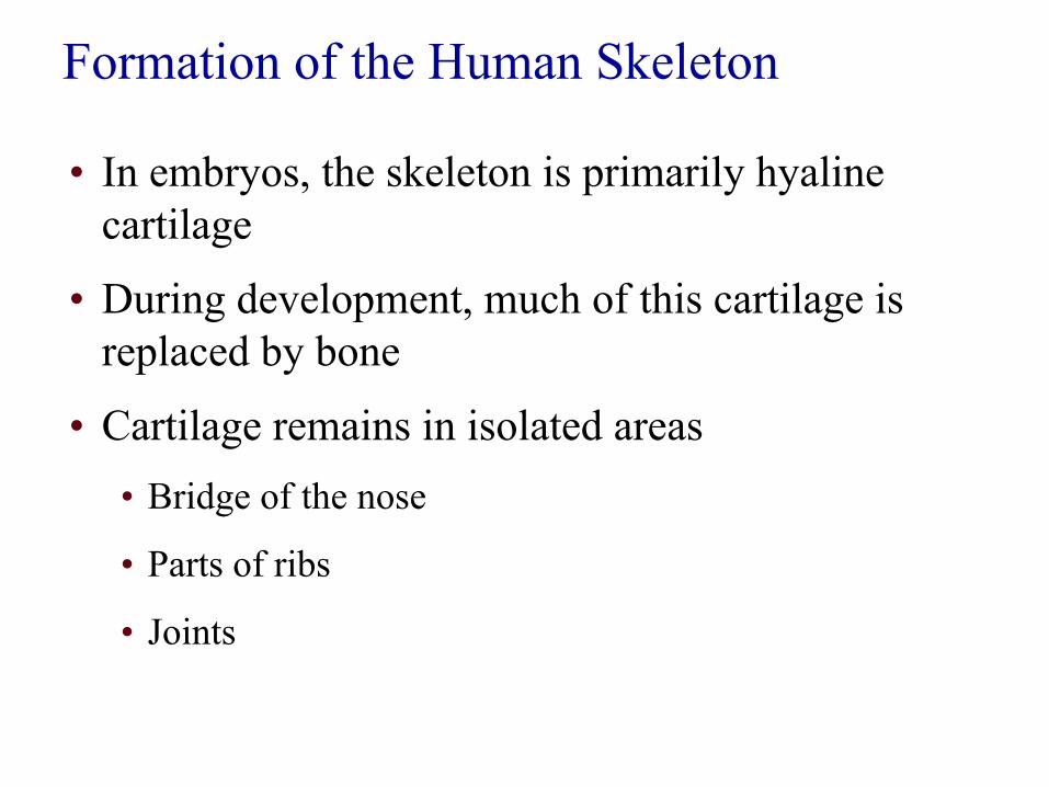
Formation of the Human Skeleton
• In embryos, the skeleton is primarily hyaline cartilage
• During development, much of this cartilage is replaced by bone
• Cartilage remains in isolated areas• Bridge of the nose
• Parts of ribs
• Joints

Bone Growth (Ossification)
Slide 5.13aCopyright © 2003 Pearson Education, Inc. publishing as Benjamin Cummings
•Epiphyseal plates allow for lengthwise growth of long bones during childhood
•New cartilage is continuously formed•Older cartilage becomes ossified
•Cartilage is broken down•Enclosed cartilage is digested away, opening up a medullary cavity•Bone replaces cartilage through the action of osteoblasts
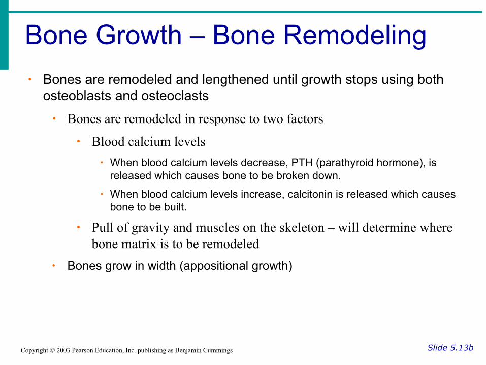
Bone Growth – Bone Remodeling
Slide 5.13bCopyright © 2003 Pearson Education, Inc. publishing as Benjamin Cummings
∙ Bones are remodeled and lengthened until growth stops using both osteoblasts and osteoclasts
∙ Bones are remodeled in response to two factors
∙ Blood calcium levels∙ When blood calcium levels decrease, PTH (parathyroid hormone), is
released which causes bone to be broken down.
∙ When blood calcium levels increase, calcitonin is released which causes bone to be built.
∙ Pull of gravity and muscles on the skeleton – will determine where bone matrix is to be remodeled
∙ Bones grow in width (appositional growth)

Figure 5.5
In a fetusIn an embryo
Bone collarHyalinecartilagemodel
Bone startingto replacecartilage
In a child
Medullarycavity
New center ofbone growth
Hyalinecartilage
Epiphysealplate cartilage
Growthin bonelength
New boneforming
Invadingbloodvessels
Epiphysealplatecartilage
Articularcartilage
Spongybone
New boneforming
Growthin bonewidth

Figure 5.5, step 1
In an embryo
Bone collarHyalinecartilagemodel
Bone startingto replacecartilage

Figure 5.5, step 2
In a fetus
Medullarycavity
New center ofbone growth
Hyalinecartilage
Growthin bonelength
Invadingbloodvessels

Figure 5.5, step 3
In a child
Epiphysealplate cartilage
New boneforming
Invadingbloodvessels
Epiphysealplatecartilage
Articularcartilage
Spongybone
New boneforming
Growthin bonewidth
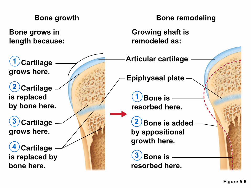
Figure 5.6
Bone growth
Bone grows inlength because:
Bone remodeling
Growing shaft isremodeled as:
Cartilagegrows here.
Cartilageis replacedby bone here.
Cartilagegrows here.
Cartilageis replaced by bone here.
1
2
3
4
1
2
3 Bone isresorbed here.
Epiphyseal plate
Articular cartilage
Bone isresorbed here.
Bone is addedby appositionalgrowth here.


• Title• Bone Fractures and
Repair
• Essential Question• What are the different
types of bone fracture, and how does the body heal a fracture?

Bone Fractures
Slide 5.16Copyright © 2003 Pearson Education, Inc. publishing as Benjamin Cummings
∙ Types of bone fractures∙Closed (simple) fracture – break that does not penetrate the skin∙Open (compound) fracture – broken bone penetrates through the skin

Common Types of Fractures
Slide 5.17Copyright © 2003 Pearson Education, Inc. publishing as Benjamin Cummings
Table 5.2

Bone Fractures
Slide 5.16Copyright © 2003 Pearson Education, Inc. publishing as Benjamin Cummings
∙ Treatment∙ reduction and immobilization∙ Realignment of the bone∙ Surgery is needed in some occasions

Repair of Bone Fractures
Slide 5.18Copyright © 2003 Pearson Education, Inc. publishing as Benjamin Cummings
1. Hematoma (blood-filled swelling) is formed
2. Break is splinted by fibrocartilage to form a callus
3. Fibrocartilage callus is replaced by a bony callus
4. Bony callus is remodeled to form a permanent patch

Stages in the Healing of a Bone Fracture
Slide 5.19Copyright © 2003 Pearson Education, Inc. publishing as Benjamin Cummings
Figure 5.5

Analyzing X-Rays

Examining X-Rays
• Your team will be given an image of an x-ray and you will need to:• Name all the bones on the x-ray
• Name the type of fracture (s)
• Provide a treatment plan
• Choose a team leader to speak for your group

INB pg 64
• Title: X Ray Example Date:• Drawing of the x-ray
• Label ALL Bones
• Name the type of fracture(s)
• Write the treatment plan

Case #1

My Expert Opinion
• Bones on X-ray: femur, tibia, fibula
• Type of Fracture: simple, impacted
• Treatment Plan: stabilize the bones and place cast on leg to immobilize the leg

Case #2

My Expert Opinion
• Bones on X-ray: femur and pelvis
• Type of Fracture: compound and comminuted
• Treatment Plan: stabilize the open wound, surgery to clean the wound, and stabilize bones with pins and screws
Case #3

My Expert Opinion
• Bones on x-ray: femur, tibia, fibula
• Type of Fracture: simple, spiral and greenstick
• Treatment: realign the bone and stabilize with a cast

Case #4

My Expert Opinion
• Bones on x-ray: skull and cervical vertebrate
• Type of Fracture: compression of the C5
• Treatment: immobilize neck with brace

Case #5

My Expert Opinion
• Bones on x-ray: humerus, radius, ulna, and phalange
• Type of Fracture: greenstick of the distal ulna
• Treatment: realign the bone and stabilize with a cast

Case #6

My Expert Opinion
• Bones on x-ray: radius, ulna, carpel, metacarpal
• Type of Fracture: simple, impacted
• Treatment: realign the bone and stabilize with a cast

Case #7
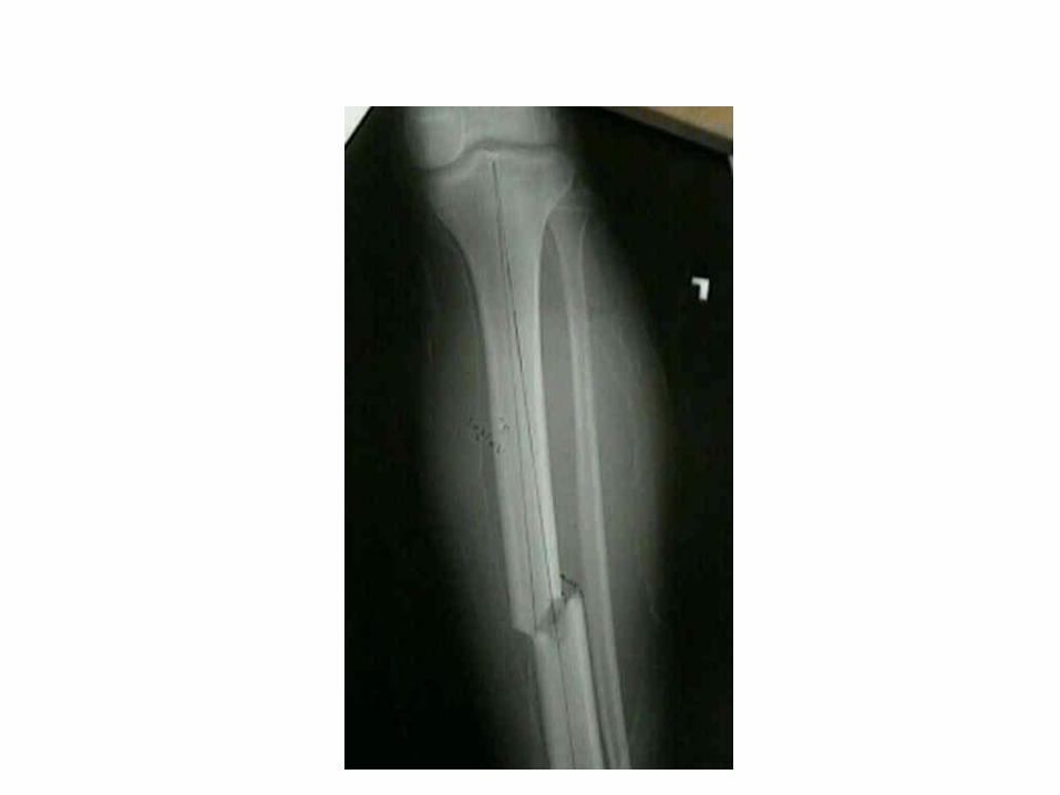
My Expert Opinion
• Bones on x-ray: humerus, ulna, and radius
• Type of Fracture: simple, spiral
• Treatment: realign the bone and stabilize with a cast


• Title• Joints
• Essential Question• Describe the structure
and function of joints.

Joints
Slide 5.43Copyright © 2003 Pearson Education, Inc. publishing as Benjamin Cummings
∙ Functions∙Hold bones together
∙Allow for mobility

Functional Classification of Joints
Slide 5.44Copyright © 2003 Pearson Education, Inc. publishing as Benjamin Cummings
∙ Synarthroses – immovable
∙ Amphiarthroses – slightly moveable
∙ Diarthroses – freely moveable

Structural Classification of Joints
Slide 5.45Copyright © 2003 Pearson Education, Inc. publishing as Benjamin Cummings
∙ Fibrous joints
∙ Cartilaginous joints
∙ Synovial joints

Fibrous Joints
Slide 5.46Copyright © 2003 Pearson Education, Inc. publishing as Benjamin Cummings
•Bones united by collagenic fibers•Types
•Sutures•Immobile•Example: Joints between skull bones
•Syndesmoses•Allows more movement than sutures but still immobile•Example: Distal end of tibia and fibula
•Gomphosis•Immobile•Example: Bind teeth to bony socket
Figure 5.27d, e

Figure 5.30a
Fibrous joints
Fibrous connective tissue
(a) Suture

Figure 5.30b
Fibrous joints
Tibia
FibulaFibrousconnective tissue
(b) Syndesmosis


Cartilaginous Joints
Slide 5.47Copyright © 2003 Pearson Education, Inc. publishing as Benjamin Cummings
•Bones connected by cartilage•Types
•Synchrondrosis •Immobile•Example: first sternocostal joint
•Symphysis •Slightly movable•Example: Pubic symphysis, intervertebral joints
Figure 5.27b, c

Figure 5.30c
Cartilaginous joints
First rib
Hyaline cartilage
Sternum
(c) Synchondrosis

Figure 5.30d
Cartilaginous joints
Vertebrae
Fibrocartilage
(d) Symphysis

Figure 5.30e
Cartilaginous joints
Pubis
Fibro- cartilage
(e) Symphysis

Synovial Joints
Slide 5.48Copyright © 2003 Pearson Education, Inc. publishing as Benjamin Cummings
• Articulating bones are separated by a joint cavity
• Synovial fluid is found in the joint cavity
•Articular cartilage (hyaline cartilage) covers the ends of bones
•Articular capsule encloses joint surfaces and lined with synovial membrane
•Joint cavity is filled with synovial fluid
•Reinforcing ligaments
•Example: Humerus with the ulna and radius
Figure 5.27f–h

Figure 5.30f
Synovial joints
ScapulaArticular capsule
Articular (hyaline) cartilageHumerus
(f) Multiaxial joint (shoulder joint)

Figure 5.30g
Synovial joints
Humerus
Articular (hyaline) cartilageArticular capsuleRadius
Ulna(g) Uniaxial joint (elbow joint)

Figure 5.30h
Synovial joints
UlnaRadiusArticular capsule
(h) Biaxial joint (intercarpal joints of hand)
Carpals
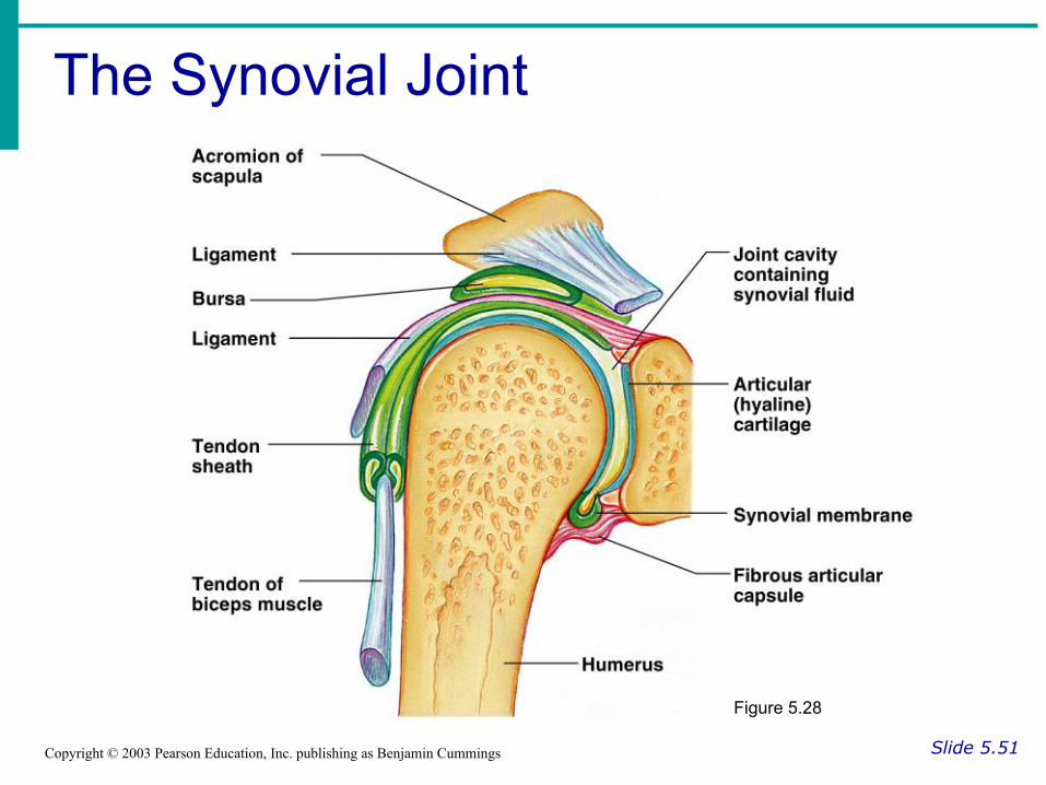
The Synovial Joint
Slide 5.51Copyright © 2003 Pearson Education, Inc. publishing as Benjamin Cummings
Figure 5.28

Types of Synovial Joints Based on Shape
Slide 5.52aCopyright © 2003 Pearson Education, Inc. publishing as Benjamin Cummings
Figure 5.29a–c

Types of Synovial Joints Based on Shape
Slide 5.52bCopyright © 2003 Pearson Education, Inc. publishing as Benjamin Cummings
Figure 5.29d–f