Embed Size (px)
Citation preview

Presence of Renin Secretory Granules in Rat Adrenal Gland and Stimulationof Renin Secretion by Angiotensin 11 but Not by AdrenocorticotropinKenji Mizuno, Loren H. Hoffman, James C. McKenzie, and Tadashi InagamiDepartments of Biochemistry and Cell Biology, Vanderbilt University School of Medicine, Nashville, Tennessee 37232;Department of Anatomy, Howard University College of Medicine, Washington, DC20059
Abstract
Renin has been identified biochemically and immunohisto-chemically in the adrenal gland. Weexamined the subcellulardistribution and behavior of adrenal renin. By differential cen-trifugation of adrenal capsules, we found renin mainly in mito-chondrial fractions. By Percoll density gradient centrifugationof this fraction, dense granules were separated from mitochon-dria and microsomes. The renin activity in the dense granulesfrom the capsules of nephrectomized rats was 15 times greaterthan that of the intact rat. Immunohistochemical studies re-vealed that the dense granules increased in number after bilat-eral nephrectomy. Immunogold staining of these granulesshowed unequivocally the presence of renin in these granules.Adrenal capsules in organ culture were found to release reninat a steady rate. Renin release from bilaterally nephrectomizedrat adrenals was 46 times faster than from the organs of intactanimals. The mechanism of the control of renin secretion fromthe adrenal gland was different from the kidney in that thesecretion was stimulated by potassium chloride (10 mM) orangiotensin II (10--10- M) but not by ACI'H (10-9-10-7M), suggesting stimulation by intracellular calcium. These re-sults provide evidence that the adrenal synthesizes renin,stores it in specific secretory granules and secretes it in a regu-lated manner.
Introduction
Renin has been recognized as a plasma enzyme released fromthe kidney. It mediates the first step of angiotensin formationby cleaving the decapeptide angiotensin I (Ang I)' from themacromolecular prohormone angiotensinogen in a highly spe-cific manner. Ang I is further converted to the active octapep-tide angiotensin II (Ang II) by a converting enzyme. Ang II is apotent vasoconstrictor and aldosterone secretagogue. Thus,renin plays a central role in blood pressure regulation. On theother hand, recent biochemical and immunological studiesrevealed that renin exists in the adrenal gland of variousmammalian species (1-5). It was also demonstrated that thisadrenal renin is primarily located in the zona glomerulosa cells
Address reprint requests to Dr. Inagami, Department of Biochemistry,Vanderbilt University School of Medicine, Nashville, TN 37232.
Receivedfor publication 15 January 1988 and in revisedform 13April 1988.
1. Abbreviations used in this paper: Ang I, angiotensin I; PMSF, phen-ylmethanesulfonyl fluoride.
rather than the fasciculata or medullary cells (6, 7). Further,the adrenal renin has been reported to increase markedly afterhigh potassium loading or after nephrectomy (7-9), whenplasma renin activity was at undetectable levels. These find-ings suggest that the adrenal renin is not merely due to con-tamination by plasma renin of renal origin but endogenous tothe adrenal. In support of this, messenger RNAfor renin hasbeen detected in the adrenal gland (10).
However, the functional significance of renin in adrenal isnot clear. Only a few reports (7, 9) suggested that the renin isinvolved in the production of aldosterone, presumablythrough intraadrenal generation of Ang II by renin, but noconcrete evidence for such function of renin in this tissue hasyet been obtained. Also unknown is whether the renin is se-creted from the adrenal. This intriguing question is raised bythe findings of the presence of renin-like activity and of immu-noreactive Ang I and Ang II in the plasma of anephric subjects(1 1-13), and the hypotensive response to converting enzymeinhibitor, captopril, by a sodium-depleted anephric subject(14), suggesting that renin or renin-like enzymes synthesized inextrarenal tissues may contribute to the generation of plasmaangiotensin. In view of these interesting issues, we tested thehypothesis that there exist renin-secretory granules in the adre-nal. Weshow here in rat adrenals that renin was identified inthe dense granules and that in the cultured adrenal capsulartissues, renin release was found and stimulated by potassiumand Ang II, but not by ACTH. The data suggest that renin islocalized in the storage granules of the adrehal presumably tobe released into the extracellular space, by a regulated mecha-nism.
Methods
Animals. Male Sprague-Dawley rats (Harlan Sprague-Dawley, India-napolis, IN) weighing 180-220 g were used for the experiments. Therats were maintained on a regular Purina chow diet (Ralston PurinaCo., St. Louis, MO)containing 0.39% sodium and 0.9% potassium andallowed free access to tap water. Bilateral nephrectomy was performedwith some of the rats under sodium pentobarbitol anesthesia (30mg/kg i.p.) 32 h before they were killed.
Fractionation by differential centrifugation. Normal and nephrec-tomized rats were anesthetized with sodium pentobarbital (30 mg/kgi.p.). The adrenal glands were removed immediately after the rats wereperfused with ice-cold saline (- 150 ml) for a period of 15 minthrough the abdominal aorta by using an infusion pump (Cole-ParmerInstrument Co., Chicago, IL) and the adrenal capsules were preparedby manual compression using nylon gauze (pore size, 100 Mm; Spec-trum Medical Industries Inc., Los Angeles, CA). All the following stepswere performed at 4°C. The capsular tissues from 10 to 12 adrenalswere cut into small pieces (- 2 X 2 mm)and homogenized gently in 6ml of ice-cold 0.25 Msucrose containing 30 mMTris-HCI and 1 mMNa2-EDTA, pH 7.4, with a motor-driven Potter-Elvehjem grinder for30 strokes at 500 rpm. The crude homogenate (hereafter called "origi-nal homogenate") was then subjected to subsequent fractionation.
Renin Granules in Rat Adrenal Gland 1007
J. Clin. Invest.© The American Society for Clinical Investigation, Inc.0021-9738/88/09/1007/10 $2.00Volume 82, September 1988, 1007-1016

After separating the unbroken cells, cell debris and capsular frag-ments (PO) by centrifugation of the original homogenate at 70 g for 10min, the supernatant was centrifuged at 600 g for 10 min. The pelletwas homogenized again in 3 ml of the sucrose solution under the sameconditions as described above and recentrifuged at 600 g for 10 min toremove the nuclear fraction (PI) in the pellet. The supernatant wascombined (- 9 ml) and further centrifuged at 5,200 g for 20 min toobtain a heavy mitochondrial fraction (P2) in the pellet. The P2 pelletwas washed twice by resuspension in 3 ml of the sucrose solution andrecentrifugation at 5,200 g for 20 min. The resulting supernatant (S2,
- 15 ml) was centrifuged at 12,000 g for 20 min to obtain a lightmitochondrial fraction (P2), and then the supernatant was furthercentrifuged at 105,000 g for 60 min to separate the microsomal frac-tion (M) from the cytosolic fraction (S).
Density gradient centrifugation. The P2 pellet was gently dispersedin 1 ml of the sucrose solution, and 0.5-ml of the solution was added to7.5 ml of 35%Percoll (Sigma Chemical Co., St. Louis, MO)containing0.25 Msucrose, 1 mMNa2-EDTA and 30 mMTris-HCl, pH 7.4. ThePercoll gradient was initiated by centrifugation at 30,000 g for 30 minin an angle-head rotor (50 Ti; Beckman Instruments, Palo Alto, CA). Atube containing standard density beads (Sigma Chemical Co.), whichhad been constituted in distilled water (0.5 ml) and then each of them(5 js) was applied to the Percoll/sucrose solution, was placed in thesame rotor and centrifuged simultaneously. The sedimentation densityof the subcellular particles within the gradient was determined bycomparison with the location of the density beads. After centrifuga-tion, the sample was fractionated into 10 fractions (0.8 ml each) bypipetting.
Measurement of enzyme activities and protein content. Samplesfrom differential and density gradient centrifugation were treated with0.1 vol of phosphate buffer (0.2 M, pH 7.0) containing 1% TritonX-100 before assay. Specific immunoreactive renin activity was deter-mined using antirenin antibody to distinguish it from nonspecificrenin-like activity of proteases according to the method previouslydetected (4, 5, 15, 16). 25 to 50 Ml of the samples were incubated with10 ,ul of diluted (1:500) rabbit antiserum raised against pure rat kidneyrenin (17) at 4VC for 20 h. This antibody used resulted in 50% inhibi-tion of renin activity (40.0 ng Ang I/h) in a rat kidney extract at adilution of 1:10,000 and complete inhibition at a dilution of 1:1,000.For controls, the same sample was incubated with preimmune rabbitserum at the same dilution. The pretreated sample was incubated at370C for 1 h (in case of fractions from differential centrifugation) or for4 h (fractions from density gradient) with 75 ul of unfractionatedplasma of bilaterally nephrectomized rats as renin substrate in 0.2 Mphosphate buffer, pH 7.0, containing 10 mMNa2-EDTA and 1 mMphenylmethanesulfonyl fluoride (PMSF). The generated Ang I wasmeasured by radioimmunoassay (18). The recovery of added Ang I(5.0 ng), incubated with original homogenates for 1 h at 370C, was92.8±3.1% (mean±SE, n = 3). Therefore, significant angiotensinaseinhibition was achieved under the conditions employed. The inter-and intraassay coefficients of variation for the renin assay over themeasured range were 7.3±1.3% and 6.2±0.9% (mean±SE, n = 10),respectively. The difference in the renin activity'of the sample prein-cubated with preimmune rabbit serum and that with anti-renin anti-serum was defined as specific renin activity. The renin-like activity ineach fraction obtained from differential and density gradient centrifu-gation was almost completely (> 98% of the total renin-like activity)inhibited by antirenin antibody under the present conditions, indicat-ing that specific renin activity comprises the majority of the renin-likeactivity. The specific renin activity is expressed in terms of nanogramsAng I * mgof protein *h-' (or 4 h-').
Acid phosphatase activity as a lysosomal marker enzyme was mea-sured at pH 4.8 with p-nitrophenyl phosphate as substrate (19), and theactivity is expressed as absorbance of -nitrophenol measured at 410nm. Succinate dehydrogenase activity was measured as a mitochon-drial marker according to the method of Slater and Bonner (20), andthe activity is reported as the decrease of absorbance of ferricyanide at450 nm. In preliminary experiments, we confirmed that these enzyme
activities were not influenced by the addition of Percoll solutionsunder conditions used in the present assay system. The protein con-centration was determined by the method of Lowry et al. (21), usingBSA (crystallized and lyophilized, Sigma Chemical Co.) as the stan-dard.
Immunocytochemistry. Immunocytochemical studies were per-formed on (a) localization of renin in the adrenal cortex to confirm thepresence of renin in the capsular (i.e., zona glomerulosa) tissues and (b)subcellular particles prepared by Percoll density gradient centrifuga-tion.
For localization of renin in the adrenal, male Sprague-Dawley rats(200-250 g) were nephrectQmized bilaterally 48 h before euthanasia byexsanguination under pentobarbital anesthesia (30 mg/kg i.p.). Ratswere perfused with 150 ml Tyrode's buffer (pH 7.4) followed by 250 mlice-cold Perfix (Fisher Scientific Co., Pittsburgh, PA). Adrenal glandswere excised, routinely embedded in Paraplast Plus (Polysciences, Inc.,Warrington, PA), sectioned at 5 Mmand mounted on acid-cleaned,gelatin/alum-coated glass slides. Immunohistochemical staining forrenin was performed using an antibody against purified rat kidneyrenin (17) at a dilution of 1:500, and ABCkits purchased from VectorLaboratories (Burlingame, CA) as previously described (22). Controlsincluded serial dilution of antibody and substitution of normal rabbitserum for antirenin antibody.
To separate subcellular particles from Percoll solutions which con-tain silica coated with polyvinylpyrrolidone, a fractionated sample wascentrifuged at 100,000 g for 90 min at 4VCwith an angle-head rotor (50Ti). The particles which remained above the hard pellet were carefullycollected by pipetting, transferred to a medium consisting of 0.25 Msucrose, 1 mMNa2-EDTA and 30 mMTris-HCI, pH 7.3, and centri-fuged again at 15,000 g for 30 min at 4°C. The supernatant wasdecanted, after which the pellet was fixed for 2 h at 4°C in 3%glutar-aldehyde buffered in 0.12 Msodium'cacodylate, pH 7.3. After bufferrinses, they were post-fixed in 1% OSO4 in the same buffer for 1 h.Samples were dehydrated with ascending concentrations of ethanol,treated with propylene oxide and embedded in Embed 812 (ElectronMicroscopy Sciences, Fort Washington, PA). Purified aldehyde solu-tions and OSO4were supplied by Ladd Research Industries, Inc., Bur-lington, VT. Thin sections showing silver-gray interference colors wereobtained from appropriate regions in the pellets, stained with uranylacetate and lead citrate, and examined at 75 kV in a Hitachi-600transmission electron microscope (Hitachi, Tokyo, Japan).
For electron microscopic immunolocalization of renin, the Percollfractions were fixed at 4°C in 4%formaldehyde/0.25% glutaraldehydein sodium cacodylate buffer for 30 min, then for an additional 6 h inthe fixative lacking glutaraldehyde. No OSO4 postfixation was em-ployed. Samples were treated with 0.5% sodium borohydride for 30min to reduce unreacted aldehyde groups, then rinsed extensively inbuffer, dehydrated as above and embedded. The epoxy resin was poly-merized at 45°C. Thin sections showing silver interference weremounted on nickel grids. These were treated for 30 min with saturatedsodium metaperiodate (23) or for 45 s with 4%potassium methoxide(24) to partially etch the epoxy resin. Grids were incubated in Tris-buffered saline (TBS, 20 mrM Tris-HCI, 150 mMNaCl, pH 7.6) con-taining 5% normal goat serum, then reacted with the rabbit anti-ratrenin antibody for 18 h at 4°C. Renin antiserum was used at a dilutionof 1:100 or 1: 1,000 in TBS/0. 1%bovine serum albumin (BSA, crystal-ized, globulin-free; Sigma Chemical Co.) containing 1% normal goatserum. Control preparations were treated with comparable dilutions ofpreimmune rabbit serum. After extensive rinses in TBS/BSA, the gridswere treated for 1 h with goat anti-rabbit IgG complexed with 10 nmgold particles (E-Y Laboratories, Inc., San Mateo, CA). The immuno-globulin-gold complex was diluted 1:20 with TBS/BSA and centri-fuged at 2,000 g for 20 min before use to remove aggregates. Afterrinsing in TBS/BSA, grids were reacted briefly with 3%glutaraldehyde,rinsed in water and stained with uranyl acetate and lead citrate.
Adrenal tissue culture. Male Sprague-Dawley rats weighing200-250 g were used for the tissue culture. The rats were maintainedon a regular Purina chow diet and allowed free access to tap water.
1008 K Mizuno, L. H. Hoffman, J. C. McKenzie, and T. Inagami

Bilateral nephrectomy was performed under sodium pentobarbital an-esthesia (30 mg/kg i.p.) 24 to 28 h before sacrifice. The adrenal cap-sules were prepared in these rats as described above and transferred toPetri dishes (9.6 cm2, capsules from three adrenals per one dish). 1 mlof RPMI 1640 medium supplemented with 0.1% BSA (RIA grade,Sigma Chemical Co.), 50 ,g/ml gentamicin sulfate and 0.5 mMPMSF(all in final concentration) were added to each dish. Cultures weremaintained at 370C under 95% air/5% CO2 for 8 h and the mediumwas totally exchanged at 2-h intervals. The medium obtained wasimmediately frozen with acetone/dry ice and stored at -80'C untilrenin assay. Since it was reported that potassium, Ang II, and ACTHinfluenced adrenal renin content (9, 25), we further examined whetherthese substances induce renin secretion from the adrenal. Potassiumchloride, Ang II (5Ile-Ang II) and ACTH (human ACTH'-24), bothfrom Peninsula Laboratories, Inc., Belmont, CA, were separatelyadded to the medium to give a final concentration of 10 mMforpotassium chloride, l0-7 to 10-9 Mfor Ang II and ACTH. The adrenalcapsules were cultured in these media for a period of 8 h, renewing themedia at 2-h intervals. Cumulative renin activity was transformedfrom these fresh 2-h samples and expressed as ng Ang I/8 h incubationwith substrate/8 h tissue incubation/adrenal. Renin activity in themedium was determined by Ang I-generating activity essentially asdescribed above except for the incubation period of renin-angiotensin-ogen reaction (8 h at 37°C).
Statistical analysis. All data are expressed as the mean±SE. Statis-tical analysis was performed on data from the tissue culture experi-ments with one-way analysis of variance followed by t test where ap-propriate for unpaired group. A P value of 0.05 was considered toindicate a significant difference.
Results
Subcellular distribution of reninTable I gives the average distribution of renin, marker en-zymes, and protein in the fractions obtained by differentialcentrifugation of the homogenate from normal rat adrenalcapsules. More than 60% of the total activity of renin wascontained in P2. Similarly, about 60% of the total activity ofsuccinate dehydrogenase was found in the P2 pellet. The acidphosphatase activity was dissociated from renin and succinatedehydrogenase activities, and more than 40%was contained inS2 supernatant. However, about 25% of the total activity stillremained in the P2, indicating that lysosomes were pelletedsimultaneously in this fraction to some extent. As comparedwith the original homogenate, a threefold enrichment in spe-cific activity of renin was obtained in P2 (3.45 ng Ang I * mgof
protein-' * h-' in original homogenate vs. 10.67 ng Ang I * mgof protein-' * h-' in P2).
In order to determine if the renin activity was associatedwith the mitochondrial fraction, we further examined the sub-cellular distribution of renin in the adrenal from nephrecto-mized rats, since nephrectomy has been known to cause amarked increase in renin content of adrenal capsular tissue (9).As shown in Table II, renin activity in the original homogenatewas very high as compared to that from normal rat adrenalsand, approximately 75% of the total activity of renin wasfound in P2. As compared with the original homogenate, a4.3-fold enrichment in specific activity of renin was obtainedin P2 (37.41 ng Ang I - mgof protein-' - h-' in original homog-enate vs. 159.47 ng Ang I * mgof protein-' * h-' in P2). Both thetotal and specific activities of renin in the P2 pellet were in-creased 15-fold over those from normal rat adrenals. On theother hand, protein contents as well as the total and specificactivities of marker enzymes in each fraction from thenephrectomized rat adrenals were practically unchanged fromthe controls. Further, the percentage distribution of renin,protein, and marker enzymes in fractions obtained from thenephrectomized groups showed an essentially similar patternto that of the normals (Tables I and II). Taken collectively,these data suggest that renin is associated with a populationdifferent from the mitochondria and lysosomes.
To further separate renin-related particles from mitochon-dria, lysosomes or other cell debris, the P2 pellet prepared fromnormal and nephrectomized rat adrenals was separately sub-jected to gradient centrifugation in 35% (vol/vol) Percoll. Aftercentrifugation, two bands were observed in the centrifugationtubes. The first band, near the top of the gradient, appearedlight brown. The second, at the bottom, appeared lighter andmore diffuse, and was much more dense in nephrectomizedgroups as compared with that of the normals. Representativeresults of the analysis of fractions collected from the top of thetubes are illustrated in Fig. 1. In the case of normal rat adrenals(Fig. 1 A), the first band, which had the lowest density, con-tained large quantities of acid phosphatase and succinate dehy-drogenase (Table III). The band at the bottom of the gradientcontained renin activity of > 60% of the total activity recov-ered from the entire gradient. This band was also partiallyassociated with acid phosphatase, and with only minor con-tamination by succinate dehydrogenase.
In the case of nephrectomized adrenals (Fig. I B), an al-
Table I. Subcellular Distribution of Immunoreactive Renin and Reference Enzymes from Adrenal Capsules of Control Rats
Fractions obtained by differential centrifugationOriginal
homogenate PO P1 P2 S2 P2 M S Recovery
Renin 20.12±0.52 0.45±0.09 0.14±0.01 10.99±0.27 1.76±0.35 1.58±0.09 1.01±0.14 1.17±0.51 85.0±1.2Acid phosphatase 11.80±2.13 0.82±0.22 0.50±0.13 2.81±0.88 4.75±0.50 0.74±0.06 0.70±0.17 0.98±0.37 96.4±4.9Succinate
dehydrogenase 6.40±0.08 0.75±0.19 1.11±0.13 3.40±0.28 0.27±0.14 0.10±0.04 0 0.10±0.05 89.7±4.7Protein 5.83±0.22 0.65±0.14 0.44±0.08 1.03±0.02 1.77±0.45 0.34±0.05 0.40±0.06 1.59±0.58 106.7±6.3
Enzyme activities and protein contents in original homogenate and in each fraction volume. Units are nanograms of angiotensin I/hour forrenin, absorbancy at 410 nm/h for acid phosphatase, decrease of absorbancy at 450 nm/16 h for succinate dehydrogenase and milligrams forprotein. Values are means±SE of three different experiments.
Renin Granules in Rat Adrenal Gland 1009
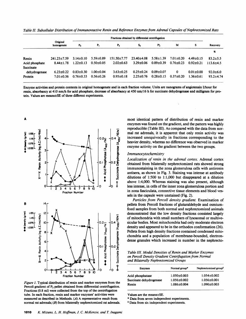
Table II. Subcellular Distribution of Immunoreactive Renin and Reference Enzymes from Adrenal Capsules ofNephrectomized Rats
Fractions obtained by differential centrifugationOriginal
homogenate PO P1 P2 S2 Pr2 M S Recovery
Renin 241.23±7.59 3.14±0.10 5.59±0.89 151.50±7.77 23.40±4.08 5.58±1.39 7.01±0.20 4.49±0.15 83.2±5.5Acid phosphate 8.44±1.78 1.22±0.13 0.50±0.05 2.02±0.63 3.29±0.06 0.89±0.39 0.76±0.23 0.92±0.21 113.6±4.5Succinate
dehydrogenase 6.23±0.22 0.83±0.30 1.00±0.04 3.63±0.25 0.25±0.24 0.09±0.07 0 0.01±0.00 92.0±6.0Protein 7.01±0.56 0.76±0.33 0.56±0.26 0.95±0.18 2.25±0.76 0.28±0.15 0.37±0.20 1.36±0.61 93.2±4.74
Enzyme activities and protein contents in original homogenate and in each fraction volume. Units are nanograms of angiotensin I/hour forrenin, absorbancy at 410 nm/h for acid phosphate, decrease of absorbancy at 450 nm/16 h for succinate dehydrogenase and milligram for pro-tein. Values are means±SE of three different experiments.
A most identical pattern of distribution of renin and markerenzymes was found on the gradient, and the pattern was highlyreproducible (Table III). As compared with the data from nor-
E -,s W mal rat adrenals, it is apparent that only renin activity was
1.11.5 E 4 0.2- 0.4 0 increased unequivocally in fractions corresponding to the* ~ I.075~ j' / \ogheavier density, whereas no difference was observed in marker
1.035L , ; '- a------ it < I genzyme activity on the gradient between the two groups.
:, /\ Al \ g g ImmunocytochemistryX.) / 0 (, i \ i|Localization of renin in the adrenal cortex. Adrenal cortex
X 0 /x x / A\\.Ig -0.2a obtained from bilaterally nephrectomized rats showed strong
cr immunostaining in the zona glomerulosa cells with antirenin
,L antisera, as shown in Fig. 3. Staining was intense at antibodyx | i dilutions of 1:500 to 1:1,000 but disappeared at a dilutionA>aNabove 1:4,000. Whereas staining was also present, although
c 1 2x-x-x-x-x 0O -0o less intense, in cells of the inner zona glomerulosa portion and2 3 4 5 6 7 8 9 10 in zona fasciculata, connective tissue elements and blood ves-Fraction Number
sels in the capsule were unstained (Fig. 2).Particles from Percoll density gradient. Examination of
B 14 - pellets from Percoll fractions of glutaraldehyde and osmium-x fixed samples from both normal and nephrectomized animalsE L155 - 12 - demonstrated that the low density fractions consisted largely
§ 1.115 of mitochondria with small numbers of lysosomal or multive-| 1.03SF E I 0.2 0.4 dsicularbodies. Mostmitochondria had only moderate electron
* 1.0 H X densityand appeared to be in the orthodox conformation (26).6 ° 8 -x Pellets from high density fractions contained condensed mito-
0I\lI 3 chondria and a population of membrane-bounded, electron-*36p \ X \ 3 | Hidense granules which increased in number in the nephrecto-6 -
=x h i~O.-0.2=04 0~~~X,4\1\ Table III. Modal Densities of Renin and Marker Enzymes
X _ \ on Percoll Density Gradient Centrifugation from Normal2 - :- and Bilaterally Nephrectomized Groupsx \ze
0 6 1 1 1 1' IJo J Ii 2 3 4 5 6 7 8 9 10
Fraction Number
Figure 1. Typical distribution of renin and marker enzymes from thePercoll gradient of P2 pellet obtained from differential centrifugation.Fractions (0.8 ml) were collected from the top of the centrifugationtube. In each fraction, renin and marker enzymes' activities were
measured as described in Methods. (A) A representative result fromnormal rat adrenals; (B) from bilaterally nephrectomized rat adrenals.
Enzymes Normal group* Nephrectomized groupt
Acid phosphatase 1.050±0.003 1.054±0.002Succinate dehydrogenase 1.056±0.002 1.056±0.001Renin 1.086±0.004 1.090±0.003
Values are the means±SE.* Data from seven independent experiments.t Data from six independent experiments.
1010 K. Mizuno, L. H. Hoffman, J. C. McKenzie, and T. Inagami

4- * Ali'C / ,rI Ve.
ilF'4~~~~~~~~~~~~4-~~~~~~~~~~~~~~~~~~~~~A
Figure 2. Immunohistochemical localization of renin in rat adrenal cortex. The first few layers of cells in the zona glomerulosa (zg) are in-tensely stained. Staining is also present, although less intense, in cells of the inner zona glomerulosa and in zona fasiculata (zf; arrowheads).Connective tissue elements and blood vessels in the capsule (cap) are unstained. X 750.
mized group (Fig. 3 A). Granules ranged from - 0.2 to 0.7 Amin diameter and presented either circular or elliptical profiles.Occasional granules contained a small membranous vesiclewithin (Fig. 3 A).
Electron microscopic examination of sections from themitochondria-rich low density fractions reacted with rabbitantirenin and goat antirabbit IgG-colloidal gold revealed asmall number of randomly scattered gold particles (Fig. 3 B).In the high density fraction with high renin activity, however,gold particles were localized to the electron-dense granules(Fig. 3 C). When reacted with a 1:100 dilution for renin anti-serum, most granules were overlain by 4-40 gold particles,although granules completely lacking gold particles were en-countered on occasion. A similar distribution of colloidal goldwas observed following treatment with the 1:1,000 dilution ofprimary antiserum, but the number of gold particles overgranules was reduced. Control sections, treated with compara-ble dilutions of preimmune rabbit serum, showed no specificreaction. Scattered gold particles were present on control sec-tions, similar to the distribution seen in low density fractionsreacted with immune serum (Fig. 3 B).
Adrenal tissue cultureThe adrenal capsular tissues prepared from normal andnephrectomized rats were cultured in RPMI 1640 supple-mented with bovine serum albumin (0.1% in final concentra-tion) for a period of 8 h.
Fig. 4 shows representative results obtained from normaland nephrectomized rats. Renin release from adrenal capsulartissues progressed in a linear fashion over the incubation pe-riod of 8 h for both normal and nephrectomized groups. Thecumulative amount of renin released into the culture mediawas significantly greater in nephrectomized rats than that innormal groups at each incubation period examined (Table IV).To further confirm if this release is a regulated process, weexamined the effect of increased concentration of extracellularpotassium (10-2 M), ACTH(10-9 to I0-` M) and Ang II (10-9to 10-' M) on the renin release form capsular tissues of normalrat adrenals.
As shown in Table IV, renin release was slightly, but signif-icantly, enhanced by increased concentration of extracellularpotassium. It was also shown that Ang II stimulated markedlythe renin release in a dose-dependent fashion; it increased therate of secretion by - +120%, +150%, and +220% over thecontrol at the concentrations of 10-9, 10-8, and 10-7 M, re-spectively. On the contrary, no significant change in reninrelease was observed by addition of various concentrations ofACTH(Table IV).
Discussion
The adrenal gland has been shown to possess renin-like activ-ity ( 1-3). Initially, it was not clear whether the activity was dueto the specific action of renin or a nonspecific action of acid
Renin Granules in Rat Adrenal Gland 1011

fi
A
,:,'"'~~~~~~A,. .w. saws.~~~~~~~~~~~3
.0
Figure 3. Electron micrographs of sections from Percoll density gra-client fractions. (A) Electron-dense granules and a condensed mito-chondrion (*) from high density fraction of nephrectomized rat adre-nal fixed in glutaraldehyde-osmium. X< 72,000. (B) Mfitochondria inorthodox conformation from low density fraction reacted with a1:1I00 dilution of rabbit anti-renin and goat anti-rabbit IgG-colloidal
gold. A scattering of gold particles is seen over the section, but thereis no specific localization. X 49,000. (C) Granules in high densityfraction reacted as in B. Numerous gold particles overlie the gran-ules. X 135,000. The diameter of the granules was between 0.2 and0.7 um, which is somewhat smaller than that of the renin secretorygranules of rat kidney cortex (28).
1012 K Mizuno, L. H. Hoffman, J. C. McKenzie, and T. Inagami
I P,

4 6Time (h)
Figure 4. Renin releasefrom cultured adrenal cap-sular tissues. Capsular tis-sues derived from normal(closed circles) and bilater-ally nephrectomized rats(open circles) were disso-
I ciated as described in theMethods section. The tis-sues were plated in Petridishes (9.6 cm2, three cap-sular tissues per dish) with1.0 ml of RPMI 1640 sup-plemented with 0.1% BSA,50 Ag/ml gentamicin sul-fate and 0.5 mMPMSF.The medium was replacedwith the fresh medium andsamples of the mediumwere removed at times 2, 4,6, and 8 h after preincuba-tion for 1 h for measure-ment of renin activity.Each data point represents
8 the mean±SE of six experi-ments.
protease(s) such as cathepsin D. It is now widely accepted thata considerable part of the renin-like activity is due to the spe-cific action of renin. The finding that nephrectomy elicits a
Table IV. Effect of Potassium Chloride, ACTH, and AngiotensinlI on Renin Release from Cultured Rat Adrenal Capsular Tissues
Number ofConditions experiments* Renin activity %control P value
Control 6 0.96±0.06 100Nephrectomy 6 4.41±0.18 461
Control 6 1.15±0.07 100Potassium <0.05
chloride lo-2 M 6 1.67±0.19 145
Control 6 1.56±0.18 100ACTH
10-9 M 6 1.63±0.16 104 NSI0-, M 6 1.94±0.39 124 NS10-7 M 6 2.02±0.35 129 NS
Control 6 0.70±0.12 100Angiotensin II
10-9 M 6 1.75±0.17 249 <0.001I0- M 6 1.97±0.23 281 <0.00 110-7 M 6 2.23±0.23 319 <0.001
Values are given as means±SE.* Each culture dish contained capsular tissue from three adrenals inI ml of medium.tCumulative values released into culture medium during entire in-cubation period of 8 h, and expressed as ng angiotensin 1/8 h incuba-tion with renin substrate/8 h incubation of tissues/adrenal.§ Compared with control values. The standardized medium con-tained 0.5 X 10² Mof KCI. The capsular tissues were prepared fromrat adrenals nephrectomized 32 h before experiments. NSdenotes P> 0.05.
marked increase in the renin activity of the adrenal cortex (6,7), which was confirmed in the present study (Table II), indi-cates that the enzyme is endogenous to the adrenal rather thandue to contamination by plasma renin of renal origin. In sup-port of this, renin mRNAhas been detected in the adrenalgland (10), giving firm evidence for intraadrenal production ofrenin.
As to regional distribution of renin in the adrenal, bio-chemical analysis has demonstrated that renin is containedmainly in the capsular (i.e., zona glomerulosa) tissue ratherthan the fasciculata-medullary portion (6, 7). The present find-ing that renin was immunostained most intensely in the zonaglomerulosa cells (Fig. 3) further supports such biochemicalobservation, permitting us to use the capsular tissues as asource of renin in the adrenal.
Although recent studies have demonstrated that the adre-nal renin is involved in the regulation of aldosterone produc-tion in vitro (6, 7, 9), the precise significance of renin in thetissue is still in dispute. In the current study, as a step towardgaining better insight into the role of renin in the adrenal, wehave examined the subcellular distribution of renin in thecapsular tissue.
The differential centrifugation results show that most ofthe renin activity is located in the mitochondrial fraction, theidentity of which was determined by the finding that most ofthe succinate dehydrogenase activity, a mitochondrial marker,was also found in this fraction. This not only is consistent withthe notion that the renin is not due to plasma contamination,but also implies that the cytosol is not the principal site ofstorage of renin in the adrenal. This hypothesis was furthertested by examining the distribution of renin and marker en-zymes in the tissue from bilaterally nephrectomized animals,since nephrectomy is known as the most potent stimulator ofadrenal renin. Our results confirm the interpretation that therenin is closely associated with components of the mitochon-drial fraction but not with other subcellular organelles, inas-much as the majority of renin activity was also found in thisfraction and the distribution pattern of the enzyme includingmarker enzymes was essentially similar to that in the controlanimals (Fig. 1).
On the basis of our previous findings, however, that reninsin the human adrenal cortex (4) and in the hog anterior pitu-itary (27) were likely to be related to granule-like compart-ments, we postulated that the adrenal renin is localized inparticles with granular structure. Indeed, in the present study itwas shown that bilateral nephrectomy elicited a marked in-crease of renin activity, but did not alter either the total orspecific activities of the mitochondrial and lysosomal markers(Tables I and II), suggesting that the nephrectomy-inducedincrement in the renin activity is associated with some com-partment other than these organelles.
Recently, we have developed a new method for purifica-tion of renin granules from homogenate of rat kidney cortexby Percoll density gradient (28). Application of a Percoll den-sity gradient centrifugation has shown successful separation ofgranules from other subcellular organelles in many tissue suchas adrenal medulla (29) and atria (30). In the current study, wethus employed this method for further resolution of the sub:cellular compartments in the mitochondrial fraction. Our re-sults clearly show that renin in rat adrenal is evidently notassociated with mitochondrial and lysosomal markers, indi-cating that the renin is not a product related to mitochondria
Renin Granules in Rat Adrenal Gland 1013
5
4
Hc
a
OD 3
.-2
c
0 2I

and lysosomes. However, the acid phosphatase activity par-tially coexisted with the renih activity, which indicates slightcontamination by high-density lysosomes (27, 30). Althoughearlier reports (31, 32) concerning renin granules in rat kidneysuggested the coexistence of renin and lysosomal enzymes in"renin granules," the present result seems to exclude the possi-bility that the renin appeared in the heavier density region islysosomes or a lysosome-related enzyme, as it was found thatonly renin activity specifically elevated after nephrectomywhereas the activity of lysosomal enzyme remained un-changed as compared with the normals (Fig. 2, A and B).Furthermore, the density of the adrenal renin band in Percoll,for both normal and nephrectomized groups, is close to, al-though slightly lower than, that of kidney renin secretorygranules (1.1 1 to 1.15 g/ml) as we previously reported (28).
However, it is also possible that the renin is containedwithin nonlysosomal endocytotic vesicles as the result of spe-cific renin uptake. Therefore, in the current study, an addi-tional effort was made to examine the structure of renin-con-taining compartments in the Percoll band by electron micro-scopic study coupled with immunochemical stainingtechnique. Our results have clearly demonstrated that thereexist dense granules in this band. More important, the popula-tion of these granules was increased in the nephrectomizedgroup as compared with the normals along witl{ a simulta-neous increase in the renin activity. The diameter of thesegranules was between 0.2 and 0.7 gm, which is somewhatsmaller than, but in the range (0.4-1.4 ,m) of, that of the reninsecretory granules of rat kidney cortex (28). Moreover, it wasshown by using immunogold labeling that most of these gran-ules reacted with specific antibody raised against pure rat kid-ney renin. Thus, these results provide evidence for the pres-ence of renin-containing specific granules in the adrenal.
In the current study, because of the limited activity ofrenin, we were not able to analyze the biochemical propertiesof renin in these granules. However, the presence of renin-containing vesicles suggests that the adrenal secretes renin intothe extracellular space. To test this hypothesis, we further ex-amined whether renin release can occur from the adrenal.
Our results demonstrate that renin is released from theadrenal tissues in a time-dependent fashion. Further, theamount of released renin was apparently greater in the culturefrom nephrectomized groups than that from normals. Assum-ing that renin in the adrenal is secreted from the granules byexocytotic mechanism, the larger amount of renin releasedfrom the nephrectomized rat adrenals is in line with the obser-vation of the increased population of renin-containing gran-ules in these animals.
It has been recognized that cellular export of proteins canoccur via two secretory pathways, referred to as "regulated"and "constitutive" (33), and proteins secreted by the regulatedpathway are stored in secretory granules and thereby becomeavailable for release in response to appropriate secretagogues.In'view of this concept, we examined the effect of potassiumchloride, Ang II, and ACTH, all of which are known to affectthe content of adrenal renin (9, 25), on renin release from theadrenal. Although our data are only preliminary, it was shownthat potassium chloride and, unexpectedly, Ang II stimulatedthe renin release whereas ACTHdid not (Table IV). The clari-fication of the exact mechanism by which the release of reninis stimulated by potassium chloride and Ang II requires furtherstudies. However, it is generally accepted that calcium stimu-
lates exocytotic release of enzymes, neurotransmitters andhormones (34) and that Ang II as well as potassium stimulatecalcium uptake and influx into intracellular space (35, 36). Weobserved recently that protein synthesis inhibitor cyclohexi-mide did not influence this Ang II-induced renin release (un-published observations). In addition, we found that the releasewas increased by calcium ionophores (A23187 or ionomycin)and synergistically by the addition of phorbol ester (unpublished observations). Ang II (10-9 to 10-' M) has been shownto increase intracellular calcium in the rat adrenal glomerulosacells in a dose-dependent manner (37). It was also shown thateither ACTHor cyclic AMP(cAMP) did not increase intracel-lular calcium in these cells (37). Furthermore, we observedrecently that adenylate cyclase stimulator, forskolin, failed tostimulate renin release from the cultured adrenal cortex (un-published observations). Thus, it would be reasonable to spec-ulate that Ang II stimulates the adrenal renin release probablyby elevating intracellular calcium rather than by stimulatingnew synthesis of renin, though the latter possibility could notentirely be eliminated. A cAMP-mediated mechanism doesnot seem operative in the release, inasmuch as ACTHfailed topromote the renin release and Ang II inhibits adenylate cyclasein most of the Ang II-responsive tissues including adrenal zonaglomerulosa cells (38, 39), vasculature (40), kidney (41), andpituitary (42). As to the effect of potassium chloride, it is wellaccepted that, in rat adrenal glomerulosa cells in contrast toother types of cells, 10 mMpotassium chloride which was usedin the present study attains the maximal potassium-dependentresponse and consequent maximal intracellular cytosolic cal-cium increase (38, 43). Thus, it is likely that calcium-mediatedmechanism is most responsible for the effect of potassiumchloride on the adrenal renin release. The lack of stimulationby Ang II and potassium rather than inhibition is in directcontrast to the renin secretory mechanisms in the kidney(44-47).
These findings strongly support the idea that renin releasefrom the adrenal is under the control of a regulated process,and thus, provide firmer evidence for the presence of renin-secretory granules in the adrenal.
Finally, accumulating evidence suggests that in extrarenaltissues or cells, renin and Ang II coexist in the same cells (22,48, 49). Further, it has been demonstrated in in vitro experi-ments using the culture system (50, 51) that it is predomi-nantly Ang II rather than renin that is secreted from some ofthe cells. These findings indicate that renin performs its intraocellular function to produce Ang II. Based on the present re-sults, however, it seems likely that the adrenal renin is secretedin response to an appropriate stimulus rather than performingits possible intracellular function to produce Ang II. Since weare not certain whether angiotensin is secreted with renin fromthe adrenal gland, further studies are required to resolve thisintriguing issue.
In summary, the data reported here have demonstrated thepresence of renin-secretory granules in the rat adrenal gland.The population of such secretory granules was increased bybilateral nephrectomy, concomitant with elevation of adrenalrenin activity evoked by the manipulation. Further, it has beenshown that the adrenal secretes renin into the extracellularspace by a controlled process, and therefore, it is possible thatthe secreted renin generates Ang II in the circulation. Thisimplies that the adrenal could be a source of renin-like activityin plasma observed in anephric humans and animals (12, 52,
1014 K. Mizuno, L. H. Hoffman, J. C. McKenzie, and T. Inagami

53). Moreover, since the renin secreted in the outer corticallayer will flow into the adrenal medulla, it is possible thatadrenal renin secretion may modulate secretion of varioushormones from the medullary cells.
Further studies will be necessary to determine the precisemechanism of synthesis of renin-secretory granules in theadrenal gland and a factor or factors that regulate its secretion.These studies could be important for our better understandingof the role of extrarenal renin including the adrenal enzyme incirculating Ang II production and, hence, in blood pressureregulation under diverse physio-pathological states.
Acknowledgments
The authors are grateful to Mr. Edward Price, Jr. and Mrs. VirginiaWinfrey for their technical assistance and Mrs. Jeanne Williams foreditorial assistance.
This study was supported by U.S. Public Health Service grantsHL-14192, HL 35323, and Center Grant HD05797 from the NationalInstitutes of Health.
References
1. Hayduck, K., R. R. Boucher, and J. Genest. 1970. Renin activitycontent in various tissues of dogs under different physio-pathologicalstate. Proc. Soc. Exp. Biol. Med. 134:252-257.
2. Ganteen, D., U. Ganten, S. Kubo, P. Granzer, W. Nowaczynski,R. R. Boucher, and J. Genest. 1974. Iso-renin in rat adrenal glands:influence of sodium, potassium and pituitary hormones. Am. J. Phys-iot. 227:224-229.
3. Naruse, M., K. Naruse, T. Inagaki, and T. Inagami. 1984. Im-munoreactive renin in mouse adrenal gland: localization in the innercortical region. Hypertension. 6:275-280.
4. Naruse, M., C. R. Sussman, K. Naruse, R. V. Jackson, and T.Inagami. 1983. Renin exists in human adrenal tissue. J. Clin. Endo-crinol. Metab. 57:482-487.
5. Mizuno, K., M. Ojima, S. Hashimoto, M. Tani, S. Niimura, N.Kunii, R. Yabe, H. Watari, T. Inagami, and S. Fukuchi. 1987. Multi-ple forms of immunoreactive renin in human adrenocortical tumourtissue from patients with primary aldosteronism. Clin. Sci. 72:699-704.
6. Doi, Y., K. Atarashi, R. Franco-Saenz, and P. J. Mulrow. 1983.Adrenal renin: a possible regulator of aldosterone production. Clin.Exp. Hypertension. A5:1119-1125.
7. Doi, Y., K. Atarashi, R. Franco-Saenz, and P. J. Mulrow. 1985.Effect of changes in sodium or potassium balances, and nephrectomy,on adrenal renin and aldosterone concentrations. Hypertension.6(Suppl. I):I-24-I-29.
8. Inagami, T., K. Pandey, M. Nakamaru, J. C. McKenzie, K. S.Misono, K. Naruse, and M. Naruse. 1984. Advances in the biochemis-try of the renin angiotensin system: intracellular renin angiotensinsystem in rat adrenal zona glomerulosa cells and rat testicular Leydigcells. In Endocrinology. F. Labrie and L. Proulx, editors. ElsevierScience Publishers, Amsterdam, 399-402.
9. Nakamaru, M., K. S. Misono, M. Naruse, R. J. Workman, andT. Inagami. 1985. A role of the adrenal renin-angiotensin system in theregulation of potassium-stimulated aldosterone production. Endocri-nology. 1 17:1772-1778.
10. Ohkubo, H., K. Nakayama, T. Tanaka, and S. Nakanishi.1986. Tissue distribution of rat angiotensinogen mRNAand structuralanalysis of its heterogeneity. J. Biol. Chem. 261:319-323.
11. Catt, K. J., M. D. Cain, and J. P. Coghlan. 1967. Measurementof angiotensin II in blood. Lancet. ii: 1005-1007.
12. Yu, R., J. Andrews, S. L. Skinner, and J. B. Best. i972. Renin inanephric man. Am. J. Med. 52:707-71 1.
13. Semple, P. F., A. S. Boyd, P. M. Dawes, and J. J. Morton. 1976.Angiotensin II and its heptapeptide (2-8), hexapeptide (3-8), and pen-tapeptide (4-8) metabolites in arterial and venous blood of man. Circ.Res. 39:671-678.
14. ManIn'TVeld, A. J., G. J. Wenting, and M. A. D. H. Schale-kamp. 1979. Does captopril lower blood pressure in anephric patients?Br. Med. J. 2:1110.
15. Mizuno, K., M. Ojima, M. Gotoh, S. Hashimoto, and S. Fuku-chi. 1985. True renin in human pituitary tissue. J. Neurochem.44:633-636.
16. Mizuno, K., M. Ojima, S. Hashimoto, and S. Fukuchi. 1985.Renin and angiotensin-converting enzyme in human neuroblastomatissue. J. Neurochem. 45:626-629.
17. Takii, Y., A. F. S. Figueiredo, and T. Inagami, 1985. Applica-tion of immunochemical methods to the identification and character-ization of rat kidney inactive renin. Hypertension. 7:236-243.
18. Haber, E., T. Koerner, L. B. Page, B. Kliman, and A. Purnode.1969. Application of a radioimmunoassay for angiotensin I to thephysiologic measurements of plasma renin activity in normal humansubjects..J. Clin. Endocrinol. Metab. 29:1349-1355.
19. Ostrowski, W., and A. Tsugita. 1961. Purification of acid phos-phomonoesterase from the human prostrate gland. Arch. Biochem.Biophys. 94:68-81.
20. Slater, E. C., and W. D. Bonner. 1952. The effect of fluoride onthe succinic oxidase system. Biochem. J. 52:185-195.
21. Lowry, 0. H., N. J. Rosebrough, A. L. Farr, and R. J. Randall.1951. Protein measurement with the Folin phenol reagent. J. Biol.Chem. 193:265-275.
22. McKenzie, J. C., K. Naruse, and T. Inagami. 1985. The renin-angiotensin system in the rat anterior pituitary: colocalization of reninand angiotensin II in gonadotrophs. Anat. Rec. 212:161-166.
23. Bendayan, M., and M. Zollinger. 1983. Ultrastructural localiza-tion of antigenic sites on osmium-fixed tissues applying the proteinA-gold technique. J. Histochem. Cytochem. 31:101-109.
24. Watson, A. Y., J. K. Anderson, K. Siminoski, J. E. Mole, andR. A. Murphy. 1985. Cellular and subcellular colocalization of nervegrowth factor and epidermal growth factor in mouse submandibularglands. Anat. Rec. 213:365-376.
25. Baba, K., Y. Doi, R. Franco-Saenz, and P. J. Mulrow. 1986.Mechanism by which nephrectomy stimulates adrenal renin. Hyper-tension. 8:997-1002.
26. Hackenbrock, C. R. 1968. Ultrastructural bases for metabolli-cally linked mechanical activity in mitochondria. II. Electron trans-port-linked ultrastructural transformations in mitochondria. J. CellBiol. 37:345-369.
27. Grammer, R. T., M. Naruse, and T. Inagami. 1983. The sub-cellular distribution of renin in hog anterior pituitary. Endocrinology.113:344-347.
28. Kawamura, M., J. C. McKenzie, L. H. Hoffman, I. Tanaka, M.Parmentier, and T. Inagami. 1986. The storage form of renin in reningranules from rat kidney cortex. Hypertension. 8:706-711.
29. Meyer, D., and M. Btirger. 1979. Isolation of a protein from theplasma membrane of adrenal medulla which binds to secretory vesi-cles. J. Biol. Chem. 254:9854-9859.
30. Thibault, G., R. Garcia, J. Gutkowska, J. Bilodeau, C. Lazure,N. G. Seidah, M. Chretien, J. Genest, and M. Cantin. 1987. Thepropeptide Asn'-Tyr'26 is the storage form of rat atrial natriuretic fac-tor. Biochem. J. 241:265-272.
31. Gomba, S. Z., and B. M. Soltesz. 1969. Histochemistry oflysosomal enzymes in juxtaglomerular cells. Experientia (Basel).25:513.
32. Fisher, E. R. 1966. Lysosomal nature of juxtaglomerular gran-ules. Science (Wash. DC). 152:1752-1753.
33. Tartakoff, A., and P. Vassalli. 1978. Comparative studies ofintracellular transport of secretory proteins. J. Cell Biol. 79:694-707.
34. Rubin, R. P. 1970. The role of calcium in the release of neuro-transmitter substances and hormones. Pharmacol. Rev. 22:389-428.
35. Fakunding, J. C., and K. J. Catt. 1977. Dependence of aldoste-
Renin Granules in Rat Adrenal Gland 1015

rone stimulation in adrenal glomerulosa cells on calcium uptake: ef-fects of lanthanum and verapamil. Endocrinology. 107:1345-1353.
36. Elliott, M. E., and T. L. Goodfriend. 1981. Angiotensin alters45Ca2+ fluxes in bovine adrenal glomerulosa cells. Proc. Natl. Acad. Sci.USA. 78:3044-3048.
37. Braley, L. M., A. I. Menachery, E. M. Brown, and G. H. Wil-liams. 1986. Comparative effect of angiotensin II, potassium, adreno-corticotropin, and cyclic adenosine 3',5'-monophosphate on cytosoliccalcium in rat adrenal cells. Endocrinology. 119:1010-1019.
38. Marie, J., and S. Jard. 1983. Angiotensin II inhibits adenylatecyclase from adrenal cortex glomerulosa zone. FEBS(Fed. Eur. Bio-chem. Soc) Lett. 159:97-101.
39. Woodcock, E. A., and C. I. Johnston. 1984. Inhibition of ade-nylate cyclase by angiotensin II in rat adrenal glomerulosa cells. Endo-crinology. 115:337-341.
40. Anand-Srivastava, M. B. 1983. Angiotensin II receptors nega-tively coupled to adenylate cyclase in rat aorta. Biochem. Biophys. Res.Commun. 117:420-428.
41. Woodcock, E. A., and C. I. Johnston. 1982. Inhibition of ade-nylate cyclase by angiotensin II in rat renal cortex. Endocrinology.111:1687-1691.
42. Marie, J., R. C. Gaillard, P. Schoenenberg, S. Jard, and J.Bockaert. 1985. Pharmacological characterization of the angiotensinreceptor negatively coupled with adenylate cyclase in rat anterior pitu-itary. Endocrinology. 116:1049-1050.
43. Connor, J. A., M. C. Cornwalls, and G. H. Williams. 1987.Spatically resolved cytosolic calcium response to angiotensin II andpotassium in rat glomerulosa cells measured by digital imaging tech-niques. J. Biol. Chem. 262:2919-2927.
44. Michelakis, A. M., J. Caudle, and G. W. Liddle. 1969. In vitro
stimulation of renin production by epinephrine, norepinephrine, andcyclic AMP. Proc. Soc. Exp. Biol. Med. 130:748-753.
45. Reid, I. A., J. R. Stockigt, A. Goldfien, and W. F. Ganong.1972. Simulation of renin secretion in dog by theophyllin. Eur. J.Pharmacol. 17:325-332.
46. Campbell, W. B., R. M. Graham, and E. K. Jackson. 1979.Role of renal prostaglandins in sympathetically mediated renin releasein the rat. J. Clin. Invest. 64:448-456.
47. Vander, A. J., and G. W. Geelhoed. 1965. Inhibition of reninsecretion by angiotensin II. Proc. Soc. Exp. Biol. Med. 120:339-343.
48. Naruse, K., T. Inagami, M. R. Celio, R. J. Workman, and Y.Takii. 1982. Immunohistochemical evidence that angiotensins I and IIare formed by intracellular mechanism in juxtaglomerular cells. Hy-pertension. 4(Suppl. II):II-70-II-74.
49. Okamura, T., D. L. Clemens, and T. Inagami. 1981. Renin,angiotensins and angiotensin-converting enzyme in neuroblastomacells: evidence for intracellular formation of angiotensins. Proc. Natl.Acad. Sci. USA. 78:6940-6943.
50. Okamura, T., D. L. Clemens, and T. Inagami. 1984. Genera-tion of angiotensins in cultured pheochromocytoma cells. Neurosci.Lett. 46:151-156.
51. Pandey, K. N., and T. Inagami. 1986. Regulation of reninangiotensins by gonadotrophic hormones in cultured Leydig tumorcells: release of angiotensin but not renin. J. Biol. Chem. 261:3934-3938.
52. Weinberger, M. H., M. B. Wade, W. Aoi, T. Usa, M. Dentino,F. Luft, and C. E. Grim. 1977. An extrarenal source of 'renin-like'activity in anephric man. Circ. Res. 40(Suppl. I):I1-14.
53. Sealey, J. E., R. P. White, J. H. Laragh, and A. L. Rubin. 1977.Plasma prorenin and renin in anephric patients. Circ. Res. 41(Suppl.II):II 17-112 ! -
1016 K. Mizuno, L. H. Hoffman, J. C. McKenzie, and T. Inagami