Embed Size (px)
Citation preview
Presentation Attack Detection for Cadaver Iris
Mateusz TrokielewiczInstitute of Control and Computation Engineering
Warsaw University of TechnologyNowowiejska 15/19, 00665 Warsaw, Poland
Adam CzajkaDepartment of Computer Science and Engineering
University of Notre DameIN, USA
Piotr MaciejewiczDepartment of OphthalmologyMedical University of Warsaw
Lindleya 4, 02005 Warsaw, [email protected]
Abstract
This paper presents a deep-learning-based method foriris presentation attack detection (PAD) when iris imagesare obtained from deceased people. Post-mortem iris recog-nition, despite being a potentially useful method that couldaid forensic identification, can also pose challenges whenused inappropriately, i.e. utilizing a dead organ of a per-son in an unauthorized way. Our approach is based onthe VGG-16 architecture fine-tuned with a database of 574post-mortem, near-infrared iris images from the Warsaw-BioBase-PostMortem-Iris-v1 database, complemented by adataset of 256 images of live irises, collected within thescope of this study. Experiments described in this papershow that our approach is able to correctly classify irisimages as either representing a live or a dead eye in al-most 99% of the trials, averaged over 20 subject-disjoint,train/test splits. We also show that the post-mortem irisdetection accuracy increases as time since death elapses,and that we are able to construct a classification systemwith APCER=0%@BPCER≈1% (Attack Presentation andBona Fide Presentation Classification Error Rates, respec-tively) when only post-mortem samples collected at least 16hours post-mortem are considered. Since acquisitions ofante- and post-mortem samples differ significantly, we ap-plied countermeasures to minimize bias in our classifica-tion methodology caused by image properties that are notrelated to the PAD. This included using the same iris sensorin collection of ante- and post-mortem samples, and analy-sis of class activation maps to ensure that discriminant irisregions utilized by our classifier are related to properties ofthe eye, and not to those of the acquisition protocol. Thispaper offers the first known to us PAD method in a post-
mortem setting, together with an explanation of the deci-sions made by the convolutional neural network. Along withthe paper we offer source codes, weights of the trained net-work, and a dataset of live iris images to facilitate repro-ducibility and further research.
1. IntroductionPost-mortem biometric identification is a field of study
well established in the scientific community, among foren-sics professionals, but also in popular culture, with sev-ered thumbs and plucked eyeballs being depicted in the bigscreen disturbingly often. With increasing importance thatbiometric authentication gains in our daily lives, fears areincreasingly common among users, regarding the possibil-ity of unauthorized access to our data, identity, or assets af-ter our demise. Law enforcement officers in the U.S. arereportedly already using the fingerprints of the deceasedto unlock the suspects’ iPhones [32], which immediatelybrings up the topic of whether liveness detection should beone of the components of Presentation Attack Detection im-plemented in such devices. With a constantly growing mar-ket share of iris recognition, and recent research provingthat iris biometrics in a post-mortem scenario can be viable[35, 34, 1, 18], these concerns are also becoming true foriris. In a recent interview for the IEEE Spectrum onlinemagazine, Czajka discussed the issue of liveness detection,which is crucial in those cases when we don’t want our bio-metric traits to be used after death [7].
To our best knowledge, there are no prior papers or pub-lished research regarding the topic of discerning live irises
978-1-5386-7180-1/18/$31.00 ©2018 IEEE
arX
iv:1
807.
0405
8v2
[cs
.CV
] 2
7 Ju
l 201
8
from dead ones. This work thus offers the first study of irisliveness detection in a post-mortem scenario and offers thefollowing contributions to the state-of-the-art:
• a method for iris liveness detection in a post-mortemsetting, using a static iris image and based on a deepconvolutional neural network (DCNN) fine-tuned witha dataset of post-mortem and live iris images,
• an analysis of iris regions or features being most usedby the network when providing its decision, employingclass activation mapping,
• a complementary dataset of live iris images, collectedwith the same equipment as the existing post-mortemiris images dataset,
• source codes and network weights for the offered so-lution.
Source codes, network weights, and a complemen-tary dataset of live iris images can be obtained athttp://zbum.ia.pw.edu.pl/EN/node/46.
This article is laid out as follows. Section 2 presents ashort discussion of presentation attack detection methodsfor iris recognition, applications of deep learning for clas-sification tasks, and techniques for explaining the reason-ing of a DCNN-based classifier. Section 3 familiarizes theReader with the dataset of live and post-mortem iris imagesused in this study. It also describes an initial experimentaiming at explaining the decisions that deep neural networkis making when classifying iris images. Sections 4 and 5provide an overview of the experimental methodology andresults, respectively. Finally, relevant conclusions are givenin Section 6.
2. Related work2.1. Presentation Attack Detection in iris recogni-
tion
Presentation attack detection is already a well estab-lished area in the field of biometrics-related research. Ex-isting methods include detection of fake representations ofirises (paper printouts, textured contact lenses, prostheticeyes, displays), or a non-conformant use of an actual eye.The most popular techniques used in iris PAD use variousimage texture descriptors (Binarized Statistical Image Fea-tures (BSIF) [16], Local Binary Patterns (LBP) [6], BinaryGabor Patterns (BGP) [17], Local Contrast-Phase Descrip-tor (LCPD) [11], Local Phase Quantization (LPQ) [28],Scale Invariant Descriptor (SID) [10], Scale Invariant Fea-ture Transform (SIFT) and DAISY [21], Weber Local De-scriptor (WLD) [11], or Wavelet Packet Transform (WPT)[2]), image quality descriptors [8], or deep-learning-based
techniques [19, 12, 21, 23]. If hardware adaptations are pos-sible one may consider multi-spectral analysis [31] or esti-mation of three-dimensional iris features [20, 13] for PAD.Making the PAD more complex, one may consider measur-ing micro-movements of an eyeball, either using Eulerianvideo magnification [24] or by using an eye-tracking device[26], or measuring pupil dynamics [4]. An extensive reviewof the state of the art in PAD for iris recognition, includ-ing a systematization of attack methodologies and counter-measures proposed, can be found in [5], and independentevaluations of algorithms detecting iris paper printouts andtextured contact lenses can be studied from the LivDet-Iriscompetition series (http://livdet.org/), which hashad the last edition in 2017 [37].
Despite the abundance of research and proposed meth-ods, there are still no published papers that would explorethe concept of liveness detection in a scenario when cadaver(post-mortem) eyes are used to perform a presentation at-tack on the biometric sensor. However, one can still envis-age such situation, in which a dead eye is used with an un-supervised biometric system to gain an unauthorized accessto the assets the system is protecting.
2.2. Deep convolutional nets in image classification
Deep convolutional neural networks (DCNN) are usefulin solving selected groups of computer vision problems,such as image classification [29], semantic segmentation[9], automatic image captioning [15], or visual question an-swering [25].
These methods learn model parameters (networkweights and biases) and hyper-parameters (network archi-tecture details and training constraints) by guessing themfrom the data that is made available to the model duringthe training phase. Thus, the model learns the data it-self, constituting a feature-learnt or data-driven approach.This is opposed to finding the model parameters by obtain-ing prior knowledge about the object being recognized andfine-tuning much fewer parameters of a less complicatedmodel, which may be called feature engineering or hand-crafted approaches. Data-driven approaches, such as theseinvolving neural networks, offer significant advantages overhand-crafted ones in situations where the knowledge aboutthe subject is either limited or difficult to be put into rela-tively simple mathematical rules that would enable buildinga feature-engineered solver.
This can be the case with processing post-mortem irissamples, either to recognize a person or to recognize a pre-sentation attack. Trokielewicz et al. revealed that althoughpost-mortem iris recognition is, to some extent, possiblewith current software, it also poses new challenges that wedo not yet have solutions to [34]. Most importantly, therecurrently are no mathematical models that would explainthe iris’ behavior over the course of post-mortem time hori-
zon, i.e. quantify and predict the changes that the iris mayundergo after one’s demise. Therefore, when aiming at dis-cerning live irises from dead ones, a potentially promisingway of solving this problem is to rely on the feature-learntapproach that utilizes the existing datasets of post-mortemand live iris images to teach itself to give the correct answer.
This has already proven promising by enabling us to pro-pose a post-mortem iris image segmentation method basedon a DCNN, which achieves performance superior to thetypical iris recognition method [33].
2.3. Explaining deep networks’ decisions
Despite spectacular successes in computer vision tasks,DCNNs all have a significant drawback, namely their in-ability to provide an intuitive, human-understandable expla-nation for their decisions. Hence, although the performanceof these solutions may be excellent, in their basic designsthey lack interpretability. This has at least two potentialdownsides: 1) if humans do not know how an expert system(e.g. a self-driving car software) works, they will not trustit, and 2) if the creators do not know how the system works,they cannot improve it.
Zhou et al. [38] propose a technique called class acti-vation mapping, or CAM, for identification of discrimina-tive image regions, i.e. these, which are decisive when itcomes to the classification output. This is said to be feasibledespite training the network only with image-level labels,and not dense, pixel-wise labels. The authors achieve thisby dropping fully-connected layers in the popular networkarchitectures (ALexNet, GoogLeNet, VGG), and replacingthem with global average pooling layers followed only bya fully-connected output softmax layer. To increase spatialresolution of the mappings, some convolutional layers arealso removed from the architectures, resulting in 14× 14 or15×15 outputs from the convolutional part of the respectivenetwork. This approach enables highlighting image regionsthat are important for discrimination, and even localize re-gions responsible for detecting patterns, such as text, andeven higher-order concepts.
Selvaraju et al. introduce improvement over the Zhou’smethod with Grad-CAM [27], a technique similar to CAM,but not requiring any changes to the network’s architecture,and thus easier to use on the already trained models. Thisapproach produces coarse localization heat-maps highlight-ing the regions that are considered important by the networkwhen predicting the concept in the image. Also, by combin-ing these low-resolution maps with high-resolution visual-ization of features learned by the network, obtained fromguided back-propagation introduced by Springenberg et al.in [30], it is possible to obtain a more fine-grained impor-tance maps, which apart from highlighting a coarse regionof the image that is considered discriminatory, also allowsinsight into which features are important.
These techniques can be important for two reasons, bothof them being explored in our paper. First, class activa-tion mapping can help analyze the potential bias in the rawdata, that can interfere with the network training, causingthe model to learn features that are not directly related to thetask at hand. For instance, learning the presence of metal re-tractors used to open cadaver eyes, and missing in live eyes,ends up with perfect accuracy albeit with no relation to PADaccuracy. Second, we hope to gain some knowledge regard-ing the iris/eye features being employed by the network fordiscriminating between live and dead irises.
3. Experimental dataset3.1. Post-mortem iris images
For the purpose of this study, we used the only, knownto us, publicly available Warsaw-BioBase-PostMortem-Iris-v1 dataset, which gathers 1,330 post-mortem iris imagescollected from 17 individuals during various times afterdeath (from 5 hours up to 34 days) [36]. These samples rep-resent ocular regions of recently deceased subjects. In ad-dition to typical, near-infrared (NIR) iris images collectedfrom cadavers, high quality visible light images are alsoavailable, however, for the purpose of this study we onlyemployed the former, as NIR samples are usually employedin commercial, deployed iris recognition systems. There are574 NIR images available in the dataset, most of them cap-tured up to two days after death, but some of the samplesextend up to 814 hours after death.
3.2. Images of live irises
Since post-mortem part of the dataset used in this studydoes not offer any ante-mortem samples, or reference im-ages of live individuals, we had to collect a complemen-tary dataset of iris images collected from live people. Tomimic the original acquisition protocol as closely as possi-ble, and thus to minimize the bias in training the DCNN, wehave employed the same iris camera as was used in the post-mortem counterpart, namely the IriTech IriShield M2120U.
3.3. Analysis of potential data bias
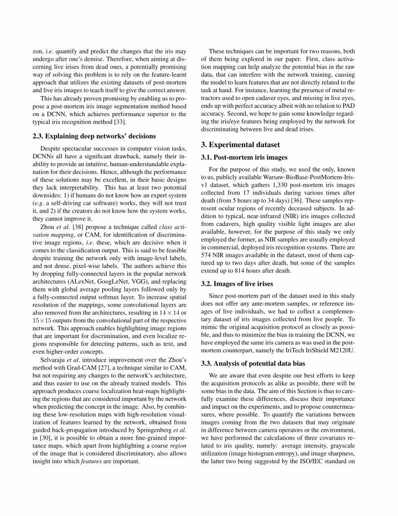
We are aware that even despite our best efforts to keepthe acquisition protocols as alike as possible, there will besome bias in the data. The aim of this Section is thus to care-fully examine these differences, discuss their importanceand impact on the experiments, and to propose countermea-sures, where possible. To quantify the variations betweenimages coming from the two datasets that may originatein difference between camera operators or the environment,we have performed the calculations of three covariates re-lated to iris quality, namely: average intensity, grayscaleutilization (image histogram entropy), and image sharpness,the latter two being suggested by the ISO/IEC standard on
Living irises Post-mortem irises
110
120
130
140
150
160
170
180Average image intensity
143.3136.8
Living irises Post-mortem irises
6
6.2
6.4
6.6
6.8
7
Grayscale utilisation [bits]
6.76.6
Living irises Post-mortem irises
0
10
20
30
40
Sharpness [ISO]
36.029.0
Figure 1: Boxplots representing differences in image quality metrics between the two datasets. Median values are shown inred, height of each boxes corresponds to an inter-quartile range (IQR) spanning from the first (Q1) to the third (Q3) quartile,whiskers span from Q1-1.5*IQR to Q3+1.5*IQR, and outliers are shown as crosses.
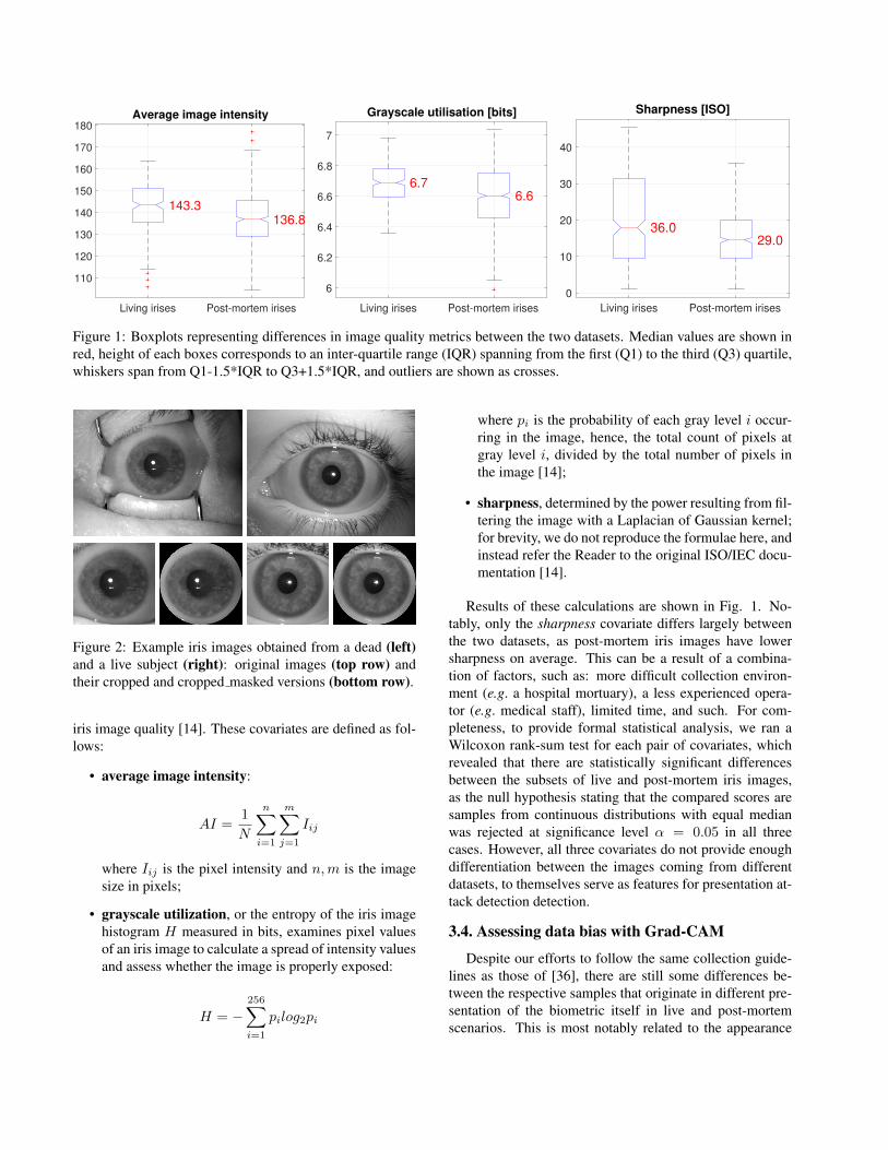
Figure 2: Example iris images obtained from a dead (left)and a live subject (right): original images (top row) andtheir cropped and cropped masked versions (bottom row).
iris image quality [14]. These covariates are defined as fol-lows:
• average image intensity:
AI =1
N
n∑i=1
m∑j=1
Iij
where Iij is the pixel intensity and n,m is the imagesize in pixels;
• grayscale utilization, or the entropy of the iris imagehistogram H measured in bits, examines pixel valuesof an iris image to calculate a spread of intensity valuesand assess whether the image is properly exposed:
H = −256∑i=1
pilog2pi
where pi is the probability of each gray level i occur-ring in the image, hence, the total count of pixels atgray level i, divided by the total number of pixels inthe image [14];
• sharpness, determined by the power resulting from fil-tering the image with a Laplacian of Gaussian kernel;for brevity, we do not reproduce the formulae here, andinstead refer the Reader to the original ISO/IEC docu-mentation [14].
Results of these calculations are shown in Fig. 1. No-tably, only the sharpness covariate differs largely betweenthe two datasets, as post-mortem iris images have lowersharpness on average. This can be a result of a combina-tion of factors, such as: more difficult collection environ-ment (e.g. a hospital mortuary), a less experienced opera-tor (e.g. medical staff), limited time, and such. For com-pleteness, to provide formal statistical analysis, we ran aWilcoxon rank-sum test for each pair of covariates, whichrevealed that there are statistically significant differencesbetween the subsets of live and post-mortem iris images,as the null hypothesis stating that the compared scores aresamples from continuous distributions with equal medianwas rejected at significance level α = 0.05 in all threecases. However, all three covariates do not provide enoughdifferentiation between the images coming from differentdatasets, to themselves serve as features for presentation at-tack detection detection.
3.4. Assessing data bias with Grad-CAM
Despite our efforts to follow the same collection guide-lines as those of [36], there are still some differences be-tween the respective samples that originate in different pre-sentation of the biometric itself in live and post-mortemscenarios. This is most notably related to the appearance
of eyelids, which in the post-mortem data are often pulledapart with a metal retracting device to keep the eye openfor image acquisition. To at least partially mitigate thesedifferences, the subjects participating in the collection ofthe reference data were asked to open their eyes as widelyas possible. However, the presence of metal parts of themedical equipment is still an issue, as these appear in post-mortem cases and do not appear in live cases.
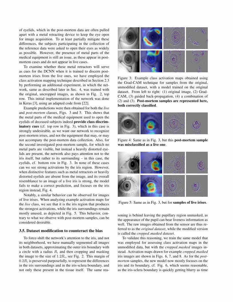
To examine whether these metal retractors will serveas cues for the DCNN when it is trained to discern post-mortem irises from the live ones, we have employed theclass activation mapping technique described in Section 2.3by performing an additional experiment, in which the net-work, same as described later in Sec. 4, was trained withthe original, uncropped images, as shown in Fig. 2, toprow. This initial implementation of the network was donein Keras [3], using an adapted code from [22].
Example predictions were then obtained for both the liveand post-mortem classes, Figs. 3 and 5. This shows thatthe metal parts of the medical equipment used to open theeyelids of deceased subjects indeed provide class discrim-inatory cues (cf. top row in Fig. 3), which in this case isstrongly undesirable, as we want our network to recognizepost-mortem irises, and not the equipment that may, or maynot accompany the post-mortem data collection. Also, forthe second investigated post-mortem sample, for which nometal parts are visible, but instead a heavily distorted eye-lids are present, the network also pays attention not to theiris itself, but rather to its surrounding – in this case, theeyelids, cf. bottom row in Fig. 3. In none of these casescan we see strong activations by the iris region. However,when distinctive features such as metal retractors or heavilydistorted eyelids are absent from the image, and its overallresemblance to an image of a live iris is strong, the modelfails to make a correct prediction, and focuses on the irisregion instead, Fig. 4.
Notably, a similar behavior can be observed for imagesof live irises. When analyzing example activation maps forthe live class, we see that it is the iris region that producesthe strongest activations, while the iris surroundings remainmostly unused, as depicted in Fig. 5. This behavior, con-trary to what we observe with post-mortem samples, can beconsidered desirable.
3.5. Dataset modification to counteract the bias
To force-shift the network’s attention to the iris, and notits neighborhood, we have manually segmented all imagesin both datasets, approximating the outer iris boundary witha circle with a radius Ri and then cropping and maskingthe image to the size of 1.2Ri, see Fig. 2. This margin of0.2Ri is preserved purposefully, to represent the differencesin the iris surroundings and in the iris-sclera boundary, andnot only these present in the tissue itself. The same rea-
Figure 3: Example class activation maps obtained usingthe Grad-CAM technique for samples from the original,unmodified dataset, with a model trained on the originaldataset. From left to right: (1) original image, (2) Grad-CAM, (3) guided back-propagation, (4) a combination of(2) and (3). Post-mortem samples are represented here,both correctly classified.
Figure 4: Same as in Fig. 3, but this post-mortem samplewas misclassified as a live one.
Figure 5: Same as in Fig. 3, but for samples of live irises.
soning is behind leaving the pupillary region unmasked, asthe appearance of the pupil can bear liveness information aswell. The raw images obtained from the sensor are thus re-ferred to as the original dataset, while the modified versionis called the cropped masked dataset.
To validate this reasoning, we train the same model thatwas employed for assessing class activation maps in theunmodified data, but with the cropped masked images in-stead. Activation maps drawn for example cropped maskediris images are shown in Figs. 6, 7, and 8. As for the post-mortem samples, the new model now mostly focuses on theiris and its boundary, cf. Fig. 6, which seems reasonable,as the iris-sclera boundary is quickly getting blurry as time
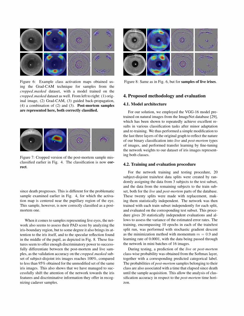
Figure 6: Example class activation maps obtained us-ing the Grad-CAM technique for samples from thecropped masked dataset, with a model trained on thecropped masked dataset as well. From left to right: (1) orig-inal image, (2) Grad-CAM, (3) guided back-propagation,(4) a combination of (2) and (3). Post-mortem samplesare represented here, both correctly classified.
Figure 7: Cropped version of the post-mortem sample mis-classified earlier in Fig. 4. The classification is now cor-rect.
since death progresses. This is different for the problematicsample examined earlier in Fig. 4, for which the activa-tion map is centered near the pupillary region of the eye.This sample, however, is now correctly classified as a post-mortem one.
When it comes to samples representing live eyes, the net-work also seems to assess their PAD score by analyzing theiris-boundary region, but to some degree it also brings its at-tention to the iris itself, and to the specular reflection foundin the middle of the pupil, as depicted in Fig. 8. These fea-tures seem to offer enough discriminatory power to success-fully differentiate between the post-mortem and live sam-ples, as the validation accuracy on the cropped masked sub-set of subject-disjoint iris images reaches 100%, comparedto less than 95% obtained for the unmodified set of the sameiris images. This also shows that we have managed to suc-cessfully shift the attention of the network towards the irisfeatures and discriminative information they offer in recog-nizing cadaver samples.
Figure 8: Same as in Fig. 6, but for samples of live irises.
4. Proposed methodology and evaluation
4.1. Model architecture
For our solution, we employed the VGG-16 model pre-trained on natural images from the ImageNet database [29],which has been shown to repeatedly achieve excellent re-sults in various classification tasks after minor adaptationand re-training. We thus performed a simple modification tothe last three layers of the original graph to reflect the natureof our binary classification into live and post-mortem typesof images, and performed transfer learning by fine-tuningthe network weights to our dataset of iris images represent-ing both classes.
4.2. Training and evaluation procedure
For the network training and testing procedure, 20subject-disjoint train/test data splits were created by ran-domly assigning the data from 3 subjects to the test subset,and the data from the remaining subjects to the train sub-set, both for the live and post-mortem parts of the database.These twenty splits were made with replacement, mak-ing them statistically independent. The network was thentrained with each train subset independently for each split,and evaluated on the corresponding test subset. This proce-dure gives 20 statistically independent evaluations and al-lows to assess the variance of the estimated error rates. Thetraining, encompassing 10 epochs in each of the train/testsplit run, was performed with stochastic gradient descentas the minimization method with momentum m = 0.9 andlearning rate of 0.0001, with the data being passed throughthe network in mini batches of 16 images.
During testing, a prediction of the live or post-mortemclass-wise probability was obtained from the Softmax layer,together with a corresponding predicted categorical label.The probabilities of post-mortem samples belonging to theirclass are also associated with a time that elapsed since deathuntil the sample acquisition. This allow the analysis of clas-sification accuracy in respect to the post-mortem time hori-zon.
92
94
96
98
100
Dete
ction a
ccura
cy [%
]Cadaver iris image detection for PAD
Median: 100.00
Mean: 98.94
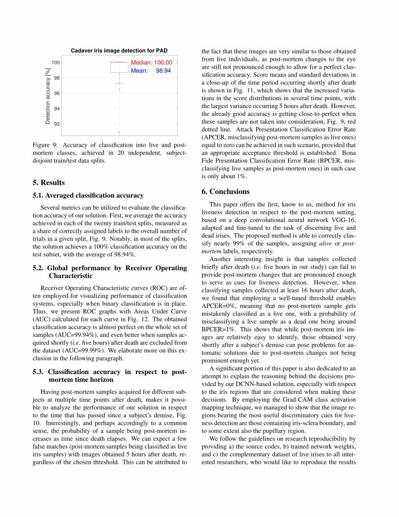
Figure 9: Accuracy of classification into live and post-mortem classes, achieved in 20 independent, subject-disjoint train/test data splits.
5. Results5.1. Averaged classification accuracy
Several metrics can be utilized to evaluate the classifica-tion accuracy of our solution. First, we average the accuracyachieved in each of the twenty train/test splits, measured asa share of correctly assigned labels to the overall number oftrials in a given split, Fig. 9. Notably, in most of the splits,the solution achieves a 100% classification accuracy on thetest subset, with the average of 98.94%.
5.2. Global performance by Receiver OperatingCharacteristic
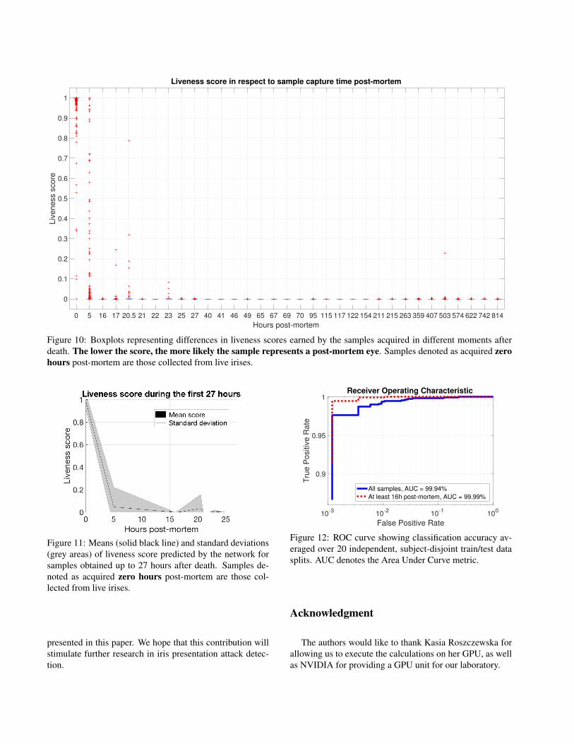
Receiver Operating Characteristic curves (ROC) are of-ten employed for visualizing performance of classificationsystems, especially when binary classification is in place.Thus, we present ROC graphs with Areas Under Curve(AUC) calculated for each curve in Fig. 12. The obtainedclassification accuracy is almost perfect on the whole set ofsamples (AUC=99.94%), and even better when samples ac-quired shortly (i.e. five hours) after death are excluded fromthe dataset (AUC=99.99%). We elaborate more on this ex-clusion in the following paragraph.
5.3. Classification accuracy in respect to post-mortem time horizon
Having post-mortem samples acquired for different sub-jects at multiple time points after death, makes it possi-ble to analyze the performance of our solution in respectto the time that has passed since a subject’s demise, Fig.10. Interestingly, and perhaps accordingly to a commonsense, the probability of a sample being post-mortem in-creases as time since death elapses. We can expect a fewfalse matches (post-mortem samples being classified as liveiris samples) with images obtained 5 hours after death, re-gardless of the chosen threshold. This can be attributed to
the fact that these images are very similar to those obtainedfrom live individuals, as post-mortem changes to the eyeare still not pronounced enough to allow for a perfect clas-sification accuracy. Score means and standard deviations ina close-up of the time period occurring shortly after deathis shown in Fig. 11, which shows that the increased varia-tions in the score distributions in several time points, withthe largest variance occurring 5 hours after death. However,the already good accuracy is getting close-to-perfect whenthese samples are not taken into consideration, Fig. 9, reddotted line. Attack Presentation Classification Error Rate(APCER, misclassifying post-mortem samples as live ones)equal to zero can be achieved in such scenario, provided thatan appropriate acceptance threshold is established. BonaFide Presentation Classification Error Rate (BPCER, mis-classifying live samples as post-mortem ones) in such caseis only about 1%.
6. ConclusionsThis paper offers the first, know to us, method for iris
liveness detection in respect to the post-mortem setting,based on a deep convolutional neural network VGG-16,adapted and fine-tuned to the task of discerning live anddead irises. The proposed method is able to correctly clas-sify nearly 99% of the samples, assigning alive or post-mortem labels, respectively.
Another interesting insight is that samples collectedbriefly after death (i.e. five hours in our study) can fail toprovide post-mortem changes that are pronounced enoughto serve as cues for liveness detection. However, whenclassifying samples collected at least 16 hours after death,we found that employing a well-tuned threshold enablesAPCER=0%, meaning that no post-mortem sample getsmistakenly classified as a live one, with a probability ofmisclassifying a live sample as a dead one being aroundBPCER=1%. This shows that while post-mortem iris im-ages are relatively easy to identify, those obtained veryshortly after a subject’s demise can pose problems for au-tomatic solutions due to post-mortem changes not beingprominent enough yet.
A significant portion of this paper is also dedicated to anattempt to explain the reasoning behind the decisions pro-vided by our DCNN-based solution, especially with respectto the iris regions that are considered when making thesedecisions. By employing the Grad-CAM class activationmapping technique, we managed to show that the image re-gions bearing the most useful discriminatory cues for live-ness detection are those containing iris-sclera boundary, andto some extent also the pupillary region.
We follow the guidelines on research reproducibility byproviding a) the source codes, b) trained network weights,and c) the complementary dataset of live irises to all inter-ested researchers, who would like to reproduce the results
0 5 16 17 20.5 21 22 23 25 27 40 41 46 49 65 67 69 70 95 115 117 122 154 211 215 263 359 407 503 574 622 742 814
Hours post-mortem
0
0.1
0.2
0.3
0.4
0.5
0.6
0.7
0.8
0.9
1
Liv
en
ess s
co
re
Liveness score in respect to sample capture time post-mortem
Figure 10: Boxplots representing differences in liveness scores earned by the samples acquired in different moments afterdeath. The lower the score, the more likely the sample represents a post-mortem eye. Samples denoted as acquired zerohours post-mortem are those collected from live irises.
Figure 11: Means (solid black line) and standard deviations(grey areas) of liveness score predicted by the network forsamples obtained up to 27 hours after death. Samples de-noted as acquired zero hours post-mortem are those col-lected from live irises.
presented in this paper. We hope that this contribution willstimulate further research in iris presentation attack detec-tion.
10-3
10-2
10-1
100
False Positive Rate
0.9
0.95
1
Tru
e P
ositiv
e R
ate
Receiver Operating Characteristic
All samples, AUC = 99.94%
At least 16h post-mortem, AUC = 99.99%
Figure 12: ROC curve showing classification accuracy av-eraged over 20 independent, subject-disjoint train/test datasplits. AUC denotes the Area Under Curve metric.
Acknowledgment
The authors would like to thank Kasia Roszczewska forallowing us to execute the calculations on her GPU, as wellas NVIDIA for providing a GPU unit for our laboratory.
References[1] D. S. Bolme, R. A. Tokola, C. B. Boehnen, T. B. Saul,
K. A. Sauerwein, and D. W. Steadman. Impact of en-vironmental factors on biometric matching during hu-man decomposition. In 2016 IEEE 8th InternationalConference on Biometrics Theory, Applications andSystems (BTAS), pages 1–8, Sept 2016.
[2] R. Chen, X. Lin, and T. Ding. Liveness detection foriris recognition using multispectral images. PatternRecognition Letters, 33(12):1513 – 1519, 2012.
[3] F. Chollet. Keras: Deep Learning library for Theanoand TensorFlow. https://keras.io/, 2015.
[4] A. Czajka. Pupil dynamics for iris liveness detec-tion. IEEE Trans. Inf. Forens. Security, 10(4):726–735, April 2015.
[5] A. Czajka and K. Bowyer. Presentation Attack De-tection for Iris Recognition: An Assessment of theState of the Art. ACM Computing Surveys (in review),https://arxiv.org/abs/1804.00194, 2018.
[6] J. S. Doyle, P. J. Flynn, and K. W. Bowyer. Automatedclassification of contact lens type in iris images. InIEEE Int. Conference on Biometrics (ICB), pages 1–6,June 2013.
[7] Eliza Strickland. Biometrics Researcher Asks: Is ThatEyeball Dead or Alive?, https://spectrum.ieee.org/the-human-os/biomedical/imaging/biometric-researcher-asks-is-that-eyeball-alive-or-dead (accessed: March20, 2018).
[8] J. Galbally, M. Savvides, S. Venugopalan, and A. A.Ross. Iris Image Reconstruction from Binary Tem-plates, pages 469–496. Springer London, London,2016.
[9] A. Garcia-Garcia, S. Orts-Escolano, S. Oprea,V. Villena-Martinez, , and J. Garcia-Rodriguez. A Review on Deep LearningTechniques Applied to Semantic Segmentation.https://arxiv.org/abs/1704.06857v1, 2017.
[10] D. Gragnaniello, G. Poggi, C. Sansone, and L. Verdo-liva. Contact lens detection and classification in irisimages through scale invariant descriptor. In Int. Con-ference on Signal-Image Technology Internet-BasedSystems (SITIS), pages 560–565, Nov 2014.
[11] D. Gragnaniello, G. Poggi, C. Sansone, and L. Ver-doliva. An investigation of local descriptors for bio-metric spoofing detection. IEEE Trans. Inf. Forens.Security, 10(4):849–863, Apr. 2015.
[12] L. He, H. Li, F. Liu, N. Liu, Z. Sun, and Z. He.Multi-patch convolution neural network for iris live-ness detection. In IEEE Intl. Conference on Biomet-rics Theory, Applications and Systems (BTAS), pages1–7, September 2016.
[13] K. Hughes and K. W. Bowyer. Detection of contact-lens-based iris biometric spoofs using stereo imaging.In Hawaii Intl. Conference on System Sciences, pages1763–1772, January 2013.
[14] ISO/IEC 29794-6. Information technology – Biomet-ric sample quality - Part 6: Iris image data (FDIS),September 2014.
[15] J. Johnson, A. Karpathy, and L. Fei-Fei. DenseCap:Fully Convolutional Localization Networks for DenseCaptioning. 2015 IEEE Conference on Computer Vi-sion and Pattern Recognition (CVPR), 2016.
[16] J. Komulainen, A. Hadid, and M. Pietikinen. Gen-eralized textured contact lens detection by extractingbsif description from cartesian iris images. In IEEEInt. Joint Conference on Biometrics (IJCB), pages 1–7, Sept 2014.
[17] Lovish, A. Nigam, B. Kumar, and P. Gupta. Ro-bust contact lens detection using local phase quanti-zation and binary gabor pattern. In G. Azzopardi andN. Petkov, editors, Int. Conference on Computer Anal-ysis of Images and Patterns (CAIP), pages 702–714.Springer International Publishing, 2015.
[18] P. M. Mateusz Trokielewicz, Adam Czajka. IrisRecognition After Death. IEEE Transactions onInformation Forensics and Security (in review),https://arxiv.org/abs/1804.01962, 2018.
[19] D. Menotti, G. Chiachia, A. Pinto, W. R. Schwartz,H. Pedrini, A. X. Falcao, and A. Rocha. Deep repre-sentations for iris, face, and fingerprint spoofing de-tection. IEEE Transactions on Information Forensicsand Security, 10(4):864–879, April 2015.
[20] A. Pacut and A. Czajka. Aliveness detection for irisbiometrics. In IEEE Int. Carnahan Conference on Se-curity Technology (ICCST), pages 122–129, October2006.
[21] F. Pala and B. Bhanu. Iris liveness detection by relativedistance comparisons. In IEEE Conference on Com-puter Vision and Pattern Recognition (CVPR) Work-shops, July 2017.
[22] V. Petsiuk. Keras implementation of Grad-CAM. https://github.com/eclique/keras-gradcam, ac-cessed on April 4, 2018.
[23] R. Raghavendra, K. B. Raja, and C. Busch. Con-tlensnet: Robust iris contact lens detection using deepconvolutional neural networks. In IEEE Winter Con-ference on Applications of Computer Vision (WACV),pages 1160–1167, March 2017.
[24] K. Raja, R. Raghavendra, and C. Busch. Videopresentation attack detection in visible spectrum irisrecognition using magnified phase information. IEEE
Trans. Inf. Forens. Security, 10(10):2048–2056, Octo-ber 2015.
[25] M. Ren, R. Kiros, and R. S. Zemel. Exploring mod-els and data for image question answering. CoRR,abs/1505.02074, 2015.
[26] I. Rigas and O. V. Komogortsev. Eye movement-driven defense against iris print-attacks. PatternRecognition Letters, 68, Part 2:316 – 326, 2015. Spe-cial Issue on Soft Biometrics.
[27] R. R. Selvaraju, M. Cogswell, A. Das, R. Vedantam,D. Parikh, and D. Batra. Grad-CAM: Visual Ex-planations from Deep Networks via Gradient-BasedLocalization. ICCV, https://arxiv.org/abs/1610.02391,2016.
[28] A. F. Sequeira, S. Thavalengal, J. Ferryman, P. Cor-coran, and J. S. Cardoso. A realistic evaluation ofiris presentation attack detection. In Int. Conferenceon Telecommunications and Signal Processing (TSP),pages 660–664, June 2016.
[29] K. Simonyan and A. Zisserman. Very Deep Convolu-tional Networks for Large-Scale Image Recognition.https://arxiv.org/abs/1409.1556, 2014.
[30] J. T. Springenberg, A. Dosovitskiy, T. Brox,and M. Riedmiller. Striving for Simplic-ity: The All Convolutional Net. ICLR,https://arxiv.org/abs/1412.6806, 2015.
[31] S. Thavalengal, T. Nedelcu, P. Bigioi, and P. Cor-coran. Iris liveness detection for next generationsmartphones. IEEE Trans. Consumer Electronics,62(2):95–102, May 2016.
[32] Thomas Brewster. Yes, Cops Are Now Open-ing iPhones With Dead People’s Fingerprints,https://www.forbes.com/sites/thomasbrewster/2018/03/22/yes-cops-are-now-opening-iphones-with-dead-peoples-fingerprints (accessed: March 20, 2018).
[33] M. Trokielewicz and A. Czajka. Data-driven Segmen-tation of Post-mortem Iris Images, 6th IAPR/IEEEInternational Workshop on Biometrics and Forensics(IWBF 2018), June 7-8, 2018, Sassari, Italy, 2018.
[34] M. Trokielewicz, A. Czajka, and P. Maciejewicz. Hu-man Iris Recognition in Post-mortem Subjects: Studyand Database, 8th IEEE International Conference onBiometrics: Theory, Applications and Systems (BTAS2016), September 6-9, 2016, Buffalo, NY, USA, 2016.
[35] M. Trokielewicz, A. Czajka, and P. Maciejewicz. Post-mortem Human Iris Recognition, 9th IAPR Interna-tional Conference on Biometrics (ICB 2016), June 13-16, 2016, Halmstad, Sweden, 2016.
[36] Warsaw University of Technology.Warsaw-BioBase-PostMortem-Iris-v1.0:http://zbum.ia.pw.edu.pl/en/node/46, 2016.
[37] D. A. Yambay, B. Becker, N. Kohli, D. Yadav, A. Cza-jka, K. W. Bowyer, S. Schuckers, R. Singh, M. Vatsa,A. Noore, D. Gragnaniello, C. Sansone, L. Verdoliva,L. He, Y. Ru, H. Li, N. Liu, Z. Sun, and T. Tan. LivDet2017 - Iris Liveness Detection Competition 2017. Bio-metrics: Theory Applications and Systems (BTAS),pages 0–5, 2017.
[38] B. Zhou, A. Khosla, A. Lapedriza, A. Oliva,and A. Torralba. Learning Deep Featuresfor Discriminative Localization. CVPR,https://arxiv.org/abs/1512.04150, 2016.

![B AUTHENTICATION UNDER THREAT: LIVENESS DETECTION … · J. G[10] proposed a probabilistic approach to reconstruct iris images from binary templates to break the iris recognition](https://img.pdfslide.net/doc/110x75/5f1247b64ebb44746965dafe/b-authentication-under-threat-liveness-detection-j-g10-proposed-a-probabilistic.jpg)


![LivDet-Iris 2013 – Iris Liveness Detection Competition 2013 · parry an attack based on iris printouts [4]. This idea was lately incorporated into the first security evaluation](https://img.pdfslide.net/doc/110x75/5f1246f3e7c2834a4e2af878/livdet-iris-2013-a-iris-liveness-detection-competition-2013-parry-an-attack-based.jpg)