Embed Size (px)
Citation preview
4/22/2016
1
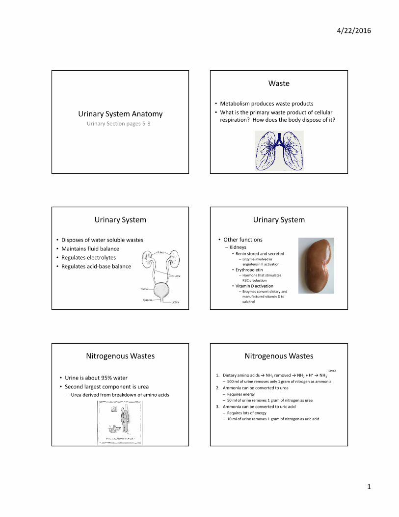
Urinary System AnatomyUrinary Section pages 5-8
Waste
• Metabolism produces waste products
• What is the primary waste product of cellular
respiration? How does the body dispose of it?
Urinary System
• Disposes of water soluble wastes
• Maintains fluid balance
• Regulates electrolytes
• Regulates acid-base balance
Urinary System
• Other functions
– Kidneys
• Renin stored and secreted
– Enzyme involved in
angiotensin II activation
• Erythropoietin
– Hormone that stimulates
RBC production
• Vitamin D activation
– Enzymes convert dietary and
manufactured vitamin D to
calcitrol
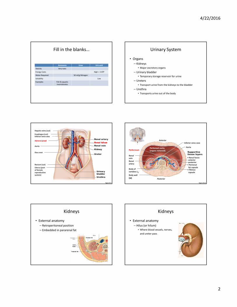
Nitrogenous Wastes
• Urine is about 95% water
• Second largest component is urea
– Urea derived from breakdown of amino acids
Nitrogenous Wastes
1. Dietary amino acids → NH2 removed → NH2 + H+ → NH3
– 500 ml of urine removes only 1 gram of nitrogen as ammonia
2. Ammonia can be converted to urea
– Requires energy
– 50 ml of urine removes 1 gram of nitrogen as urea
3. Ammonia can be converted to uric acid
– Requires lots of energy
– 10 ml of urine removes 1 gram of nitrogen as uric acid
TOXIC!
4/22/2016
2
Fill in the blanks…
Ammonia Urea Uric acid
Toxicity Very toxic
Energy Costs High = 3 ATP
Water Required 50 ml/g Nitrogen
Solubility Low
Examples Fish & aquatic
invertebrates
Urinary System
• Organs
– Kidneys
• Major excretory organs
– Urinary bladder
• Temporary storage reservoir for urine
– Ureters
• Transport urine from the kidneys to the bladder
– Urethra
• Transports urine out of the body
Figure 25.1
Esophagus (cut)Inferior vena cava
Adrenal gland
Hepatic veins (cut)
Renal artery
Renal hilum
Renal vein
Iliac crest
Kidney
Ureter
Urinary
bladder
Urethra
Aorta
Rectum (cut)
Uterus (part
of female
reproductive
system)
Figure 25.2a
Body wall
• Perirenal
fat capsule
Renal
artery
Renal
vein
Inferior vena cava
Aorta
• Fibrous
capsule
• Renal fasciaanteriorposterior
Supportivetissue layers
Body of
vertebra L2
PeritoneumPeritoneal cavity
(organs removed)
Anterior
Posterior(a)
Kidneys
• External anatomy
– Retroperitoneal position
– Embedded in pararenal fat
Kidneys
• External anatomy
– Hilus (or hilum)
• Where blood vessels, nerves,
and ureter pass
4/22/2016
3
Kidneys
• External anatomy
– 3 tissue layers surround & support
• Fibrous capsule
• Perirenal fat capsule
• Renal fascia
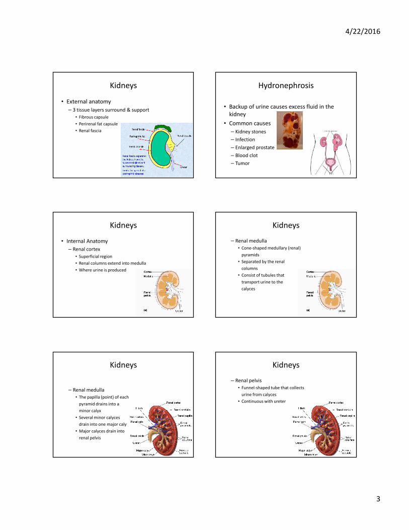
Hydronephrosis
• Backup of urine causes excess fluid in the
kidney
• Common causes
– Kidney stones
– Infection
– Enlarged prostate
– Blood clot
– Tumor
Kidneys
• Internal Anatomy
– Renal cortex
• Superficial region
• Renal columns extend into medulla
• Where urine is produced
Kidneys
– Renal medulla
• Cone-shaped medullary (renal)
pyramids
• Separated by the renal
columns
• Consist of tubules that
transport urine to the
calyces
Kidneys
– Renal medulla
• The papilla (point) of each
pyramid drains into a
minor calyx
• Several minor calyces
drain into one major calyx
• Major calyces drain into
renal pelvis
Kidneys
– Renal pelvis
• Funnel-shaped tube that collects
urine from calyces
• Continuous with ureter
4/22/2016
4
Figure 25.3
Renal cortex
Renal medulla
Major calyx
Papilla of
pyramid
Renal pelvis
Ureter
Minor calyx
Renal column
Renal pyramid
in renal medulla
Fibrous capsule
Renal
hilum
(a) Photograph of right kidney, frontal section (b) Diagrammatic view
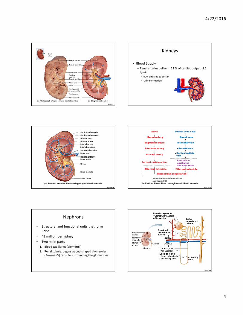
Kidneys
• Blood Supply
– Renal arteries deliver ~ 22 % of cardiac output (1.2
L/min)
• 90% directed to cortex
• Urine formation
Figure 25.4a
Cortical radiate vein
Cortical radiate artery
Arcuate vein
Arcuate artery
Interlobar vein
Interlobar artery
Segmental arteries
Renal artery
Renal vein
Renal pelvis
Ureter
Renal medulla
Renal cortex
(a) Frontal section illustrating major blood vessels
Figure 25.4b
Aorta
Renal artery
Segmental artery
Interlobar artery
Arcuate artery
Cortical radiate artery
Afferent arteriole
Glomerulus (capillaries)
Nephron-associated blood vessels
(see Figure 25.8)
Inferior vena cava
Renal vein
Interlobar vein
Arcuate vein
Cortical radiate
vein
Peritubular
capillaries
and vasa recta
Efferent arteriole
(b) Path of blood flow through renal blood vessels
Nephrons
• Structural and functional units that form
urine
• ~1 million per kidney
• Two main parts
1. Blood capillaries (glomeruli)
2. Renal tubule: begins as cup-shaped glomerular
(Bowman’s) capsule surrounding the glomerulus
Figure 25.5
4/22/2016
5
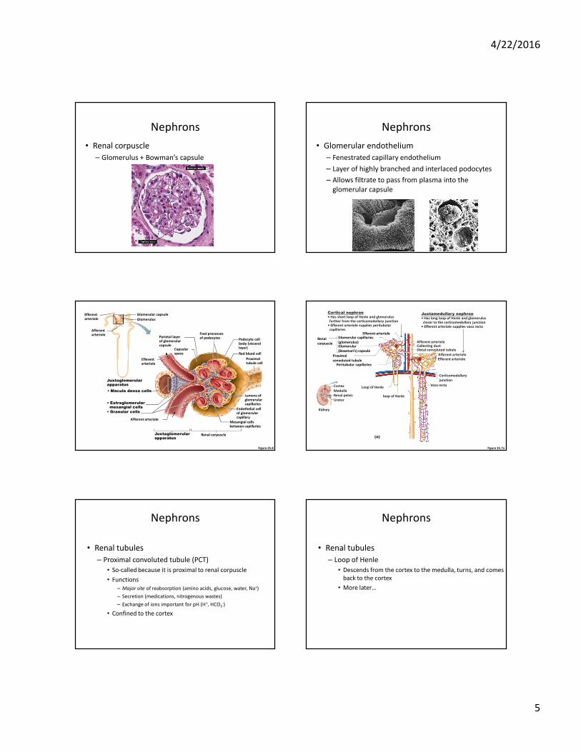
Nephrons
• Renal corpuscle
– Glomerulus + Bowman’s capsule
Nephrons
• Glomerular endothelium
– Fenestrated capillary endothelium
– Layer of highly branched and interlaced podocytes
– Allows filtrate to pass from plasma into the
glomerular capsule
Figure 25.8
Glomerulus
Glomerular capsule
Afferent arteriole
Efferent arteriole
Red blood cell
Podocyte cell body (visceral layer)
Foot processesof podocytesParietal layer
of glomerularcapsule
Proximaltubule cell
Lumens of glomerularcapillaries
Endothelial cellof glomerularcapillary
Efferent arteriole
• Macula densa cells
• Granular cells
• Extraglomerularmesangial cells
Afferent arteriole
Capsularspace
Renal corpuscleJuxtaglomerularapparatus
Mesangial cellsbetween capillaries
Juxtaglomerularapparatus
Figure 25.7a
Cortical nephron• Has short loop of Henle and glomerulusfurther from the corticomedullary junction
• Efferent arteriole supplies peritubularcapillaries
Juxtamedullary nephron• Has long loop of Henle and glomeruluscloser to the corticomedullary junction
• Efferent arteriole supplies vasa recta
Corticomedullary
junction
Ureter
Renal pelvis
Kidney
Cortex
Medulla
(a)
Afferent arteriole
Afferent arteriole
Collecting ductDistal convoluted tubule
Efferent arteriole
Vasa rectaLoop of Henle
Peritubular capillaries
Glomerular capillaries
(glomerulus)Glomerular
(Bowman’s) capsule
Renal
corpuscle
loop of Henle
Efferent arteriole
Proximal
convoluted tubule
Nephrons
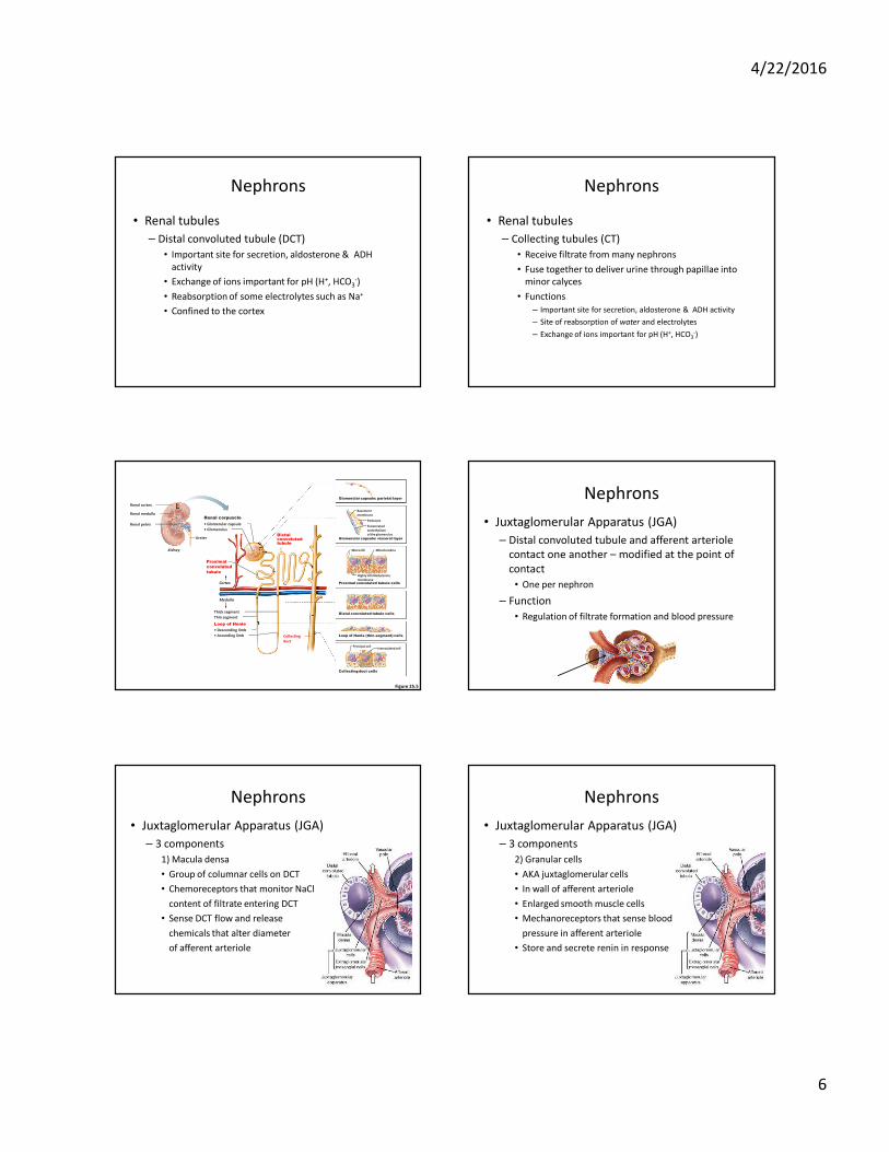
• Renal tubules
– Proximal convoluted tubule (PCT)
• So-called because it is proximal to renal corpuscle
• Functions
– Major site of reabsorption (amino acids, glucose, water, Na+)
– Secretion (medications, nitrogenous wastes)
– Exchange of ions important for pH (H+, HCO3-)
• Confined to the cortex
Nephrons
• Renal tubules
– Loop of Henle
• Descends from the cortex to the medulla, turns, and comes
back to the cortex
• More later…
4/22/2016
6
Nephrons
• Renal tubules
– Distal convoluted tubule (DCT)
• Important site for secretion, aldosterone & ADH
activity
• Exchange of ions important for pH (H+, HCO3-)
• Reabsorption of some electrolytes such as Na+
• Confined to the cortex
Nephrons
• Renal tubules
– Collecting tubules (CT)
• Receive filtrate from many nephrons
• Fuse together to deliver urine through papillae into
minor calyces
• Functions
– Important site for secretion, aldosterone & ADH activity
– Site of reabsorption of water and electrolytes
– Exchange of ions important for pH (H+, HCO3-)
Figure 25.5
Fenestrated
endothelium
of the glomerulus
Microvilli
Cortex
Medulla
Podocyte
Basement
membrane
Mitochondria
Highly infolded plasma
membrane
Proximal
convoluted
tubule
Distalconvolutedtubule
• Descending limb
Loop of Henle
• Ascending limb
• Glomerular capsule
Renal corpuscle
• Glomerulus
Thick segment
Collecting
duct
Intercalated cellPrincipal cell
Thin segment
Proximal convoluted tubule cells
Glomerular capsule: parietal layer
Glomerular capsule: visceral layer
Distal convoluted tubule cells
Loop of Henle (thin-segment) cells
Collecting duct cells
Renal cortex
Renal medulla
Renal pelvis
Ureter
Kidney
Nephrons
• Juxtaglomerular Apparatus (JGA)
– Distal convoluted tubule and afferent arteriole
contact one another – modified at the point of
contact
• One per nephron
– Function
• Regulation of filtrate formation and blood pressure
Nephrons
• Juxtaglomerular Apparatus (JGA)
– 3 components
1) Macula densa
• Group of columnar cells on DCT
• Chemoreceptors that monitor NaCl
content of filtrate entering DCT
• Sense DCT flow and release
chemicals that alter diameter
of afferent arteriole
Nephrons
• Juxtaglomerular Apparatus (JGA)
– 3 components
2) Granular cells
• AKA juxtaglomerular cells
• In wall of afferent arteriole
• Enlarged smooth muscle cells
• Mechanoreceptors that sense blood
pressure in afferent arteriole
• Store and secrete renin in response
4/22/2016
7
Nephrons
• Juxtaglomerular Apparatus (JGA)
– 3 components
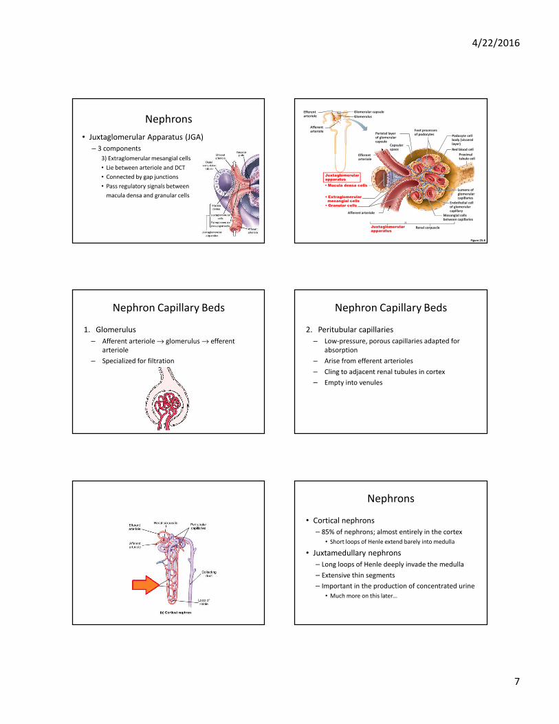
3) Extraglomerular mesangial cells
• Lie between arteriole and DCT
• Connected by gap junctions
• Pass regulatory signals between
macula densa and granular cells
Figure 25.8
Glomerulus
Glomerular capsule
Afferent arteriole
Efferent arteriole
Red blood cell
Podocyte cell body (visceral layer)
Foot processesof podocytesParietal layer
of glomerularcapsule
Proximaltubule cell
Lumens of glomerularcapillaries
Endothelial cellof glomerularcapillary
Efferent arteriole
• Macula densa cells
• Granular cells
• Extraglomerularmesangial cells
Afferent arteriole
Capsularspace
Renal corpuscleJuxtaglomerularapparatus
Mesangial cellsbetween capillaries
Juxtaglomerularapparatus
Nephron Capillary Beds
1. Glomerulus
– Afferent arteriole → glomerulus → efferent
arteriole
– Specialized for filtration
Nephron Capillary Beds
2. Peritubular capillaries
– Low-pressure, porous capillaries adapted for
absorption
– Arise from efferent arterioles
– Cling to adjacent renal tubules in cortex
– Empty into venules
Nephrons
• Cortical nephrons
– 85% of nephrons; almost entirely in the cortex
• Short loops of Henle extend barely into medulla
• Juxtamedullary nephrons
– Long loops of Henle deeply invade the medulla
– Extensive thin segments
– Important in the production of concentrated urine
• Much more on this later…
4/22/2016
8
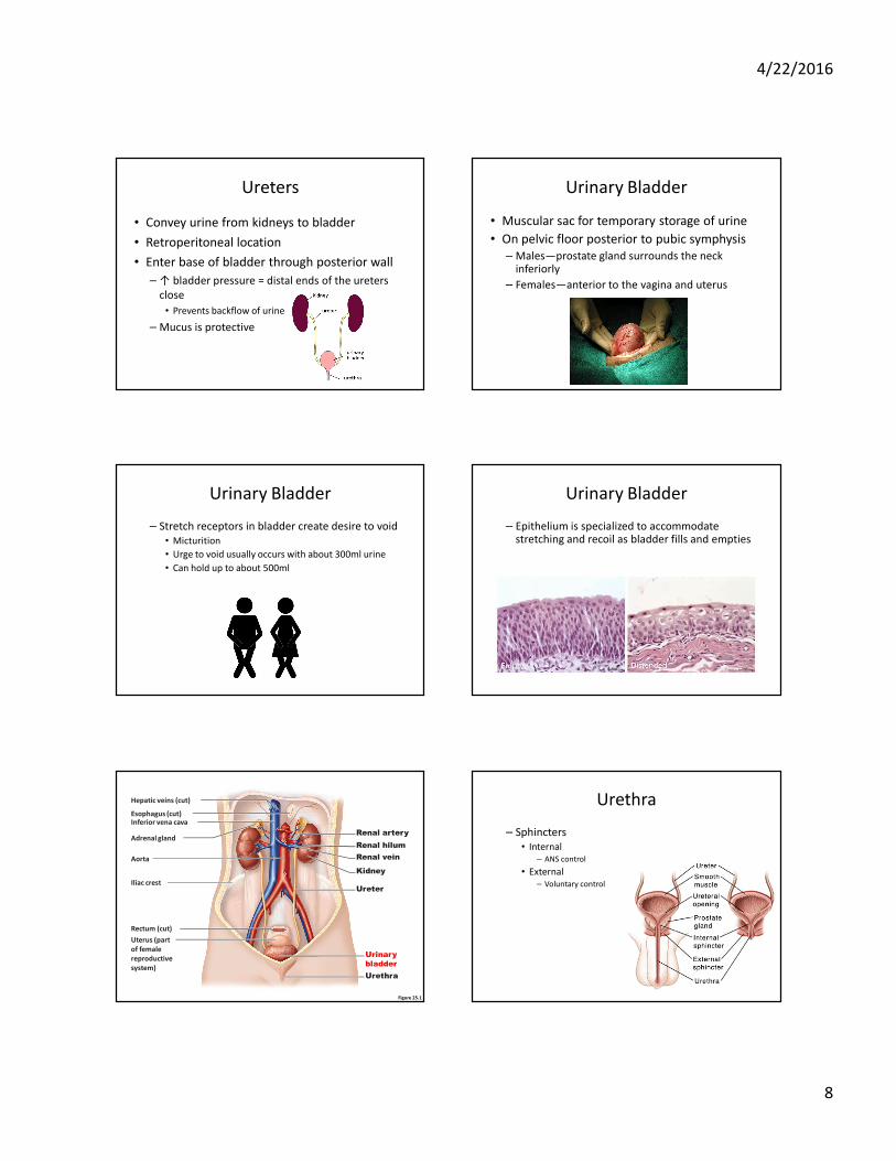
Ureters
• Convey urine from kidneys to bladder
• Retroperitoneal location
• Enter base of bladder through posterior wall
– ↑ bladder pressure = distal ends of the ureters
close
• Prevents backflow of urine
– Mucus is protective
Urinary Bladder
• Muscular sac for temporary storage of urine
• On pelvic floor posterior to pubic symphysis
– Males—prostate gland surrounds the neck inferiorly
– Females—anterior to the vagina and uterus
Urinary Bladder
– Stretch receptors in bladder create desire to void
• Micturition
• Urge to void usually occurs with about 300ml urine
• Can hold up to about 500ml
Urinary Bladder
– Epithelium is specialized to accommodate stretching and recoil as bladder fills and empties
Figure 25.1
Esophagus (cut)Inferior vena cava
Adrenal gland
Hepatic veins (cut)
Renal artery
Renal hilum
Renal vein
Iliac crest
Kidney
Ureter
Urinary
bladder
Urethra
Aorta
Rectum (cut)
Uterus (part
of female
reproductive
system)
Urethra
– Sphincters
• Internal
– ANS control
• External
– Voluntary control
4/22/2016
9
Figure 25.21b
Ureter
Trigone
Peritoneum
Rugae
Detrusor
muscle
Bladder neck
Internal urethral
sphincter
External urethral
sphincter
Urogenital diaphragm
Urethra
External urethral
orifice
Ureteric orifices
(b) Female.
Figure 25.21a
Ureter
Trigone of bladder
Prostate
Membranous urethra
Prostatic urethra
Peritoneum
Rugae
Detrusor muscle
Bladder neck
Internal urethral sphincter
External urethral sphincter
Urogenital diaphragm
Spongy urethra
Erectile tissue of penis
Ureteric orifices
Adventitia
(a) Male. The long male urethra has three
regions: prostatic, membranous and spongy.
External urethral orifice
Figure 25.22
Somatic motor
nerve activity
External urethral
sphincter opens
Sympathetic
activity
Parasympathetic
activity
Urinary bladder
filling stretches
bladder wall
Spinal
cord
Promotes micturition
by acting on all three
spinal efferents
Inhibits micturition
by acting on all three
spinal efferents
Allow or inhibit micturition
as appropriate
Brain
Simple
spinal
reflex
Spinal
cord
Inhibits
Parasympathetic activity
Sympathetic activitySomatic motor nerve activity
Pontine micturition
center
Pontine storage
center
Higher brain
centers
Detrusor muscle
contracts; internalurethral sphincter
opens
Afferent impulses
from stretch
receptors
Micturition
Incontinence
• Damage to spinal cord
• Frequent micturition in infants
– Incontinence is normal: control of the voluntary urethral sphincter develops with the nervous system
Pathway of Urine Flow
Urine formed in nephrons
Calyces
Renal pelvis
Ureters
Bladder
Urethra