Embed Size (px)
Citation preview
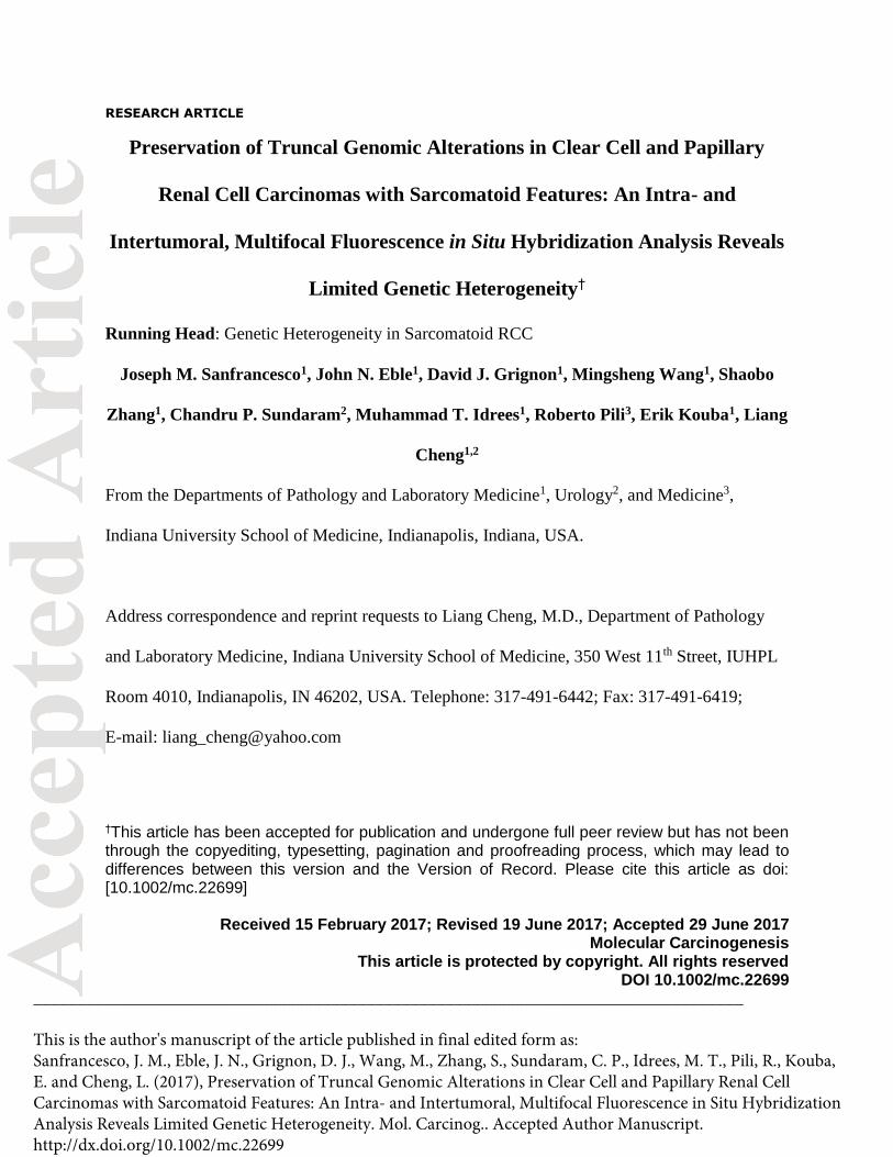
RESEARCH ARTICLE
Preservation of Truncal Genomic Alterations in Clear Cell and Papillary
Renal Cell Carcinomas with Sarcomatoid Features: An Intra- and
Intertumoral, Multifocal Fluorescence in Situ Hybridization Analysis Reveals
Limited Genetic Heterogeneity†
Running Head: Genetic Heterogeneity in Sarcomatoid RCC
Joseph M. Sanfrancesco1, John N. Eble1, David J. Grignon1, Mingsheng Wang1, Shaobo
Zhang1, Chandru P. Sundaram2, Muhammad T. Idrees1, Roberto Pili3, Erik Kouba1, Liang
Cheng1,2
From the Departments of Pathology and Laboratory Medicine1, Urology2, and Medicine3,
Indiana University School of Medicine, Indianapolis, Indiana, USA.
Address correspondence and reprint requests to Liang Cheng, M.D., Department of Pathology
and Laboratory Medicine, Indiana University School of Medicine, 350 West 11th Street, IUHPL
Room 4010, Indianapolis, IN 46202, USA. Telephone: 317-491-6442; Fax: 317-491-6419;
E-mail: [email protected]
†This article has been accepted for publication and undergone full peer review but has not been through the copyediting, typesetting, pagination and proofreading process, which may lead to differences between this version and the Version of Record. Please cite this article as doi: [10.1002/mc.22699]
Received 15 February 2017; Revised 19 June 2017; Accepted 29 June 2017 Molecular Carcinogenesis
This article is protected by copyright. All rights reserved DOI 10.1002/mc.22699
_________________________________________________________________________________ This is the author's manuscript of the article published in final edited form as: Sanfrancesco, J. M., Eble, J. N., Grignon, D. J., Wang, M., Zhang, S., Sundaram, C. P., Idrees, M. T., Pili, R., Kouba, E. and Cheng, L. (2017), Preservation of Truncal Genomic Alterations in Clear Cell and Papillary Renal Cell Carcinomas with Sarcomatoid Features: An Intra- and Intertumoral, Multifocal Fluorescence in Situ Hybridization Analysis Reveals Limited Genetic Heterogeneity. Mol. Carcinog.. Accepted Author Manuscript. http://dx.doi.org/10.1002/mc.22699
2
This article is protected by copyright. All rights reserved
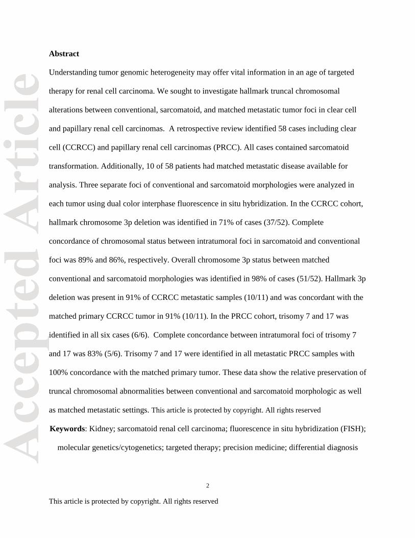
Abstract
Understanding tumor genomic heterogeneity may offer vital information in an age of targeted
therapy for renal cell carcinoma. We sought to investigate hallmark truncal chromosomal
alterations between conventional, sarcomatoid, and matched metastatic tumor foci in clear cell
and papillary renal cell carcinomas. A retrospective review identified 58 cases including clear
cell (CCRCC) and papillary renal cell carcinomas (PRCC). All cases contained sarcomatoid
transformation. Additionally, 10 of 58 patients had matched metastatic disease available for
analysis. Three separate foci of conventional and sarcomatoid morphologies were analyzed in
each tumor using dual color interphase fluorescence in situ hybridization. In the CCRCC cohort,
hallmark chromosome 3p deletion was identified in 71% of cases (37/52). Complete
concordance of chromosomal status between intratumoral foci in sarcomatoid and conventional
foci was 89% and 86%, respectively. Overall chromosome 3p status between matched
conventional and sarcomatoid morphologies was identified in 98% of cases (51/52). Hallmark 3p
deletion was present in 91% of CCRCC metastatic samples (10/11) and was concordant with the
matched primary CCRCC tumor in 91% (10/11). In the PRCC cohort, trisomy 7 and 17 was
identified in all six cases (6/6). Complete concordance between intratumoral foci of trisomy 7
and 17 was 83% (5/6). Trisomy 7 and 17 were identified in all metastatic PRCC samples with
100% concordance with the matched primary tumor. These data show the relative preservation of
truncal chromosomal abnormalities between conventional and sarcomatoid morphologic as well
as matched metastatic settings. This article is protected by copyright. All rights reserved
Keywords: Kidney; sarcomatoid renal cell carcinoma; fluorescence in situ hybridization (FISH);
molecular genetics/cytogenetics; targeted therapy; precision medicine; differential diagnosis
3
This article is protected by copyright. All rights reserved
Introduction
The morphological and genetic complexities of renal cell carcinoma (RCC) have been
extensively studied. Additionally, several recent studies have used genomic sequencing to
highlight intratumoral genetic heterogeneity.[1-6] Such complexities are compounded by the
predisposition of a subset of RCC to undergo sarcomatoid differentiation. RCC with sarcomatoid
features, regardless of the conventional morphologic subtype, has been shown to be associated
with an overall worse prognosis.[7] Accordingly, studies have attempted to characterize the
extent of intratumoral heterogeneity within sarcomatous tumor components and compare the
differences between conventional (e.g. epithelial) and sarcomatous elements.[8,9]
Intratumoral genetic heterogeneity, particularly in the setting of divergent morphologic
features and/or high clinical stage, may have significant impact on treatment options for patients,
especially those with metastatic disease[10]. Hallmark chromosomal gains and losses in primary
RCC have been characterized, most notably the presence of chromosome 3p deletion in clear cell
RCC (CCRCC) and trisomy 7 and 17 in papillary RCC (PRCC)[11-20]. Prior studies have
attempted to compare intratumoral concordance of hallmark chromosomal gains and losses and
subsequent concordance with matched metastases.[21-23] We herein investigate the preservation
of hallmark chromosomal abnormalities along with intratumoral concordance in the setting of
matched sarcomatous transformation and matched metastatic disease in CCRCC and PRCC.
Materials and Methods
Fifty-eight patients diagnosed with either CCRCC (52 patients) or PRCC (6 patients)
with sarcomatoid transformation between 1995 and 2016 were retrieved from our institution’s
surgical pathology files. Histopathologic features of each case were evaluated by a single senior
4
This article is protected by copyright. All rights reserved
genitourinary pathologist (LC) and confirmed using diagnostic criteria set forth in the World
Health Organization Classification of Tumors of the Urinary System and Male Genital
Organs.[24] Appropriate sections containing both conventional and sarcomatoid tumor foci were
selected for subsequent analysis. Specimens of biopsy/resection-proven metastatic tumors of the
patients were also identified and lesions were matched with primary lesions for analysis. This
study was approved by the Institutional Review Board.
Formalin-fixed, paraffin-embedded tissue blocks of each tumor were obtained based on
correlation with the reviewed hematoxylin and eosin–stained glass slides demonstrating
conventional (epithelial) and sarcomatoid morphologies. Three separate foci of conventional and
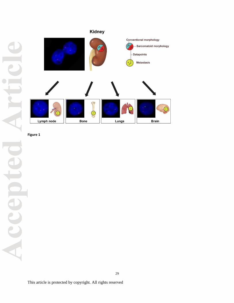
sarcomatoid morphologies were analyzed in each primary tumor. When available, three foci of
metastatic RCC were marked, analyzed, and compared to their primary RCC (Figure 1).
Fluorescence in situ hybridization (FISH) analysis was performed on each of these selected foci
to identify the presence of hallmark chromosomal abnormalities associated with CCRCC and
PRCC including deletion of 3p and trisomy 7 and 17, respectively, as described
previously.[25,26]
Selected slides were deparaffinized with two washes of xylene, 15 min each; washed
twice with absolute ethanol, 10 min each; and air-dried in a fume hood. Subsequently, the slides
were treated with 0.1mM citric acid (pH 6.0) at 95 degrees for 10 min, rinsed in distilled water
for 3 min, and followed by a wash of standard saline citrate (2 × SSC) for 5 min. Tissue
digestion was performed by applying 0.4 ml of pepsin (Sigma, St. Louis, MO, USA) solution (4
mg/ml in 0.9% NaCL in 0.01N HCl) to each slide and incubating the slides in humidified box for
40 min at 37 °C. The slides were rinsed with distilled water for 5 min, washed with SSC for 5
min, and air-dried.
5
This article is protected by copyright. All rights reserved
The alterations in chromosomes 7 and 17 were assessed using a probe cocktail containing
probe CEP7 (green) and CEP17 (orange). The CEP7/CEP17 probe set was diluted with
tDenHyb1 (Insitus, Albuquerque, NM, USA) in ratios of 1:50 and 1:100, respectively. Deletion
of chromosome 3p was assessed using a probe cocktail containing BAC clone probe to
chromosome 3p25 (RP11-572 M14, Green; Empire Genomics, Buffalo, NY, USA) and CEP3
(CEP3-Orange; Abbott, Downers Grove, IL, USA). The VHL gene was mapped at 3p25.3
(chr3:10,141,008-10,153,670). The BAC clone RP11-572M14 (chr3: 10,011,785-10,180,797)
covers 100% of the gene VHL. The 3p25/CEP3 probe set was diluted with tDenHyb2 (Insitus) in
ratios of 1:50. The diluted probe (5 μl) was applied to each slide under reduced light conditions.
The slides were then covered with a 22 × 22 mm coverslip and sealed with rubber cement.
Denaturation was achieved by incubating the slides at 83 °C for 10 min in a humidified box and
hybridization at 37 °C overnight. The coverslips were removed, and the slides were washed
twice with 0.1 × SSC/1.5 M urea at 45 °C (20 min for each), followed by a wash with 2 × SSC
for 20 min, and with 2 × SSC/0.1% NP-40 for 10 min at 45 ºC. The slides were further washed
with 2 × SSC at room temperature for 5 min. The slides were air dried and counterstained with
10 μl 4,6-diamidino-2-phenylindole (Insitus), covered with coverslips, and sealed with nail
polish.
The hybridized slides were observed and documented using a MetaSystem FISH system
(MetaSystem, Newton, MA, USA) under ×100 oil objective. The images were acquired with a
cool box camera and analyzed with MetaSystem Isis software (MetaSystem). The following
filters were used: SP-100 for DAPI, FITC MF-101 for spectrum green, and Gold 31003 for
spectrum gold signals. Signals from each color channel (probe) were counted under false color,
with computerized translation of each color channel into blue, green, and red. Four sequential
6
This article is protected by copyright. All rights reserved
focus stacks with 0.3-μm intervals were acquired and integrated into a single image to reduce
thickness-related artifacts.
Analysis was performed identifying 100–150 nuclei from tumor tissue for each slide and
was scored for probe signals under the fluorescence microscope with ×1000
magnification[16,27]. Definitions of chromosomal trisomy for chromosomes 7 and 17 were
based on the Gaussian model and were related to the nonneoplastic renal cortex control cell
signals. The cutoff values were set for each probe at the mean plus 3 standard deviations of the
control values. The method of analysis for 3p25 deletion was based on previous studies of
chromosome deletions at 1p and 19q in oligodendrogliomas. The cutoff value for 3p deletion
was defined as a 3p25/CEP3 ratio of ≤0.7. Chromosome 3p deletion was considered to be
characteristic of CCRCC and trisomy of chromosomes 7 and/or 17 was considered characteristic
of PRCC.
Results
We identified 58 patients with CCRCC (n=52) or PRCC (n=6) tumors with sarcomatoid
features, additionally 13 samples (CCRCC n = 11; PRCC n = 2) of metastatic disease from 10
different patients were available for comparative analysis to the primary tumor. All tumors were
the International Society of Urological Pathology (ISUP; formerly Fuhrman) nuclear/nucleolar
grade 4 of 4. The male-to-female ratio was 1.5:1 and the mean age was 57 years at the time of
the primary resection (median, 57; range, 28–77 years). The degree of sarcomatoid features in
the primary tumor ranged from <5–90%. Primary renal tumor sizes ranged from 2.0–20.5 cm and
metastatic sites included lymph nodes, bone, gastrointestinal tract, lung, brain, and liver. A
chromosomal aberration in at least one tumor focus was considered adequate to classify a
7
This article is protected by copyright. All rights reserved
morphologic component as deletion (e.g., chromosome 3p) or polysomy (e.g., chromosome
7/17).
Clear Cell Renal Cell Carcinoma
The study cohort of CCRCC with sarcomatoid features included 52 patients with a male-
to-female ratio of 1.3:1 and mean and median ages of 57 and 59 years, respectively. The primary
tumor sizes ranged from 4.3–20.5 cm (mean, 10.6 cm). Pathologic stages of primary tumors
(when available) were T1b (n=3), T2a (n=1), T2b (n=2), T3a (n=30), T3b (n=2), and T4 (n=4).
The percentage of sarcomatoid features in the primary CCRCC tumors ranged from <5–90%.
Eleven metastatic tumor site samples from 8 different patients were available for comparison to
their primary CCRCC including bone (n=3), brain (n=2), gastrointestinal (n=2), liver (n=2),
lymph node (n=1), and lung (n=1) [Table 1].
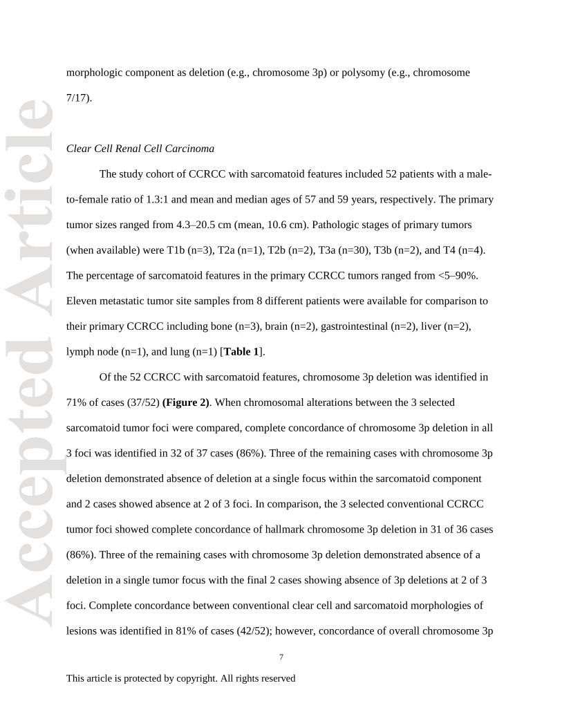
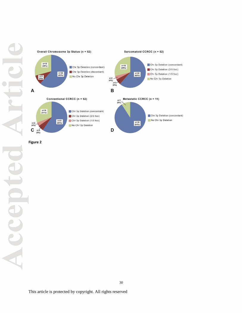
Of the 52 CCRCC with sarcomatoid features, chromosome 3p deletion was identified in
71% of cases (37/52) (Figure 2). When chromosomal alterations between the 3 selected
sarcomatoid tumor foci were compared, complete concordance of chromosome 3p deletion in all
3 foci was identified in 32 of 37 cases (86%). Three of the remaining cases with chromosome 3p
deletion demonstrated absence of deletion at a single focus within the sarcomatoid component
and 2 cases showed absence at 2 of 3 foci. In comparison, the 3 selected conventional CCRCC
tumor foci showed complete concordance of hallmark chromosome 3p deletion in 31 of 36 cases
(86%). Three of the remaining cases with chromosome 3p deletion demonstrated absence of a
deletion in a single tumor focus with the final 2 cases showing absence of 3p deletions at 2 of 3
foci. Complete concordance between conventional clear cell and sarcomatoid morphologies of
lesions was identified in 81% of cases (42/52); however, concordance of overall chromosome 3p
8
This article is protected by copyright. All rights reserved
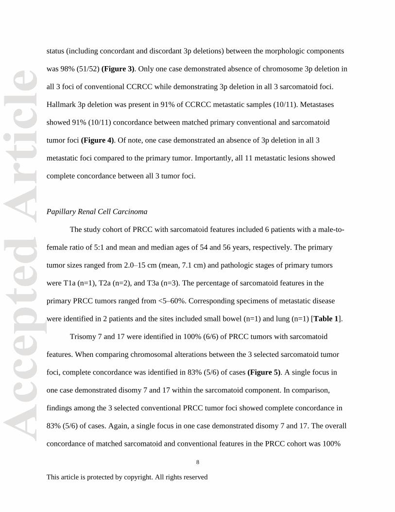
status (including concordant and discordant 3p deletions) between the morphologic components
was 98% (51/52) (Figure 3). Only one case demonstrated absence of chromosome 3p deletion in
all 3 foci of conventional CCRCC while demonstrating 3p deletion in all 3 sarcomatoid foci.
Hallmark 3p deletion was present in 91% of CCRCC metastatic samples (10/11). Metastases
showed 91% (10/11) concordance between matched primary conventional and sarcomatoid
tumor foci (Figure 4). Of note, one case demonstrated an absence of 3p deletion in all 3
metastatic foci compared to the primary tumor. Importantly, all 11 metastatic lesions showed
complete concordance between all 3 tumor foci.
Papillary Renal Cell Carcinoma
The study cohort of PRCC with sarcomatoid features included 6 patients with a male-to-
female ratio of 5:1 and mean and median ages of 54 and 56 years, respectively. The primary
tumor sizes ranged from 2.0–15 cm (mean, 7.1 cm) and pathologic stages of primary tumors
were T1a (n=1), T2a (n=2), and T3a (n=3). The percentage of sarcomatoid features in the
primary PRCC tumors ranged from <5–60%. Corresponding specimens of metastatic disease
were identified in 2 patients and the sites included small bowel (n=1) and lung (n=1) [Table 1].
Trisomy 7 and 17 were identified in 100% (6/6) of PRCC tumors with sarcomatoid
features. When comparing chromosomal alterations between the 3 selected sarcomatoid tumor
foci, complete concordance was identified in 83% (5/6) of cases (Figure 5). A single focus in
one case demonstrated disomy 7 and 17 within the sarcomatoid component. In comparison,
findings among the 3 selected conventional PRCC tumor foci showed complete concordance in
83% (5/6) of cases. Again, a single focus in one case demonstrated disomy 7 and 17. The overall
concordance of matched sarcomatoid and conventional features in the PRCC cohort was 100%
9
This article is protected by copyright. All rights reserved
(6/6). Trisomy 7 and 17 was present in all PRCC metastases (2/2) and showed 100%
concordance with primary conventional and sarcomatoid tumor foci (Figure 6).
Discussion
In this study, we evaluated the presence and maintenance of truncal chromosomal
aberrations of multiple tumor foci in matched conventional and sarcomatous components of
primary RCC. CCRCC is the most common RCC subtype and mutations of von Hippel Lindau
gene (VHL) and the loss of chromosome 3p are fundamental events in development.[13,14,28]
Previously, discordant 3p deletion patterns have been shown in multifocal CCRCC (intrarenal
metastatic spread)[26]; however, Gerlinger et al have demonstrated preservation of chromosome
3p deletion sampling multiple regions of 10 CCRCC cases.[4] Our study includes 58 different
patients and considering the multifocal sampling performed, we were able to evaluate the
chromosomal status of 374 primary tumor foci. With 13 samples of metastatic tumor from 10 of
58 patients, our total foci for comparison became 387. While recent studies have highlighted
variations in the subclonal population identified by genomic sequencing, our multifocal intra-
and intertumoral evaluation of hallmark chromosomal aberrations serves to highlight impressive
concurrence of chromosomal status in the setting of divergent morphology and metastasis.[29]
Sarcomatoid divergence in RCC is associated with aggressive behavior, high
pathologic/clinical stage, and overall poor prognosis.[7] Approximately 5–10% of RCC have
some component of sarcomatoid morphology.[7] The underlying mechanisms of transition to
sarcomatoid differentiation have been extensively hypothesized; however, regardless of such
purported mechanisms causing this divergence, limited prior study shows chromosomal
aberrations of the conventional components appear to be maintained.[30] Prior landmark
10
This article is protected by copyright. All rights reserved
genomic studies in RCC subtypes typically excluded cases with divergent morphology, which
explains the paucity of data evaluating preservation of these recurrent chromosomal aberrations
in the setting of sarcomatoid differentiation [9].
Cancer evolution remains an extensively studied concept and encompasses a complicated
series of molecular events. While hypotheses and opinions differ, it appears that RCCs arise from
a single clonal event which subsequently branch (or “braid”) into subclonal events. A key point
of contention is how these subclones interact, if at all, as the tumor evolves [17,20,31]. Herein
lays the debate between linear versus braided evolution from a truncal event in
tumorigenesis.[32] Furthermore, the “braided” model hypothesizes that spatially heterogenous
mutations may happen at different points in time but the overall genomic profile inevitably
becomes similar [33]. Given that many subclones are point mutations or short segment gains and
losses, FISH study methodology is crucial. Analytical resolution of FISH is determined by probe
size and labeling methods[16]. By using plasmid or oligo probes, detection could increase
resolution to 2-3 KB level. These types of probes are especially useful for the detection of micro-
deletions but with reduced sensitivity. It is still unclear how these small genetic lesions interact
with clinical tumor progression or contribute to possible resistance to treatment.[21] Regardless,
many of these subclones, while being genetically distinct, do share identical driver mutations
[29,34,35].
Regardless of the role of subclones in tumor evolution, chromosome 3p abnormalities in
CCRCC and trisomy 7/17 in PRCC remain significant in tumorigenesis and targeted treatment
[8,25]. In CCRCC, these abnormalities directly relate to the VHL gene, which is located at 3p25.
Variations in VHL expression have direct consequences to three key pathways including the
hypoxia-inducible factor (HIF), mammalian target of rapamycin (MTOR), and overexpression of
11
This article is protected by copyright. All rights reserved
vascular endothelial growth factor/platelet-derived growth factor (VEGF) [14,17,22,32]. VHL is
a ligase responsible for degrading HIF1/2A. Given the vascular nature of CCRCC, the
angiogenesis seen in these lesions is likely a direct result of this uninhibited activation.[36] The
importance of chromosome 3p deletion and VHL genetic alterations stems from their function as
truncal drivers in the development of CCRCC [4,16,17,19]. Mutations in PBRM1, SETD2, and
BAP1 have been identified in CCRCC and are notably located on the short arm of chromosome
3p (all within a 50-Mb region), highlighting further importance in assessing 3p
mutations/deletions [14,37].
Similar to the hallmark chromosome 3p deletion in CCRCC, the study by Kovac et al
describes similar evolution of PRCC from a truncal chromosomal alteration, such as trisomy
chromosome 7 and/or 17, followed by subsequent subclonal mutations.[12] While types 1 and 2
PRCC have been shown to be genetically and clinically distinct, a substantial majority show
chromosomal gains of 7 and 17.[38] PRCC mutations also include MET, NF2 (in the Hippo
signaling pathway), and PNKD which occur on chromosome 7 [39]. Drugs such as foretinib
which is a mutikinase agent targeting MET, AXL, and other receptors have been used [40].
Some evidence has been shown to suggest chemotherapeutic agents used to treat CCRCC,
including sunitinib, sorafenib, and mTOR inhibitors such as everolimus and temsirolimus, also
show efficacy in other RCC subtypes including PRCC and chromophobe renal cell
carcinoma[41]. Interestingly, Foretinib appears to benefit patients with germline MET mutations
compared to all PRCC patients [40].
In current models of RCC evolution and of cancer progression, prior data suggest that
conventional and sarcomatoid tumor components likely arise from a common progenitor cell
with subsequent subclonal populations arising as the tumor progresses.[8,9] It has been reported
12
This article is protected by copyright. All rights reserved
that areas of sarcomatoid differentiation in RCC have increased mutations and greater loss of
heterozygosity.[42] Gulati et al among other previous studies evaluating intratumoral
heterogeneity, particularly at the subclonal level, have argued that single focus sampling is
insufficient for true molecular classification of individual tumors.[10,43,44] Previously, Kouba
et al have compared mutational variability between primary and matched metastatic lesions,
which have shown similarities in a majority of tumor pairs.[21,45] Conversely, a study by Huang
et al demonstrated chromosome 3p deletions in metastatic RCC that were not present in the
primary tumor, which was identified in a single case within our current study.[5] Ito et al have
demonstrated foci of sarcomatoid morphology or matched metastatic sites may contain high rates
of chromosomal imbalances, but we have demonstrated that truncal hallmark drivers are often
maintained.[6] Other histologic types of RCC, such as chromophobe RCC, have been shown to
contain similar preservation of genetic abnormalities between primary and matched metastatic
lesions.[46-48] Several studies have argued that metastatic lesions are less heterogeneous, but
also completely different genetically, from their primary lesion.[5] Our study highlighted
complete chromosomal concordance between all tumor foci within each metastatic lesion in both
CCRCC and PRCC.
Overall, the 5-year survival for RCC ranges from 61–72%.[49,50] Regardless of the
histologic subtype, high-stage RCC disease has a 5-year survival rate of 53% and only 8% with
metastatic disease.[51] Adjuvant chemotherapy has historically been reserved for RCC patients
with metastatic disease.[52,53] Ravaud et al have suggested that adjuvant treatment with high-
risk nonmetastatic RCC could provide significantly longer disease-free survival.[54] VHL
mutations are associated with dysregulated angiogenesis. Tyrosine kinase inhibitors, including
sunitinib, sorafenib, pazopanib, axitinib, levatinib, and cabozantinib and remain first line therapy
13
This article is protected by copyright. All rights reserved
in metastatic RCC, have been used in treatment of advanced RCCs since the early 21st
century.[11,55-57] Success using these chemotherapeutic agents appears to stem from targeting
activating mutations of angiogenesis by way of VEGF receptors and the MTOR[33,58,59]. Some
evidence has been shown to suggest chemotherapeutic agents used to treat CCRCC, including
sunitinib, sorafenib, and mTOR inhibitors such as everolimus and temsirolimus, could also show
efficacy in other RCC subtypes including PRCC and chromophobe renal cell
carcinoma[14,41,60]. The VHL gene, located on chromosome 3p25, has been shown to be a
tumor suppressor gene; therefore, deletion or inactivation has been attributed to downstream
effects on HIF.[14,61] Demonstration of preserved chromosome 3p deletions in intratumoral and
metastatic foci is of importance given the first line targeted therapies in chemotherapeutic
regimens. In addition to its correlation with VHL, chromosome 3p deletion has been attributed
with better overall survival in CCRCC patients. Of note, correlating overall survival as it relates
to chromosome 14q (which harbors the HIF1A gene) status has also been evaluated.[62]
As previously stated, studies have postulated that intratumoral heterogeneity contributes
to therapeutic resistance likely secondary to intratumoral genetic heterogeneity by a combination
of genomic instability and Darwinian cancer evolution.[2,10,29] Furthermore, these studies
maintain that genomic/genetic reprofiling of metastatic lesions could be a novel approach to
identify causes of resistance to anti-VEGF inhibitors.[23] Genetic overlap between CCRCC and
PRCC, upwards of 100 genes, has been described.[63] Of note, resistance to first line VEGF
inhibitors has been attributed to mutations in tyrosine kinase pathways (e.g. MET or AXL).
Combination therapy or use of drugs that target both VEFG and MET pathways (e.g.
cabozantinib) could in theory address this issue, but toxicity with agents remains a serious issue
in RCC treatment[53].While both tumors have similar treatment modalities in the metastatic
14
This article is protected by copyright. All rights reserved
setting, a study by Voss et al postulated that the decreased effectiveness of treatment of
metastatic PRCC is likely secondary more to the use of antiangiogenic therapies in a tumor that
typically lacks VHL loss rather than resistance secondary to tumor heterogeneity.[64]
Unfortunately, challenges in treating metastatic PRCC appear to go beyond targeting a truncal
clonal chromosomal alteration (e.g., trisomy 7 or 17). While combination chemotherapeutic
drugs like foretinib, a MET/VEGF inhibitor, may show efficacy in patients with PRCC (as the
MET gene is located on chromosome 7), the complex genetic landscape of these tumors
continually poses a challenge to treatment.[40,53,65]
Our current study has corroborated many prior molecular findings in CCRCC and PRCC,
such as hallmark abnormalities and relative preservation of chromosomal status by multifocal
analysis. Unfortunately, this has not elucidated why efficacy of chemotherapeutic agents is so
poor. Given that mutations/alterations in CCRCC, including lesser known mutations such as
BAP1 and SETD2, typically occur on the short arm of 3p, current regiments targeting these
regions have done little to improve overall survival and disease-free survival[14]. While we may
have been able to prove that CCRCC and PRCC tumors retain similar chromosomal status
regardless of tumor divergence or metastasis, this unfortunately also highlights that tumor
progression likely goes beyond the pathways described above.
One potential limitation identified in this study is the ability to identify all possible
chromosome 3p genetic alterations in clear cell renal cell carcinoma by FISH studies[16-19]. In
the current study, the BAC clone probe used was 169K in size. According to other metaphase
and molecular studies, the majority of the 3p deletions in CCRCC were in the pattern of 3p13-
ptar or 3p25-pter deletion, and microdeletions were not typically reported in CCRCC
[4,13,14,66,67]. Since the previously reported deletions were far greater in size than that of the
15
This article is protected by copyright. All rights reserved
probe (169KB), we felt a vast majority would be detectable by the probe. The histograms
(Figure 3G and 6E) represent the signal pattern distribution from 100 typical tumor cells from
CCRCC (3G) and PRCC (6E). The x-axis represents the signal patterns and the y-axis represents
the cell numbers that baring a specific signal pattern. The 3p25 deletion histogram demonstrated
that more than 60% of the tumor cells presented as 1G2R pattern (3p25 deletion). A small
population of cells also showed disomic (2G2R), section truncations (1G/1R or 2G1R) or more
than 3 of G/R signals, which may represent the DNA replication in the population of mitotic
cells. For the chromosome 7/17 enumeration, the histogram showed that about 80% of cells
demonstrated trisomy (all populations with 3G signal pattern, while the chromosome 17 trisomy
frequency was about 70% of total tumor cell populations. The histogram shows that the current
threshold could effectively detect the chromosome deletion and gains.
While a majority of allelic loss occurs by simple deletion, a subset of tumors have been
shown to incur loss of chromosome region by uniparental disomy (UPD) or copy-neutral loss of
heterozygosity (LOH)[68]. Traditionally UPD, which occurs when a person inherits two
chromosomal copies from the same parent and none from the other, had been associated with
inherited-type diseases [18,69]. However, prior studies have shown this can occur in an acquired
fashion in hematopoietic and solid organ malignancies [70-72]. Unfortunately, these LOH or
copy number-neutral changes could not be detectable by current FISH platforms, despite using a
BAC clone probe (RP11-572M14, chr3: 10,011,785-10,180,797) that covers the entire sequence
of VHL gene (VHL, chr3:10,141,008-10,153,670) [16,17,25,26,69,73]. Further molecular
analysis, particularly by high-resolution single-nucleotide polymorphism microarrays, may serve
to identify an even higher percentage of alterations in 3p chromosomal regions in future studies
[8,16,17,26,68].
16
This article is protected by copyright. All rights reserved
RCC with sarcomatoid features is an aggressive, heterogeneous subset of tumors that
frequently present at a high clinical stage. We evaluated 387 tumor foci of 58 separate RCCs
within conventional, sarcomatoid, and metastatic components. Overall, our study demonstrates
impressive preservation of chromosomal status across multiple sampled foci within similar
tumor components as well as comparatively between divergent morphologies and in the
metastatic setting. While concordance of chromosomal status was preserved in a majority of
cases, we did identify heterogeneity in a subset. Further study of tumor heterogeneity,
particularly at the subclonal level, may offer vital information in an age of targeted therapy
where efficacy remains suboptimal.
Acknowledgements: The authors would like to thank Natasha Gibson for excellent editorial
assistance.
17
This article is protected by copyright. All rights reserved
References:
1. McGranahan N, Swanton C. Clonal heterogeneity and tumor evolution: past, present, and
the future. Cell 2017;168:613-628.
2. Andor N, Graham TA, Jansen M et al. Pan-cancer analysis of the extent and
consequences of intratumor heterogeneity. Nat Med 2016;22:105-113.
3. Gerlinger M, Rowan AJ, Horswell S et al. Intratumor heterogeneity and branched
evolution revealed by multiregion sequencing. N Engl J Med 2012;366:883-892.
4. Gerlinger M, Horswell S, Larkin J et al. Genomic architecture and evolution of clear cell
renal cell carcinomas defined by multiregion sequencing. Nat Genet 2014;46:225-233.
5. Huang Y, Gao S, Wu S et al. Multilayered molecular profiling supported the monoclonal
origin of metastatic renal cell carcinoma. Int J Cancer 2014;135:78-87.
6. Ito T, Pei J, Dulaimi E et al. Genomic copy number alterations in renal cell carcinoma
with sarcomatoid features. J Urol 2016;195:852-858.
7. de Peralta-Venturina M, Moch H, Amin M et al. Sarcomatoid differentiation in renal cell
carcinoma: a study of 101 cases. Am J Surg Pathol 2001;25:275-284.
8. Jones TD, Eble JN, Wang M, Maclennan GT, Jain S, Cheng L. Clonal divergence and
genetic heterogeneity in clear cell renal cell carcinomas with sarcomatoid transformation.
Cancer 2005;104:1195-1203.
9. Malouf GG, Ali SM, Wang K et al. Genomic characterization of renal cell carcinoma
with sarcomatoid dedifferentiation pinpoints recurrent genomic alterations. Eur Urol
2016;70:348-357.
10. Gerlinger M, Swanton C. How Darwinian models inform therapeutic failure initiated by
clonal heterogeneity in cancer medicine. Br J Cancer 2010;103:1139-1143.
18
This article is protected by copyright. All rights reserved
11. Brunelli M, Fiorentino M, Gobbo S et al. Many facets of chromosome 3p cytogenetic
findings in clear cell renal carcinoma: the need for agreement in assessment FISH
analysis to avoid diagnostic errors. Histol Histopathol 2011;26:1207-1213.
12. Kovac M, Navas C, Horswell S et al. Recurrent chromosomal gains and heterogeneous
driver mutations characterise papillary renal cancer evolution. Nat Commun
2015;6:6336.
13. The Cancer Genome Atlas Network. Comprehensive molecular characterization of clear
cell renal cell carcinoma. Nature 2013;499:43-49.
14. Frew IJ, Moch H. A clearer view of the molecular complexity of clear cell renal cell
carcinoma. Annu Rev Pathol 2015;10:263-289.
15. The Cancer genome Atlas Network. Comprehensive molecular characterization of
papillary renal-cell carcinoma. N Engl J Med 2016;374:135-145.
16. Cheng L, Zhang S, Wang L, MacLennan GT, Davidson DD. Fluorescence in situ
hybridization in surgical pathology: principles and applications. J Pathol Clin Res
2017;3:73-99.
17. Cheng L, Zhang S, MacLennan GT, Lopez-Beltran A, Montironi R. Molecular and
cytogenetic insights into the pathogenesis, classification, differential diagnosis, and
prognosis of renal epithelial neoplasms. Human pathology 2009;40:10-29.
18. Cheng LZ, DY.; Eble, JN. Molecular Genetic Patholgoy. New York, NY: Springer; 2013.
19. Cheng L, Eble JN. Molecular Surgical Pathology. New York, NY: Springer; 2013.
20. Cheng L, Williamson SR, Zhang S, Maclennan GT, Montironi R, Lopez-Beltran A.
Understanding the molecular genetics of renal cell neoplasia: implications for diagnosis,
prognosis and therapy. Expert review of anticancer therapy 2010;10:843-864.
19
This article is protected by copyright. All rights reserved
21. Kouba EJ, Eble JN, Simper N et al. High fidelity of driver chromosomal alterations
among primary and metastatic renal cell carcinomas: implications for tumor clonal
evolution and treatment. Mod Pathol 2016;29:1347-1357.
22. Wang L, Williamson SR, Wang M et al. Molecular subtyping of metastatic renal cell
carcinoma: implications for targeted therapy. Mol Cancer 2014;13:39.
23. Massari F, Ciccarese C, Bria E et al. Reprofiling metastatic samples for chromosome 9p
and 14q aberrations as a strategy to overcome tumor heterogeneity in clear-cell renal cell
carcinoma. Appl Immunohistochem Mol Morphol 2017;25:39-43.
24. Moch H, Humphrey PA, Ulbright TM, Reuter VE. WHO classification of tumors of the
urinary system and male genital organ. Lyon: LARC Press; 2016.
25. Jones TD, Eble JN, Wang M et al. Molecular genetic evidence for the independent origin
of multifocal papillary tumors in patients with papillary renal cell carcinomas. Clin
Cancer Res 2005;11:7226-7233.
26. Cheng L, MacLennan GT, Zhang S et al. Evidence for polyclonal origin of multifocal
clear cell renal cell carcinoma. Clin Cancer Res 2008;14:8087-8093.
27. Halat S, Eble JN, Grignon DJ et al. Multilocular cystic renal cell carcinoma is a subtype
of clear cell renal cell carcinoma. Mod Pathol 2010;23:931-936.
28. McGranahan N, Swanton C. Biological and therapeutic impact of intratumor
heterogeneity in cancer evolution. Cancer Cell 2015;27:15-26.
29. Burrell RA, Swanton C. Re-evaluating clonal dominance in cancer evolution. Trends
Cancer 2016;2:263-276.
30. Bi M, Zhao S, Said JW et al. Genomic characterization of sarcomatoid transformation in
clear cell renal cell carcinoma. Proc Natl Acad Sci U S A 2016;113:2170-2175.
20
This article is protected by copyright. All rights reserved
31. Venkatesan S, Swanton C. Tumor evolutionary principles: How intratumor heterogeneity
influences cancer treatment and outcome. Am Soc Clin Oncol Educ Book 2016;35:e141-
149.
32. Hsieh JJ, Manley BJ, Khan N, Gao J, Carlo MI, Cheng EH. Overcome tumor
heterogeneity-imposed therapeutic barriers through convergent genomic biomarker
discovery: A braided cancer river model of kidney cancer. Semin Cell Dev Biol (2016, in
press).
33. Hsieh JJ, Purdue MP, Signoretti S et al. Renal cell carcinoma. Nat Rev Dis Primers
2017;3:17009.
34. Schwartz R, Schaffer AA. The evolution of tumour phylogenetics: principles and
practice. Nat Rev Genet 2017;advance online publication.
35. McDonald O, Li X, Saunders T et al. Epigenomic reprogramming during pancreatic
cancer progression links anabolic glucose metabolism to distant metastasis. Nat Genet
(2017, In press).
36. Semenza GL. HIF-1 mediates metabolic responses to intratumoral hypoxia and
oncogenic mutations. J Clin Invest 2013;123:3664-3671.
37. Cohen HT, McGovern FJ. Renal-cell carcinoma. N Engl J Med 2005;353:2477-2490.
38. The Cancer Genome Atlas Research Network. Comprehensive molecular characterization
of papillary renal-cell carcinoma. N Engl J Med 2016;374:135-145.
39. Gonzalez Del Alba A, Arranz JA, Puente J et al. Recent advances in genitourinary
tumors: A review focused on biology and systemic treatment. Crit Rev Oncol Hematol
2017;113:171-190.
21
This article is protected by copyright. All rights reserved
40. Choueiri TK, Vaishampayan U, Rosenberg JE et al. Phase II and biomarker study of the
dual MET/VEGFR2 inhibitor foretinib in patients with papillary renal cell carcinoma. J
Clin Oncol 2013;31:181-186.
41. Abbosh P, Sundararajan S, Millis S et al. Molecular and genomic profiling to identify
actionable targets in chromophobe renal cell cancer. Eur Urol Focus 2017:(in press).
42. Manley BJ, Hsieh JJ. Sarcomatoid renal cell carcinoma: genomic insights from
sequencing of matched sarcomatous and carcinomatous components. Transl Cancer Res
2016:S160-S165.
43. Gulati S, Martinez P, Joshi T et al. Systematic evaluation of the prognostic impact and
intratumour heterogeneity of clear cell renal cell carcinoma biomarkers. Eur Urol
2014;66:936-948.
44. Haddad AQ, Margulis V. Tumour and patient factors in renal cell carcinoma-towards
personalized therapy. Nat Rev Urol 2015;12:253-262.
45. Goswami RS, Patel KP, Singh RR et al. Hotspot mutation panel testing reveals clonal
evolution in a study of 265 paired primary and metastatic tumors. Clin Cancer Res
2015;21:2644-2651.
46. Brunelli M, Gobbo S, Cossu-Rocca P et al. Chromosomal gains in the sarcomatoid
transformation of chromophobe renal cell carcinoma. Mod Pathol 2007;20:303-309.
47. Abbosh P, Sundararajan S, Millis S et al. Molecular and genomic profiling to identify
actionable targets in chromophobe renal cell cancer. Eur Urol Focus (2017, in press).
48. Davis CF, Ricketts CJ, Wang M et al. The somatic genomic landscape of chromophobe
renal cell carcinoma. Cancer Cell 2014;26:319-330.
22
This article is protected by copyright. All rights reserved
49. Znaor A, Lortet-Tieulent J, Laversanne M, Jemal A, Bray F. International variations and
trends in renal cell carcinoma incidence and mortality. Eur Urol 2015;67:519-530.
50. Capitanio U, Montorsi F. Renal cancer. The Lancet;387:894-906.
51. Siegel RL, Miller KD, Jemal A. Cancer statistics, 2017. CA Cancer J Clin 2017;67:7-30.
52. Gore ME, Szczylik C, Porta C et al. Final results from the large sunitinib global
expanded-access trial in metastatic renal cell carcinoma. Br J Cancer 2015;113:12-19.
53. Choueiri TK, Motzer RJ. Systemic therapy for metastatic renal-cell carcinoma. N Engl J
Med 2017;376:354-366.
54. Ravaud A, Motzer RJ, Pandha HS et al. Adjuvant sunitinib in high-risk renal-cell
carcinoma after nephrectomy. N Engl J Med 2016;375:2246-2254.
55. Motzer RJ, Hutson TE, Cella D et al. Pazopanib versus sunitinib in metastatic renal-cell
carcinoma. N Engl J Med 2013;369:722-731.
56. Escudier B, Porta C, Schmidinger M et al. Renal cell carcinoma: ESMO clinical practice
guidelines for diagnosis, treatment and follow-up. Ann Oncol 2016;27:v58-v68.
57. Haas NB, Manola J, Uzzo RG et al. Adjuvant sunitinib or sorafenib for high-risk, non-
metastatic renal-cell carcinoma (ECOG-ACRIN E2805): a double-blind, placebo-
controlled, randomised, phase 3 trial. Lancet 2016;387:2008-2016.
58. Yap TA, Gerlinger M, Futreal PA, Pusztai L, Swanton C. Intratumor heterogeneity:
seeing the wood for the trees. Sci Transl Med 2012;4:127ps110.
59. Zhang J, Yang PL, Gray NS. Targeting cancer with small molecule kinase inhibitors. Nat
Rev Cancer 2009;9:28-39.
23
This article is protected by copyright. All rights reserved
60. Buchler T, Bortlicek Z, Poprach A et al. Outcomes for patients with metastatic renal cell
carcinoma achieving a complete response on targeted therapy: a registry-based analysis.
Eur Urol 2016;70:469-475.
61. Klatte T, Rao PN, de Martino M et al. Cytogenetic profile predicts prognosis of patients
with clear cell renal cell carcinoma. J Clin Oncol 2009;27:746-753.
62. Kroeger N, Klatte T, Chamie K et al. Deletions of chromosomes 3p and 14q molecularly
subclassify clear cell renal cell carcinoma. Cancer 2013;119:1547-1554.
63. Chen F, Zhang Y, Senbabaoglu Y et al. Multilevel genomics-based taxonomy of renal
cell carcinoma. Cell Rep 2016;14:2476-2489.
64. Voss MH, Molina AM, Chen YB et al. Phase II Trial and Correlative Genomic Analysis
of Everolimus Plus Bevacizumab in Advanced Non-Clear Cell Renal Cell Carcinoma. J
Clin Oncol 2016;34:3846-3853.
65. Albiges L, Guegan J, Le Formal A et al. MET is a potential target across all papillary
renal cell carcinomas: result from a large molecular study of pRCC with CGH array and
matching gene expression array. Clin Cancer Res 2014;20:3411-3421.
66. Beuselinck B, Job S, Becht E et al. Molecular subtypes of clear cell renal cell carcinoma
are associated with sunitinib response in the metastatic setting. Clin Cancer Res
2015;21:1329-1339.
67. Fenner A. Genetics: a molecular atlas of clear cell renal cell carcinoma. Nat Rev Clin
Oncol 2013;10:485.
68. Sato Y, Yoshizato T, Shiraishi Y et al. Integrated molecular analysis of clear-cell renal
cell carcinoma. Nat Genet 2013;45:860-867.
24
This article is protected by copyright. All rights reserved
69. Saeki H, Kitao H, Yoshinaga K et al. Copy-neutral loss of heterozygosity at the p53 locus
in carcinogenesis of esophageal squamous cell carcinomas associated with p53
mutations. Clin Cancer Res 2011;17:1731-1740.
70. Murthy SK, DiFrancesco LM, Ogilvie RT, Demetrick DJ. Loss of heterozygosity
associated with uniparental disomy in breast carcinoma. Mod Pathol 2002;15:1241-1250.
71. Andersen CL, Wiuf C, Kruhoffer M, Korsgaard M, Laurberg S, Orntoft TF. Frequent
occurrence of uniparental disomy in colorectal cancer. Carcinogenesis 2007;28:38-48.
72. Ogiwara H, Kohno T, Nakanishi H, Nagayama K, Sato M, Yokota J. Unbalanced
translocation, a major chromosome alteration causing loss of heterozygosity in human
lung cancer. Oncogene 2008;27:4788-4797.
73. Castronovo C, Valtorta E, Crippa M et al. Design and validation of a pericentromeric
BAC clone set aimed at improving diagnosis and phenotype prediction of supernumerary
marker chromosomes. Molecular cytogenetics 2013;6:45.
25
This article is protected by copyright. All rights reserved
Figure Legends
Figure 1. Sampling of tumor foci for fluorescence in situ hybridization (FISH) studies. (A)
Primary renal cell carcinoma tumors were sampled to identify three separate tumor foci in
conventional regions and three additional foci in sarcomatoid regions for FISH. Additionally,
three tumor foci were marked for FISH analysis in each matched metastatic sites including
lymph node, bone, brain, and lung.
Figure 2. Chromosome 3p status in primary clear cell renal cell carcinoma (including
conventional and sarcomatoid tumor foci) with matched metastatic sites. (A) Overall
chromosome 3p status including concordant and discordant chromosome 3p deletions as well as
clear cell renal carcinoma with no deletion of chromosome 3p. (B-C) Sarcomatoid and
conventional clear cell renal carcinoma morphologies with concordant (all foci) and discordant
(1 or 2 of 3 foci) deletions of chromosome 3p. A subset with no deletion of chromosome 3p in
any focus was identified in both morphologies. (D) Concordant deletion (all tumor foci) of
chromosome 3p in all matched metastatic sites was identified in 10 of 11 cases.
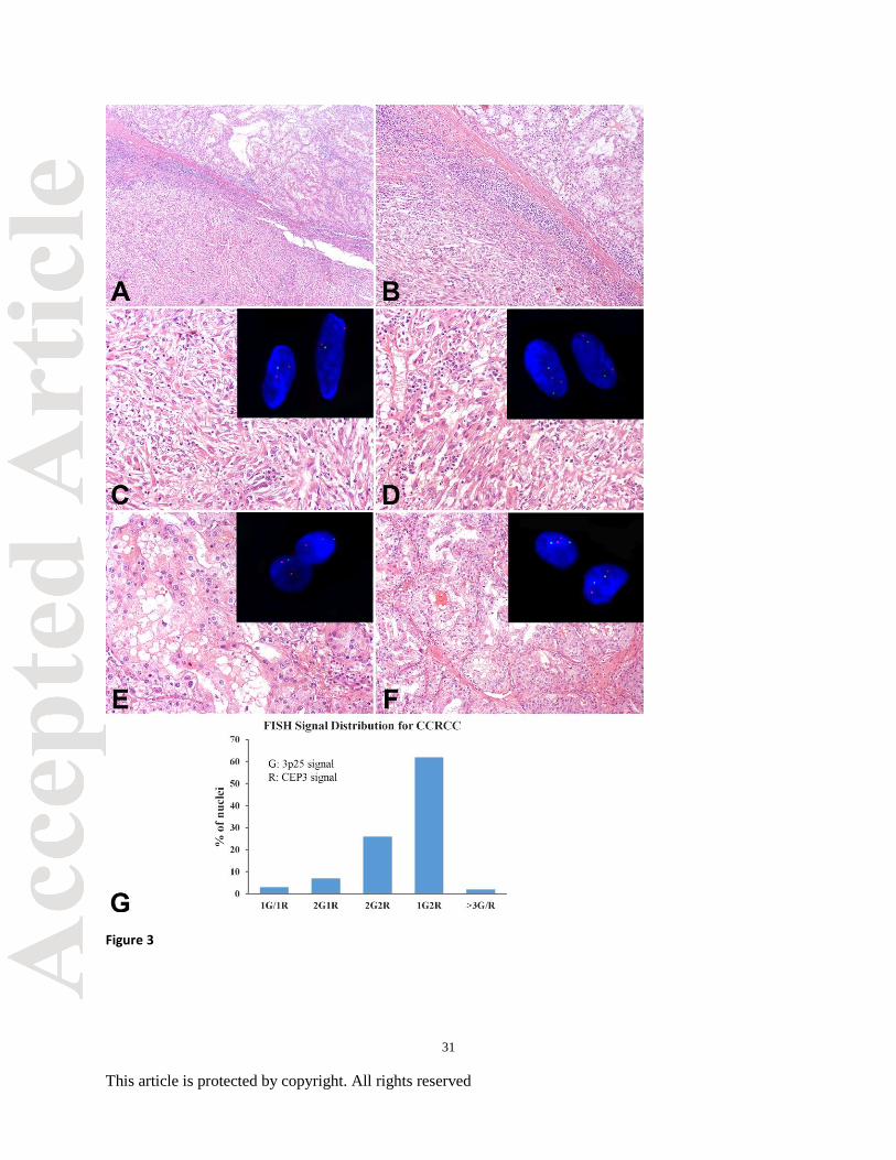
Figure 3. Primary clear cell renal cell carcinoma (CCRCC), sarcomatoid and conventional
morphologies. (A-B) Transition between conventional and sarcomatoid morphologies, low
power (A) and high power (B). (C) CCRCC with sarcomatoid features with loss of chromosome
3p using interphase fluorescence in situ hybridization (FISH; see inset). (D) Sarcomatoid
CCRCC with no deletion of chromosome 3p (see inset). (E) Conventional CCRCC with loss of
chromosome 3p by FISH (see inset). (F) Conventional CCRCC with no deletion of chromosome
3p (see inset). (G) Histogram of FISH signal distribution of a typical CCRCC. G: Green signal
26
This article is protected by copyright. All rights reserved
corresponding 3p25; R: Red signal corresponding to CEP3. Vertical axis represents the
percentage of cells showing the specific signal pattern, The signal pattern was denominated as
#G#R, which present as number of Green signal (3p25) and number of Red signal (CEP3).
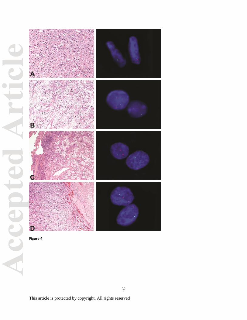
Figure 4. Primary clear cell renal cell carcinoma (CCRCC; conventional and sarcomatoid,
multiple foci) with matched metastatic sites. (A) CCRCC with sarcomatoid features with
chromosomal 3p deletion in two of three foci using interphase fluorescence in situ hybridization
(FISH). (B) Conventional CCRCC with chromosomal 3p deletion in two of three foci by FISH.
(C) Matched metastatic CCRCC to lung, 7 years status post nephrectomy, with chromosomal 3p
deletion by FISH. (D) Matched metastatic CCRCC to brain, 11 years status post nephrectomy,
with chromosomal 3p deletion by FISH.
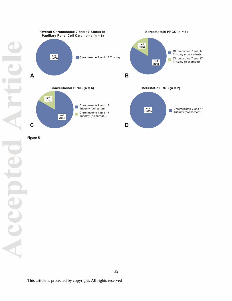
Figure 5. Chromosome 7 and 17 statuses in papillary renal cell carcinoma (PRCC; including
conventional and sarcomatoid tumor foci) with matched metastatic sites. (A) Overall, all six
PRCC carcinoma tumors demonstrated trisomy 7 and 17 in conventional and sarcomatoid tumor
foci. (B-C) Trisomy 7 and 17 was identified in single tumor focus in both the sarcomatoid and
conventional sampling demonstrating minor discordance. (D) Matched metastatic PRCC
demonstrated trisomy 7 and 17 in concordance with the primary lesions.
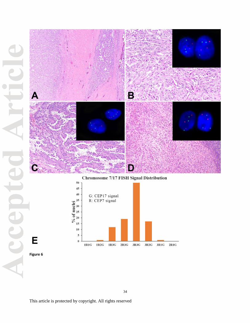
Figure 6. Primary (conventional and sarcomatoid) and matched metastatic papillary renal cell
carcinoma (PRCC). (A) Transition between conventional and sarcomatoid PRCC, low power.
(B) Sarcomatoid PRCC, multiple foci, with trisomy 7 and 17 using interphase fluorescence in
situ hybridization (FISH; inset). (C) Conventional PRCC, multiple foci, with trisomy 7 and 17
27
This article is protected by copyright. All rights reserved
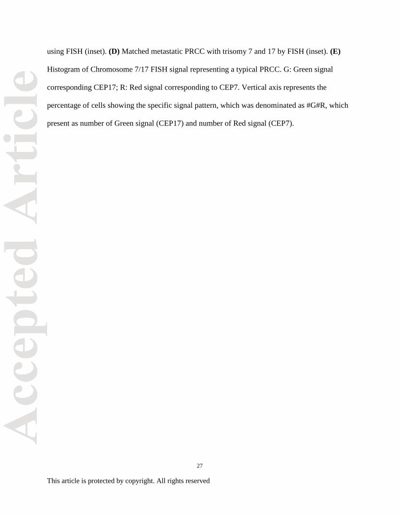
using FISH (inset). (D) Matched metastatic PRCC with trisomy 7 and 17 by FISH (inset). (E)
Histogram of Chromosome 7/17 FISH signal representing a typical PRCC. G: Green signal
corresponding CEP17; R: Red signal corresponding to CEP7. Vertical axis represents the
percentage of cells showing the specific signal pattern, which was denominated as #G#R, which
present as number of Green signal (CEP17) and number of Red signal (CEP7).
28
This article is protected by copyright. All rights reserved
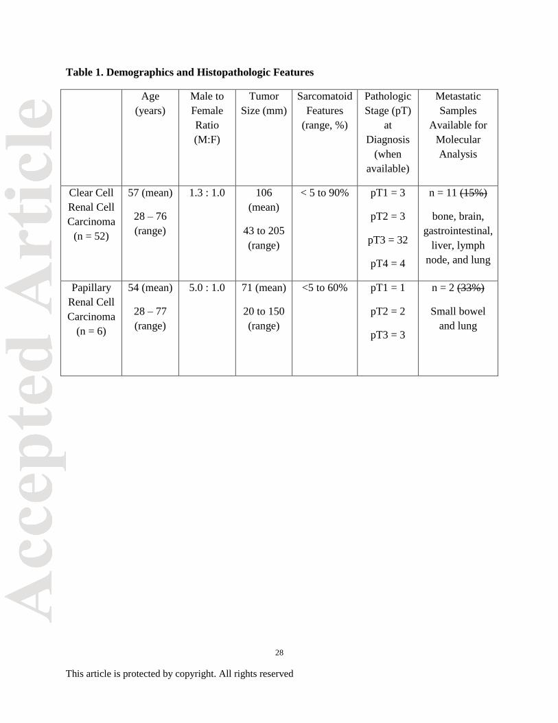
Table 1. Demographics and Histopathologic Features
Age
(years)
Male to
Female
Ratio
(M:F)
Tumor
Size (mm)
Sarcomatoid
Features
(range, %)
Pathologic
Stage (pT)
at
Diagnosis
(when
available)
Metastatic
Samples
Available for
Molecular
Analysis
Clear Cell
Renal Cell
Carcinoma
(n = 52)
57 (mean)
28 – 76
(range)
1.3 : 1.0 106
(mean)
43 to 205
(range)
< 5 to 90% pT1 = 3
pT2 = 3
pT3 = 32
pT4 = 4
n = 11 (15%)
bone, brain,
gastrointestinal,
liver, lymph
node, and lung
Papillary
Renal Cell
Carcinoma
(n = 6)
54 (mean)
28 – 77
(range)
5.0 : 1.0 71 (mean)
20 to 150
(range)
<5 to 60% pT1 = 1
pT2 = 2
pT3 = 3
n = 2 (33%)
Small bowel
and lung
29
This article is protected by copyright. All rights reserved
Figure 1
30
This article is protected by copyright. All rights reserved
Figure 2
31
This article is protected by copyright. All rights reserved
Figure 3
32
This article is protected by copyright. All rights reserved
Figure 4
33
This article is protected by copyright. All rights reserved
Figure 5
34
This article is protected by copyright. All rights reserved
Figure 6


![Malassezia Folliculitis versus Truncal Acne Vulgaris ... · 278 Malassezia Folliculitis versus Truncal Acne Vulgaris (Clinical and Histopathological Study) support the diagnosis [5,6,10]](https://img.pdfslide.net/doc/110x75/5cdf712988c99399558c9005/malassezia-folliculitis-versus-truncal-acne-vulgaris-278-malassezia-folliculitis.jpg)