Embed Size (px)
Citation preview

Presurgical Nasoalveolar Molding for Correction ofCleft Lip Nasal Deformity: Experience FromNorthern India
Brijesh Mishra, MS, MCh, DNB,a Arun K. Singh, MCh, MNAMS, FICS, MNASc,a
Javed Zaidi, DMC,a G. K. Singh, BDS, MDS,b Rajiv Agrawal, MCh, DNB,a andVijay Kumar, MCha
aDepartment of Plastic Surgery and bDepartment of Orthodontics, Chhatrapati Shahuji MaharajMedical University (Erst while King Georges Medical College), Lucknow, Uttar Pradesh, India
Correspondence: [email protected] July 23, 2010
Context: The cleft lip type nasal deformity presents one of the most complex surgi-cal challenges. The long-term postoperative results are still not satisfactory despite anemphasis on primary nasal correction. This is attributed to tissue memory and healing.Nasoalveolar molding is used effectively to reshape the nasal cartilage and to mold themaxillary arch before cleft lip repair. Aims: This study was undertaken to evaluate therole of presurgical nasoalveolar molding in correction of cleft lip nasal deformity forpatients with unilateral and bilateral clefts of the lip. Settings and Design: Twenty-three cases of clefts of lip and palate with nasal deformity were subjected to presentstudy from May 2004 to May 2006. These cases were initially treated on outpatientbasis, and they were admitted at the time of operation. All of these patients were chil-dren of less than 1 year of age, belonging to north Indian population. Material andMethods: Study consisted of patients of cleft lip and palate who were given presur-gical nasoalveolar splints at early age. Lip repair was done after at least 2 months ofmolding. These patients along with control group (without presurgical nasoalveolarmolding) were followed up for 1 year. Measurements were taken at different inter-vals in study over dental cast and on patients. Data obtained from comparison of 2groups were analyzed using “MSTAT” analysis software (developed by Dr Russel Freed,Professor & Director, Crop & Soil Sciences Department, Michigan State University,East Lansing, Michigan). Results: In our study, we found that nostril height was morein patients of experimental group (P = .18), while nostril width and alar perimeter werenot changed significantly. Children with nasoalveolar molding had significant lengthen-ing of columella (P = .02). Patients of unilateral cleft lip had more reduction in alveolargap (P = .08) than bilateral group (P = .15). Conclusions: Nasoalveolar molding canbe a useful adjunct for treatment of cleft lip nasal deformity. It is a cost-effective tech-nique that can reduce the number of future surgeries such as alveolar bone grafting andsecondary rhinoplasties.
443

ePlasty VOLUME 10
The management of cleft patients has evolved dramatically in recent years. Outcomeis improving because of better surgical techniques, timing, and incorporation of procedureslike presurgical orthopedics.
Presurgical infant orthopedics was first introduced by McNeil1 in 1950. Since then,techniques are changing and so are the results. Active molding and repositioning of thenasal cartilages take advantage of the plasticity of cartilage in the newborn infant.2,3
In the last decade, it has been shown that correction of nasal deformity by stretchingof the nasal mucosal lining, and achievement of nonsurgical columella elongation can becombined with molding of the alveolar process in cleft patients.4,5
Multiple reports have come from around the world about efficacy and utility of na-soalveolar molding with different opinions; however, studies on Indian populations arelacking. Objective of this study was to evaluate the role of nasoalveolar molding for correc-tion of cleft lip nasal deformity in Indian patients and to see efficacy of molding in differentage groups.
SUBJECTS AND METHODS
Twenty-three cases of clefts of lip and palate with nasal deformity were subjected to presentstudy from May 2004 to May 2006. These cases were initially treated on outpatient basis,and they were admitted at the time of operation. All of these patients were children of lessthan 1 year of age, belonging to north Indian population.
Their parents were explained about the cleft deformity and various stages of treatment.They were specially told about the procedure of nasoalveolar molding, the technique,requirement for periodic checkups, and sequential correction. Local patients or patientsfrom nearby places who consented for weekly follow-ups were chosen for study.
Preparation of splint
The impression was obtained with the infant fully awake, in prone position without anes-thesia. Before impressions, child was kept nil orally for about 2 hours. Impressions weretaken on dental chair with child in the lap of his or her parents. Impression should be takenvery carefully and is always done after insuring the availability of anesthesia team.
First, the impression tray was checked in the mouth of patient. After selection of aproper size tray, alginate paste was made, loaded in the tray, and inserted in the mouth. Soonafter this, alginate paste was applied over the plate by hand up to root of the nose. Child’slower jaw was pulled down, and precautions were taken to avoid falling of impressionmaterial into oral cavity. After some time (15-20 s), this nose, lip, and alveolus negativeimpression was removed in a single piece. Oral and nasal cavities were inspected for anyremaining particles.
After impression, a dental stone cast was made by filling it with paste of dental stonematerial. It was allowed to fix. Dental stone model was made for purpose of measurementsand fabrication of appliance. These dental stone casts were labeled with patient’s name,age/sex, and date.
A conventional molding plate was fabricated on the maxillary cast using clear acrylicresin with a nasal stent wire passed from it going superiorly toward nose. The tip of wire
444

MISHRA ET AL
was covered with hard and then soft acrylic. At the active tip of nasal stent, the acrylic wascovered with a thin layer of soft denture lining material to insure that tissue irritation doesnot occur when pressure is applied for nasoalveolar molding (Fig 1).
Figure 1. Dental model and fabricationof nasoalveolar moulding splint.
After the nasoalveolar molding plate was ready, it was examined for any rough areas.Plate was handed over to parents, and they were explained about maintaining oral hygiene,cleaning, insertion, and removal of plate.
Patients were called at weekly intervals to gradually change the direction of nasal wire.At every visit, local area was examined for any ulceration or pressure points. Mea-
surements of different nasal parameters and alveolus were taken on prepared dental stonemodel as well as on patients directly with the help of thread and artery forceps, and wererecorded. It was done for the purpose of accuracy. Measurements on patients were matchedwith measurements on dental models, and they were found to be almost similar.
Figure 2. Measurements before lip repair.
Following Reference points were used for different measurements (Figs 2 and 3):
a. alar base noncleft sideb. columellar base noncleft sidec. midpoint of a-b, centre of floor of the nosed. the highest point on the alar rim noncleft sidee. midpoint at the base of columellaf. the highest point in the midline of columella
445
ePlasty VOLUME 10
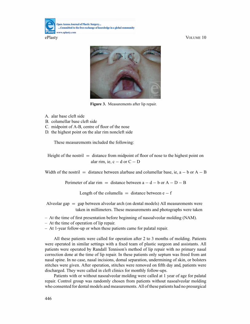
Figure 3. Measurements after lip repair.
A. alar base cleft sideB. columellar base cleft sideC. midpoint of A-B, centre of floor of the noseD. the highest point on the alar rim noncleft side
These measurements included the following:
Height of the nostril = distance from midpoint of floor of nose to the highest point onalar rim, ie, c − d or C − D
Width of the nostril = distance between alarbase and columellar base, ie, a − b or A − B
Perimeter of alar rim = distance between a − d − b or A − D − B
Length of the columella = distance between e − f
Alveolar gap = gap between alveolar arch (on dental models) All measurements weretaken in millimeters. These measurements and photographs were taken
– At the time of first presentation before beginning of nasoalveolar molding (NAM).– At the time of operation of lip repair.– At 1-year follow-up or when these patients came for palatal repair.
All these patients were called for operation after 2 to 3 months of molding. Patientswere operated in similar settings with a fixed team of plastic surgeon and assistants. Allpatients were operated by Randall Tennison’s method of lip repair with no primary nasalcorrection done at the time of lip repair. In these patients only septum was freed from antnasal spine. In no case, nasal incisions, domal separation, undermining of skin, or bolstersstitches were given. After operation, stitches were removed on fifth day and, patients weredischarged. They were called in cleft clinics for monthly follow-ups.
Patients with or without nasoalveolar molding were called at 1 year of age for palatalrepair. Control group was randomly chosen from patients without nasoalveolar moldingwho consented for dental models and measurements. All of these patients had no presurgical
446

MISHRA ET AL
nasoalveolar molding, and these were operated by same team of surgeons using the similartechnique of repair with no primary nasal correction. Dental models were made for thesepatients, and measurements were taken on dental models as well as patients.
Statistical evaluation
The data obtained were subjected to statistical analysis. Statistical analysis was done with“MSTAT” analysis software (Michigan State University, East Lansing, Michigan). Mean(SD) was calculated for all groups. Paired t test and unpaired t test were used to test thesignificance of change in variables and difference in 2 groups. Values are presented aremean (SD) and percentage.
RESULTS
These 23 patients of cleft lip nose deformity of experimental group were of age ranging from10 to 360 days. The experimental group had 17 patients of unilateral cleft nose deformityand 6 patients of bilateral cleft nose deformity. The duration of the NAM ranged from 2 to3 months (average 2 months 10 days) (Figs 4–8).
In unilateral clefts difference in nostril height on cleft side was higher in experimentalgroup than control group (P = .18), while noncleft side in both groups were almost similar(P = .85). In bilateral clefts nostril height was increased in both sides in experimentalgroup than control group (P = .30 for both sides) (Table 1).
Table 1. Comparison of nostril height at 1-year follow-up after lip repair
Cases with NAM, Controls without NAM,mean (SD), mm mean (SD), mm t P
Unilateral cases (N = 17) Noncleft 6.000 (0.9354) 6.0588 (0.9824) 0.18 .85Cleft 5.4412 (0.8993) 5.0000 (0.9683) 1.38 .18
Bilateral cases (N = 6) Right 5.0833 (1.1583) 4.2500 (1.4405) 1.10 .30Left 4.8333 (1.2910) 4.0000 (1.4491) 1.05 .30
Abbreviation: NAM, nasoalveolar molding.
Columellar length was found significantly higher for cases both in unilateral andbilateral clefts (P = .05 for both unilateral and bilateral cases). Relative comparison ofcolumellar lengthening shows that lengthening of columella is significantly higher in uni-lateral cases than bilateral cases (P = .02) (Table 2).
Table 2. Comparison of length of columella at 1-year follow-up after lip repair
Cases with NAM, Controls without NAM,mean (SD), mm mean (SD), mm t P
Unilateral cases (N = 17) 5.2647 (0.7524) 4.5882 (0.8703) 2.42 <.05Bilateral cases (N = 6) 4.3333 (0.5164) 3.4167 (0.6646) 2.67 <.05
Abbreviation: NAM, nasoalveolar molding.
447

ePlasty VOLUME 10
After 1 year of lip repair, alveolar gap was found higher in control group both inunilateral (P = .08) and bilateral cases (right, P = .15; left, P = .15). Relative comparisonwithin the experimental group shows that presence of alveolar gap is higher in bilateralcases on both sides in comparison with gap in unilateral cases (right, P = .45; left, P = .15)(Table 3).
Table 3. Comparison of presence of alveolar gap at 1-year follow-up after lip repair
Alveolar gap cases with Alveolar gap controls withoutNAM, mean (SD), mm NAM, mean (SD), mm t P
Unilateral cases (N = 17) 1.5294 (1.6999) 2.7059 (1.7946) 1.96 .08Bilateral cases (N = 6) Right 2.1667 (1.8348) 3.6667 (1.2111) 1.67 .15
Left 2.1667 (1.5055) 4.080 (1.4142) 1.58 .15
Abbreviation: NAM, nasoalveolar molding.
Patients were divided in 4 groups according to their age of starting the NAM. Maximumchange was observed in first 2 groups. Infant up to 6 weeks had maximum effect of theNAM, especially, on cleft side, that is, 72% increase from pretreatment level. This increaseis 66.7% in group II, 38% in group III, and 47% in group IV. It signifies that molding ismost effective if done in early age groups (Table 4).
Table 4. Analysis of nostril height in unilateral cases of experimental group according to ageof starting of nasoalveolar molding
Pretreatment, Preoperative, Change, meanSubgroup age mean (SD), mm mean (SD), mm (SD), mm % change
Group I (birth-6 wk)Noncleft 3.333 (0.7528) 4.4167 (0.6646) 1.0833 (0.5845) 32.5Cleft 2.6661 (0.7528) 4.5853 (0.6646) 1.9167 (0.7360) 72.0
Group II (7 wk-3 mo)Noncleft 3.667 (0.6055) 4.500 (0.9487) 0.8333 (0.9309) 22.73Cleft 2.7500 (0.2739) 4.5833 (1.0206) 1.8333 (1.0328) 66.7
Group III (4 mo-6 mo)Noncleft 4.1667 (0.7600) 4.8333 (0.7638) 0.6667 (0.2887) 16.0Cleft 3.500 (0.5000) 4.8333 (0.7638) 1.3333 (0.2887) 38.0
Group IV (7 mo-1 y)Noncleft 6.000 (2.8284) 6.2500 (2.4749) 0.2500 (0.3536) 4.2Cleft 4.2500 (2.4749) 6.2500 (2.4749) 2.0000 (0.4210) 47.1
Gain in columellar length was maximum in group I (42.1%), and gain in lengthdecreases as the age of starting of molding increases (30% in group II, 26% in group III,and 19.1% in group IV). Reduction in alveolar gap is maximum in group I (50.1). Changein alveolar gap in other group was 36% in group II, 33.3% in group III, and 50% in groupIV (Table 5).
448

MISHRA ET AL
Table 5. Analysis of columellar length and alveolar gap in unilateral cases of experimentalgroup according to age of starting of nasoalveolar molding
Pretreatment, Preoperative, Change, meanSubgroup age mean (SD), mm mean (SD), mm (SD), mm % change
Group I (birth-6 wk)Col length 3.1667 (0.7528) 4.500 (0.8367) 1.3333 (0.5164) 42.1Alv gap 6.8333 (2.1370) 3.4167 (1.4287) 3.4167 (1.0206) 50.1
Group II (7 wk-3 mo)Col length 3.3333 (0.6055) 4.333 (0.8165) 1.000 (0.8367) 30Alv gap 7.500 (3.9370) 4.8333 (1.3292) 2.6667 (3.4448) 36
Group III (4 mo-6 mo)Col length 3.8333 (0.2887) 4.8333 (0.7683) 1.00 (0.50) 26.1Alv gap 4.0000 (6.9282) 2.6667 (4.6182) 1.333 (2.3094) 33.3
Group IV (7 mo-1 y)Col length 5.2500 (2.4749) 6.25 (2.4749) 1.000 (0.000) 19.1Alv gap 4.000 (5.6549) 2.00 (2.8284) 2.000 92.8284) 50.0
Abbreviations: Alv gap, alveolar gap; Col length, columellar length.
DISCUSSION
Bardach and Cutting6 in 1990 described the NAM by acrylic intraoral molding plate witha nasal stent of acrylic, rising from the labial vestibular flange. Similar procedure wasdescribed by Hotz and Gnoinski7 as Zurich type molding plate, but only for alveolus.Cutting and Bardach gradually added small amount of acrylic resin to lift the alar domecartilage on the cleft side to achieve normal elevation and symmetry.
Our appliance is nearly same except that we used an orthodontic wire covered byan acrylic bulb to give pressure for active molding. Our appliance is more cost-effective,because it does not need any further addition of acrylic every week, only wire angle isincreased a bit to increase the pressure exerted.
Matsuo and Hirose2,3showed role of preoperative molding in changing the cartilagememory of deformed nasal cartilage, because these cartilages have higher amount ofhyaluronic acid, which gradually diminishes after few months of birth.
In our study we found that changes because of molding were most significant in theage group of birth to 6 weeks, and they were better in first 3 months of life. It shows thatbeneficial effect of molding is maximum in the youngest children.
Maull and colleague,5 Cutting and colleague,8 and Grayson and colleague,9 studiedlong-term effects of the NAM on 3-dimensional nasal shape in unilateral clefts by usingnasal cast of the subjects. They scanned these casts in 3 dimensions, and a numericalasymmetry score was determined. The mean asymmetry index for the NAM group was0.74, and for the control group it was 1.21. This difference was statistically significant(P < .05). They concluded presurgical NAM significantly increases the symmetry of thenose.
Grayson and Maull10 and Cutting and colleague11 evaluated the financial impact of 2treatment approaches to the unilateral cleft alveolus. They compared NAM, and gingivope-riosteoplasty at the time of lip repair with the traditional approach of lip repair followed by
449

ePlasty VOLUME 10
secondary alveolar bone graft. Of the 16 patients treated by NAM, gingivoperiosteoplasty,lip repair, and primary nasal repair, 10 required no further treatment of the unilateral cleftalveolus; 6 patients required secondary alveolar bone graft.
Our study also shows a trend of reduced alveolar gap in experimental patients afterNAM that may lead to avoidance of surgery in future like alveolar bone grafting andsecondary rhinoplasties.
Da Silveira et al12 have also described a similar appliance with metallic wire and foundit to be useful and more easy to manipulate.
Liou EJ et al13 assessed 25 infants for the progressive changes of nasal symmetry,growth, and relapse by direct linear measurements on photographs and concluded that thenasal asymmetry was significantly improved after nasoalveolar molding and was furthercorrected to symmetry after primary cheiloplasty. After the primary cheiloplasty, the nasalasymmetry significantly relapsed in the first year postoperatively and then remained stableand well afterward. The relapse was the result of a significant differential growth betweenthe cleft and noncleft sides in the first year postoperatively.
Pai BC et al14 in their study concluded that molding improved symmetry of the nosein width, height, and columella angle, as compared with their presurgical status. There wassome relapse of nostril shape in width (10%), height (20%), and angle of columella (4.7%)at 1 year of age.
In our study the change in nostril height on cleft side of nostril was significant inexperimental group at the time of lip repair (P = .001), and at 1 year of age it wasslightly less significant (P = .18) when compared with control group, while there wasinsignificant change observed in nostril width and nasal alar perimeter. Nostril widthwas slightly increased in bilateral cases.
Doruk and Kilic15 suggested extra oral nasal molding appliance for presurgical NAMin newborns, but in our experience an extra oral appliance is very difficult to retain on theseinfants and compliance was poor in such cases.
Deng et al16 observed clinical effect of presurgical NAM in infants with completecleft lip and palate. After 108 to 152 days of therapy, the average width of alveolar cleftdecreased by 5.3 mm in 26 patients with unilateral cleft lip and palate. Nasal profile wasimproved in 76% of cases. In 12 patients with bilateral cleft lip and palate, the average widthof left cleft decreased by 4.7 mm and that of the right decreased by 4.2 mm. The distancebetween right and left cleft increased by 5.1 mm. Nasal profile was improved in 66% ofcases.
Our findings are correlating closely with their studies showing better nostril heightand better profile in unilateral cases then bilateral cases.
Ziai, Mandana et al17 described natal/neonatal teeth in an article. They showed thatthe teeth interfered with the fabrication and application of the NAM appliance, they wereremoved, and the NAM device was placed without difficulty. We also encountered a pa-tient who returned after 1 week with a small swelling over the margin of alveolar cleft.Molding plate was withdrawn, inflamed swelling subsided in a week but slight eleva-tion remained. Parents refused for further molding treatment and patient was operated(Fig 9).
We accept the limitations of present study in terms of small sample size, variation insample size, and smaller follow-up period. This study involves periodic clinic visits, patientsneed to wait for impression, fabrication of cast, plates and measurements. This is often not
450

MISHRA ET AL
possible for patients of rural and remote areas. Study was started with much wider patientbase, but many of them did not come back. Mostly, local patients were included for studyfor obvious reasons. Such studies require educated parents, dedicated paramedics staff, andinfrastructure for better reception and effective time management for these patients. All ofthe factors are the potential causes for limitation of our study. It will definitely be better tohave a larger data from many centers with longer follow-ups to produce more scientificallyjustified reports.
Figure 4. Case 1 : Pt of left unilateral cleft lip and palate before moulding, after mouldingand after lip repair.
451

ePlasty VOLUME 10
Figure 5. Case 2 : Pt of right unilateral cleft lip and palate before moulding, after mouldingand after lip repair.
452

MISHRA ET AL
Figure 6. Case 3 : Pt of left unilateral cleft lip and palate before moulding, after mouldingand after lip repair.
453
ePlasty VOLUME 10
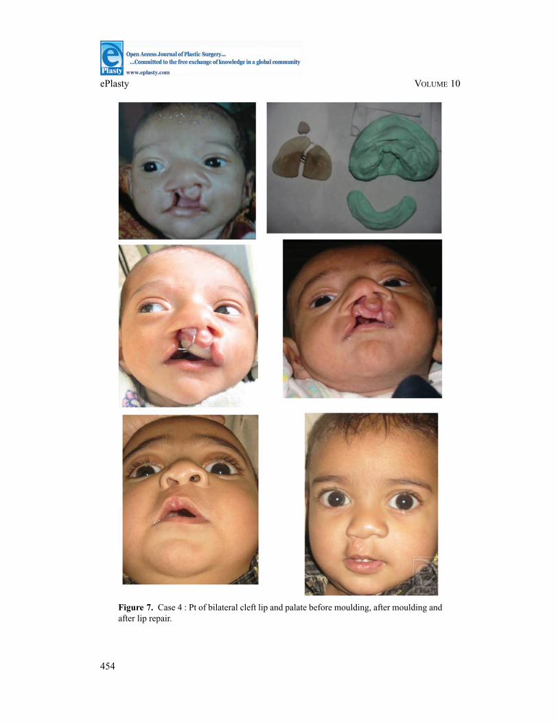
Figure 7. Case 4 : Pt of bilateral cleft lip and palate before moulding, after moulding andafter lip repair.
454

MISHRA ET AL
Figure 8. Case 5 : Pt of bilateral cleft lip and palate before moulding, after moulding andafter lip repair.
455

ePlasty VOLUME 10
Figure 9. Ulceration after application of molding appliance.
CONCLUSION
Nasoalveolar molding can be useful adjunct for treatment of cleft lip nasal deformity. Itis possible to incorporate presurgical NAM at centers where basic plastic surgery servicesand support of orthodontist/prosthodontist is present. It can prove to be a cost-effectivetechnique by reducing number of future surgeries in cleft patients. Studies with widerpatient base and longer follow-ups are needed for definitive results.
456

MISHRA ET AL
REFERENCES
1. McNeil C. Orthodontic procedures in the treatment of congenital cleft palate. Dent Rec. 1950;70:126-32.2. Matsuo K, Hirose T. Non surgical correction of cleft lip nasal deformity in the early neonate. Ann Acad
Med Singapore. 1988;17(3):358-5.3. Matsuo K, Hirose T. Preoperative non surgical over correction of cleft lip nasal deformity. Br J Plast Surg.
1991;44(1):5-11.4. Grayson BH, Santiago P, Brecht L, Cutting CB. Presurgical naso-alveolar molding in patients with cleft lip
and palate. Cleft Palate Craniofac J. 1999;5:139-9.5. Maull D, Grayson B, Cutting C, et al. Long-term effects of nasoalveolar molding on three-dimensional
nasal shape in unilateral clefts. Cleft Palate Craniofac J. 1999;36:391-7.6. Bardach J, Cutting C. Anatomy of unilateral and bilateral cleft lip and nose. In: Bardach J, Morris HL, eds.
Multidisciplinary Management of Cleft Lip and Palate. Philadelphia, Pa: WB Saunders; 1990: 154-8.7. Hotz M, Gnoinski W. Comprehensive care of the Cleft lip and palate children at Zurich University: a
preliminary report, Am J Orthod. 1976;70:481-504.8. Santiago PE, Grayson BH, Cutting CB, Gianoutsos MP, Brecht LE, Kwon SM. Reduced need for alveolar
bone grafting by presurgical orthopedics and primary gingivoperiosteoplasty. Cleft Palate Craniofac J.1998;35:77-80.
9. Grayson BH, Santiago PE, Brecht LE, Cutting CB. Presurgical nasoalveolar molding in infants with cleftlip and palate. Cleft Palate Craniofac J. 1999;36(6):486-98.
10. Grayson BH, Maull D. Nasoalveolar molding for infants born with clefts of the lip, alveolus, and palate.Clin Plast Surg. 2004;31(2):149-58, vii.
11. Pfeifer TM, Grayson BH, Cutting CB. Nasoalveolar molding and gingivoperiosteoplasty versus alveolarbone graft: an outcome analysis of costs in the treatment of unilateral cleft alveolus. Cleft Palate CraniofacialJ. 2002;39(5):570
12. Da Silveira AC, Oliveira N, Gonzalez S, Shahani M, Reisberg D, Daw JL Jr. Modified nasal alveolarmolding appliance for management of cleft lip defect. J Craniofac Surg. 2003;14(5):700-3.
13. Liou EJ, Subramanian M, Chen PK, Huang CS. The progressive changes of nasal symmetry and growthafter nasoalveolar molding: a three-year follow-up study. Plast Reconstr Surg. 2004;114(4):858-64.
14. Pai BC, Ko EW, Huang CS, Liou EJ. Symmetry of the nose after presurgical nasoalveolar molding in infantswith unilateral cleft lip and palate: a preliminary study. Cleft Palate Craniofac J. 2005;42(6):658-63.
15. Doruk C, Kilic B. Extraoral nasal molding in a newborn with unilateral cleft lip and palate: a case report.Cleft Palate Craniofac J. 2005;42(6):699-702.
16. Deng XH, Zhai JY, Jiang J, Li F, Pei X, Wang HT. A clinical study of presurgical nasoalveolar molding ininfants with complete cleft lip and palate. Zhonghua Kou Qiang Yi Xue Za Zhi. 2005;40(2):144-6.
17. Ziai MN, Bock DJ, Da Silveira A, Daw JL, Natal teeth: a potential impediment to nasoalveolar molding ininfants with cleft lip and palate. J. Craniofac Surg. 2005;16(2):262-6.
457

![Correlation between Nasoalveolar Molding and Surgical ... · with one (for unilateral cleft lip and/or palate [UCLP]) or two (for bilateral cleft lip and/or palate [BCLP]) nasal stents](https://img.pdfslide.net/doc/110x75/5f6006d40abc5d40510400bd/correlation-between-nasoalveolar-molding-and-surgical-with-one-for-unilateral.jpg)