Embed Size (px)
Citation preview

1
PREVALENCE AND ASSOCIATED FACTOR
FOR POSITIVE CHEST X-RAY DURING
TUBERCULOSIS SCREENING AMONG HIGH
RISK GROUPS IN KEDAH
DR AHMAD HANIS AHMAD SHUSHAMI
UNIVERSITI SAINS MALAYSIA
2017

2
PREVALENCE AND ASSOCIATED FACTOR
FOR POSITIVE CHEST X-RAY DURING
TUBERCULOSIS SCREENING AMONG HIGH
RISK GROUPS IN KEDAH
By
DR AHMAD HANIS AHMAD SHUSHAMI
Research Project Report submitted in partial
fulfilment of the requirement for the degree of
Master of Public Health
MAY 2017

ii
ACKNOWLEDGEMENT
I would never have been able to finish my dissertation without the continuous
support from my beloved wife, Nurul Shahida Binti Ahmad who patiently manages
the family alone without my presence. Special thanks for all my co-researchers from
Tuberculosis and Leprosy Unit, Kedah State Health Department, Dr. Zahariyah
Yaacob and Hj.Khairol Nizam bin Ibrahim for assistance given to complete this
research. Not to forget, our radiologist, Dr. Nik Fatimah Salwati Nik Malek and Dr.
Siti Maimunah Binti Jamaludin.
I would like to express the deepest gratitude to my supervisor, Associate Professor
Dr. Wan Mohd Zahiruddin Bin Wan Mohammad for the continuous support,
assistance and patience during my research project. His excellent guidance helped
me from the beginning of the research until the end.
I would also like to extend my appreciation to all tuberculosis in charge officer from
districts of Kedah that involved in this study. Without their assistance in data
collection, this study won’t be completed. Finally, special thanks to all my friends
especially Dr. Mohammad Fadhil bin Marzuki. Without all their precious support, I
would not able to complete this research.

iii
TABLE OF CONTENTS
TABLE PAGE
ACKNOWLEDGEMENT ....................................................................................... ii
TABLE OF CONTENTS ........................................................................................ iii
LIST OF TABLES ................................................................................................... x
LIST OF FIGURES ................................................................................................ xi
LIST OF ABBREVIATIONS ................................................................................ xii
LIST OF SYMBOLS ............................................................................................ xiii
LIST OF APPENDICES ....................................................................................... xiv
ABSTRAK ............................................................................................................. xv
ABSTRACT ......................................................................................................... xvii
CHAPTER 1: INTRODUCTION
1.1 Introduction ....................................................................................................... 1
1.1.1 Tuberculosis in Malaysia ........................................................................ 1
1.1.2 The importance of screening for case detection ...................................... 2
1.1.3 High Risk Groups .................................................................................... 4
1.1.4 Increasing number of high risk population ............................................. 4
1.1.5 Huge incidence gap ................................................................................. 5
1.1.6 Chest X-ray as a screening tool ............................................................... 6
1.2 Problem Statement and Rationale of study ........................................................ 8
1.3 Research questions ............................................................................................ 8
1.4 Objectives .......................................................................................................... 9

iv
1.4.1 General objective .................................................................................... 9
1.4.2 Specific objectives .................................................................................. 9
1.5 Hypotheses ................................................................................................. 9
CHAPTER 2: LITERATURE REVIEW
2.1 Socio-demographic factor for chest x-ray positivity ....................................... 10
2.1.1 Age ........................................................................................................ 10
2.1.2 Sex ......................................................................................................... 11
2.1.3 Nationality ........................................................................................ 12
2.1.4 Ethnicity ........................................................................................... 14
2.2 Symptoms ........................................................................................................ 15
2.3 High Risk Group for TB ................................................................................ 15
2.3.1 Person with Human Immunodeficiency Virus ................................. 15
2.3.2 Diabetes Mellitus .............................................................................. 16
2.3.3 Smokers ............................................................................................ 17
2.3.4 Institutionalised individuals ............................................................. 17
2.3.5 End Stage Renal Failure ................................................................... 19
2.3.6 Chronic Obstructive Pulmonary Disease ......................................... 19
2.3.7 Contact with TB patient ................................................................... 20
2.3.8 Substance abuse ................................................................................ 20

v
2.4 Conceptual framework .................................................................................... 21
CHAPTER 3: METHODOLOGY
3.1. Study design ................................................................................................... 23
3.2 Study duration.................................................................................................. 23
3.3 Study location .................................................................................................. 23
3.4 Reference population ....................................................................................... 26
3.5 Source population ............................................................................................ 26
3.6 Sampling frame................................................................................................ 26
3.7 Study criteria.................................................................................................... 27
3.8 Sample size estimation .................................................................................... 27
3.8.1 Objective 1: ........................................................................................... 27
3.8.2 Objective 3: ........................................................................................... 28
3.10 Sampling method ........................................................................................... 29
3.9 Research tools .................................................................................................. 31
3.9.1 Checklist proforma ................................................................................ 31
3.10.2 TB Information System for screening high risk group ................. 32
3.10 Data collection ............................................................................................... 32
3.11 Operational definition .................................................................................... 33
3.11.1 Criteria for positive chest x-ray........................................................... 33
3.11.2 Criteria of TB symptoms ..................................................................... 34

vi
3.11.3 Criteria for symptomatic patients ........................................................ 35
3.11.4 Age groups .......................................................................................... 35
3.11.5 Ethnicity .............................................................................................. 36
3.11.6 Risk Groups ......................................................................................... 36
3.11.6(a) HIV/Substance Abuse ..................................................................... 36
3.11.6(b)Contact of TB patient ...................................................................... 36
3.11.6(c) Institutionalised............................................................................... 37
3.11.6(d) Smoker ............................................................................................ 37
3.11.6(f) Diabetes ........................................................................................... 37
3.11.6(g) End Stage Renal Failure on Dialysis/ Chronic Obstructive Airway
Disease on treatment ...................................................................................... 37
3.11.6(h) Others (Low Risk) .......................................................................... 38
3.12 Statistical analyses ......................................................................................... 38
3.13 Ethical issue ................................................................................................... 41
3.14 Flow of study ................................................................................................. 42
CHAPTER 4: RESULT
4.1 Socio demographic characteristics .................................................................. 43
4.2 Clinical Presentation ........................................................................................ 45
4.3 Risk Group ....................................................................................................... 45

vii
4.4 Chest X-Ray Result ........................................................................................ 47
4.5 Prevalence of Positive Chest X-Ray .............................................................. 47
4.6 Other details of sample................................................................................... 49
4.7 Associated factor for positive chest x-ray during high risk group screening . 50
4.7.1 Simple logistic regression (Univariable analysis) ............................ 50
4.8 Associated factor for positive chest x-ray during high risk group screening .. 35
4.8.1 Multiple Logistic Regression ........................................................... 35
CHAPTER 5 DISCUSSION AND LIMITATIONS
5.1 Socio demographic factors and clinical characteristics of patients in high risk
group TB screening ........................................................................................ 39
5.1.2 Gender .............................................................................................. 40
5.1.3 Nationality and Ethnicity .................................................................. 40
5.1.4 Clinical presentation ......................................................................... 41
5.1.5 Risk group ................................................................................................ 41
5.1.5 (a) Contacts of TB patient ..................................................................... 41
5.1.5 (b) Smoking .................................................................................................. 42
5.1.5 (c) HIV/Substance Abuse ............................................................................. 42
5.1.5 (d) End stage renal failure / Chronic obstructive airway disease ................. 43
5.1.5(e) Institutionalised ........................................................................................ 43
5.1.5 (f) Diabetes ................................................................................................... 44
viii
5.2 Prevalence of positive chest x-ray.................................................................. 45
5.2.1 Age ................................................................................................... 45
5.2.2 Gender ...................................................................................................... 45
5.2.3 Nationality and ethnicity .......................................................................... 45
5.2.4 Clinical presentation ......................................................................... 45
5.2.5 High Risk Groups ............................................................................. 46
5.3 Associated factor for positive chest x-ray ...................................................... 46
5.3.1 Age ................................................................................................... 46
5.3.2 Gender .............................................................................................. 47
5.3.3 Nationality and ethnicity .................................................................. 47
5.3.4 Clinical presentation ......................................................................... 48
5.3.5 Risk Groups ...................................................................................... 49
5.3.5(a) Smoking ............................................................................................ 50
5.3.5(b) HIV/Substance abuse........................................................................ 50
5.3.5(c) ESRF/COAD .................................................................................... 51
5.3.5(d) Institutionalised ................................................................................ 52
5.3.5(e) Diabetes ............................................................................................ 52
5.4 Strength and limitations ................................................................................... 53
5.3.3 Nationality and ethnicity .................................................................. 55

ix
5.3.4 Clinical presentation ......................................................................... 56
5.3.5 Risk Groups ...................................................................................... 57
5.3.5(a) Smoking ............................................................................................ 57
5.3.5(b) HIV/Substance abuse........................................................................ 58
5.3.5(c) ESRF/COAD .................................................................................... 59
5.3.5(d) Institutionalised ................................................................................ 59
5.3.5(f) Diabetes ........................................................................................ 60
CHAPTER 6: CONCLUSION AND RECOMMENDATION ........................ 61
6.1 Conclusion ....................................................................................................... 61
6.2 Recommendation ............................................................................................. 61
6.2.1 Immediate Measures ......................................................................... 61
6.2.1(a) Utilisation of mobile x-ray................................................................ 61
6.2.1(b) Financial support .............................................................................. 62
6.2.1(c) Criteria for high risk group ............................................................... 62
6.2.2 Long term measures ......................................................................... 63
6.2.2(a) Multisector and intra agency collaboration ...................................... 63
REFERENCES....................................................................................................... 61
APPENDICES ....................................................................................................... 72

x
LIST OF TABLES
Table 3.1 Summary of sample size calculation for each associated factors
of positive chest x-ray in this screening.
28
Table 4.1 Socio demographic characteristics 44
Table 4.2 Clinical presentations 45
Table 4.3 Diabetic control in high risk group 46
Table 4.4 Proportion of high risk group 46
Table 4.5 Proportion of chest x-ray characteristics 47
Table 4.6 Characteristics of positive chest x-ray 48
Table 4.7 High risk group with positive chest x-ray 48
Table 4.8 Period of chest x-ray collection 49
Table 4.9 Distribution of chest x-ray among sample district 49
Table 4.10 Univariate analysis (binary logistic regression) of factors
associated with positive chest x-ray among high risk group
51
Table 4.11 Multivariate analysis of factors associated with positive chest x-
ray among risk group
36

xi
LIST OF FIGURES
Figure 1.1 TB Notification Rate (All Case), Malaysia (1990-2015),
Adapted from Kedah State Health Department Annual Report
2015
1
Table 1.2 TB Notification Rate (All Case), Kedah (2005-2015), (Adapted
from Kedah State Health Department Annual Report 2015)
2
Figure 1.3 : Reported HIV Cases per Transmission Mode, Malaysia 1988-
2010, Adapted from National Strategic Plan for AIDS, MOH
2016)
2
Figure 1.4 Trend of Diabetes, from NHMS 2006, 2011 & 2015 shows
increasing trend of Diabetes prevalence among adults, adapted
from National Health Morbidity Survey Factsheet, 2015
3
Figure 2.1 Conceptual framework 22
Figure 3.1 Map of Kedah 24
Figure 3.2 Flow chart of study 42

xii
LIST OF ABBREVIATIONS
AFB Acid Fast Bacilli
CCRC Cure and Care Centre
COAD Chronic Obstructive Airway Disease
DM Diabetes Mellitus
DOTS Directly Observed Treatment, short course
ESRF End Stage Renal Failure
HIV Human Immunodeficiency Virus
HRG High Risk Group
JKNK Jabatan Kesihatan Negeri Kedah
KKM Kementerian Kesihatan Malaysia
NHMS National Health and Morbidity Survey
OR Odds Ratio
RM Ringgit Malaysia
ROC Receiver Operating Characteristic
SD Standard Deviation
TNF Tumour Necrosis Factor
UN United Nation
USM Univeristi Sains Malaysia
WHO World Health Organisation

xiii
LIST OF SYMBOLS
< Less Then
> More then
= Equal to
≤ Less than equal to
≥ More than equal to
Alpha
Β Beta
% Percentage
∆ Precision/Delta
& And
xiv
LIST OF APPENDICES
Appendix A Universiti Sains Malaysia Ethical Approval Letter
Appendix B Online Proforma
Appendix C TBIS 104 A Microsoft Excel template
Appendix D Chest X-Ray Reporting Format for High Risk Group Screening
Appendix E Ministry of Health (MOH) Ethical Approval Letter

xv
ABSTRAK
PREVALENS DAN FAKTOR-FAKTOR BERKAITAN DENGAN
PENEMUAN X-RAY POSITIF SEMASA SARINGAN TUBERKULOSIS
DALAM KALANGAN GOLONGAN BERISIKO TINGGI DI KEDAH 2016
Latar Belakang: Program pemeriksaan tuberkulosis (TB) dalam kalangan kumpulan
risiko tinggi menggunakan sinar-x dada telah dilaksanakan oleh Kementerian
Kesihatan Malaysia tetapi kadar pengesanan kes tidak menggalakkan. Langkah
mengenalpasti faktor-faktor yang menyumbang kepada pemeriksaan sinar-x yang
positif adalah penting supaya kes-kes positif TB tidak akan tercicir dan dapat
membantu menghalang rangkaian penularan TB dalam masyarakat.
Kaedah Kajian: Kajian keratan rentas telah dijalankan ke atas pesakit-pesakit yang
disaring semasa saringan golongan berisiko tinggi di Negeri Kedah. Pesakit-pesakit
yang terlibat adalah daripada senarai yang telah dimasukkan di dalam Sistem
Maklumat Tibi (TBIS) 104 A yang merupakan sistem pemberitahuan bagi saringan
ini. Kajian ini melibatkan 1417 orang yang melibatkan fasiliti yang telah dipilih
secara rawak, daripada enam buah daerah yang juga dipilih secara rawak. Analisa
regresi lojistik mudah dan analisa lojistik pelbagai digunakan untuk mengenalpasti
faktor-faktor yang dikaitkan dengan keputusan x-ray dada yang positif dengan
mengambil kira nilai p <0.05 dalam model akhir
Keputusan: Dalam kajian ini kebanyakan pesakit adalah pada usia lewat dewasa.
Min (SD) bagi umur ialah 49.19 (18.2) tahun. Pembahagian antara Lelaki dan
perempuan adalah 51.2% dan 48.8%. Majoriti adalah warganegara Malaysia, yang
terdiri daripada 72.3% Melayu, 9.8% India, 8.6% Cina dan 0.9% lain-lain. Prevalens
x-ray dada positif terdiri daripada beberapa kumpulan. Dalam kumpulan umur,

xvi
warga emas mempunyai prevalens yang paling tinggi, diikuti dewasa dan kanak-
kanak. Lelaki lebih ramai berbanding perempuan dan jika mengikut kumpulan etnik,
Cina merupakan kumpulan yang mempunyai prevalens tertinggi bagi x-ray dada
positif. Individu yang mempunyai gejala mempunyai prevalens yang tinggi jika
dibandingkan dengan yang tiada gejala. Dalam kalangan golongan berisiko tinggi,
merokok merupakan kumpulan yang mempunyai prevalens tertinggi diikuti
HIV/penyalahgunaan substan, penyakit buah pinggang/penyakit paru-paru kronik,
lain-lain, institusi, kencing manis dan kontak. Selepas pelarasan faktor penyebab,
faktor penentu yang signifikan bagi x-ray dada positif ialah umur, AOR (95% CI)
1.03 (1.01-1.04), gejala AOR (95% CI) 3.8 (2.72-5.50), institusi AOR (95% CI) 2.1
(1.09-4.25 dan HIV/Penyalahgunaan substan AOR (95% CI) 3.6 (1.35-10.0)
Kesimpulan: Penemuan x-ray dada positif semasa saringan golongan berisiko tinggi
TB dipengaruhi oleh faktor seperti pertambahan umur, dan gejala-gejala TB. Antara
dua belas golongan berisiko yang telah dikaji, dua kumpulan risiko telah dikenalpasti
sebagai kumpulan penting yang perlu diberikan priority iaitu institusi and
HIV/Penyalahgunaan substan. Oleh yang demikian, ini akan mempermudahkan
saringan ini supaya dijalankan dengan lebih efisyen
KATA KUNCI:
tuberkulosis, golongan berisiko tinggi dan faktor perkaitan, x-ray dada positif.

xvii
ABSTRACT
PREVALENCE AND ASSOCIATED FACTOR FOR POSITIVE CHEST X-
RAY DURING TB SCREENING AMONG HIGH RISK GROUPS IN KEDAH
2016
Background: Tuberculosis screening program among high risk groups using chest x-
ray have been implemented by Ministry of health Malaysia but the case detection are
not encouraging. More prioritization is needed to identify factors that contribute to a
chest x-ray screening positive so that the positive cases would not be missed and
may help in halting the chain of transmission of TB in the society.
Methodology: This was a cross sectional study involving patients who were screened
during TB screening for high risk groups in Kedah in 2016. The patients involved
were from the list that has been included in the TB information system (TBIS) 104 A
which is a notification system for this screening. The study involved 1417 people
involving facilities which were randomly selected from six regions that were also
selected randomly. Data analysis was conducted using SPSS version 22 for
descriptive and inferential analysis. Simple logistic regression analysis and multiple
logistic analysis was used to identify factors associated with chest x-ray result at p-
value of <0.05 in the final model
Results: In this study, most of the people involved were at their late adulthood. The
mean (SD) of age was 49.19 (18.2) years. Male accounts for 726 people (51.2%).
Majority of the sample population were Malaysian 1298 (91.6%) which comprise of
Malay 1024 (72.3%) followed by Indian 139 (9.8%), Chinese 122 (8.6%) and other

xviii
races 13 (0.9. Majority of the sample population were asymptomatic 1036 (73.1%).
Diabetes were the largest proportion of risk group screened 638 (45.0 %), followed
by contact 334 (23.6%), ‘others unspecified’ 204 (14.4%), institutionalised 124
(8.8), clients of quit smoking clinic 57 (4.0%), End Stage Renal Failure/Chronic
Obstructive Airway Disease 33 (2.3%) and lastly HIV/Substance abuse 27 (1.9%).
Prevalence of positive x-ray was divided into few groups. In age group, elderly has
highest prevalence followed by adult and children. According to gender, male has
higher prevalence than female. If according to ethnicity, Chinese has highest
prevalence of positive chest x-ray among all ethnic. Symptomatic people have higher
prevalence if compared to asymptomatic. Among high risk group individual,
smoking has highest prevalence of positive chest x-ray (28%) followed by
HIV/Substance abuse (25.9%), ESRF/COAD (24.2%), ‘Other unspecified’ (21%),
Institutionalized (16.9%), Diabetes (12.6%) and Contacts (7.1%). After other
cofounders were adjusted, the important risk factors are age AOR (95% CI) 1.03
(1.01-1.04), symptoms AOR (95% CI) 3.8 (2.72-5.50), institutionalised AOR (95%
CI) 2.1 (1.09-4.25) and HIV/Substance abuse, AOR (95% CI) 3.6 (1.35-10.0).
Conclusion: The discovery of positive chest x-ray during screening for high risk
groups affected by factors such as age, and symptoms of TB. Among the twelve-risk
factors that have been studied, two risk factors have been identified as an important
factor that should be given priority which are institutionalized and HIV/Substance
abuse. Consequently, this will facilitate the screening to be carried out more
efficiently
KEYWORDS:
tuberculosis, high risk groups and associated factors, positive chest x-ray

1
CHAPTER 1
INTRODUCTION
1.1 Introduction
1.1.1 Tuberculosis in Malaysia
TB is still a major health threat to world and Malaysia. There were estimated 10.4
million new (incident) TB cases worldwide (WHO, 2016). It contributed to the top
10 causes of death worldwide in 2015, and was responsible for more deaths than
HIV and malaria (WHO, 2016). Our country is known as an Intermediate Burden of
Tuberculosis due to the incidence rate that was less than 100/100,000 population.
The latest notification rate for Tuberculosis (all Form) in Malaysia was
79.44/100,000 population in 2015 (Figure 1.1), while in Kedah TB notification rate
(all form) at the same year (Figure 1.2) was lower at 61.73/100,000 (MOH, 2016)
Figure 1.1: TB Notification Rate (All Case), Malaysia (1990-2015), Adapted from
Kedah State Health Department Annual Report 2015

2
Figure 1.2: TB Notification Rate (All Case), Kedah (2005-2015), (Adapted from
Kedah State Health Department Annual Report 2015)
1.1.2 The importance of screening for case detection
If we look at the trend of cases in Figure 1and Figure 2, we can see that despite a lot
of measures done to detect cases, there was still a slow increase of notification rate
for the past 10 years for both in Kedah and Malaysia. This is worrying because it
means that the untreated patient still lingers in the society and will transmit to others
who are susceptible to the disease. To better control this disease, the notification rate
should have increased exponentially for the past 10 years but after we have treated
most of the patients, the transmission rate would have gone down. We can take
example from HIV/AIDS control program that concentrated on screening program
during early 90’s (MOH, 2011; MOH, 2016). The reported cases of HIV increased
dramatically from 1988 and peaked in 2002, and then the rate declined slowly after
(Figure 1.3).
Every year TB sector, MOH came out with the target number for case detection rate
(CDR) calculation, which is the estimated number of new patient that should be
detected in that year that becomes the denominator for CDR calculation. The

3
numerator will be the actual case detected in that year. The target case detection rate
(CDR) was 95% and is one of the indicator that hardly achieved by all of states in
Malaysia except Johor which have exceeded 95% target in 2015. Ideally, the number
of new cases detected should exceed the number of estimated cases, but that’s never
happen. For detection of cases, we are too dependent on symptomatic individual who
seek for treatment in hospital (MOH, 2016). As we already know, for TB to exert the
full-blown symptoms, it must reach certain amount of tubercle bacilli in the patient’s
lung and by the time the patient came to seek healthcare, he has already coughed out
tubercle bacilli and spread it to the society. What we should do is to further expand
our detection of cases towards asymptomatic or people who are having less
symptoms as well. Therefore, something must be done to further strengthen the
strategy to increase case detection. Part of the solution is, to concentrate TB
screening in high risk individuals followed by prompt treatment in reducing the
spread of TB in the country (WHO, 2013a).
Figure 1.1: Reported HIV Cases per Transmission Mode, Malaysia 1988-2010,
Adapted from National Strategic Plan for AIDS, MOH 2016)

4
1.1.3 High Risk Groups
For intermediate burden country like Malaysia, TB may only concentrate on a
specific group of people, that we called high risk group or populations. Usually they
do not seek for treatment because they don’t recognize the symptoms or they have no
symptom at all because being in immunocompromise state. They are also belongs to
a group of people who usually have difficult access to healthcare such as elderly,
immigrant, drug abuser, prisoner, and people who are having underlying disease
such as COPD or people who are exposed to occupational hazard that predispose to
TB infection such as healthcare worker and miners (WHO, 2013a). They are those a
group of people who are marginalized and being neglected by society and family,
especially elderly, HIV, institutionalized and substance abuser (WHO, 2013a)
1.1.4 Increasing number of high risk population
In recent years, there was substantial Increase in number of high risk population in
Malaysia. Particularly in Kedah, the rate was alarming. These include an increased
number of people who involved in substance abuse (new & old case) which was
19,532 in 2011 and increased to 26,668 in 2015. Kedah is also have highest
prevalence of new cases of substance abuse in Malaysia (Agensi Antidadah
Kebangsaan, 2016). There was an increased number of diabetic case in Malaysia
including Kedah (Institut Kesihatan Umum, 2016). In 2005 there were 13,000
patients on dialysis in Malaysia and the number have reached 20,000 by 2008 (Hooi,
2006). We also can see there was shifting of population towards elderly population
and there was influx of immigrants from high burden country to Malaysia

5
1.1.5 Huge incidence gap
There was huge gap between estimated TB incidences by the WHO and Malaysia.
Notification rate for TB (all form) in Malaysia for the year 2015 was 79.44/100,000
population while WHO estimates was at 104/100,000 population in 2014 (TB &
Leprosy Sector, 2016). With the gap of 2.4 million of undetected TB patients, it
means that we are not aggressive enough in detecting new cases. There were few
limitations as highlighted by the WHO which include limitation of sputum smear
microscopy (WHO, 2013b). Sputum smear microscopy was unreliable in
asymptomatic patients because bacterial load for them was very low and cannot be
detected (Nobuyuki, 2013). Those in high risk group are often asymptomatic as they
will not seek treatment and even if they have symptoms, it is unlikely for them to
seek for treatment until they develop severe complication (Nobuyuki, 2013). Sputum
smear microscopy is also time consuming for both patient and clinician especially to
the former since it requires many steps. Therefore, chest x-ray has been selected by
MOH Malaysia as the screening tool for the high-risk group population.
Figure 1.4: Trend of Diabetes, from NHMS 2006, 2011 & 2015 shows increasing
trend of Diabetes prevalence among adults, adapted from National Health Morbidity
Survey Factsheet, 2015

6
1.1.6 Chest X-ray as a screening tool
The advantages of chest x-ray are relatively easier, not time consuming and will be
able to detect more sputum AFB negative but culture positive patient (Nobuyuki,
2013). There were also few disadvantages which are poor detection outcome (Miller
et al., 1998) and the yield is too low and not economical (Gottridge, 1989). Recently
in Kedah, since screening for high risk group of TB started in 2014, the yield was
around 4% (MOH, 2016). There were few issues highlighted in which one of it is
improper chest x-ray selection among high risk group who were screened. There
were also no proper assessment and risk prioritization where clinicians just screened
everybody who are at risk.
WHO has developed guidelines on screening for active TB. It suggests that
screening, if done in the right way and targeting the right people, may reduce
suffering and death (WHO, 2013a). Active case detection using chest x-ray is one of
the recommended tools for TB screening by WHO and has been adapted by MOH.
Historically in 1974, after reviewing the results of several decades of TB screening,
the ninth report by WHO’s Expert Committee on TB recommended that
indiscriminate TB case-finding using miniature mass radiography should be
abandoned due to its inefficiency (WHO, 2013a). This is supported by studies done
in early 90s that found out chest x-ray are of value in 0% to 1.3% (Gottridge, 1989).
After a decade, WHO have looked back towards chest x-ray for TB screening and
found out that its useful only if the TB prevalence among the target population is
high (Nobuyuki, 2013). Other studies also found out that targeted screening using x-
ray is an effective tool for the early detection of active TB in hard-to-reach
populations (Story, 2012). Therefore, chest x-ray that specifically targeting high-risk

7
groups was useful in the early detection of active disease (Fuentes, 2014). As
mentioned earlier, the high-risk group are usually immunocompromised as they did
not have profound symptoms. Therefore, high risk group screening that has been
adapted by MOH is to screen all high risk group individual, including all
symptomatic and asymptomatic individual (TB & Leprosy Sector, 2016a; WHO,
2013a).
Prevalence of chest x-ray positive among high risk groups in other countries with a
low incidence of TB is almost like in Malaysia. In a study done in Spain, they found
out of 3654 x-rays done among high risk groups, 227 (6.21 %) were positive
(Fuentes, 2014). Similar finding also reported by (Miller et al., 1998) in Routine
Emergency Department Chest Radiograph done in high risk group of TB in New
Jersey, United States, 2% had chest radiographic findings considered to be
meaningful for further investigation to confirm diagnosis of TB.
Previous studies found few associated factors that influence positive chest X-rays.
(Boon, 2006) reported that TB patients with HIV are more likely have positive
radiographic findings which are atypical. Other study conclude that the older the
patient in a high risk group, the more chest x-ray positivity seen, as reported by
Miller et al. (1998). This mean that the radiographic changes was significantly higher
if the patient is older. However, there were also findings in studies that shows no
significant different between chest x-ray finding for those among high risk group or
not, as reported by Bacakog˘lu (2001) who conclude that diabetes does not affect
radiological features of pulmonary infiltrates and diabetic patients had a higher
prevalence of typical x-ray presentations but no significant difference compared with
non-diabetics (Paquette et al., 2014)

8
1.2 Problem Statement and Rationale of study
TB incidence is still high in Malaysia and still increasing. There was a huge
detection gap between our true incidence and WHO estimated incidence particularly
among high risk group. Chest X-Ray is an effective tool, but have poor yield and it’s
not being used effectively. Therefore, screening must be very selective to detect
more cases. We want to identify which group among the high-risk individuals who
are more likely to have positive chest X-Ray and possible contributing factors as
well.
This study aimed to identify which are the higher risk group and what are the major
factors that have higher odds of positive chest x-ray finding. By obtaining the
information we will be able recognize which group we must prioritize for chest x-ray
screening. Hopefully with this study, we can detect more TB cases and with more
case detected, we will able to break the chain of transmission in the society and will
benefit our country as a whole
1.3 Research questions
1. What is the prevalence of positive chest x-ray among high risk groups in
Kedah?
2. What are the associated factors of positive chest x-ray among high risk
groups?

9
1.4 Objectives
1.4.1 General objective
To study the prevalence and associated factors of positive chest x-ray in high risk
group for tuberculosis (TB) in Kedah
1.4.2 Specific objectives
1. To determine the prevalence of positive chest x-ray for TB screening among
high risk group in Kedah.
2. To describe the socio demographic factors (age, sex, and ethnicity) and
clinical characteristics (symptoms, co-morbidities) of high risk group for TB
in Kedah.
3. To identify the associated factors of positive chest x-ray in high risk group of
high risk group for TB in Kedah.
1.5 Hypotheses
There are significant association between symptoms, co-morbidities and socio
demographic factors (age, sex, and ethnicity) and the positivity of chest x-ray among
high risk groups in Kedah.

10
CHAPTER 2
LITERATURE REVIEW
The search for literature review was done with subscribed USM online tools such as
Science Direct, Springer Link, JAMA, Scopus, OVID and ProQuest. Outsource
search engines were also used which include PubMed and Google Scholar. Few of
the references also manually searched in Online MOH Archive and from the library
in Kedah State Health Department. Based on literature review, the risk factors of
chest x-ray positivity have been identified and the relevant risk factors for Malaysia
have been selected for this study.
2.1 Socio-demographic factor for chest x-ray positivity
2.1.1 Age
Many studies have found that the risk of getting TB increased by increasing age
(MOH, 2012a; WHO, 2016) However not many studies were done to evaluate chest
x-ray positivity towards TB-meaningful finding. But there are many studies done to
evaluate chest x-ray screening towards positive TB either by sputum smear
microscopy or GENE X-Pert (Casas et al., 2013)
Bacakoğlu (2001) did a retrospective cohort study among diabetics and non-diabetics
who acquire TB. They found that diabetics have lower mean age compared to non-
diabetics to have abnormal X-Ray. Age is important even within a risk group itself,
where a study done among TB contact in children by Khalilzadeh et al. (2003) reveal

11
that the risk for positive radiological findings were higher in 10-14 years old
compared to 5-9 years old.
There was a study done by Goetsch et al. (2012) in Germany, who screened
homeless and illicit drug users for TB using chest x-ray and proceed to sputum
microscopy to see if there were positive x-ray finding. The study concluded that age
was the only variable to be associated with the risk of smear positive TB for both
risk group. Different risk group also has difference in age for the manifestation of
positive chest x-ray. Another study done among high risk individual in Spain by
Goetsch et al. (2012) stated that drug users became smear positive TB at a younger
age compared to homeless people. Age is also an important predictor of positive
chest x-ray, even among low risk individual. As reported by Yeshurun et al. (1996)
during compulsory screening of approximately 5000 Israeli Defence Force, they
found out that abnormal findings were influenced by age. Another study done using
routine chest x-ray among 481 asymptomatic high risk groups individual (drugs,
alcohol abuse and emergency psychiatric illness) that attended the emergency
department in New Jersey in 6-month period, also stated that the patient with
positive radiographic changes was significantly older (Miller et al., 1998). As a
conclusion, age is an important factor in determining positivity of chest x-ray, even
among high risk and non-high risk population and within the particular high risk
group itself.
2.1.2 Sex
Male gender is known to have higher risk in getting pulmonary TB (MOH, 2012a;
WHO, 2016). The male to female ratio was 15:10, based on a study done in Kedah,
Malaysia (Ismail, 2004). Similar findings also was found in a Cambodian prevalence

12
study involving 37,417 individuals by Mao et al. (2014) where there were 15.1:10
ratio of males to females among smear positive TB cases and, 14.8:10 ratio of males
to females for smear-negative, culture-positive TB.
A similar picture is seen in chest x-ray from a cross sectional study done in Uganda
involving 863 symptomatic from outpatient department, which showed significant
gender difference with x-ray was suggestive of TB in 66.5% of males and in 44.8%
of female (p<0.001). For confirmed Pulmonary TB patients, males have higher odds
of having abnormal chest x-ray (OR 5.0, 95% CI: 3.29, 7.57, p <0.001) (Boum et al.,
2014).
However, Zhang et al. (2015) reported that there were no differences in sex for
positive chest x-ray during TB screening using chest x-ray involving 8418 elderly
that was classified in high risk individuals (symptomatic, close contact and diabetes).
In other study done in emergency department by (Miller et al., 1998) mentioned
earlier, also found that there was no significant difference in sex for positivity of
chest radiograph among the risk groups.
Fuentes (2014) also found that gender is not a significant factor for positive TB in
screening for active TB in high-risk groups done in Spain. In another study done
among diabetic TB patients, there was also no significant difference in gender for TB
among diabetics and when compared with non-diabetics, the gender difference is
also insignificant (Tatar et al., 2009).
2.1.3 Nationality
There are studies that looked into positive chest x-ray and relation with nationality in
screening programs for immigrants and refugees. Pulmonary TB accounts for 22.4%

13
of disease detected by FOMEMA sdn bhd in 1998 and 1999 using chest radiographs
(Leong, 2006). In an active case finding among high risk group individual using
chest x-ray in Spain, they reported that the prevalence of TB detected during
screening in immigrants were significantly higher compared to poor people, drug
user, foreigners >2 years and native born (Fuentes, 2014).
In regards to confirmed case of pulmonary TB, foreigners are at higher risk, when
compared to Malaysian (Nissapatorn et al., 2007). This is particularly true for those
who are from high burden countries such as Indonesia, Philippine and Myanmar. The
finding is similar in other countries as well such as United States, where the
incidence rate of TB in foreign-born were higher than U.S citizen (Talbot et al.,
2000). Another study also in Unites States also found that refugees in their first year
of arrival has highest incidence rate of TB compared to other foreign-born residents
(Lobato, 2008). This situation is almost like in Kedah because there was sudden
influx of Rohingya ethnic immigrants from Myanmar who arrived in the end of
2015, in which majority of them still resides in Belantik Immigration Detention
Depot and some of them have already been released to designated villages all over
Kedah with UNHCR identification card. In Malaysia, 12.3% of notified TB cases in
2015 were among the immigrant population (MOH, 2016). In Sabah, immigrants
contributed more than 24% of the newly detected TB cases (Dony et al., 2004) and
the figures became larger due to huge influx of immigrants to Sabah in recent years.
We are surrounded by high TB burden country and most of immigrants that came to
Malaysia are from these countries. Therefore, it is important to know the impact of
nationality on TB screening program in our country.

14
From an evaluation done in Selangor, incidence rate for all forms of TB according to
nationality were 126.7/ 100,000 population for foreigners, compared to overall
incidence rate for Selangor for 2001, at 43.1/ 100,000 population (Venugopalan,
2004). However, even though foreigners might have higher risk to get TB infection,
most of them present as symptomatic patient, rather than from screening, such as by
FOMEMA. This is supported by a study in Netherland by Verver et al. (2001) who
found that foreigners who undergone TB screening using chest x-ray were less likely
to have positive sputum smear microscopy.
2.1.4 Ethnicity
In a screening for pulmonary TB using mobile digital chest radiography done in
London, Unite Kingdom, revealed that there was no significant difference in chest x-
ray positivity among white, black african, black caribbean, south asian and others
(Story, 2012). Similar finding also found during Chest x-ray screening for TB done
in a Hong Kong prison, revealed that there was no significant different in yield of
CXR screening among Chinese and other races (Leung et al., 2005).
In Malaysia, chest x-ray positivity among different ethnicity is unclear because it
was barely discussed in the studies that have been done. However, pertaining to
differences among ethnicity in active TB, one study done in Selangor clearly
described the difference of incidence rates among all Malaysian ethnics, which are
indian: 41.6/ 100,000 population, Malays: 39.7/ 100,000 population, chinese: 29.3/
100,000 population and others: 24.8/ 100,000 (Venugopalan, 2004).

15
2.2 Symptoms
The availability of TB symptoms also plays a major role in chest x-ray positivity, as
reported in previous study that sensitivity of chest x-ray can be up to 100% when
combined with presence of symptoms (Hoog, 2012; Joshi, 2012).
Cavitary lung disease is a common presentation and they typically present with
prolonged cough, associating fever and/or night sweats and weight loss. This
cavitation is easily detected by trained personnel through chest x-ray (Heemskerk,
2015). Chest x-ray when combined with symptoms screening, will increase the
number of TB detected (Churchyard et al., 2010).
2.3 High Risk Group for TB
2.3.1 Person with Human Immunodeficiency Virus
Person with Human Immunodeficiency virus (HIV) infection is a known risk factor
for TB, as stated in a study that people with HIV are prone to get TB, due to their
immune-compromised state (Baughman, 1999). In terms of chest x-ray finding, most
of HIV patient have normal chest x-ray, as reported by Akinbami et al. (2012). In a
prevalence survey done in Georgia, United States by Hoog (2012), they concluded
that chest x-ray abnormality for HIV infected person was lower compared to HIV
uninfected person. Furthermore, for the HIV infected individual, the chest x-ray
finding of ‘any abnormality’ can yield more smear or culture positive cases,
compared to chest x-ray with ‘only pulmonary or pleural abnormality’. It means that

16
HIV infected person often presents with atypical chest x-ray finding that does not
confine to lung parenchyma alone.
Similar finding was reported by Aderaye et al. (2004) in a study involving TB
patient from outpatient department in Ethiopia, in which they found that chest x-ray
findings for a HIV infected person who was diagnosed to have TB were mostly
atypical and can be normal or with minimal changes. Similar findings also reported
by Palmieri et al. (2002b) in a study done among HIV patient in Italy. Elliott et al.
(1990) also mentioned that TB patient who are HIV positive are more likely to be
sputum smear negative and less likely to show classical upper lobe involvement and
cavitation. As a conclusion, HIV is a risk to acquire TB infection, but it’s the other
way around for chest x-ray finding.
2.3.2 Diabetes Mellitus
Diabetes is a known risk factor for TB, as mentioned by several studies. From a
retrospective cohort study in Korea using nationwide database of 331, 601 patients,
higher incidence of TB was found among patient with diabetes and most of them was
likely to be diagnosed in the first year of diagnosis (Heo et al., 2015). It is consistent
with Jabbar et al. (2006) who reported in a study involving adults with diabetes in
Pakistan, which stated that patient with diabetes mellitus are ten times more common
to have TB, compared to those without diabetes.
Regarding the chest x-ray finding, a study done in Turkey by Bacakog˘lu, (2001),
found that the clinical and radiological presentation of TB among diabetics is
insignificant. Al-Wabel et al. (1997) also reported similar findings where chest x-ray
involvement including cavitary disease was again similar between diabetics and non-

17
diabetics. However, if we compare the findings of the x-ray, non-diabetic patient
usually presents with upper lobe involvement and cavitation, but diabetic patients
often presents with lower lobe involvement. A comparative study done by Perez-
Guzman et al. (2001) in Mexico, compared 192 TB patients with underlying diabetes
mellitus with 130 TB patients without diabetes, found that the chest x-ray was
mostly atypical. They also noted that the presentation was mostly lower lung lesion
and its frequency increased by age.
2.3.3 Smokers
Smokers were known to impose higher risk of developing TB. A cross-sectional
study done in Spain, involving 13,038 from their TB registry found smokers have
1.5 odds of having pulmonary TB compared to non-smokers (Altet-Gomez et al.,
2005). Other study also reported similar findings, where smokers were significantly
associated with TB, especially among younger population (Oh et al., 2016). Similar
finding also mentioned by Leung et al., 2003 in a study involving 851 patients from
Hong Kong’s TB registry.
In terms of chest x-ray finding, smokers were also prone to get abnormal chest x-ray.
In a study done by Altet-Gomez et al. (2005) that mentioned earlier, they also found
that TB who smoke have 1.9 odds to develop cavity lesions in their x-ray finding,
compared to non-smokers.
2.3.4 Institutionalised individuals
Institutionalised individuals are defined by persons living in an institution, which in
this study, are imprisoned, inmates of immigration detention centre and old folk’s
home. These groups of people cannot control the condition which they live and are

18
prone to get many diseases, mainly airborne disease like TB. People in institutions
pose higher risk than the normal population, evidenced by a study by Southern et al.
(1999) in London, where they screened homeless people at 35 shelters, and found a
high prevalence of TB, which were 17.2 per 1000 person screened, more than the
normal population. This is because they were living in a crowded place and confined
space, where the ventilation is poor.
The rates are even higher among prisoners, where in 1992, the reported incidence
rate among prisoners in U.S was 156.2/100,000 compared to 10.4/100,000 for the
normal population (Valway et al., 1994). It also was reported a ten times higher risk
to have TB, compared to the normal population (Puisis et al., 1996; Valway et al.,
1994). The finding was similar to a cohort study by Story (2012) among TB patient
in London, where the study reported highest prevalence of TB were among the
homeless (788/100,000 population), followed by drug users (345/100,000
population) and prisoners (208/100,000). They were significantly higher, compared
with overall prevalence which was 27.1/10,000 population. In a screening using
chest x-ray for TB in a Hong Kong prison by Leung et al. (2005), among the risk
groups included in the study, the yield for positive TB was highest, which are 1.23%
for prisoners, followed by 0.98% for contacts and 0.32% for HIV infected
individuals.
Regarding chest x-ray positivity among prisoners, Leung et al. (2005) found that
prisoner have higher yield of positive chest x-ray which was 6.51 %, compared with
other mass x-ray screening involving low risk population, which ranged from one to
four percent (Gottridge, 1989; WHO, 2013b).

19
2.3.5 End Stage Renal Failure
If we compare findings of the National Health Morbidity Survey (NHMS) in 2006
and 2015 (Institut Kesihatan Umum, 2016), we can see that the prevalence of
diabetes in Malaysia is increasing and diabetic renal disease account for 80% of all
dialysis patient. Renal failure patients are known to have higher risk to get TB,
because they are immuncompromised (Nantha, 2014). They also have higher risk of
TB reactivation, and also faced with the risk of transmission during dialysis (Lee et
al., 2010). Shajahan et al. (2016) also reported similar findings whereby renal
impairment can predispose patient to TB.
2.3.6 Chronic Obstructive Pulmonary Disease
Having the diseases, Chronic Obstructive Pulmonary Disease (COPD) is listed as
one of the associated factor of TB, according to Malaysia Clinical Practice Guideline
(CPG) (MOH, 2012a). They are also prone to require long term use of steroids.
Therefore, this makes them more vulnerable to get TB infection (Shajahan et al.,
2016). Other studies also find similar findings as well, such as from a population
based study involving 115, 867 COPD patients from Swedish hospitals by
Inghammar et al. (2010), the odds of having TB was three times more than the
normal population.
From x-ray screenings involving 546 COPD patients in United Kingdom, 13% of all
x-ray done among COPD patients have TB features, most of them are old PTB
(Wallace et al., 2009). This also have larger yield compared with other mass x-ray
screening involving low risk population, which ranged from one to four percent
(Gottridge, 1989; WHO, 2013b).

20
2.3.7 Contact with TB patient
Contacts have higher risk to be infected by TB, either via household or social contact
(MOH, 2012a). The contacts were also being followed up until 18 month just to
make sure they did not get infected TB, because as we already know, the incubation
period of TB can be more than one year. From a study done in United Kingdom by
Underwood et al. (2003a), where they compared the yield of TB in between 643
contacts of active TB with 322 immigrants being screened. They found the yield for
contact screening was significantly higher, 7.7% and 3.3 % respectively. This is
because contact screening is done among the group of family members and
neighbourhood that already have higher risk to get TB.
Kilicaslan et al. (2009) also mentioned similar findings in the study involving 6188
household contacts for 1570 index cases in Istanbul, where the incidence rate of
active TB was higher than normal population, especially for those who are in the 15-
34 age group. For latent TB, Moran-Mendoza et al. (2010) concluded in his study
that household contact has highest odd of getting latent TB.
2.3.8 Substance abuse
Substance abuse is a known risk factor to get TB (MOH, 2012a). It has been
mentioned in previous study that it is an important risk group among other risk
group. In a study done by Story (2012), who screened high risk group individual for
TB, substance abuse has higher odds of getting TB, when compared to homeless and
people who live in shelters. This was because they also have other risk factors, such
as improper housing, being poor, malnourished, has HIV and other co morbidities as
21
well. For example, a study in Russia mentioned that HIV infection is primarily
related to intravenous drug use (Fleming et al., 2006).
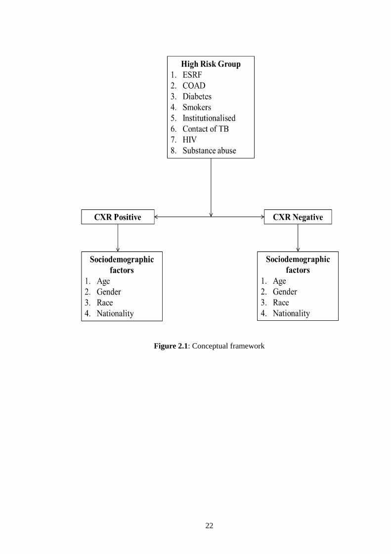
2.4 Conceptual framework
The conceptual framework below explains the factors included in the study. Those
factors influence the positivity of the chest x-ray are symptoms of the patient,
whether symptomatic or asymptomatic and the types of symptoms experienced by
the patient. The socio demographic factors studied are age, gender, race and
nationality. The high-risk groups that included in the study are the risk group defined
by MOH to be included for x-ray screening. The groups are end stage renal failure,
chronic obstructive airway disease, diabetes, smoker, institutionalised people,
contact of TB patient, HIV patient, and substance abuse. Rheumatoid arthritis and
anti-TNF is not included because since the directive is still new, many hospitals did
not have adequate data.
22
Figure 2.1: Conceptual framework

23
CHAPTER 3
METHODOLOGY
3.1. Study design
This was a cross sectional study using the screening registry for high risk groups of
TB patient retrieved from TBIS 104 A and the chest x-ray reporting report from the
facilities of Kedah.
3.2 Study duration
This study was done from December 2016 to March 2017.
3.3 Study location
This study was conducted in TB Unit, Kedah State Health Department, Radiology
unit Hospital Sultanah Bahiyah Alor Setar, along with other Hospitals and Health
Clinics which are selected in the study. State of Kedah is the northern state of
Malaysia beside Perlis. It borders to Thailand and Perlis from the north, Penang and
Perak from south and Kelantan from the east. The widths are 250,000 km squares,
almost equivalence to Kelantan state. It consists of 11 districts which are:
1. Langkawi Island
2. Kubang Pasu
3. Padang Terap

24
4. Kota Setar
5. Pendang
6. Sik
7. Baling
8. Kuala Muda
9. Yan
10. Kulim
11. Bandar Bharu
Figure 3.1: Map of Kedah