Embed Size (px)
Citation preview
371
Braz. J. Vet. Res. Anim. Sci., São Paulo, v. 47, n. 5, p. 371-379, 2010
Introduction
Cardiac arrhythmia is defined as a disturbance in heart rate or rhythm, as well as abnormalities of im-pulse formation and impulse conduction (either alone or in combination)1,2. Impulse conduction may or may not change heart rhythm. The electrocardiogram (ECG) is essential for diagnosing most cardiac arrhyth-mias and conduction disturbances and it can be used to aid cardiac evaluation to establish a cause, an anatomic and physiologic diagnosis and a prognosis. Also, it is recommended preoperatively in older animals3.
In 1960, Patterson et al.4 showed that among 3000 dogs which were brought to one veterinary clinic dur-
ing 18 months, 124 had disturbances of rhythm and 15 different arrhythmias were detected. Larsson and Schwartz5 observed 106 cases of cardiac arrhythmias among 533 dogs with cardiac disease. Schwartz et al.6 analyzed 353 ECGs of dogs and showed 27 different
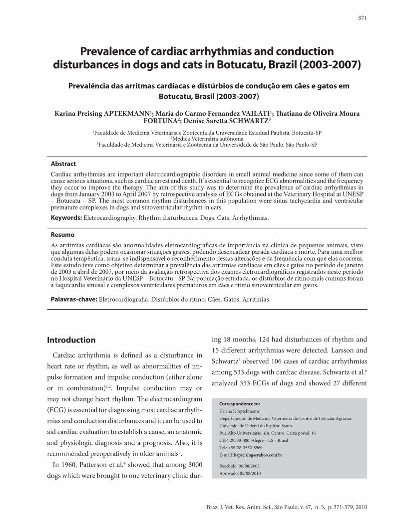
Prevalence of cardiac arrhythmias and conduction disturbances in dogs and cats in Botucatu, Brazil (2003-2007)
Prevalência das arritmas cardíacas e distúrbios de condução em cães e gatos em Botucatu, Brasil (2003-2007)
Karina Preising APTEKMANN1; Maria do Carmo Fernandez VAILATI1; Thatiana de Oliveira Moura FORTUNA2; Denise Saretta SCHWARTZ3
1Faculdade de Medicina Veterinária e Zootecnia da Universidade Estadual Paulista, Botucatu-SP2Médica Veterinária autônoma
3Faculdade de Medicina Veterinária e Zootecnia da Universidade de São Paulo, São Paulo-SP
Correspondence to:
Karina P. Aptekmann Departamento de Medicina Veterinária do Centro de Ciências Agrárias Universidade Federal do Espírito Santo Rua Alto Universitário, s/n, Centro. Caixa postal: 16 CEP: 29500-000. Alegre – ES – Brasil Tel.: +55-28-3552-8960 E-mail: [email protected]
Recebido: 06/08/2008 Aprovado: 05/08/2010
Abstract
Cardiac arrhythmias are important electrocardiographic disorders in small animal medicine since some of them can cause serious situations, such as cardiac arrest and death. It’s essential to recognize ECG abnormalities and the frequency they occur to improve the therapy. The aim of this study was to determine the prevalence of cardiac arrhythmias in dogs from January 2003 to April 2007 by retrospective analysis of ECGs obtained at the Veterinary Hospital at UNESP – Botucatu – SP. The most common rhythm disturbances in this population were sinus tachycardia and ventricular premature complexes in dogs and sinoventricular rhythm in cats.Keywords: Eletrocardiography. Rhythm disturbances. Dogs. Cats. Arrhythmias.
Resumo
As arritmias cardíacas são anormalidades eletrocardiográficas de importância na clínica de pequenos animais, visto que algumas delas podem ocasionar situações graves, podendo desencadear parada cardíaca e morte. Para uma melhor conduta terapêutica, torna-se indispensável o reconhecimento dessas alterações e da frequência com que elas ocorrem. Este estudo teve como objetivo determinar a prevalência das arritmias cardíacas em cães e gatos no período de janeiro de 2003 a abril de 2007, por meio da avaliação retrospectiva dos exames eletrocardiográficos registrados neste período no Hospital Veterinário da UNESP – Botucatu - SP. Na população estudada, os distúrbios de ritmo mais comuns foram a taquicardia sinusal e complexos ventriculares prematuros em cães e ritmo sinoventricular em gatos.
Palavras-chave: Eletrocardiografia. Distúrbios do ritmo. Cães. Gatos. Arritmias.
Braz. J. Vet. Res. Anim. Sci., São Paulo, v. 47, n. 5, p. 371-379, 2010
372
combinations of rhythms. In 1996, 700 healthy dogs were submitted to ECG evaluation and 49 of them showed electrocardiographic abnormalities7.
The objective of this study was to assess the type and frequency of cardiac arrhythmias and conduction dis-turbances occurring spontaneously in dogs and cats brought to a Veterinary Teaching Hospital in a period of 52 months, without considering the underlying disease process.
Material and Method
A total of 1426 electrocardiogram tracings of dogs and cats performed in the Veterinary Hospital at the School of Veterinary Medicine and Animal Science at São Paulo State University, Botucatu - SP from Janu-ary/2003 until April/2007 were retrospectively ana-lyzed. Tracings were printed for interpretation from previously recorded ECGs from a commercially avail-able computerized ECG analyzer and software pro-gram (ECG-PC TEB®, Brazil). All animals had been positioned in right lateral recumbency with standard ECG leads on the limbs in agreement with Tilley´s3 recommendation. Breed, age and gender were not considered, as some ECGs did not have the complete identification.
The heart rate, rhythm, mean electrical axis, QRS com-plexes and PR intervals duration and the relationship be-tween P waves and QRS complexes were determined in order to classify the rhythm or conduction disturbance. Measurements of QT interval, alterations in ST segment and T wave were not considered in this study.
All ECGs of dogs and cats were analyzed, regard-less of the primary disease presented by the animal. Only the first electrocardiographic exam was used for animals that had more than one ECG tracing. ECGs showing many artifacts that affected the analysis were excluded, as well as ECGs without any animal identifi-cation information were not used. ECG tracings were accessed by each of three authors separately and the
results were compared and discussed. The results were submitted to descriptive statistical analysis.
Results
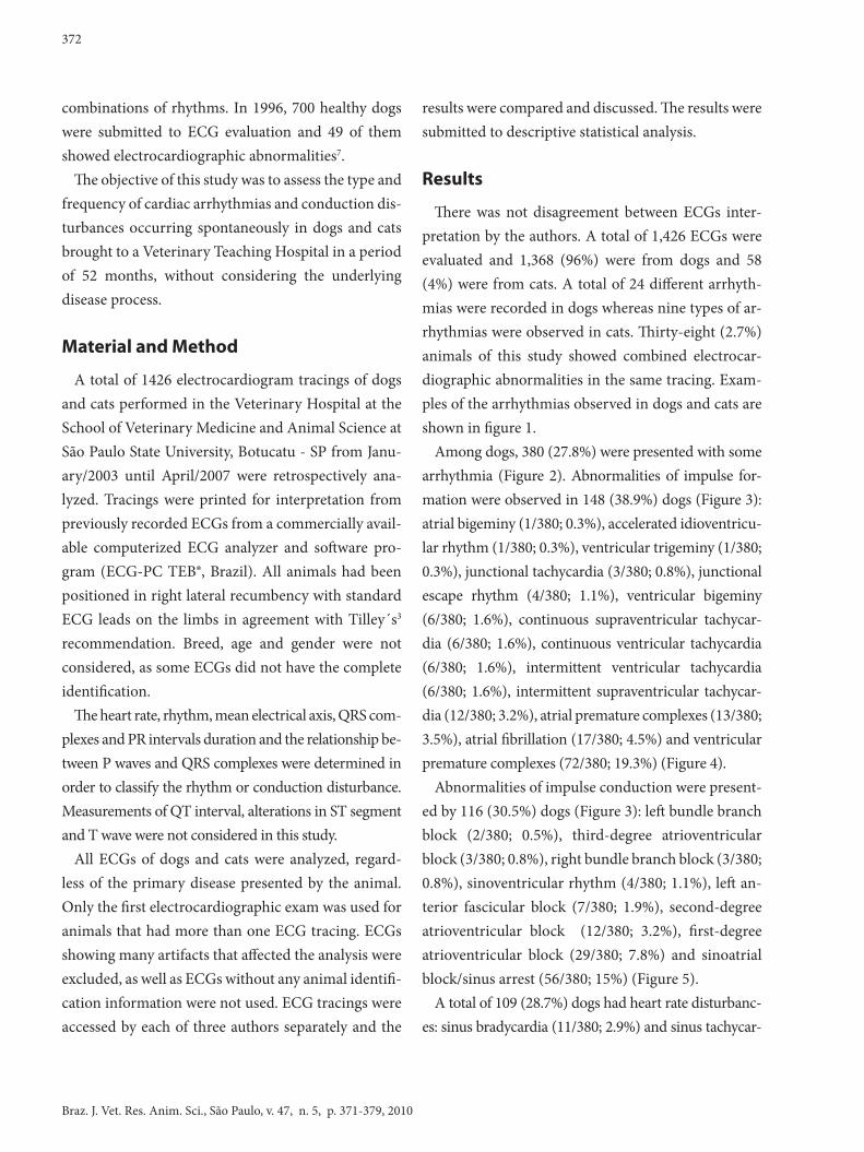
There was not disagreement between ECGs inter-pretation by the authors. A total of 1,426 ECGs were evaluated and 1,368 (96%) were from dogs and 58 (4%) were from cats. A total of 24 different arrhyth-mias were recorded in dogs whereas nine types of ar-rhythmias were observed in cats. Thirty-eight (2.7%) animals of this study showed combined electrocar-diographic abnormalities in the same tracing. Exam-ples of the arrhythmias observed in dogs and cats are shown in figure 1.
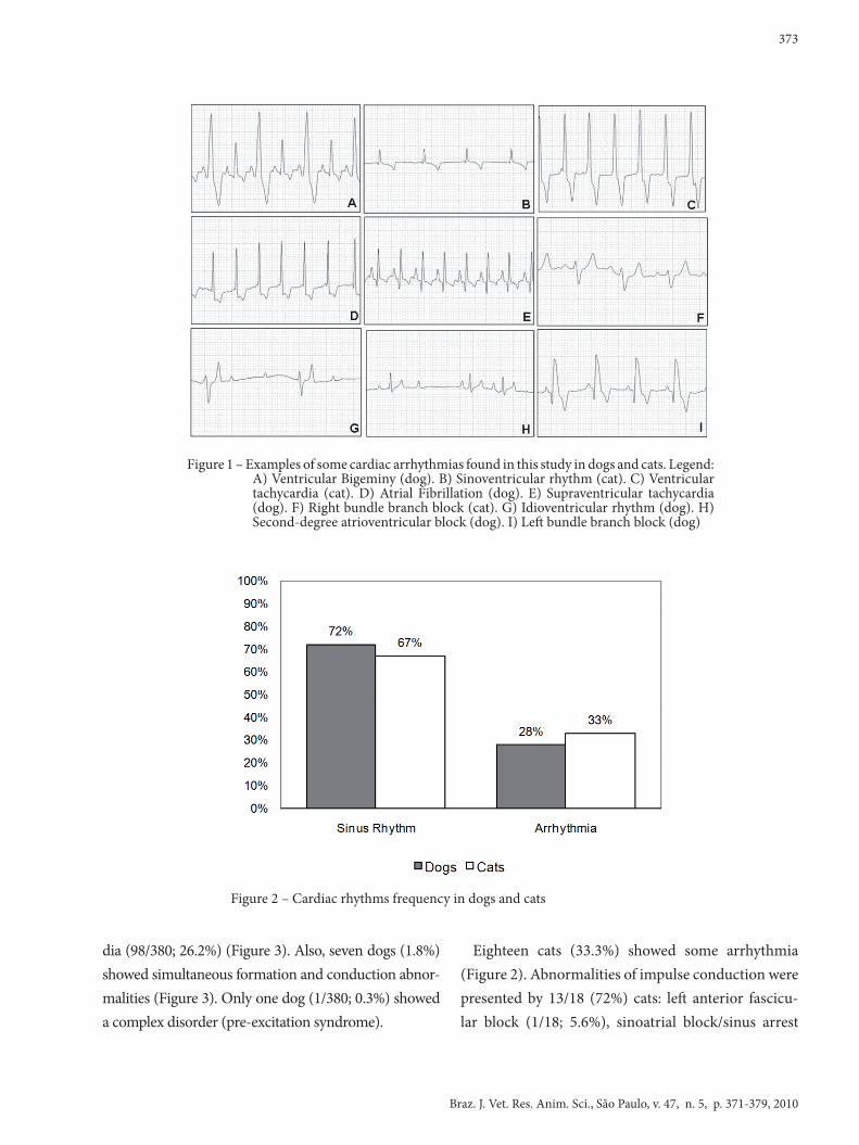
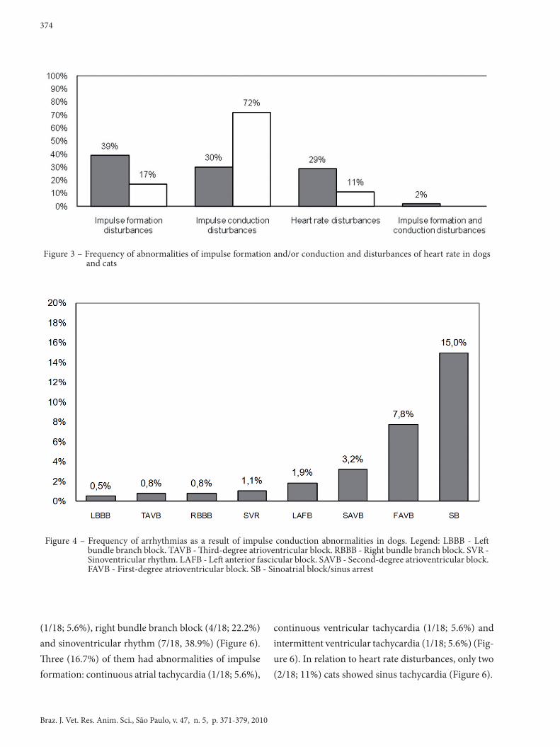
Among dogs, 380 (27.8%) were presented with some arrhythmia (Figure 2). Abnormalities of impulse for-mation were observed in 148 (38.9%) dogs (Figure 3): atrial bigeminy (1/380; 0.3%), accelerated idioventricu-lar rhythm (1/380; 0.3%), ventricular trigeminy (1/380; 0.3%), junctional tachycardia (3/380; 0.8%), junctional escape rhythm (4/380; 1.1%), ventricular bigeminy (6/380; 1.6%), continuous supraventricular tachycar-dia (6/380; 1.6%), continuous ventricular tachycardia (6/380; 1.6%), intermittent ventricular tachycardia (6/380; 1.6%), intermittent supraventricular tachycar-dia (12/380; 3.2%), atrial premature complexes (13/380; 3.5%), atrial fibrillation (17/380; 4.5%) and ventricular premature complexes (72/380; 19.3%) (Figure 4).
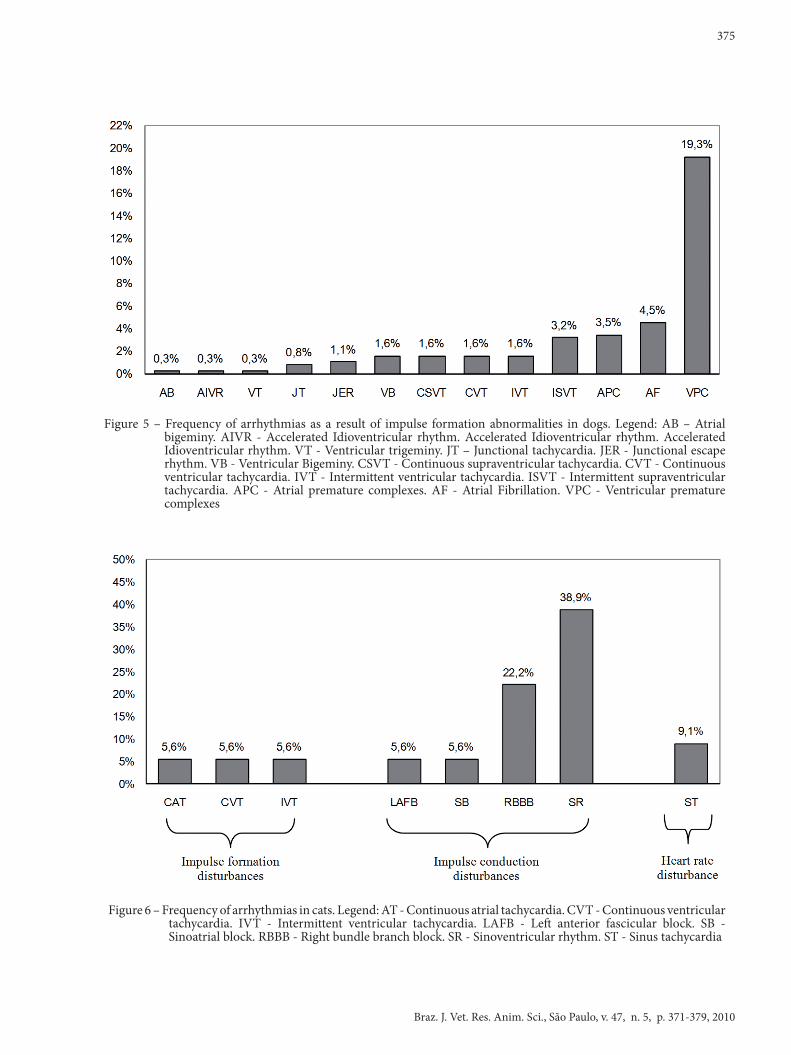
Abnormalities of impulse conduction were present-ed by 116 (30.5%) dogs (Figure 3): left bundle branch block (2/380; 0.5%), third-degree atrioventricular block (3/380; 0.8%), right bundle branch block (3/380; 0.8%), sinoventricular rhythm (4/380; 1.1%), left an-terior fascicular block (7/380; 1.9%), second-degree atrioventricular block (12/380; 3.2%), first-degree atrioventricular block (29/380; 7.8%) and sinoatrial block/sinus arrest (56/380; 15%) (Figure 5).
A total of 109 (28.7%) dogs had heart rate disturbanc-es: sinus bradycardia (11/380; 2.9%) and sinus tachycar-
373
Braz. J. Vet. Res. Anim. Sci., São Paulo, v. 47, n. 5, p. 371-379, 2010
Figure 1 – Examples of some cardiac arrhythmias found in this study in dogs and cats. Legend: A) Ventricular Bigeminy (dog). B) Sinoventricular rhythm (cat). C) Ventricular tachycardia (cat). D) Atrial Fibrillation (dog). E) Supraventricular tachycardia (dog). F) Right bundle branch block (cat). G) Idioventricular rhythm (dog). H) Second-degree atrioventricular block (dog). I) Left bundle branch block (dog)
Figure 2 – Cardiac rhythms frequency in dogs and cats
dia (98/380; 26.2%) (Figure 3). Also, seven dogs (1.8%) showed simultaneous formation and conduction abnor-malities (Figure 3). Only one dog (1/380; 0.3%) showed a complex disorder (pre-excitation syndrome).
Eighteen cats (33.3%) showed some arrhythmia (Figure 2). Abnormalities of impulse conduction were presented by 13/18 (72%) cats: left anterior fascicu-lar block (1/18; 5.6%), sinoatrial block/sinus arrest
Braz. J. Vet. Res. Anim. Sci., São Paulo, v. 47, n. 5, p. 371-379, 2010
374
(1/18; 5.6%), right bundle branch block (4/18; 22.2%) and sinoventricular rhythm (7/18, 38.9%) (Figure 6). Three (16.7%) of them had abnormalities of impulse formation: continuous atrial tachycardia (1/18; 5.6%),
continuous ventricular tachycardia (1/18; 5.6%) and intermittent ventricular tachycardia (1/18; 5.6%) (Fig-ure 6). In relation to heart rate disturbances, only two (2/18; 11%) cats showed sinus tachycardia (Figure 6).
Figure 3 – Frequency of abnormalities of impulse formation and/or conduction and disturbances of heart rate in dogs and cats
Figure 4 – Frequency of arrhythmias as a result of impulse conduction abnormalities in dogs. Legend: LBBB - Left bundle branch block. TAVB - Third-degree atrioventricular block. RBBB - Right bundle branch block. SVR - Sinoventricular rhythm. LAFB - Left anterior fascicular block. SAVB - Second-degree atrioventricular block. FAVB - First-degree atrioventricular block. SB - Sinoatrial block/sinus arrest
375
Braz. J. Vet. Res. Anim. Sci., São Paulo, v. 47, n. 5, p. 371-379, 2010
Figure 5 – Frequency of arrhythmias as a result of impulse formation abnormalities in dogs. Legend: AB – Atrial bigeminy. AIVR - Accelerated Idioventricular rhythm. Accelerated Idioventricular rhythm. Accelerated Idioventricular rhythm. VT - Ventricular trigeminy. JT – Junctional tachycardia. JER - Junctional escape rhythm. VB - Ventricular Bigeminy. CSVT - Continuous supraventricular tachycardia. CVT - Continuous ventricular tachycardia. IVT - Intermittent ventricular tachycardia. ISVT - Intermittent supraventricular tachycardia. APC - Atrial premature complexes. AF - Atrial Fibrillation. VPC - Ventricular premature complexes
Figure 6 – Frequency of arrhythmias in cats. Legend: AT - Continuous atrial tachycardia. CVT - Continuous ventricular tachycardia. IVT - Intermittent ventricular tachycardia. LAFB - Left anterior fascicular block. SB - Sinoatrial block. RBBB - Right bundle branch block. SR - Sinoventricular rhythm. ST - Sinus tachycardia
Braz. J. Vet. Res. Anim. Sci., São Paulo, v. 47, n. 5, p. 371-379, 2010
376
Discussion
There are only a few publications on studies about prevalence of cardiac arrhythmias in dogs4,5,6,7 and there is a lack of these studies in cats from hospital population. Only case-reports were found in the lit-erature regarding arrhythmias in cats8,9,10,11,12,13,14, or reports of cats with specific arrhythmias12,15,16, or studies on Holter monitoring in unsedated healthy cats17,18,19. Some studies performed in distinct dog breeds showed a variation in prevalence of cardiac arrhythmias that could be associated with the most common diseases in those populations4,7.
In this study, the number of ECGs evaluated was sig-nificant (n = 1,426) and showed a larger prevalence of arrhythmias among dogs (27.8%) when compared with two other studies (7%7 and 4.1%4, respectively). This fact could be explained by the difference between inclusion criteria adopted in each study. This study included all ECGs of dogs performed in that period, regardless of the primary disease presented by the animal, while other studies included only dogs with evidence of cardiac ab-normalities4 or clinically normal7. The results showed that disturbances of excitability and impulse formation were the most common causes of arrhythmias in dogs. Other research has reported similar results4.
Sinus tachycardia was the most common abnormal-ity found in dogs (26.2%). In most cases, it could be related to physiological conditions as fear or stress that explain the large prevalence of dogs presented with this rhythm3. As a disturbance of heart rate, sinus bradycardia was less common than tachycardia and only 11 dogs (2.9%) presented this abnormality. Slow sinus rhythm can be a normal finding or be associated with cardiac and systemic alterations2.
Concerning disturbances of impulse formation, ventricular premature complexes (VPCs) were the most common abnormalities (including ventricular bigeminy and trigeminy) recorded in 79 dogs (21.1%) in this study, also demonstrated by Patterson et al.4.
Another study showed that 97.5% of dogs with id-iopathic dilated cardiomyopathy had VPCs20. In the present study, ventricular tachycardia (VT) occurred in 3.2% of dogs (six with continuous VT and six with intermittent VT). Patterson et al.4 showed a larger prevalence (6.4%), but only represented by intermit-tent ventricular tachycardia. Considering specific populations, dogs with idiopathic dilated cardiomy-opathy20 and healthy Boxers21,22 also showed a larger prevalence of ventricular arrhythmias (45%, 58.3% and 70%, respectively). Accelerated idioventricular rhythm (AIVR) was not common in this study. A prevalence of 5.5% had been showed by Guglielmini et al.23. Causes of VPCs, VT and AIVR include any cardiac or systemic disorder, with the most common of these including primary cardiac diseases1,2.
Other studies reported a 25.5% prevalence of atri-al fibrillation (AF)5 in dogs with cardiac disease. In the present study only 4.5% of dogs (with or without cardiac disease) had this disturbance. The difference between the percentages could be explained by the distinct population of dogs. AF most often occurs in dogs when marked atrial enlargement is present sec-ondary to a primary cardiac disease2,20, although this was not the purpose of the study, most of these dogs did have cardiac disease.
Atrial premature complexes, including atrial bi-geminy, were recorded in 3.8% of dogs, a lesser preva-lence than found by Patterson et al.4 (11.3%). Eighteen dogs (4.8%) had supraventricular tachycardia (SVT) (continuous or intermittent). Patterson et al.4 showed a prevalence of 2.4% of dogs with intermittent SVT. Causes of these abnormalities are most commonly related to a structural cardiac atrial lesion2 and dogs with cardiomyopathy20.
The pre-excitation syndrome is a complex disorder involving abnormalities of both excitability and con-duction. Most of the times, it is an incidental and clini-cally silent finding. In this study, only one dog showed this syndrome, as observed by Patterson et al.4.
377
Braz. J. Vet. Res. Anim. Sci., São Paulo, v. 47, n. 5, p. 371-379, 2010
Junctional tachycardia was found in three dogs and could be associated with digitalis toxicity or cardiac disease3. Junctional escape rhythm was considered an impulse formation disturbance, however it is sec-ondary to all causes of sinus bradycardia, sinus ar-rest, atrioventricular (AV) block, digitalis toxicity, increased vagal tone and sick sinus syndrome3. Four dogs showed this disturbance in this study in conse-quence to third-degree AV block.
As the most common disturbance of impulse con-duction, sinus arrest was observed in 14.5% of dogs, a greater prevalence compared with a healthy dog population in other study (3.4%)7. Most of the time, sinus arrest was associated with pronounced sinus arrhythmia. However, it could not be differentiated from sinoatrial block due to difficulty in determin-ing the type of disturbance of the sinus impulse in a routine ECG. It could be a normal incidental find-ing in brachycephalic breeds or it could be associ-ated to pathologic conditions that cause stimulation of the vagus nerve, atrial conditions and sick sinus syndrome3.
Other conduction disturbances to be considered although not necessarily related to rhythm distur-bance, are the bundle branch blocks (BBB), which were not common. Three dogs (0.8%) presented right BBB, and two dogs (0.5%) presented left BBB. There are many causes of BBB and they may be due to a variety of pathologic changes including hyper-trophy, dilation and inflammation1. Although the left anterior fascicular (LAF) block is more common in cats1, seven dogs (1.9%) showed this disturbance of impulse conduction.
Atrioventricular blocks are defined as delays or stoppages of conduction between the atria and the ventricles1. Both first and second-degree AV blocks may occur in dogs that are clinically normal and healthy3. Also, the second-degree AV blocks could be associated with causes of increased vagal tone3. Third-degree AV blocks are sometimes functional,
but are commonly associated with a structural le-sion, inflammatory or degenerative1. Atrioventricu-lar blocks are reported on the cardiomyopathy in the English Cocker Spaniel24. The results showed that a total of 44 dogs (11.8%) presented with some type of AV block and the first-degree AV block was the most common (7.8%). Patterson et al.4 showed a fre-quency of 0.9% of AV block, with similar frequency between first and second-degree AV block (0.4%) and 0.1% of third-degree AV block.
Sinoventricular rhythm was not common but was recorded in four dogs (1%). This rhythm is secondary to systemic diseases3.
In the study reported here, a large percentage of ECGs of cats showed arrhythmias. Disturbances of conduction were the most important causes of ar-rhythmia in cats, an opposite result compared with ECGs abnormalities found in cats with heart disease, which showed a prevalence of 6.6% in conduction abnormalities and 60.5% in abnormalities of impulse formation25.
The sinoventricular rhythm was the most common disturbance of conduction in this study. It was probably in consequence of hyperkalemia, as evidenced by Tag and Day26. Urethral obstruction was also very common and it is probably the most common cause of sinoven-tricular rhythm in cats. As mentioned above, the LAF block is the type of conduction block most common in cats and commonly found in cats with feline idiopathic cardiomiopathy27, however, only one cat (5.6%) had LAF block in this study. Four cats (22.2%) had right BBB, a higher prevalence than showed by Riesen et al.25 in 0.3% of cats with cardiac disease.
Among abnormalities of impulse formation, ven-tricular tachycardia (intermittent or continuous) was the arrhythmia most often recorded in cats, but it was represented by only two cats. No cats had VPCs in this study, while VPCs were observed even in healthy cats in three different studies during Holter monitor-ing17,18,19. In the present study, one cat had continuous
Braz. J. Vet. Res. Anim. Sci., São Paulo, v. 47, n. 5, p. 371-379, 2010
378
1. CÔTÉ, E.; ETTINGER, S. J. Electrocardiography and cardiac arrhythmias. In: ETTINGER, S. J.; FELDMAN, E. C. Textbook of veterinary internal medicine: diseases of the dog and cat. 6. ed. St. Louis: Elsevier, 2005. p. 1040-1076.
2. NELSON, R. W.; COUTO, C. G. Cardiac rhythm disturbances and antiarrhythmic therapy. In: NELSON, R. W.; COUTO, C. G. Small animal internal medicine. 3. ed. Philadelphia: Mosby, 2003. p. 73-97.
3. TILLEY, L. P. Essentials of canine and feline electrocardiography: interpretation and treatment. 3. ed. Philadelphia: Lea & Febiger, 1992. 470 p.
4. PATTERSON, D. F.; DETWEILER, D. K.; HUBBEN, M. S. K.; BOTTS, R. P. Spontaneous abnormal cardiac arrhythmias and conduction disturbances in the dog. American Journal of Veterinary Research, v. 22, p. 355-369, 1960.
5. LARSSON, M. H. M. A.; SCHWARTZ, D. S. Estudo das arritmias mais frequentemente diagnosticadas em cães. In: CONFERêNCIA ANUAL DA SOCIEDADE PAULISTA DE MEDICINA VETERINáRIA, 45., 1990, São Paulo. Anais... São Paulo, 1990. p. 8.
6. SCHWARTZ, D. S.; LARSSON, M. H. M. A.; FIGUEIREDO, C.; FRANCO, S. R. V. S.; CURI, P. R. Estudo clínico das arritmias cardíacas em cães. In: CONGRESSO BRASILEIRO DA ANCLIVEPA, 15., 1993, Rio de Janeiro. Anais... Rio de Janeiro, 1993. p. 123.
7. SKOULA, C. M.; GORDON, B. E. Retrospective clinical study of computer-detected electrocardiogram abnormalities in a random-source dog population (abstract). Laboratory Animal Science, v. 46, n. 4, p. 462, 1996.
8. MEURS, K. M.; MILLER, M. W.; MACKIE, J. R.; MATHISON, P. Syncope associated with cardiac lymphoma in a cat. Journal of the American Animal Hospital Association, v. 30, n. 6, p. 583-585, 1994.
9. GAVAGHAN, B. J.; KITTLESON, M. D.; MCALOOSE, D. Persistent atrial standstill in a cat. Australian Veterinary Journal, v. 77, n. 9, p. 574-579, 1999.
10. CÔTÉ, E.; HARPSTER, N. K.; LASTE, N. J.; MACDONALD, K. A.; KITTLESON, M. D.; BOND, B. R.; BARRETT, K. A.; ETTINGER, S. J.; ATKINS, C. E. Atrial fibrillation in cats: 50 cases (1979-2002). Journal of the American Animal Hospital Association, v. 225, n. 2, p. 256-260, 2004.
11. HARVEY, A. M.; BATTERSBY, I. A.; FAENA, M.; FEWS, D.; DARKE, P. G. G.; FERASIN, L. Arrhythmogenic right
ventricular cardiomyopathy in two cats. Journal of Small Animal Practice, v. 46, n. 3, p. 151-156, 2005.
12. NORMAN, B. C.; CÔTÉ, E.; BARRETT, K. A. Wide-complex tachycardia associated with severe hyperkalemia in three cats. Journal of Feline Medicine and Surgery, v. 8, n. 6, p. 372-378, 2006.
13. ROLAND, R. M.; ESTRADA, A. H. ECG of the month. Ventricular preexcitation. Journal of the American Animal Hospital Association, v. 228, n. 10, p. 1500-1502, 2006.
14. TICEHURST, K.; ZAKI, S.; MADDERN, K.; LINGARD, A.; BARRS, V.; MALIK, R. Bradyarrhythmia in an anaesthetised, elderly, hypertensive cat. Journal of Feline Medicine and Surgery, v. 9, n. 6, p. 521-525, 2007.
15. JOHNSON, L.; SISSON, D. Atrioventricular block in cats. Compendium on Continuing Education for the Practicing Veterinarian, v. 15, n. 10, p. 1356-1367, 1993.
16. KELLUM, H. B.; STEPIEN, R. L.Third-degree atrioventricular block in 21 cats (1997-2004). Journal of Veterinary Internal Medicine, v. 20, n. 1, p. 97-103, 2006.
17. WARE, W. Twenty-four-hour ambulatory electrocardiography in normal cats. Journal of Veterinary Internal Medicine, v. 13, n. 3, p. 175-180, 1999.
18. YAMAKI, F. L.; LARSSON, M. H. M. A.; SOARES, E. C.; PEREIRA, G. G.; YAMATO, R. J.; SARRAFF, A. P. P.; BRITO, F. S.; PINTO, A. C. B. F.; LEOMIL NETO, M. Padronização de monitorização eletrocardiográfica ambulatorial (monitorização Holter) por 24 horas de felinos clinicamente normais. In: CONGRESSO BRASILEIRO DA ANCLIVEPA, 24., 2003. Belo Horizonte, Anais… Belo Horizonte: ANCLIVEPA, 2003. 1 CD-ROM.
19. HANÅS, S.; TIDHOLM, A.; EGENVALL, A.; HOLST, B. S. Twenty-four hour Holter monitoring of unsedated healthy cats in the home environment. Journal of Veterinary Cardiology, v. 11, n. 1, p. 17-22, 2009.
20. YAMAKI, F. L.; SOARES, E. C.; PEREIRA, G. G.; OLIVEIRA, V. M. C.; LARSSON, M. H. M. A. Monitorização eletrocardiográfica ambulatorial por 24-horas em cães com cardiomiopatia dilatada idiopática. Arquivo Brasileiro de Medicina Veterinária e Zootecnia, v. 59, n. 6, p. 1417-1424, 2007.
21. NOGUEIRA, R. B.; MUZZI, R. A. L.; HERRERA, D. S.; FALCO, I. R.; CAVALCANTE, G. A. O. Avaliação do ritmo cardíaco em cães da raça Boxer saudáveis pela eletrocardiografia contínua
References
atrial tachycardia. Supraventricular and ventricular tachycardia were reported in cats with hyperkale-mia12,26 and cardiac disease25. One case of persistent atrial standstill not related with hyperkalemia is pre-sented in the literature9. A study reported 50 cats with atrial fibrillation that was detected when signs of de-compensated cardiac disease were evident, but also was commonly identified as an incidental finding10. In the present study no cases of atrial fibrillation were observed among cats. This fact is related to the popu-
lation studied. Most cats included in this study had urethral obstruction, and not cardiac disease.
Conclusion
The most common rhythm disturbances in cats and dogs brought to the Veterinary Hospital at the School of Veterinary Medicine and Animal Science, Botucatu - SP were sinus tachycardia and ventricular premature complexes in dogs and sinoventricular rhythm in cats in the population studied.
379
Braz. J. Vet. Res. Anim. Sci., São Paulo, v. 47, n. 5, p. 371-379, 2010
(Holter). Arquivo Brasileiro de Medicina Veterinária e Zootecnia, v. 58, n. 1, p. 133-136, 2006.
22. LEOMIL NETO, M.; LARSSON, M. H. M. A.; PEREIRA, L.; BRITO, F. S. Padronização da monitorização eletrocardiográfica por 24 horas em cães. Arquivo Brasileiro de Medicina Veterinária e Zootecnia, v. 54, n. 2, p. 133-138, 2002.
23. GUGLIELMINI, C.; DIANA, A.; CIVITELLA, C.; DIANA, D.; LUCIANI, A.; CIPONE, M. Accelerated idioventricular rhythm in 9 dogs. Veterinary Research Communications, v. 30, p. 305-307, 2006. Supplement 1.
24. PEREIRA, L.; LARSSON, M. H. M. A.; NETO, M. L.; BRITO, F. S. Cardiomiopatia de cães da raça Cocker Spaniel Inglês:
aspectos clínicos, eletrocardiográficos, radiográficos e ecocardiográficos. Ciência Rural, v. 34, n. 2, p. 419-424, 2004.
25. RIESEN, S. C.; KOVACEVIC, A.; LOMBARD, C. W.; AMBERGER, C. Prevalence of heart disease in symptomatic cats: an overview from 1998 to 2005. Schweizer Archiv Fur Tierheilkunde, v. 149, n. 2, p. 65-71, 2007.
26. TAG, T. L.; DAY, T. K. Electrocardiographic assessment of hyperkalemia in dogs and cats. Journal of Veterinary Emergency and Critical Care, v. 18, n. 1, p. 61-67, 2008.
27. FERASIN, L.; STURGESS, C. P.; CANNON, M. J.; CANEY, S. M. A.; GRUFFYDD-JONES, T. J.; WOTTON, P. R. Feline idiopathic cardiomyopathy: a retrospective study of 106 cats (1994–2001). Journal of Feline Medicine and Surgery, v. 5, p. 151-159, 2003.