Embed Size (px)
Citation preview

Dana Sladkus
Prevalence of Sarcocystis neurona and Neospora Hughesi in horses
from Kentucky based on presence of serum antibodies to parasite surface
antigen

Review of Literature: Origin • EPM is fairly common in North America
• 60% of horses in the U.S. have been exposed to S. Neurona
• However only 1% of horses may develop EPM
https://encrypted-tbn0.gstatic.com/images?q=tbn:ANd9GcTl48Gw3eSCl0lIO4WiewfMMxcbg1c2KFlvxr12mzikQmpusJrJpQ
(Ellison et al. 2007)

Review of Literature: Symptoms
Symptoms include:
• Gait abnormalities • Ataxia • Mentation• Soreness• Lethargy
http
://w
ww
.epm
hors
e.or
g/Re
sour
ces/
Phot
o_Vi
deo.
htm
Carr et al. 2014

Review of Literature: Effect on the Body
• Horses diagnosed with EPM based on the presence of S. Neurona antibodies in the serum. (bodily fluids, blood)
• Horses with EPM had lower proliferation responses.
(Norton et al. 2006)
http://largeanimal.vethospitals.ufl.edu/files/2012/06/U
F-Equine-Neuro-Exam
-3a.jpg

The Parasite
Parasite enters and travels to the brain or spinal cord.
Nervous system of horse is effected (Dubey et al 2001)
Horses come in contact with the parasite through contaminated feed or water (Mackay et al 2012)
http
://e
n.w
ikip
edia
.org
/wik
i/Eq
uine
_pro
tozo
al_m
yelo
ence
phal
itis

http
://a
rs.u
sda.
gov/
imag
es/d
ocs/
1102
8_11
222/
sarc
olife
cycl
e.jp
g

Review of literature : DNA markers: S. Neurona
• Random-amplified polymorphic DNA techniques were used to enlarge DNA.
• Isolates of S. Neurona were taken from the infected horse. (Tanhauser, 1999)
• DNA sequence analysis of polymerase chain reaction (PCR) products.
• This was then used to design PCR primers to amplify specific Sarcocystis DNA products (Murphy et al, 2005)

The parasite: Neospora Hughesi• Known to be another parasite that causes EPM in horses.
• Now being identified in horses across the United states. (horses in 24 states tested positive for antibodies)
• Tested serums and CSF of horses to see if they came into contact with this parasite. (Yowell, 2010)

Hosts of EPM Parasite: The Opossum: Raccoon
• The opossum is the most common host.
• Both schizonts and merozoits carried by the opossum.(Dubey et al., 2001; Granstrom et al. 1998)
• Stages of S. neurona may be found in many parts
• of the raccoons body: (Stanek et al.2002) - Blood- Intestines- tissues
http://ww
w.hoxtonradio.com
/wp-content/uploads/2012/11/O
possom.jpeg

How do you test for EPM? Histopathology of the horse:
Tissue samples mounted on microscopic slides
Immunohistochemistry-an assay that shows specific antigens in tissues by the use of markers that are either fluorescent dyes or enzymes
PCR- copying DNA strands
• Immunohistochemical findings- schizonts, merozoites found.
(Mullaney et al. 2005)( Jones. et al. 2002)
https://encrypted-tbn3.gstatic.com/images?
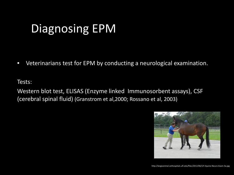
Diagnosing EPM
• Veterinarians test for EPM by conducting a neurological examination.
Tests: Western blot test, ELISAS (Enzyme linked Immunosorbent assays), CSF (cerebral spinal fluid) (Granstrom et al,2000; Rossano et al, 2003)
http://largeanimal.vethospitals.ufl.edu/files/2012/06/UF-Equine-Neuro-Exam-3a.jpg
Hypothesis
H0 – Horses that come into contact with the parasites S. neurona or N. Hughesi are less likely to form antigens of this parasite.
H1 - If horses come into contact with the parasite S. Neurona or N. Hughesi, they are more likely to form antibodies against the antigens of this parasite.

Methods • Sera was taken from 133 horses living in the pastures of University of
Kentucky’s farm.
• ELISAS conducted on both parasites S. neurona and Neospora Hughesi
https://www.pinterest.com/pin/199847302191370992/

Procedure
• Each sera was put into individual tubes. The tubes were then mixed with antigens of the parasites S. neurona and N. Hughesi.
• This was to determine the Seroprevalence of antibodies that were against the parasites surface antigens

Procedure
Solutions used:
• For washing well plates, antibody incubation solution, as well as substrate used in plates : Block solution (PBS, normal goat serum (second antibody), dry milk, and tween)
• stop reagent: Diluted sulfuric acid
• Recombinant protein was then diluted: left incubated over night.

Procedure• After ELISA protocol was completed well plates were checked for blue
coloring.
• Sulfuric Acid trhen added to turn the color yellow so that the ELISA reader could calculate the dilution of each well.


ELISA

MY Mentor: Dr. Howe• Gluck Equine Research Center
Department of Veterinary ScienceUniversity of Kentucky
• Sarcocystis neurona Genome Sequencing Project
• Examination of SnSAG Gene Family in Sarcosystis neurona
• Enzyme linked Immunosorbent Assay (ELISAs) Based on Sarcocystis neurona Surface Antigens (SnSAGs)
http
s://
ww
w.g
oogl
e.co
m/s
earc
h?q=
Dr.+
dani
el+h
owe+
univ
ersi
ty+o
f+ke
ntuc
ky&
biw
=192
0&b

Bibliography
•
• Dubey, J.P., Lindsay, D.S., Saville, W.J., Reed, S.M., Granstrom, D.E., Speer, C.A., 2001b. A review of Sarcocystis neurona and equine protozoal myeloencephalitis (EPM). Vet. Parasitol. 95, 89–131].
• Mullaney , thomas. "Evidence to support horses as natural intermediate hosts for Sarcocystis neurona." T. Mullaney et al. / Veterinary Parasitology 133 (2005) 27–36. 133. (2005): 27-36. Print. <http://www.sciencedirect.com/science/article/pii/S0304401705002402>.
• Stanek, J.F., Dubey, J.P., Oglesbee, M.J., Reed, S.M., Lindsay, D.S., Capitini, L.A., Njoku, C.J., Vittitow, K.L., Saville, W.J., 2002. Life cycle of Sarcocystis neurona in its natural intermediate host, the raccoon, Procyon lotor. J. Parasitol. 88, 1151–1158].
• Tanhauser, S.M., Yowell, C.A., Cutler, T.J., Greiner, E.C., MacKay, R.J., Dame, J.B., 1999. Multiple DNA markers differentiate Sarcocystis neurona and Sarcocystis falcatula. J. Parasitol. 85, 221–228
• Yowell, C.A., Cutler, T.J., Greiner, E.C., MacKay, R.J., Dame, J.B., 1999. Multiple DNA markers differentiate Sarcocystis neurona and Sarcocystis falcatula. J. Parasitol. 85, 221–228
• Yeargan et al , . "exposer to sarcosystis neurona in horses determined by western blot analysis using s. neurons merozoits." 185. (2012): n. page. Print.

(cont.)
• Cosme et al , "prevelenc eof antibody of sarcosystos neuron in horses." 29. (2013): n. page. Print.
• Howe et al , . "improved detection of equine antibody against s neurona using ELISAs against the parasite." 176. (2011): n. page. Print.
• Yang et al, "immune response to s. neurons infected in natural infected horses with EPM." 138. (2006): n. page. Print.
• Granstrom, "A review of s. neurona ad equine protozoal myeloencephalitis." 95. (2001): n. page. Print.


![Research Article Effects of Experimental Sarcocystis neurona ...S. neurona Culture. Merozoites were obtained by a technique described by Ellison et al. [ , ]. Brie y, SnSAG merozoites](https://img.pdfslide.net/doc/110x75/6130cc351ecc5158694453ac/research-article-effects-of-experimental-sarcocystis-neurona-s-neurona-culture.jpg)