Embed Size (px)
Citation preview
Preventing Complications
Associated with
Tube Feedings
Norma A. Metheny, RN, PhD, FAAN
Saint Louis University

Potential for Malpositioned Feeding Tubes
Outside the GI Tract:
Lung
Mediastinum
Abdominal Cavity
Brain
Within the GI Tract:
Esophagus
Stomach (if gastric
emptying delayed)
Scope of Problem:
Over 1 million feeding
tubes inserted annually
Most by blind passage
Usually placed by nurses
Can easily be inserted
into undesirable site

Most Frequent Site for Malpositioned Tube
Approximately 4% of
blind tube insertions
enter the respiratory
tract
Tip can end in the
tracheobronchial tree
or the pleural space

1987-1989: Auscultation and pH
1991-1994: pH and Aspirate Appearance
1995-1998: pH, Enzymes and Bilirubin
Studies Related to Feeding Tube Placement
R01 NR01669 National Institute of Nursing Research
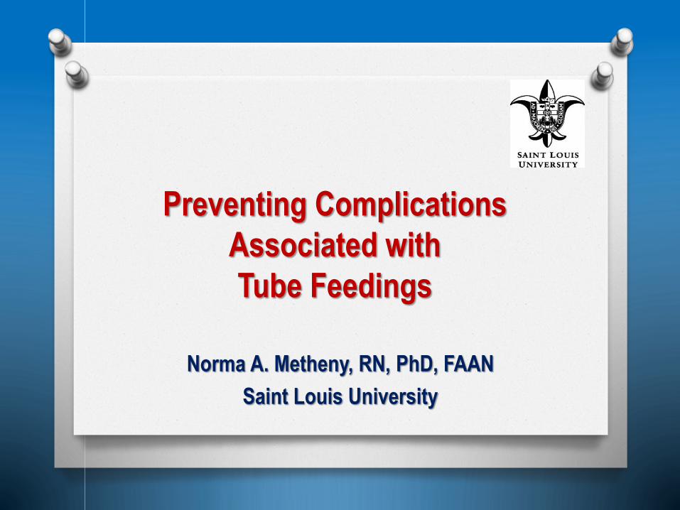
Auscultatory Method
No distinction between
sounds in stomach and
small bowel
Nursing Research, 1990 Heart & Lung, 1990
Failed in 8 of 9 cases to
identify tubes in lung

Anecdotal Report
Two RNs reported hearing air over
epigastrium following air insufflation of tube
in brain Am J Nursing, 2002
18 Fr polyvinyl
chloride tube placed
through sinus into
patient’s brain

hSource Examples of Other Anecdotal Reports
London
Evening
Standard,
Jan 8, 2014
Auscultation - Physician placed tube and
tested placement by the ‘Whoosh’ test. Ignored
nurses request for an x-ray to confirm tube
placement. Feedings delivered – removed
about 2 L of fluid from lung (death)
Chest, 1981Auscultation - tube in left mainstem bronchus.
Sepsis and empyema after infusion of 4 L of
formula (death)

Guidelines Regarding Auscultation
Organization Recommendtion
Am Assoc Crit Care Nurses (AACN):
Verification of Feeding Tube
Placement Practice Alert, 2010
Recognize air bolus method is
unreliable
NHS: UK.Patient Safety Alert:
Reducing Harm Caused by
Misplaced Nasogastric Feeding
Tubes in Adults & Children, 2011
Never use the ‘whoosh’ test to
confirm the nasogastric tube
position
Group of radiologists and GI
physicians: Gastroenterology, 2011;
141(2):742-765
Be aware that tubes in
inappropriate locations may be
mistaken as properly positioned
by auscultation
National Association of Children’s
Hospitals: Child Health Patient
Safety Organization. August 2012
Immediately discontinue insertion
of an air bolus to assess/verify NG
tube placement

Rationale for pH Method
Fasting gastric secretions
normally have a low pH
Intestinal secretions
normally have a high pH
Tracheobronchial
secretions and pleural
fluid normally have a high
pH
Note: Patients fasting
for at least 4 hours

pH & Tube Site in Adults
Tube Site Mean pH
Stomach, no acid-inhibitors (n=235) 3.33 ± 0.10
Stomach, acid-inhibitors (n=445) 4.34 ± 0.14
Small Bowel (n=578) 7.14 ± 0.03
Pleural fluid, tracheobronchial
secretions (n=280)7.64 ± 0.03
Note: Patients fasting for at least 4 hours
Am J Nursing, 2001
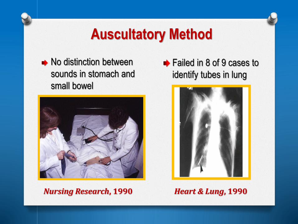
Is age a factor?
O In a study of 53 infants, the mean
gastric pH was:
5.4 at 15 minutes after birth
3.1 at 1 hour after birth
2.2 by 5 to 6 hours after birth

pH and Tube Site in Children
Gastric pH According to Use of Gastric Acid Inhibitors
Acid Inhibitor Absent
(n=2482 aspirates)
Acid Inhibitor Present
(n=1152 aspirates)
≤ 4.0 74.1% 55.1%
≤ 5.0 89.9% 76.7%
≤ 5.5 95.6% 84.8%
≤ 6.0 98.5% 93.1%
Information from: Gilbertson et al: J Parenteral & Enteral
Nutrition, 2011
Note: Feedings absent for at least one
hour at time of data collection

pH Method: Measurements
Method Comments
Electronic pH meter
Accurate
Impractical in most
clinical settings
Colorimetric pH strips (0-10):
Calibrated in units of one
Calibrated in units of 0.5
Calibrated in units of 0.2 or 0.3
Subjective
Point of Care testing
requirements may be
imposed
Litmus paper Inappropriate

Examples of Recommended pH Cut-Points
Source Recommendation
UK. National Health Service:
Patient Safety Alert:
Reducing the harm caused
by misplaced NG feeding
tubes in adults, children and
infants, Patient Safety
Agency, 2011
‘For determining correct
placement of feeding tubes, pH
testing is the first-line method ‘
‘Safe range is 1 to 5.5’
Gilbertson et al: J of
Parenteral & Enteral
Nutrition, 2011
Cut point of 5.0 to distinguish
between gastric and respiratory
tube site.
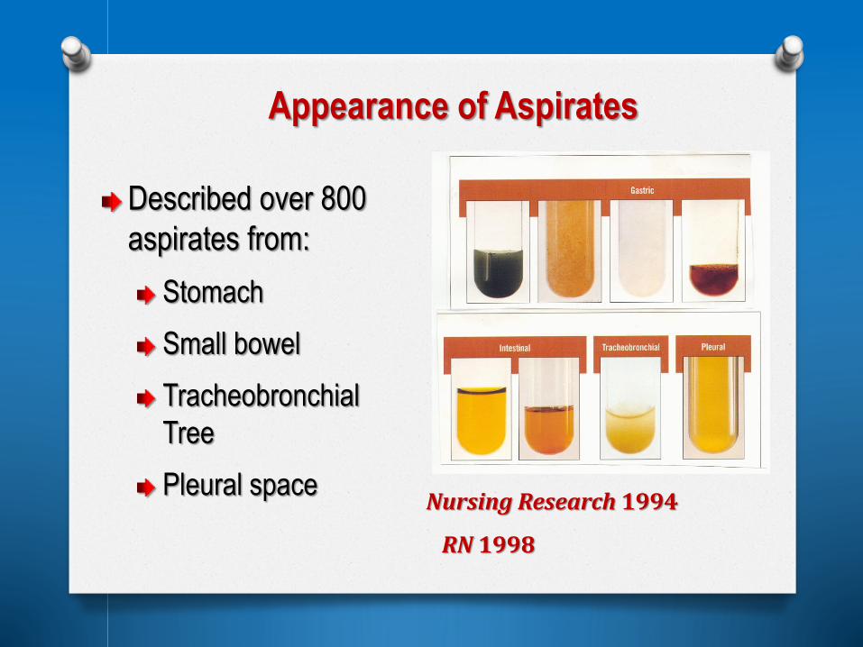
Appearance of Aspirates
Described over 800
aspirates from:
Stomach
Small bowel
Tracheobronchial
Tree
Pleural space Nursing Research 1994
RN 1998

Combination of pH & Appearance:
Stomach vs. Small Bowel
pH 7 pH 2
Bile-Stained Colorless

Appearance of Respiratory Aspirate
(Pleural Fluid)
pH 7

Identify Tube Site by Viewing Aspirates?
Photographed 106 aspirates: Viewed by
staff nurses who were able to identify
approximately:
• 90% of gastric aspirates
• 70% of small bowel aspirates
• 50% of respiratory aspirates
Nursing Research, 1994

Examples of Guidelines for Aspirate Appearance
Source Recommendation
Verification of Feeding Tube
Placement Practice Alert, 2010
(AACN)
Observe appearance of
aspirates if feedings are
interrupted for more than a few
hours.
NHS Patient Safety Alert:
Reducing Harm Caused by
Misplaced Nasogastric
Feeding Tubes in Adults &
Children, 2011
Do not observe the
appearance of a feeding tube
aspirate as an indication of
placement of a NG tube

Study Sample/Method Findings
Gast
Nsg,
2007
Device attached to ET
tube and then to NG
tube in 7 infants
Readings 0 mm Hg from NG
tube, 32-61 mm Hg from ET
tube
NCP,
2008
Colorimetric device
used during 424 blind
tube insertions
Correctly identified over 99% of
gastric placements; failed to
detect 2 of 4 tubes in lung
CO2 MonitorsDesigned to detect tube placement in respiratory
tract by showing presence of carbon dioxide
Relies on tube’s ports being freely exposed to gas
during insertion procedure

Examples of Guidelines for RadiographicConfirmation of Tube Site
Guidelines that Recommend
Radiography for ALL Blindly Inserted Tubes
Source Recommendation
Practice guidelines for GI
access for enteral nutrition;
Gastroenterology, 2011
“After blind insertion of a tube, every
patient should undergo radiography to
confirm proper position of the tube
before feeding is started”
American Association of
Critical Care Nurses: Practice
Alert – Verification of Feeding
Tube Placement
“Obtain radiographic confirmation of
correct placement of any blindly
inserted tube prior to its initial
use for feedings or medication
administration”

Guidelines that Refer to Use of
Radiography as Second Line Test
Source Recommendation
National Health Service.
Patient Safety Alert:
Reducing Harm Caused by
Misplaced Nasogastric
Feeding Tubes in Adults &
Children, 2011
Radiography only used as a
second-line test when no aspirate
can be obtained or pH indicator
paper has not confirmed position
of the NG tube

AFTER feedings started, check tube location at 4-hour
intervals:
Observe for a change in length of the external portion of
the feeding tube
Review routine chest and abdominal x-ray reports to look
for notations about tube location.
If pH strips are available, measure pH of feeding tube
aspirates if feedings are interrupted for more than a few
hours.
Observe the appearance of feeding tube aspirates if
feedings are interrupted for more than a few hours.
AACN Practice Alert, 2010

Tests Used in Clinical Practice
X-Ray AuscultationAspirate
Appearance
pH Capnography

Survey of American Association of Critical Care Nurses [AACN] n=2298
Question:
“Does your ICU require
radiographic proof of tube
placement before it is used
for the first time?”
Am J Critical Care, 2012

AACN Survey (continued)
Question: “What bedside
methods are used to check
tube placement prior to an x-
ray?”
Auscultatory method most
common response
Often used in combination
with aspirate appearance
and observation for distress
Am J Critical Care, 2012

AACN Survey (continued)
Question: ‘What single
method would you use to
test tube placement when
x-ray not used?’
161 nurses responded
Auscultation most common
response
Am J Critical Care, 2012

National Survey of Pediatric Nurses (n=95)
0102030405060708090
Auscultation AspirateAppearance
pH
Hospital Protocol
Unpublished data, 2012
%

Reasons pH Method not Widely Used
Confusion about reliable pH cut-point
Point-of-Care Testing not allowed -or pH strips not available on unit
Requires extra time and effort

Reasons Auscultation & Aspirate
Appearance Widely Used
Don’t require extra effort or time
Don’t require extra equipment
‘Way its always been done’

Bringing About Change in Practice?
Publications and guidelines: Minimal effect in
discouraging use of auscultatory method
Mandatory protocols needed:
Catastrophic event: Major incentive for
hospitals to update protocols
Magnet Hospitals: More likely to adopt
research-based protocols

Other Markers for Tube Placement?
Site pH Bilirubin Pepsin Trypsin
Stomach
Intestine
Lung
JPEN, 1997; Nursing Research, 1999

Bedside Test for Bilirubin
Used bilirubin standards to develop scale
Tested on specimens from 626 patients:
Stomach, n=328
Small bowel, n=303
Trachea, n=177
Pleural space, n=32
Good predictive ability
Nursing Research, 2000

Large company developed prototype of
device that could allow concurrent readings
of pH, bilirubin, pepsin and trypsin.
Simple algorithm provided good
predictive ability
Device not moved to production: Market
survey of nurses showed they were
satisfied with auscultatory method
What About Electromagnetic Guidance Placement Devices?
Designed to allow visualization of tube’s track
during the insertion procedure
Multiple studies regarding efficacy in
distinguishing between gastric and small bowel
placement
Relatively few studies refer to method’s efficacy
in detecting tube placement in respiratory tract
Question: Are these devices sufficiently accurate
to preclude additional placement tests?

Am J Crit Care, 2014
Review of 6 Studies (2007-2012)
Review of FDA MAUDE Database (2007-2012)
Collectively, device
evaluated in over 1700
patients
No cases of pneumothorax
Majority of individuals who
used the device in these
studies had advanced skills
and lengthy training
Search yielded reports of
2 deaths and 17 cases of
pneumothorax
Database relies on
voluntary reports – thus,
other cases may have
occurred and not been
reported

Source Warning
U.K. NHS Patient
Safety Alert.
Reference number: NHS/PSA/W/2013/001
“Placement devices for nasogastric
tube insertion DO NOT replace initial
position checks.” December 2013
U.S. FDA letter to a
manufacturer of an
electromagnetic
placement device,
April 2013
(posted on internet)
Between January 10, 2012 and
October 5, 2012, your firm received
nine (9) complaints alleging “lung
placement” while utilizing your
system to aid and confirm the proper
placement of nasogastric tubes. The
majority resulted in pneumothorax.’
Warnings

1999-2001: Evaluation of aspiration detection methods
2002-2005: Identification of risk factors for aspiration
2006-2008: Evaluation of interventions to prevent aspiration
Studies Related to Aspiration
R01 NR05007, National Institute of Nursing Research

Aspiration
Major risk in tube-fed patients
Macro aspirations rare
Micro aspirations common – difficult to
detect clinically
Lack of a simple test for aspiration has
hindered research

Detection of Small-Volume Aspirations:
Animal Model (1999-2002)
Used 182 New Zealand White rabbits to evaluate 3 aspiration detection methods:
1. Blue Dye
2. Glucose
3. Trace amounts of Pepsin
161 anesthetized and ventilated experimental
rabbits
21 anesthetized and ventilated control rabbits
3 forced small-volume aspirations:
Dye-stained enteral formulas mixed with
human gastric juice (experimental animals)
Normal saline (control animals)
Suctioned every 2 hours over 6 hour period
Methods

Dye Method: Low Sensitivity
Dye visible in less than half of suctioned secretions
Dye method rarely used in recent years
0.0 ml/L 0.8 ml/L 1.5 ml/L
CHEST, 2002

Demise of Blue Dye Method
Photograph of 12 month old boy who
received enteral formula tinted with FD&C
blue dye #1 published in New England
Journal of Medicine, 2000
U.S. Food and Drug Administration (FDA)
issued an alert in 2003 about possible
toxicity with use of FD&C blue dye #1 in
enteral feedings

Premise of Glucose Method
Tracheal secretions normally contain little
or no glucose
Most enteral formulas contain sizable
quantity of glucose
Therefore, finding glucose in
tracheobronchial secretions signals
aspiration of glucose-rich enteral formula.

Glucose Method: Low Specificity
0
10
20
30
40
2-hour 4-hour 6-hour
Time of measurement
% o
f G
luco
se V
ari
an
ce E
xp
lain
ed Blood
Glucose
GastricGlucose
FormulaGlucose
Glucose concentration in formula not significant (no
variance according to low, moderate, or high G formulas)
Blood glucose major contributor to tracheal glucose
MEDSURG Nursing, 2005
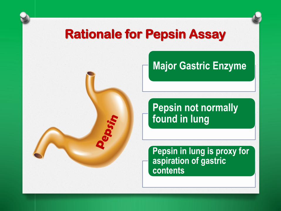
Rationale for Pepsin Assay
Major Gastric Enzyme
Pepsin not normally found in lung
Pepsin in lung is proxy for aspiration of gastric contents
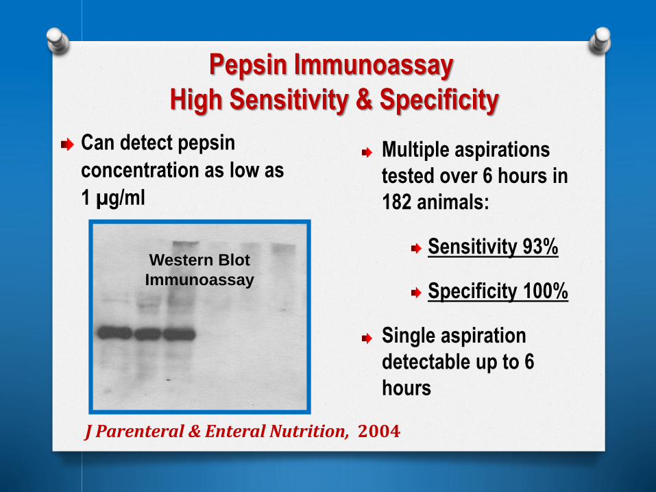
Pepsin Immunoassay
High Sensitivity & Specificity
Can detect pepsin
concentration as low as
1 µg/ml
Multiple aspirations
tested over 6 hours in
182 animals:
Sensitivity 93%
Specificity 100%
Single aspiration
detectable up to 6
hours
J Parenteral & Enteral Nutrition, 2004
Western Blot
Immunoassay

Development of a
Bedside Test for Aspiration (2009-2011)
Aim:
Develop a simple bedside immunoassay for
pepsin in tracheal secretions
Developed 5-minute assay that worked well
on pepsin standards and serum
However, not able to use test on tracheal
secretions (due to presence of competing
proteins)

Where to go from here?
Bedside Detection Methods?
• None currently available
Focus on PREVENTION
• Identify modifiable risk factors
• Evaluate interventions

Descriptive Clinical Study of
Aspiration (2002-2005)
Objectives:
Describe frequency of
aspiration in 360 ICU patients
Describe outcomes of
aspiration in ICU patients
Describe risk factors for
aspiration in ICU patients
St Louis University Hospital
Critical Care Medicine, 2006

RISK FACTORS
Decreased LOC
Heavy sedation
Low HOB
Gastric feeding site
High GRVs; vomiting
RISK FACTORS
Decreased LOC
Heavy sedation
Low HOB
Prolonged MV
Co-Morbidities 2
Immunosuppression
TUBE FEEDING
ASPIRATION OF
GASTRIC
CONTENTS
Defined as
presence of pepsin
in tracheal
secretions
PNEUMONIA
Defined as
Clinical Pulmonary
Infection Score 6
Framework

Frequency of Micro-Aspiration
Tested 5,857 tracheal secretions
(31% were pepsin-positive)
320 of 360 (89%) patients
aspirated at least once
Separated into two groups:
High Aspiration (>25% pepsin-
positive tracheal secretions)
Low Aspiration (<25% pepsin-
positive tracheal secretions)
Pneumonia Absent (n=187) Pneumonia Present (n=173)
5
10
15
20
25
30
35
40
45
50
As
pir
ate
s C
on
tain
ing
Pe
ps
in (
%)
N=175 N-185

Relationship Between
Aspiration and Pneumonia
High Aspiration Group had
four times greater risk for
pneumonia
Incidence of pneumonia
increased from Day 1 to
Day 3 as aspiration events
accumulated
Critical Care Medicine, 20060
1
2
3
4
5
6
Day 1 Day 2 Day 3
Mean
C
um
ulative # P
PT
S* p
er
Patien
t
0
10
20
30
40
50
60
70
80
90
100
%P
neu
mo
nia
Aspiration Events
Pneumonia

Risk Factors for Aspiration
VariableLow Aspiration
Group (n=175)
High Aspiration
Group (n=185)p-value
Mean age (years) 53 52 0.5
Mean APACHE II Score 22.3 23.4 .09
Mean GCS 7.5 6.9 .03
% pts with HOB
< 30 degrees56% 68% .02
Gastric feeding site 44% 56% .02

Gastric Residual Volumes
Only 20 of the 182
gastric-fed patients
had two or more GRVs
> 250 ml
15 of the 20 were in
the High Aspiration
Group, p = .08
n=5
n=15

Conclusions
Micro-aspirations common in critically ill, MV
tube-fed patients
Frequent micro-aspirations associated with poor
outcomes
Possible modifiable risk factors:
Head of bed elevation
Feeding tube location
Residual volumes

Prevention of Aspiration (2006-2008)
Objective:
Evaluate effectiveness of 3-pronged intervention
(ARRP: Aspiration Risk Reduction Protocol)
Design
Two-group quasi-experimental study
Usual Care Group, n=329 (2002-2005)
ARRP Group, n=145, (2006-2008)
Setting:
Same ICUs in both arms of study

Aspiration Risk Reduction Protocol (ARRP)
Elevate HOB to ≥ 30 degrees
Place tube in small bowel, as indicated
Implement algorithm for GRVs

HOB: Modifiable Risk Factor
Encouraged physicians to write orders for
elevated HOB (at least 30 degrees)
Added space on chart for hourly HOB elevation
notation
Researchers present 16 hours/day to reinforce
intervention
Distributed HOB recommendations to staff

HOB Elevation RecommendationsOrganization Guideline
Canadian Clinical Practice
Guidelines for Nutrition Support in
Mechanically Ventilated Critically Ill
Adult Patients (2003)
Elevate HOB to 45o unless
contraindicated
CDC and Healthcare Infection
Control Practices Advisory
Committee (2004)
Elevate HOB from 30o to 45o
unless contraindicated
Society of Critical Care Medicine
and American Society for Parenteral
and Enteral Nutrition (2009)
Elevate HOB from 30o to 45o
unless contraindicated
Practice Alert: Prevention of
Aspiration. American Association of
Critical Care Nurses (2011)
Elevate HOB from 30o to 45o
unless contraindicated

Effect of Intervention on HOB Elevation

Feeding Site: Modifiable Risk Factor
Distal
small bowel
For patients with:
Slowed gastric
emptying
Poor tolerance
to HOB elevation
Low level of
consciousness

Tube Placement Protocol
Physicians:
Asked to write orders for small-bowel tube
placements (when indicated by clinical condition)
Advance Practice Nurse:
Present 40 hours per week to instruct RNs in ICUs
on procedure for placing small bowel feeding
tubes

Effect of Intervention on Tube Site
Nursing Research, 2010
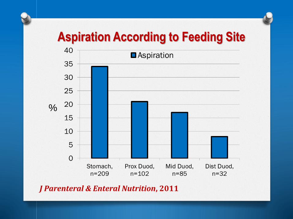
Aspiration According to Feeding Site
J Parenteral & Enteral Nutrition, 2011
%

GRVs: Modifiable Risk Factor
Algorithm Defined high GRV
as > 250 mlInstructions for use of
prokinetics
Instructions for moving tube
to small bowel if needed
Instructions for returning
aspirate to patient
Pepsin

Effect of Intervention on GRVs
Unable to test algorithm
Physicians held differing views on
how to handle GRVs
Nurses followed physician orders
instead of algorithm

Effect of ARRP on Primary Outcomes
ARRP resulted in
significant decrease in:
% patients with at
least one aspiration
event
% patients with
pneumonia

Dangers of Esophageal Placement
Significantly increases risk for aspiration
Case Example:
4 liters of bowel-prep solution administered via tube with
ports in esophagus
Caused severe aspiration in both lungs
Difficult to detect esophageal placement with bedside tests
Auscultation fails
pH fails (can be acidic if aspirate is refluxed gastric fluid, or
can be alkaline if aspirate is swallowed saliva)

High Gastric Residual Volumes
Slowed Bowel Sounds
Vomiting
Gastrointestinal Intolerance

Controversy about Significance ofGastric Residual Volumes
Pepsin
Risk of
Aspiration
Risk of
Under-
Feeding
0
50
100
150
200
250
300
350
400
450
500
ml

Possible Explanations for Disagreements
Different outcomes?
Aspiration
Pneumonia
Same outcome, different definition?
Different sample sizes?
Different types of patients?
Measurement problems?

GRV Measurement Error?

645 dual measurements from
14-18 Fr multi-port tubes and
10 Fr single-port feeding
tubes in 62 patients
Large-bore tubes identified
‘high’ GRVs 3 to 6 times more
often than 10 Fr feeding tubes
However, at times, GRVs
higher from 10 Fr tubes ≥ 250 ml
≥ 200 ml
≥ 150 ml
10 Fr 14-18 Fr
J Parenteral & Enteral Nutrition, 2005
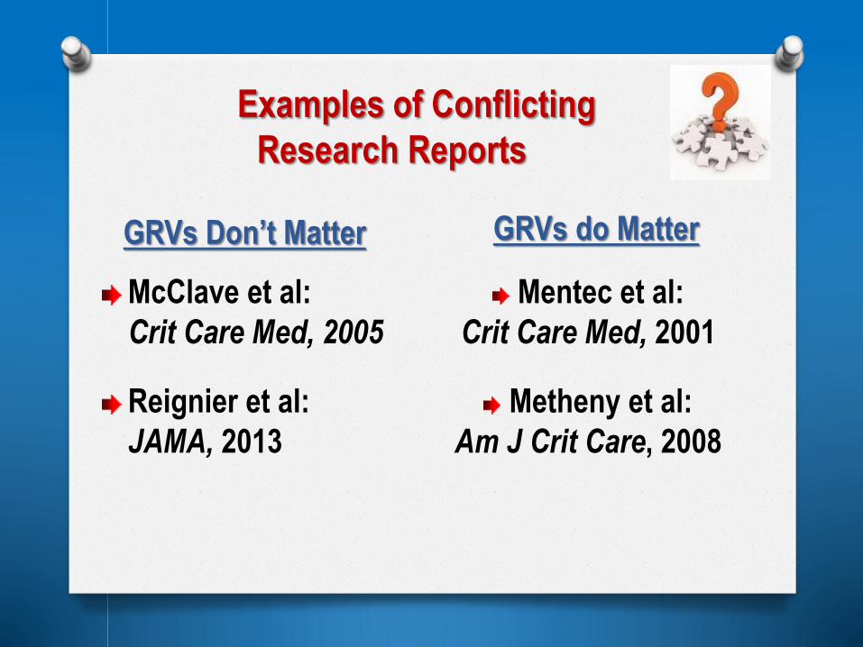
Examples of Conflicting
Research Reports
GRVs Don’t Matter GRVs do Matter
McClave et al:
Crit Care Med, 2005
Reignier et al:
JAMA, 2013
Mentec et al:
Crit Care Med, 2001
Metheny et al:
Am J Crit Care, 2008

McClave et al, Crit Care Med, 2005
Description
Sample 40 ICU patients
Tubes 21 NG (8-12 Fr); 19 gastrostomy tubes
Primary
Outcome
Aspiration: Yellow beads added to formula; tracheal
aspirates viewed via fluoroscopy for yellow
discoloration
Methods
High GRVs categorized from 0 to 400 ml +
Interested in comparing 200 ml and 400 ml levels
Followed patients from 3 days; GRVs every 4 hours
Compared % aspiration according to GRV level (n=587)

(McClave et al, continued)
0
5
10
15
20
25
30
35
40
% A
sp
iratio
n
0-50 51-100 101-150 151-200 201-299 300-399 400+
Range of GRVs
N
Category of GRV Volume
Percent Aspiration per
GRV Category
92% of aspirates ≤ 100 ml
Only 3.9% > 200 ml
Only 1.4% ≥ 400 ml

Reignier et al, JAMA, 2013
Description
Sample Randomized multi-site study (ICUs)
227 patients in intervention group; 222 in control group
Tubes Not described
OutcomesVentilator Associated Pneumonia
Caloric Intake
Methods
Intervention group – no GRV monitoring. Fed at full rate until
vomiting occurred.
Control group – GRVs measured every 6 hours, 250 ml
considered high
Results
No difference in pneumonia rates
Intervention group had higher caloric intake
Intervention group vomited more frequently

(continued)
Comments
Excluded patients who had surgery in the previous month – thus
excluded an important segment of the critically ill population.
While nutritional intake was greater in the ‘not checking GRV’ group,
differences were minimal
Study may have been under-powered to rule out harm to patients
from vomiting.
Clinicians providing care not blinded to group assignment
Investigators did not report how many high GRVs were encountered
in the control group – or how often feedings were interrupted
Not clear that this study is adequate to convince clinicians to follow
the investigators’ recommendation to stop measuring GRVs
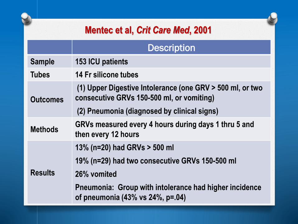
Mentec et al, Crit Care Med, 2001
Description
Sample 153 ICU patients
Tubes 14 Fr silicone tubes
Outcomes
(1) Upper Digestive Intolerance (one GRV > 500 ml, or two
consecutive GRVs 150-500 ml, or vomiting)
(2) Pneumonia (diagnosed by clinical signs)
MethodsGRVs measured every 4 hours during days 1 thru 5 and
then every 12 hours
Results
13% (n=20) had GRVs > 500 ml
19% (n=29) had two consecutive GRVs 150-500 ml
26% vomited
Pneumonia: Group with intolerance had higher incidence
of pneumonia (43% vs 24%, p=.04)

Metheny et al, Am J Crit Care 2008
Sample206 critically ill patients receiving gastric tube feedings
(SICU, MICU, NICU)
Tubes 10 Fr (40%); 14-20 Fr (60%)
Methods
Followed for 3 consecutive days
GRVs measured every 4 hours
Categorized into 3 overlapping GRV groups:(at least 150
mL, at least 200 mL, and at least 250 mL)
Outcome
Aspiration (pepsin-assay)
Categorized into High and Low Aspiration Groups
Compared GRVs to Aspiration Group

Backward regression:
O GRV categories
O Mean Glasgow Coma Score
O Mean sedation score
O Mean HOB elevation
O Mean APACHE II Score
Following categories remained in model:
O 2 or more GRVs of at least 200 ml
O 1 or more GRVs of at least 250 mL
O 2 or more GRVs of at least 250 ml
(Continued)

Relationship between percent aspiration
and frequency of high GRVs
%A
s
p
I
r
a
t
I
o
n
Am J Critical Care, 2008
Recommend
measuring
GRVs
although
imprecise

Canadian Clinical Practice Guidelines
“Use of and Threshold for Gastric Residual Volumes”
March 2013
“Recommendation: There are insufficient data to
make a recommendation for not checking gastric
residual volumes or a specific gastric residual
volume threshold.”
“Based on two level 2 studies, a gastric residual
volume of either 250 or 500 mLs (or somewhere in
between) is acceptable as a strategy to optimize
delivery of enteral nutrition in critically ill patients.”

Preventing Tube Clogging
Compared efficacy of
water, Coca-Cola, and
cranberry juice in keeping
feeding tubes patent
Funded by STTI, 1986 Cranberry juice
usually causes
tubes to clog
Water & Coca-Cola
work equally well
(Nursing Research,
1988)