Embed Size (px)
Citation preview
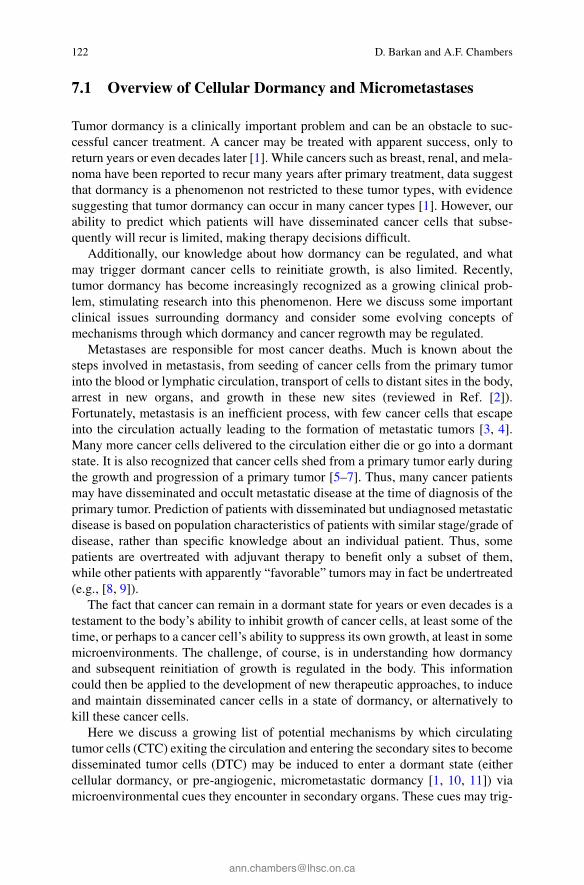
121© Springer Science+Business Media New York 2016 R.J. Cote, R.H. Datar (eds.), Circulating Tumor Cells, Current Cancer Research, DOI 10.1007/978-1-4939-3363-1_7
Chapter 7 Prevention of Conversion of Tumor Dormancy into Proliferative Metastases
Dalit Barkan and Ann F. Chambers
Abstract Late recurrences of cancer are believed to be due to dormant disease that can persist for long periods following apparently successful treatment of a primary tumor. Clinical tumor dormancy thus creates uncertainty for cancer patients and their physicians, who cannot be certain that their cancer will not recur. We have a poor understanding about which individual patients are at risk for cancer recurrence following a period of tumor dormancy. Thus, in spite of the clinical importance of tumor dormancy, much remains to be learned about the mechanisms responsible for induction of, and release from, dormancy. Here we consider the clinical problem of tumor dormancy and discuss evolving ideas of how tumor dormancy and reinitiation of growth may be regulated, both naturally in the body and therapeutically. A better understanding of mechanisms by which dormancy can be regulated may suggest new therapeutic approaches to either eliminate dormant cancer cells or promote the maintenance of dormancy.
Keywords Metastasis • Tumor dormancy • Disseminated tumor cells • Molecular characterization • Tumor microenvironment • Cellular dormancy • Angiogenesis • Immune regulation
D. Barkan , Ph.D. (*) Laboratory of Tumor Dormancy and Metastasis, Department of Biology and Human Biology, Faculty of Natural Sciences , University of Haifa , Mount Carmel , Haifa 31905 , Israel e-mail: [email protected]
A. F. Chambers , Ph.D. Departments of Oncology, Medical Biophysics and Pathology & Laboratory Medicine, London Regional Cancer Program , London Health Sciences Centre/University of Western Ontario , 790 Commissioners Road East , London , ON , N6A 4L6 Canada
122
7.1 Overview of Cellular Dormancy and Micrometastases
Tumor dormancy is a clinically important problem and can be an obstacle to suc-cessful cancer treatment. A cancer may be treated with apparent success, only to return years or even decades later [ 1 ]. While cancers such as breast, renal, and mela-noma have been reported to recur many years after primary treatment, data suggest that dormancy is a phenomenon not restricted to these tumor types, with evidence suggesting that tumor dormancy can occur in many cancer types [ 1 ]. However, our ability to predict which patients will have disseminated cancer cells that subse-quently will recur is limited, making therapy decisions diffi cult.
Additionally, our knowledge about how dormancy can be regulated, and what may trigger dormant cancer cells to reinitiate growth, is also limited. Recently, tumor dormancy has become increasingly recognized as a growing clinical prob-lem, stimulating research into this phenomenon. Here we discuss some important clinical issues surrounding dormancy and consider some evolving concepts of mechanisms through which dormancy and cancer regrowth may be regulated.
Metastases are responsible for most cancer deaths. Much is known about the steps involved in metastasis, from seeding of cancer cells from the primary tumor into the blood or lymphatic circulation, transport of cells to distant sites in the body, arrest in new organs, and growth in these new sites (reviewed in Ref. [ 2 ]). Fortunately, metastasis is an ineffi cient process, with few cancer cells that escape into the circulation actually leading to the formation of metastatic tumors [ 3 , 4 ]. Many more cancer cells delivered to the circulation either die or go into a dormant state. It is also recognized that cancer cells shed from a primary tumor early during the growth and progression of a primary tumor [ 5 – 7 ]. Thus, many cancer patients may have disseminated and occult metastatic disease at the time of diagnosis of the primary tumor. Prediction of patients with disseminated but undiagnosed metastatic disease is based on population characteristics of patients with similar stage/grade of disease, rather than specifi c knowledge about an individual patient. Thus, some patients are overtreated with adjuvant therapy to benefi t only a subset of them, while other patients with apparently “favorable” tumors may in fact be undertreated (e.g., [ 8 , 9 ]).
The fact that cancer can remain in a dormant state for years or even decades is a testament to the body’s ability to inhibit growth of cancer cells, at least some of the time, or perhaps to a cancer cell’s ability to suppress its own growth, at least in some microenvironments. The challenge, of course, is in understanding how dormancy and subsequent reinitiation of growth is regulated in the body. This information could then be applied to the development of new therapeutic approaches, to induce and maintain disseminated cancer cells in a state of dormancy, or alternatively to kill these cancer cells.
Here we discuss a growing list of potential mechanisms by which circulating tumor cells (CTC) exiting the circulation and entering the secondary sites to become disseminated tumor cells (DTC) may be induced to enter a dormant state (either cellular dormancy, or pre-angiogenic, micrometastatic dormancy [ 1 , 10 , 11 ]) via microenvironmental cues they encounter in secondary organs. These cues may trig-
D. Barkan and A.F. Chambers
123
ger the cells to resume active growth after a period of dormancy. An improved understanding of ways by which cancer cells can enter and leave a functional state of dormancy may lead to new opportunities to target therapy directed against dor-mant cancer cells, to either destroy them or to maintain them in a non-growing state.
7.2 Mechanisms Underlying Quiescence and Survival of Dormant Tumor Cells
Metastasis, the spread of tumor cells, is an ineffi cient process where few of the dis-seminated tumor cells will successfully survive their journey. DTCs that survive the hemodynamic forces and the immune surveillance may seed secondary sites, encountering a new microenvironment that will determine their fate [ 1 , 12 , 13 ]. The DTCs may survive, become dormant, or progressively grow to form metastases [ 10 ]. The majority of the DTCs do not survive the initial colonization, whereas those that adapt and survive may persist to reside in a quiescent state (cellular dor-mancy) for many years (reviewed in Refs. [ 1 , 12 , 14 ]). This long term survival and quiescence of the DTCs may account for the latent recurrence years and decades after primary tumor resection and adjuvant therapy [ 15 ].
Three scenarios have been proposed to induce quiescence and survival of DTCs [ 16 ]. These include (1) the tumor microenvironment at the secondary site, (2) the tumor microenvironment at the primary site, and (3) early dissemination of tumor cells. We consider evidence in support of each of these scenarios.
7.2.1 Tumor Microenvironment at the Secondary Site
The idea that the tissue microenvironment at a secondary site may play a role in determining the fate of cancer cells that have spread throughout the body is a con-cept that was put forward over a century ago by Stephen Paget. Paget proposed that metastasis will occur only when the tumor cell (the “seed”) and the microenviron-ment of a given organ (the “soil”) are compatible [ 17 ]. Willis and Hadfi eld further developed this concept [ 18 ]. They coined the term “tumor dormancy” and specifi ed tumor dormancy as a process involving growth restraints exerted by the ectopic tis-sue leading to reversible mitotic arrest (reviewed in Ref. [ 13 ]). Hadfi eld noted that “ When the interval (between surgical excision and appearance of secondary tumors) is prolonged to six years or more it seems impossible to escape the conclusion that the cells of the dormant growth are in a state of temporary mitotic arrest, no matter how long the period may be ” [ 18 ]. Consistent with this concept, it has been demon-strated in experimental models that cancer cells may be seeded throughout the body, where they may remain dormant, only growing in specifi c “favorable” organs (e.g., [ 19 , 20 ]). Hence, a foreign, ectopic microenvironment may promote quiescence (cellular dormancy) of some DTCs.
7 Prevention of Conversion of Tumor Dormancy into Proliferative Metastases
124
Several mechanisms underlying DTC quiescence and long-term survival have recently been proposed. We previously demonstrated potential mechanisms by which the microenvironment may regulate tumor dormancy [ 21 – 23 ]. Solitary tumor dormancy and the transition to proliferation were recapitulated in vitro by utilizing a 3D in vitro culture system constituted from growth factor-reduced basement mem-brane extract (BME), to mimic components of the extracellular matrix (ECM). Our results revealed that in the 3D culture system, cells with dormant behavior in vivo remained cell cycle arrested with elevated nuclear expression of p16 and p27. Our fi ndings that the ECM can impose growth inhibitory signals on tumor cells were in concordance with previous reports [ 24 , 25 ] (Fig. 7.1 ). Interestingly, the dormant tumor cells displayed distinct cytoskeletal organization with evidence of only tran-sient adhesion to the ECM [ 21 ]. However, we demonstrated that the switch from quiescence to proliferative metastatic growth was strongly infl uenced by interac-tions with the ECM as a result of cytoskeletal reorganization and formation of actin stress fi bers. During this transition the tumor cells formed actin stress fi bers via β1 integrin signaling and downstream phosphorylation of myosin light chain by myo-sin light chain kinase [ 21 , 26 ]. These fi ndings are consistent with previous work implicating β1 integrins in microenvironmental regulation of cell behavior [ 27 ] and were subsequently confi rmed by others [ 28 ], emphasizing the important role of the full engagement of the dormant tumor cell with the ECM as a mechanism to escape tumor dormancy [ 21 , 23 ].
These observations are also consistent with previous studies in which downregu-lation of the urokinase receptor was shown to mediate signaling through α5β1 inte-
Primary tumor
Early dissemination
Cellular dormancy phase
Microenvironmentalsignals promotingsurvival and quiescenceof DTC
BMP-7
CXCL12 TRAIL
GAS6
TGFβ-2
BMETSP1BMP4
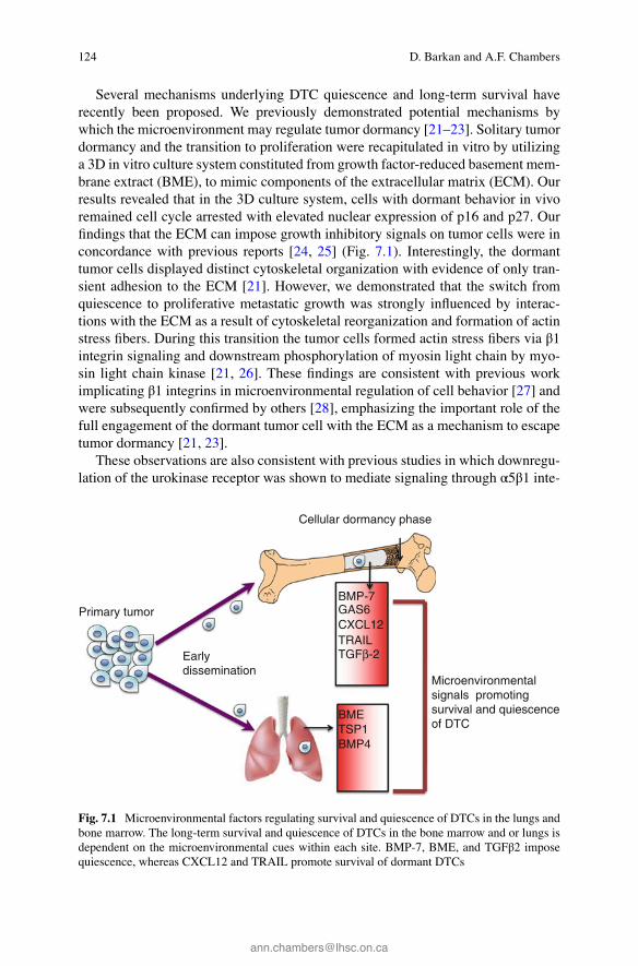
Fig. 7.1 Microenvironmental factors regulating survival and quiescence of DTCs in the lungs and bone marrow. The long-term survival and quiescence of DTCs in the bone marrow and or lungs is dependent on the microenvironmental cues within each site. BMP-7, BME, and TGFβ2 impose quiescence, whereas CXCL12 and TRAIL promote survival of dormant DTCs
D. Barkan and A.F. Chambers
125
grin, forcing the cells into dormancy [ 29 , 30 ]. Furthermore, in transgenic mouse models for mammary or pancreatic beta cell cancer, knockdown of β1 integrin resulted in inhibition of proliferation of the mammary tumor cells and senescence of the pancreatic beta tumor cells [ 31 , 32 ]. Thus, multiple lines of evidence indicate that lack of adhesion of the tumor cell to the ECM via integrins can lead a tumor cell to enter a dormant phase. A solitary dormant tumor cell that fails to properly adhere to the ECM can initiate, under these stress conditions, mechanisms that lead to its long-term survival. Pioneering work by the Aguirre-Ghiso laboratory demonstrated that endoplasmic reticulum (ER) stress signaling pathways contribute to growth arrest and survival programs during tumor cell dormancy. They showed that failure of squamous carcinoma cells (HEp3) to engage with the ECM led to inhibition of ERK1/2 signaling and activation of p38α/β signaling pathways. The reduction in ERK/p38 signaling ratio induced the stress adaptive response known as the unfolded protein response (UPR) [ 33 – 35 ]; and reviewed in Ref. [ 16 ]. These signals lead to an epigenetic reprogramming and induction of quiescence, by activation of RNA- dependent protein kinase–like ER kinase (PERK) [ 33 , 34 ], survival and adaptation of dormant HEp3 (D-HEp3) cells in vivo by activation of ATF6alpha-Rheb-mTOR signaling independent on Akt signaling [ 36 ]. Interestingly, several metastasis sup-pressor genes which selectively inhibit the growth at secondary sites, such as MKK4 and MKK6, are activated by stress signals and are upstream activators of p38 [ 37 ]. The transcription factors BHLHB3/41/Sharp1 and NR2F1 are regulated by p38α/β and are required for dormancy of tumor cells in vivo [ 37 ]. Therefore, the growing family of metastasis suppressor genes, including KISS1, MKK6, BHLHLB3/Sharp- 1, and Nm23-H1 among others, may inhibit the growth of DTC at secondary sites (reviewed in Ref. [ 38 ]), further supporting the notion that the microenviron-mental cues can regulate DTC quiescence.
Indeed, quiescent DTCs are found in the bone marrow (BM) of patients [ 39 ]. Several recent studies have demonstrated how the BM could produce factors that will impose dormancy of their residing DTC (Fig. 7.1 ). Bone morphogenic protein 7 (BMP7) in the BM was shown to trigger dormancy of prostate DTCs by activating p38 signaling, upregulating the metastasis suppressor gene NRDG1, and thus induc-ing reversible growth arrest [ 40 ]. Secretion of growth arrest-specifi c 6 (GAS6) by osteoblasts and tumor cells was shown to induce dormancy of prostate cancer tumor cells [ 41 ]. Recently, Bragado et al. have demonstrated that transforming growth factor-beta2 (TGF-β2) highly expressed in the bone marrow induced ERK/p38 low signaling ratio resulting in induction of quiescence of highly malignant DTCs [ 42 ]. Intriguingly, in addition to growth factors regulating tumor dormancy in the BM, a recent report demonstrated that the transfer of miRNAs from BM stroma to breast cancer cells induced quiescence of the breast cancer cells [ 43 ]. Hence, microenvi-ronmental factors in the BM may defi ne metastasis-restrictive microenvironment activating stress signals in DTC leading to their quiescence (Fig. 7.1 ).
Collectively, DTCs residing at secondary sites can be exposed directly to stress signals upon their failure to properly adhere to the ECM, and or their exposure to factors defi ning restrictive microenvironment. These stress conditions may initiate mechanisms that will promote their quiescence and survival. However, can these mechanisms initiate programs that will ensure quiescent DTC long-term survival?
7 Prevention of Conversion of Tumor Dormancy into Proliferative Metastases
126
Autophagy is a highly regulated self-digestion process that produces nutrients and energy for the cell through the breakdown of cytosolic components, and can lead to long-term cell survival under stress conditions [ 44 ]. Evidence in the literature sug-gests that abrogated adhesion of epithelial cells to the ECM may induce autophagy through growth factor and nutrient sensing pathways, energy-sensing pathways, and integrated stress response [ 45 ]. Thus, restrictive microenvironments and induction of stress signals may trigger autophagy, thereby promoting long-term survival of the quiescent DTC [ 46 ].
In addition to the stress signals generated by microenvironment that may regu-late DTC quiescence and long-term survival, there are additional microenvironmen-tal factors that can promote the survival of DTCs. CXCL12 and TRAIL were shown to induce the survival of disseminated breast tumor cells in bone by upregulating Akt signaling via c-Src [ 47 ]. Similarly we have shown previously that activation of Src and ERK signaling is required for the switch of dormant breast cancer cells to metastatic growth [ 22 ], and combined inhibition of Src and MEK signaling was shown recently to reduce the survival of the dormant tumor cells in the lungs [ 48 ].
Overall the microenvironment at the secondary sites can promote stress regu-lated signals in the DTCs, directly or indirectly, thus determining their fate.
7.2.2 Tumor Microenvironment at the Primary Site
The microenvironment at the primary tumor site may prime the disseminated tumor cells to enter a quiescent state that will be maintained once the cells will colonize the distant site with matching microenvironmental cues. Gene signatures present in the primary tumors have been shown to predict long-term metastatic relapse [ 45 , 49 , 50 ]. Furthermore, gene expression signatures from surrounding histologically nor-mal tissue proximal to the tumor were also shown to predict breast cancer patient survival [ 51 ]. It is possible that these gene signatures may be generated by stress signals present at the primary site such as hypoxia. These stress signals were shown to promote autophagy of the tumor cells, thus promoting the induction of quies-cence and survival signals [ 44 , 52 , 53 ] that may protect tumor cells from pro-grammed cell death induced upon cell detachment from extracellular matrix (anoikis) [ 45 ]. Hence, one can envision that a subset of cells in a primary tumor that disseminate from a hypoxic microenvironment may already be in a dormant state. These cells may be already primed with survival mechanisms such as autophagy and or gene expression patterns that may be enable their successful seeding of dis-tant sites and their continued survival in a quiescent state.
7.2.3 Early Dissemination of Tumor Cells
Early-disseminated tumor cells may not possess the genetic input required to initi-ate growth at secondary sites [ 6 , 54 , 55 ]. Therefore, tumor cells that disseminate early from the primary site may be an additional instigator of DTC dormancy. There
D. Barkan and A.F. Chambers
127
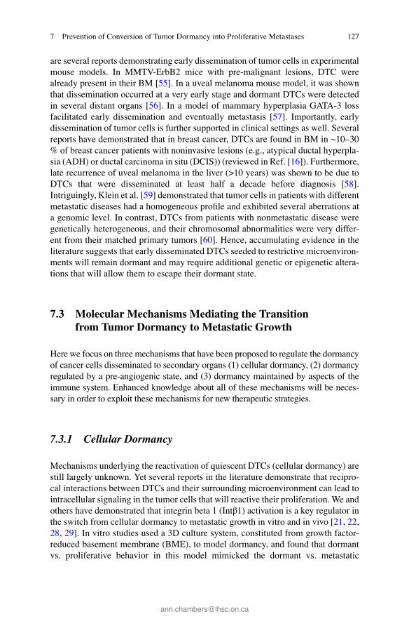
are several reports demonstrating early dissemination of tumor cells in experimental mouse models. In MMTV-ErbB2 mice with pre-malignant lesions, DTC were already present in their BM [ 55 ]. In a uveal melanoma mouse model, it was shown that dissemination occurred at a very early stage and dormant DTCs were detected in several distant organs [ 56 ]. In a model of mammary hyperplasia GATA-3 loss facilitated early dissemination and eventually metastasis [ 57 ]. Importantly, early dissemination of tumor cells is further supported in clinical settings as well. Several reports have demonstrated that in breast cancer, DTCs are found in BM in ~10–30 % of breast cancer patients with noninvasive lesions (e.g., atypical ductal hyperpla-sia (ADH) or ductal carcinoma in situ (DCIS)) (reviewed in Ref. [ 16 ]). Furthermore, late recurrence of uveal melanoma in the liver (>10 years) was shown to be due to DTCs that were disseminated at least half a decade before diagnosis [ 58 ]. Intriguingly, Klein et al. [ 59 ] demonstrated that tumor cells in patients with different metastatic diseases had a homogeneous profi le and exhibited several aberrations at a genomic level. In contrast, DTCs from patients with nonmetastatic disease were genetically heterogeneous, and their chromosomal abnormalities were very differ-ent from their matched primary tumors [ 60 ]. Hence, accumulating evidence in the literature suggests that early disseminated DTCs seeded to restrictive microenviron-ments will remain dormant and may require additional genetic or epigenetic altera-tions that will allow them to escape their dormant state.
7.3 Molecular Mechanisms Mediating the Transition from Tumor Dormancy to Metastatic Growth
Here we focus on three mechanisms that have been proposed to regulate the dormancy of cancer cells disseminated to secondary organs (1) cellular dormancy, (2) dormancy regulated by a pre-angiogenic state, and (3) dormancy maintained by aspects of the immune system. Enhanced knowledge about all of these mechanisms will be neces-sary in order to exploit these mechanisms for new therapeutic strategies.
7.3.1 Cellular Dormancy
Mechanisms underlying the reactivation of quiescent DTCs ( cellular dormancy ) are still largely unknown. Yet several reports in the literature demonstrate that recipro-cal interactions between DTCs and their surrounding microenvironment can lead to intracellular signaling in the tumor cells that will reactive their proliferation. We and others have demonstrated that integrin beta 1 (Intβ1) activation is a key regulator in the switch from cellular dormancy to metastatic growth in vitro and in vivo [ 21 , 22 , 28 , 29 ]. In vitro studies used a 3D culture system, constituted from growth factor-reduced basement membrane (BME), to model dormancy, and found that dormant vs. proliferative behavior in this model mimicked the dormant vs. metastatic
7 Prevention of Conversion of Tumor Dormancy into Proliferative Metastases
128
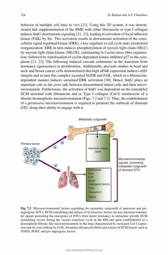
behavior of multiple cell lines in vivo [ 21 ]. Using this 3D system, it was demon-strated that supplementation of the BME with either fi bronectin or type I collagen induces Intβ1 downstream signaling [ 21 , 22 ], leading to activation of focal adhesion kinase (FAK) by Src. This activation results in downstream activation of the extra-cellular signal regulated kinase (ERK), a key regulator in cell cycle and cytoskeletal reorganization. ERK in turn induces phosphorylation of myosin light chain (MLC) by myosin light chain kinase (MLCK), culminating in f-actin stress fi ber organiza-tion, followed by translocation of cyclin-dependent kinase inhibitor p27 to the cyto-plasm [ 21 , 22 ]. The following induced cascade culminates in the transition from dormancy (quiescence) to proliferation. Additionally, previous studies in head and neck and breast cancer cells demonstrated that high uPAR expression induces α5β1 integrin and in turn this complex recruited EGFR and FAK, which in a fi bronectin-dependent manner induces sustained ERK activation [ 30 ]. Hence, Intβ1 plays an important role in the cross talk between disseminated tumor cells and their micro-environment. Furthermore, the activation of Intβ1 was dependent on the remodeled ECM enriched with fi bronectin and or Type I collagen (Col-I) reminiscent of a fi brotic/desmoplastic microenvironment (Figs. 7.2 and 7.3 ). Thus, the establishment of a permissive microenvironment is required to promote the outbreak of dormant DTC along their ability to engage with it.
Primary tumor
Metastatic outgrowth
Microenvironmental signals promoting metastatic outgrowth of dormant DTC Fibronectin
Col-I LOX
POST Angiogenic factors
Coco
Tenascin-C
osteolytic viciouscycle
TGFβ1
Fig. 7.2 Microenvironmental factors regulating the metastatic outgrowth of quiescent and pre- angiogenic DTCs. ECM remodeling and release of its bioactive factors are key microenvironmen-tal signals promoting the emergence of DTCs from tumor dormancy to metastatic growth. ECM remodeling occurs during the vicious osteolytic cycle in the BM and upon establishment of a desmoplastic/fi brotic like microenvironment in the lung characterized by increased Col-I expres-sion and its cross linking by LOX, formation fi bronectin fi brils and release of ECM factors such as TGFβ1, POST, and pro-angiogenic factors
D. Barkan and A.F. Chambers
129
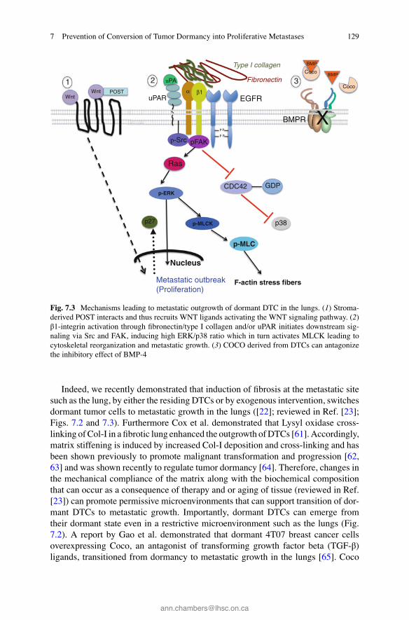
Indeed, we recently demonstrated that induction of fi brosis at the metastatic site such as the lung, by either the residing DTCs or by exogenous intervention, switches dormant tumor cells to metastatic growth in the lungs ([ 22 ]; reviewed in Ref. [ 23 ]; Figs. 7.2 and 7.3 ). Furthermore Cox et al. demonstrated that Lysyl oxidase cross- linking of Col-I in a fi brotic lung enhanced the outgrowth of DTCs [ 61 ]. Accordingly, matrix stiffening is induced by increased Col-I deposition and cross-linking and has been shown previously to promote malignant transformation and progression [ 62 , 63 ] and was shown recently to regulate tumor dormancy [ 64 ]. Therefore, changes in the mechanical compliance of the matrix along with the biochemical composition that can occur as a consequence of therapy and or aging of tissue (reviewed in Ref. [ 23 ]) can promote permissive microenvironments that can support transition of dor-mant DTCs to metastatic growth. Importantly, dormant DTCs can emerge from their dormant state even in a restrictive microenvironment such as the lungs (Fig. 7.2 ). A report by Gao et al. demonstrated that dormant 4T07 breast cancer cells overexpressing Coco, an antagonist of transforming growth factor beta (TGF-β) ligands, transitioned from dormancy to metastatic growth in the lungs [ 65 ]. Coco
p-MLCK
p-MLC
Fibronectin
Type I collagen
α β1
uPA
uPAR
PPPP
p-Src pFAK
p-ERK
Ras
F-actin stress fibersMetastatic outbreak(Proliferation)
p38
CDC42
EGFR
GDP
Nucleus
p27
X
Coco
BMP
Coco BMP
Wnt POSTWnt
BMPR
1 32
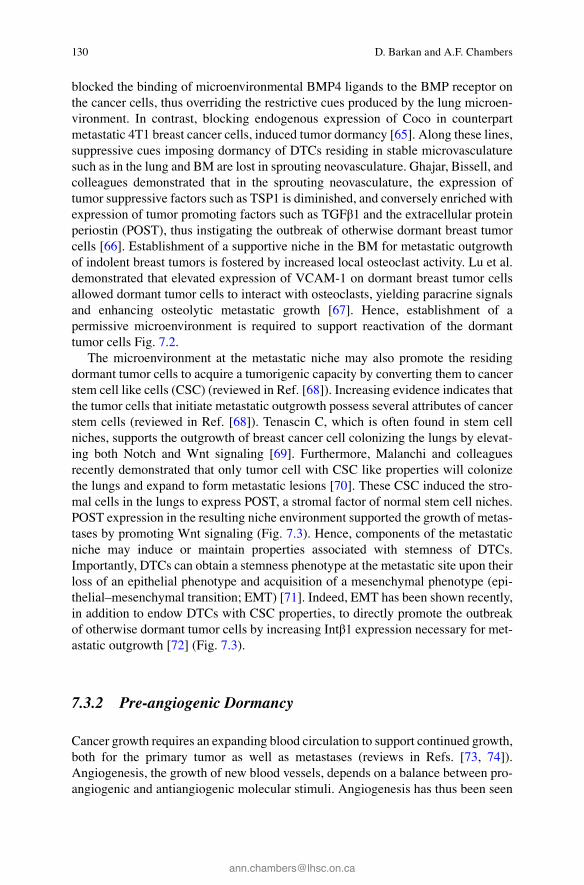
Fig. 7.3 Mechanisms leading to metastatic outgrowth of dormant DTC in the lungs. ( 1 ) Stroma- derived POST interacts and thus recruits WNT ligands activating the WNT signaling pathway. ( 2 ) β1-integrin activation through fi bronectin/type I collagen and/or uPAR initiates downstream sig-naling via Src and FAK, inducing high ERK/p38 ratio which in turn activates MLCK leading to cytoskeletal reorganization and metastatic growth. ( 3 ) COCO derived from DTCs can antagonize the inhibitory effect of BMP-4
7 Prevention of Conversion of Tumor Dormancy into Proliferative Metastases
130
blocked the binding of microenvironmental BMP4 ligands to the BMP receptor on the cancer cells, thus overriding the restrictive cues produced by the lung microen-vironment. In contrast, blocking endogenous expression of Coco in counterpart metastatic 4T1 breast cancer cells, induced tumor dormancy [ 65 ]. Along these lines, suppressive cues imposing dormancy of DTCs residing in stable microvasculature such as in the lung and BM are lost in sprouting neovasculature. Ghajar, Bissell, and colleagues demonstrated that in the sprouting neovasculature, the expression of tumor suppressive factors such as TSP1 is diminished, and conversely enriched with expression of tumor promoting factors such as TGFβ1 and the extracellular protein periostin (POST), thus instigating the outbreak of otherwise dormant breast tumor cells [ 66 ]. Establishment of a supportive niche in the BM for metastatic outgrowth of indolent breast tumors is fostered by increased local osteoclast activity. Lu et al. demonstrated that elevated expression of VCAM-1 on dormant breast tumor cells allowed dormant tumor cells to interact with osteoclasts, yielding paracrine signals and enhancing osteolytic metastatic growth [ 67 ]. Hence, establishment of a permissive microenvironment is required to support reactivation of the dormant tumor cells Fig. 7.2 .
The microenvironment at the metastatic niche may also promote the residing dormant tumor cells to acquire a tumorigenic capacity by converting them to cancer stem cell like cells (CSC) (reviewed in Ref. [ 68 ]). Increasing evidence indicates that the tumor cells that initiate metastatic outgrowth possess several attributes of cancer stem cells (reviewed in Ref. [ 68 ]). Tenascin C, which is often found in stem cell niches, supports the outgrowth of breast cancer cell colonizing the lungs by elevat-ing both Notch and Wnt signaling [ 69 ]. Furthermore, Malanchi and colleagues recently demonstrated that only tumor cell with CSC like properties will colonize the lungs and expand to form metastatic lesions [ 70 ]. These CSC induced the stro-mal cells in the lungs to express POST, a stromal factor of normal stem cell niches. POST expression in the resulting niche environment supported the growth of metas-tases by promoting Wnt signaling (Fig. 7.3 ). Hence, components of the metastatic niche may induce or maintain properties associated with stemness of DTCs. Importantly, DTCs can obtain a stemness phenotype at the metastatic site upon their loss of an epithelial phenotype and acquisition of a mesenchymal phenotype (epi-thelial–mesenchymal transition; EMT) [ 71 ]. Indeed, EMT has been shown recently, in addition to endow DTCs with CSC properties, to directly promote the outbreak of otherwise dormant tumor cells by increasing Intβ1 expression necessary for met-astatic outgrowth [ 72 ] (Fig. 7.3 ).
7.3.2 Pre-angiogenic Dormancy
Cancer growth requires an expanding blood circulation to support continued growth, both for the primary tumor as well as metastases (reviews in Refs. [ 73 , 74 ]). Angiogenesis , the growth of new blood vessels, depends on a balance between pro- angiogenic and antiangiogenic molecular stimuli. Angiogenesis has thus been seen
D. Barkan and A.F. Chambers
131
as a target for anticancer therapy, and the complexities of this approach are well recognized [ 75 ]. Angiogenesis has been shown to play a role in regulating cancer growth and dormancy. Folkman and colleagues documented that antiangiogenic factors secreted by a primary tumor could restrict distant micrometastatic growth, holding the metastases in an “active” state of functional dormancy in which cell division and apoptosis were balanced, with no net increase in metastatic tumor size [ 11 , 76 ]. Tumors in a state of pre-angiogenic dormancy thus are distinct from quies-cent, dormant tumor cells, and consequently may present a distinct therapeutic tar-get [ 10 ]. Antiangiogenic therapies thus have the potential to inhibit tumor growth (at the primary or metastatic sites), and also to maintain pre-angiogenic microme-tastases in a functionally dormant, non-expanding state.
Recent work from Naumov and colleagues have shown, in mouse models of primary tumor growth, that the angiogenic phenotype may be plastic and regulat-able, raising hopes for development of agents that could revert vascularized metas-tases to a pre-angiogenic, non-growing state [ 77 ]. Along these lines, Almog and colleagues recently identifi ed a set of 19 small noncoding RNA molecules (miR-NAs) that control the phenotypic switch of human dormant breast carcinoma, glio-blastoma, osteosarcoma, and liposarcoma tumors to exponential growth [ 78 ]. Downregulation of 16 of the highly expressed miRNAs correlated with the switch of dormant tumor to the fast-growing angiogenic tumor. Moreover, reconstitution of miR-580, 588, or 190 promoted prolonged tumor dormancy of otherwise actively proliferation angiogenic tumors. Hence, metastasis may potentially be maintained long-term in a pre-angiogenic dormant state by antiangiogenic therapy as was dem-onstrated previously [ 79 ] and as was predicted recently by the mathematical model-ing by Benzekry et al. [ 80 ].
7.3.3 Dormancy Regulated by the Innate and Adaptive Immune System
Micrometastatic dormancy is characterized by active equilibrium between prolifer-ation and apoptosis. This equilibrium was suggested to be regulated by immune surveillance in addition to the angiogenic switch [ 81 ]. In a mouse model of mela-noma, the outgrowth of early DTCs at distant sites was controlled partially by CD8+ T cells. CD8+ T cells inhibited the growth of disseminated tumor cells, surprisingly, not by cytotoxic effects, but through cytostatic effects and their depletion led in turn to the emergence of DTCs from their dormant state [ 56 ]. Accordingly, recent reports demonstrated the role of T-lymphocytes as regulator of tumor dormancy [ 82 ] and active suppression of T cells by IFN-γ or IL-12 blocking induces escape from dor-mancy (reviewed in Ref. [ 83 ]).
In the DA1-3b mouse model of acute myeloid leukemia, dormant tumor cells were resistant to cytotoxic lymphocytes (CTL) by overexpressing B7-H1 and B7.1. B7-H1 interacts with programmed death-1 (PD-1) expressed on T cells, and inhibits T-cell activation and CTL-mediated lysis [ 84 ]. Hence, dormant tumor cells may become more resistant to specifi c CTL mediated killing. Indeed, recent reports have
7 Prevention of Conversion of Tumor Dormancy into Proliferative Metastases
132
demonstrated that PDL-1 (the ligand of PD-1) was upregulated in irradiated tissue. In contrast, administration of anti-PD-L1 enhanced the effi cacy of ionizing irradia-tion (IR) through a CTL-dependent mechanisms leading to antitumor immunity in mice [ 85 , 86 ]. Along these lines, methylation of suppressor of cytokine signaling (SOCS1) and its downregulation in dormant tumor cells was reported to deregulate JAK/STAT pathways within the dormant tumor cells, thus promoting resistance to CTL-mediated killing [ 87 ]. Hence, inhibition of T-lymphocytes and preventing the resistance of the dormant tumor cells to CTL mediated killing may be the mecha-nisms accounting for the escape of the dormant tumor cells from the immune response.
Overall, the studies described above in several animal models of tumor dormancy support the potential role of the immune system in keeping the micrometastases indolent for prolonged periods of time. However, controversies exist regarding the role of the immune system in regulating tumor dormancy in the clinical settings (reviewed in Ref. [ 88 ]). Furthermore, a recent report by Magnus et al. adds another complexity to the role of the immune system in regulating tumor dormancy [ 89 ]. They demonstrated that expression of Tissue Factor in indolent human glioma cells led to a stepwise transition of dormant tumor cells to metastatic outgrowth, a pro-cess that was preceded by recruitment of vascular (CD105+) and myeloid (CD11b+ and F4/80+) cells, thus demonstrating that the immune system might actually aug-ment an escape of tumor cells from dormancy.
7.4 Conclusions of Metastatic Tumor Dormancy as a Clinical Target
Metastasis continues to be responsible for the majority of cancer deaths, in spite of our enhanced understanding of tumor biology. When cancer is detected early, before it has spread, it is more likely to be successfully treated, while metastatic disease is considerably more diffi cult to treat. Compounding this diffi culty is the ability of an apparently successfully treated cancer to recur, sometimes years or decades later, following a protracted period of tumor dormancy. Here we consider some of the clinical and biological issues about tumor dormancy, and our relatively limited understanding of how dormancy may be regulated.
An increase in recent years in studies on mechanisms contributing to regulation of tumor dormancy is providing a growing wealth of information about dormancy. Clearly, tumor dormancy is a complex and multifaceted problem, and we have much to learn about how dormancy arises and persists, as well as how cancer cells can be released from dormancy and reinitiate growth. The fact that cancer can be naturally maintained in a state of dormancy gives hope that these processes can be studied and utilized in future therapies. However, the complexity of factors that contribute to dormancy and release from dormancy will make this approach challenging. Here we outline some of the factors that have been identifi ed as contributers to tumor
D. Barkan and A.F. Chambers
133
dormancy, and thus suggesting ways to either maintain cancer in a dormant state or kill dormant cancer cells. It is clear that many aspects of the tissue microenviron-ment surrounding dormant metastatic disease contribute to the dormant phenotype. Potential therapeutic approaches to prevent dormant cancer cells from reinitiating growth include blocking microenvironmental signals that promote tumor growth, inhibiting angiogenic stimulation of micrometastatic growth, and enhancing immune regulation of dormancy. We have much to learn about dormancy and its regulation, but models are becoming increasingly available for experimental study. Additionally, there is a growing recognition that we need to learn much more about dormancy in patients. Which patients harbor dormant cells, and which patients can be considered cured of their disease? In patients who do have persistent cancer cells, what factors—either inherent to the tumor cell or modifi able factors in the patient—contribute to maintenance of dormancy vs. reinitiation of tumor growth? In order to address the clinical problem of tumor dormancy, we need continued and enhanced experimental efforts to understand the biology of tumor dormancy, coupled with increased understanding of the clinical status of disseminated disease in patients. This enhanced knowledge is crucial to improve the survival of cancer patients.
References
1. Goss PE, Chambers AF (2010) Does tumour dormancy offer a therapeutic target? Nat Rev Cancer 10(12):871–877
2. Chambers AF, Naumov GN, Varghese HJ, Nadkarni KV, MacDonald IC, Groom AC (2001) Critical steps in hematogenous metastasis an overview. Surg Oncol Clin N Am 10(2):243–255, vii
3. Weiss L (1990) Metastatic ineffi ciency. Adv Cancer Res 54:159–211 4. Tarin D, Vass AC, Kettlewell MG, Price JE (1984) Absence of metastatic sequelae during
long-term treatment of malignant ascites by peritoneo-venous shunting. A clinico-pathological report. Invasion Metastasis 4(1):1–12
5. Klein CA (2003) The systemic progression of human cancer a focus on the individual dissemi-nated cancer cell—the unit of selection. Adv Cancer Res 89:35–67
6. Klein CA (2009) Parallel progression of primary tumours and metastases. Nat Rev Cancer 9(4):302–312
7. Oskarsson T, Batlle E, Massague J (2014) Metastatic stem cells sources, niches, and vital pathways. Cell Stem Cell 14(3):306–321
8. Gnant M, Steger GG (2009) Fighting overtreatment in adjuvant breast cancer therapy. Lancet 374(9707):2029–2030
9. Albain KS, Barlow WE, Ravdin PM, Farrar WB, Burton GV, Ketchel SJ, Cobau CD, Levine EG, Ingle JN, Pritchard KI, Lichter AS, Schneider DJ, Abeloff MD, Henderson IC, Muss HB, Green SJ, Lew D, Livingston RB, Martino S, Osborne CK (2009) Adjuvant chemotherapy and timing of tamoxifen in postmenopausal patients with endocrine-responsive, node-positive breast cancer a phase 3, open-label, randomised controlled trial. Lancet 374(9707):2055–2063
10. Chambers AF, Groom AC, MacDonald IC (2002) Dissemination and growth of cancer cells in metastatic sites. Nat Rev Cancer 2(8):563–572
11. Holmgren L, O’Reilly MS, Folkman J (1995) Dormancy of micrometastases balanced prolif-eration and apoptosis in the presence of angiogenesis suppression. Nat Med 1(2):149–153
7 Prevention of Conversion of Tumor Dormancy into Proliferative Metastases
134
12. Aguirre-Ghiso JA (2007) Models, mechanisms and clinical evidence for cancer dormancy. Nat Rev Cancer 7(11):834–846
13. Klein CA (2011) Framework models of tumor dormancy from patient-derived observations. Curr Opin Genet Dev 21(1):42–49
14. Wells A, Griffi th L, Wells JZ, Taylor DP (2013) The dormancy dilemma quiescence versus balanced proliferation. Cancer Res 73(13):3811–3816
15. Wikman H, Vessella R, Pantel K (2008) Cancer micrometastasis and tumour dormancy. Acta Pathol Microbiol Immunol Scand 116(7–8):754–770
16. Bragado P, Sosa MS, Keely P, Condeelis J, Aguirre-Ghiso JA (2012) Microenvironments dic-tating tumor cell dormancy. Recent Results Cancer Res 195:25–39
17. Paget S (1989) The distribution of secondary growths in cancer of the breast. 1889. Cancer Metastasis Rev 8(2):98–101
18. Hadfi eld G (1954) The dormant cancer cell. Br Med J 2(4888):607–610 19. Suzuki M, Mose ES, Montel V, Tarin D (2006) Dormant cancer cells retrieved from metastasis-
free organs regain tumorigenic and metastatic potency. Am J Pathol 169(2):673–681 20. Nguyen DX, Bos PD, Massague J (2009) Metastasis from dissemination to organ-specifi c
colonization. Nat Rev Cancer 9(4):274–284 21. Barkan D, Kleinman H, Simmons JL, Asmussen H, Kamaraju AK, Hoenorhoff MJ, Liu ZY,
Costes SV, Cho EH, Lockett S, Khanna C, Chambers AF, Green JE (2008) Inhibition of meta-static outgrowth from single dormant tumor cells by targeting the cytoskeleton. Cancer Res 68(15):6241–6250
22. Barkan D, El Touny LH, Michalowski AM, Smith JA, Chu I, Davis AS, Webster JD, Hoover S, Simpson RM, Gauldie J, Green JE (2010) Metastatic growth from dormant cells induced by a col-I-enriched fi brotic environment. Cancer Res 70(14):5706–5716
23. Barkan D, Green JE, Chambers AF (2010) Extracellular matrix a gatekeeper in the transition from dormancy to metastatic growth. Eur J Cancer 46(7):1181–1188
24. Henriet P, Zhong ZD, Brooks PC, Weinberg KI, DeClerck YA (2000) Contact with fi brillar collagen inhibits melanoma cell proliferation by up-regulating p27KIP1. Proc Natl Acad Sci U S A 97(18):10026–10031
25. Roth JM, Akalu A, Zelmanovich A, Policarpio D, Ng B, MacDonald S, Formenti S, Liebes L, Brooks PC (2005) Recombinant alpha2(IV)NC1 domain inhibits tumor cell-extracellular matrix interactions, induces cellular senescence, and inhibits tumor growth in vivo. Am J Pathol 166(3):901–911
26. Barkan D, Chambers AF (2011) β1-integrin a potential therapeutic target in the battle against cancer recurrence. Clin Cancer Res 17(23):7219–7223
27. Roskelley CD, Desprez PY, Bissell MJ (1994) Extracellular matrix-dependent tissue-specifi c gene expression in mammary epithelial cells requires both physical and biochemical signal transduction. Proc Natl Acad Sci U S A 91(26):12378–12382
28. Shibue T, Weinberg RA (2009) Integrin beta1-focal adhesion kinase signaling directs the pro-liferation of metastatic cancer cells disseminated in the lungs. Proc Natl Acad Sci U S A 106(25):10290–10295
29. Aguirre Ghiso JA, Kovalski K, Ossowski L (1999) Tumor dormancy induced by downregula-tion of urokinase receptor in human carcinoma involves integrin and MAPK signaling. J Cell Biol 147(1):89–104
30. Aguirre-Ghiso JA, Liu D, Mignatti A, Kovalski K, Ossowski L (2001) Urokinase receptor and fi bronectin regulate the ERK(MAPK) to p38(MAPK) activity ratios that determine carcinoma cell proliferation or dormancy in vivo. Mol Biol Cell 12(4):863–879
31. White DE, Kurpios NA, Zuo D, Hassell JA, Blaess S, Mueller U, Muller WJ (2004) Targeted disruption of beta1-integrin in a transgenic mouse model of human breast cancer reveals an essential role in mammary tumor induction. Cancer Cell 6(2):159–170
32. Kren A, Baeriswyl V, Lehembre F, Wunderlin C, Strittmatter K, Antoniadis H, Fassler R, Cavallaro U, Christofori G (2007) Increased tumor cell dissemination and cellular senescence in the absence of beta1-integrin function. EMBO J 26(12):2832–2842
D. Barkan and A.F. Chambers
135
33. Ranganathan AC, Zhang L, Adam AP, Aguirre-Ghiso JA (2006) Functional coupling of p38- induced up-regulation of BiP and activation of RNA-dependent protein kinase-like endoplas-mic reticulum kinase to drug resistance of dormant carcinoma cells. Cancer Res 66(3):1702–1711
34. Ranganathan AC, Ojha S, Kourtidis A, Conklin DS, Aguirre-Ghiso JA (2008) Dual function of pancreatic endoplasmic reticulum kinase in tumor cell growth arrest and survival. Cancer Res 68(9):3260–3268
35. Adam AP, George A, Schewe D, Bragado P, Iglesias BV, Ranganathan AC, Kourtidis A, Conklin DS, Aguirre-Ghiso JA (2009) Computational identifi cation of a p38SAPK-regulated transcription factor network required for tumor cell quiescence. Cancer Res 69(14):5664–5672
36. Schewe DM, Aguirre-Ghiso JA (2008) ATF6alpha-Rheb-mTOR signaling promotes survival of dormant tumor cells in vivo. Proc Natl Acad Sci U S A 105(30):10519–10524
37. Taylor J, Hickson J, Lotan T, Yamada DS, Rinker-Schaeffer C (2008) Using metastasis sup-pressor proteins to dissect interactions among cancer cells and their microenvironment. Cancer Metastasis Rev 27(1):67–73
38. Hedley BD, Allan AL, Chambers AF (2006) Tumor dormancy and the role of metastasis sup-pressor genes in regulating ectopic growth. Future Oncol 2(5):627–641
39. Alix-Panabieres C, Riethdorf S, Pantel K (2008) Circulating tumor cells and bone marrow micrometastasis. Clin Cancer Res 14(16):5013–5021
40. Kobayashi A, Okuda H, Xing F, Pandey PR, Watabe M, Hirota S, Pai SK, Liu W, Fukuda K, Chambers C, Wilber A, Watabe K (2011) Bone morphogenetic protein 7 in dormancy and metastasis of prostate cancer stem-like cells in bone. J Exp Med 208(13):2641–2655
41. Shiozawa Y, Pedersen EA, Patel LR, Ziegler AM, Havens AM, Jung Y, Wang J, Zalucha S, Loberg RD, Pienta KJ, Taichman RS (2010) GAS6/AXL axis regulates prostate cancer inva-sion, proliferation, and survival in the bone marrow niche. Neoplasia 12(2):116–127
42. Bragado P, Estrada Y, Parikh F, Krause S, Capobianco C, Farina HG, Schewe DM, Aguirre- Ghiso JA (2013) TGF-beta2 dictates disseminated tumour cell fate in target organs through TGF-beta-RIII and p38alpha/beta signalling. Nat Cell Biol 15(11):1351–1361
43. Lim PK, Bliss SA, Patel SA, Taborga M, Dave MA, Gregory LA, Greco SJ, Bryan M, Patel PS, Rameshwar P (2011) Gap junction-mediated import of microRNA from bone marrow stromal cells can elicit cell cycle quiescence in breast cancer cells. Cancer Res 71(5):1550–1560
44. Gewirtz DA (2009) Autophagy, senescence and tumor dormancy in cancer therapy. Autophagy 5(8):1232–1234
45. Lock R, Debnath J (2008) Extracellular matrix regulation of autophagy. Curr Opin Cell Biol 20(5):583–588
46. Sosa MS, Bragado P, Debnath J, Aguirre-Ghiso JA (2013) Regulation of tumor cell dormancy by tissue microenvironments and autophagy. Adv Exp Med Biol 734:73–89
47. Zhang XH, Wang Q, Gerald W, Hudis CA, Norton L, Smid M, Foekens JA, Massague J (2009) Latent bone metastasis in breast cancer tied to Src-dependent survival signals. Cancer Cell 16(1):67–78
48. El Touny LH, Vieira A, Mendoza A, Khanna C, Hoenerhoff MJ, Green JE (2014) Combined SFK/MEK inhibition prevents metastatic outgrowth of dormant tumor cells. J Clin Invest 124(1):156–168
49. Villanueva A, Hoshida Y, Toffanin S, Lachenmayer A, Alsinet C, Savic R, Cornella H, Llovet JM (2010) New strategies in hepatocellular carcinoma genomic prognostic markers. Clin Cancer Res 16(19):4688–4694
50. Kim RS, Avivar-Valderas A, Estrada Y, Bragado P, Sosa MS, Aguirre-Ghiso JA, Segall JE (2012) Dormancy signatures and metastasis in estrogen receptor positive and negative breast cancer. PLoS One 7(4):e35569
51. Troester MA, Lee MH, Carter M, Fan C, Cowan DW, Perez ER, Pirone JR, Perou CM, Jerry DJ, Schneider SS (2009) Activation of host wound responses in breast cancer microenviron-ment. Clin Cancer Res 15(22):7020–7028
7 Prevention of Conversion of Tumor Dormancy into Proliferative Metastases
136
52. Levine B, Kroemer G (2008) Autophagy in the pathogenesis of disease. Cell 132(1):27–42 53. Lu Z, Luo RZ, Lu Y, Zhang X, Yu Q, Khare S, Kondo S, Kondo Y, Yu Y, Mills GB, Liao WS,
Bast RC Jr (2008) The tumor suppressor gene ARHI regulates autophagy and tumor dormancy in human ovarian cancer cells. J Clin Invest 118(12):3917–3929
54. Pantel K, Brakenhoff RH (2004) Dissecting the metastatic cascade. Nat Rev Cancer 4(6):448–456
55. Husemann Y, Geigl JB, Schubert F, Musiani P, Meyer M, Burghart E, Forni G, Eils R, Fehm T, Riethmuller G, Klein CA (2008) Systemic spread is an early step in breast cancer. Cancer Cell 13(1):58–68
56. Eyles J, Puaux AL, Wang X, Toh B, Prakash C, Hong M, Tan TG, Zheng L, Ong LC, Jin Y, Kato M, Prevost-Blondel A, Chow P, Yang H, Abastado JP (2010) Tumor cells disseminate early, but immunosurveillance limits metastatic outgrowth, in a mouse model of melanoma. J Clin Invest 120(6):2030–2039
57. Kouros-Mehr H, Bechis SK, Slorach EM, Littlepage LE, Egeblad M, Ewald AJ, Pai SY, Ho IC, Werb Z (2008) GATA-3 links tumor differentiation and dissemination in a luminal breast can-cer model. Cancer Cell 13(2):141–152
58. Ossowski L, Aguirre-Ghiso JA (2010) Dormancy of metastatic melanoma. Pigment Cell Melanoma Res 23(1):41–56
59. Klein CA, Schmidt-Kittler O, Schardt JA, Pantel K, Speicher MR, Riethmuller G (1999) Comparative genomic hybridization, loss of heterozygosity, and DNA sequence analysis of single cells. Proc Natl Acad Sci U S A 96(8):4494–4499
60. Klein CA, Blankenstein TJ, Schmidt-Kittler O, Petronio M, Polzer B, Stoecklein NH, Riethmuller G (2002) Genetic heterogeneity of single disseminated tumour cells in minimal residual cancer. Lancet 360(9334):683–689
61. Cox TR, Bird D, Baker AM, Barker HE, Ho MW, Lang G, Erler JT (2013) LOX-mediated collagen crosslinking is responsible for fi brosis-enhanced metastasis. Cancer Res 73(6):1721–1732
62. Paszek MJ, Zahir N, Johnson KR, Lakins JN, Rozenberg GI, Gefen A, Reinhart-King CA, Margulies SS, Dembo M, Boettiger D, Hammer DA, Weaver VM (2005) Tensional homeosta-sis and the malignant phenotype. Cancer Cell 8(3):241–254
63. Levental KR, Yu H, Kass L, Lakins JN, Egeblad M, Erler JT, Fong SF, Csiszar K, Giaccia A, Weninger W, Yamauchi M, Gasser DL, Weaver VM (2009) Matrix crosslinking forces tumor progression by enhancing integrin signaling. Cell 139(5):891–906
64. Schrader J, Gordon-Walker TT, Aucott RL, van Deemter M, Quaas A, Walsh S, Benten D, Forbes SJ, Wells RG, Iredale JP (2011) Matrix stiffness modulates proliferation, chemothera-peutic response, and dormancy in hepatocellular carcinoma cells. Hepatology 53(4):1192–1205
65. Gao H, Chakraborty G, Lee-Lim AP, Mo Q, Decker M, Vonica A, Shen R, Brogi E, Brivanlou AH, Giancotti FG (2012) The BMP inhibitor Coco reactivates breast cancer cells at lung meta-static sites. Cell 150(4):764–779
66. Ghajar CM, Peinado H, Mori H, Matei IR, Evason KJ, Brazier H, Almeida D, Koller A, Hajjar KA, Stainier DY, Chen EI, Lyden D, Bissell MJ (2013) The perivascular niche regulates breast tumour dormancy. Nat Cell Biol 15(7):807–817
67. Lu X, Mu E, Wei Y, Riethdorf S, Yang Q, Yuan M, Yan J, Hua Y, Tiede BJ, Haffty BG, Pantel K, Massague J, Kang Y (2011) VCAM-1 promotes osteolytic expansion of indolent bone micrometastasis of breast cancer by engaging alpha4beta1-positive osteoclast progenitors. Cancer Cell 20(6):701–714
68. Giancotti FG (2013) Mechanisms governing metastatic dormancy and reactivation. Cell 155(4):750–764
69. Oskarsson T, Acharyya S, Zhang XH, Vanharanta S, Tavazoie SF, Morris PG, Downey RJ, Manova-Todorova K, Brogi E, Massague J (2011) Breast cancer cells produce tenascin C as a metastatic niche component to colonize the lungs. Nat Med 17(7):867–874
70. Malanchi I, Santamaria-Martinez A, Susanto E, Peng H, Lehr HA, Delaloye JF, Huelsken J (2012) Interactions between cancer stem cells and their niche govern metastatic colonization. Nature 481(7379):85–89
D. Barkan and A.F. Chambers
137
71. Mani SA, Guo W, Liao MJ, Eaton EN, Ayyanan A, Zhou AY, Brooks M, Reinhard F, Zhang CC, Shipitsin M, Campbell LL, Polyak K, Brisken C, Yang J, Weinberg RA (2008) The epithelial- mesenchymal transition generates cells with properties of stem cells. Cell 133(4):704–715
72. Wendt MK, Taylor MA, Schiemann BJ, Schiemann WP (2011) Down-regulation of epithelial cadherin is required to initiate metastatic outgrowth of breast cancer. Mol Biol Cell 22(14):2423–2435
73. Folkman J (2003) Fundamental concepts of the angiogenic process. Curr Mol Med 3(7):643–651
74. Weis SM, Cheresh DA (2011) Tumor angiogenesis molecular pathways and therapeutic tar-gets. Nat Med 17(11):1359–1370
75. Cook KM, Figg WD (2010) Angiogenesis inhibitors current strategies and future prospects. CA Cancer J Clin 60(4):222–243
76. O’Reilly MS, Holmgren L, Chen C, Folkman J (1996) Angiostatin induces and sustains dor-mancy of human primary tumors in mice. Nat Med 2(6):689–692
77. Rogers MS, Novak K, Zurakowski D, Cryan LM, Blois A, Lifshits E, Bo TH, Oyan AM, Bender ER, Lampa M, Kang SY, Naxerova K, Kalland KH, Straume O, Akslen LA, Watnick RS, Folkman J, Naumov GN (2014) Spontaneous reversion of the angiogenic phenotype to a nonangiogenic and dormant state in human tumors. Mol Cancer Res 12(5):754–764
78. Almog N, Ma L, Schwager C, Brinkmann BG, Beheshti A, Vajkoczy P, Folkman J, Hlatky L, Abdollahi A (2012) Consensus micro RNAs governing the switch of dormant tumors to the fast-growing angiogenic phenotype. PLoS One 7(8):e44001
79. Folkman J, Watson K, Ingber D, Hanahan D (1989) Induction of angiogenesis during the tran-sition from hyperplasia to neoplasia. Nature 339(6219):58–61
80. Benzekry S, Gandolfi A, Hahnfeldt P (2014) Global dormancy of metastases due to systemic inhibition of angiogenesis. PLoS One 9(1):e84249
81. Koebel CM, Vermi W, Swann JB, Zerafa N, Rodig SJ, Old LJ, Smyth MJ, Schreiber RD (2007) Adaptive immunity maintains occult cancer in an equilibrium state. Nature 450(7171):903–907
82. Romero I, Garrido C, Algarra I, Collado A, Garrido F, Garcia-Lora AM (2014) T lymphocytes restrain spontaneous metastases in permanent dormancy. Cancer Res 74(7):1958–1968
83. Quesnel B (2008) Tumor dormancy and immunoescape. Acta Pathol Microbiol Immunol Scand 116(7–8):685–694
84. Saudemont A, Quesnel B (2004) In a model of tumor dormancy, long-term persistent leukemic cells have increased B7-H1 and B7.1 expression and resist CTL-mediated lysis. Blood 104(7):2124–2133
85. Liang H, Deng L, Burnette B, Weichselbaum RR, Fu YX (2013) Radiation-induced tumor dormancy refl ects an equilibrium between the proliferation and T lymphocyte-mediated death of malignant cells. Oncoimmunology 2(9):e25668
86. Deng L, Liang H, Burnette B, Beckett M, Darga T, Weichselbaum RR, Fu YX (2014) Irradiation and anti-PD-L1 treatment synergistically promote antitumor immunity in mice. J Clin Invest 124(2):687–695
87. Saudemont A, Hamrouni A, Marchetti P, Liu J, Jouy N, Hetuin D, Colucci F, Quesnel B (2007) Dormant tumor cells develop cross-resistance to apoptosis induced by CTLs or imatinib mesylate via methylation of suppressor of cytokine signaling 1. Cancer Res 67(9):4491–4498
88. Uhr JW, Pantel K (2011) Controversies in clinical cancer dormancy. Proc Natl Acad Sci U S A 108(30):12396–12400
89. Magnus N, Garnier D, Meehan B, McGraw S, Lee TH, Caron M, Bourque G, Milsom C, Jabado N, Trasler J, Pawlinski R, Mackman N, Rak J (2014) Tissue factor expression provokes escape from tumor dormancy and leads to genomic alterations. Proc Natl Acad Sci U S A 111(9):3544–3549
7 Prevention of Conversion of Tumor Dormancy into Proliferative Metastases