Embed Size (px)
Citation preview
ASMASE REGULATES AUTOPHAGY AND LYSOSOMALMEMBRANE PERMEABILIZATION AND ITS INHIBITIONPREVENTS EARLY STAGE NONALCOHOLIC STEATOHEPATITIS
Raquel Fucho1,2, Laura Martínez1,2, Anna Baulies1,2, Sandra Torres1,2, Nuria Tarrats1,2,Anna Fernandez1,2, Vicente Ribas1,2, Alma M. Astudillo3, Jesús Balsinde3, Pablo Garcia-Rovés4, Montserrat Elena5, Ina Bergheim6, Sophie Lotersztajn7, Christian Trautwein8,Hanna Appelqvist9, Adrienne W. Paton10, James C. Paton10, Mark J. Czaja11, NeilKaplowitz12, Jose C. Fernandez-Checa1,2,12, and Carmen García-Ruiz1,2,12
1Department of Cell Death and Proliferation, IIBB-CSIC, Barcelona, Spain
2Liver Unit-Hospital Clinic-IDIBAPS, and CIBEREHD, Barcelona, Spain
3Institute of Molecular Biology and Genetics CSIC, Medical School of University of Valladolid,CIBERDEM, Valladolid, Spain
4Diabetes and Obesity Laboratory, IDIBAPS-Hospital Clinic de Barcelona, Barcelona, Spain
5Biochemial Service, Hospital Clinic, Barcelona, Spain
6Friedrick-Schiller-University Jena, Department of Nutritional Sciences, Jena, Germany
7Université Paris-Est, UMR-S955, UPEC, Créteil, France
8Department of Internal Medicine III, University Hospital, RWTH Aachen, Aachen, Germany
9Experimental Pathology, Department of Clinical and Experimental Medicine, Faculty of HealthSciences, Linköping University, Linköping, Sweden
10Research Centre for Infectious Diseases, School of Molecular and Biomedical Science,University of Adelaide, Australia
11Department of Medicine, Albert Einstein College of Medicine, Bronx, NY
12Southern California Research Center for ALPD and Cirrhosis, Keck School of Medicine of theUniversity of Southern California, Los Angeles, CA
Abstract
Background & Aim—Acid sphingomyelinase (ASMase) is activated in nonalcoholic
steatohepatitis (NASH). However, ASMase’s contribution to NASH is poorly understood and
© 2014 European Association of the Study of the Liver. Published by Elsevier B.V. All rights reserved.
To whom correspondence should be addressed: Jose C Fernandez-Checa, [email protected], or Carmen Garcia-Ruiz,[email protected].
Publisher's Disclaimer: This is a PDF file of an unedited manuscript that has been accepted for publication. As a service to ourcustomers we are providing this early version of the manuscript. The manuscript will undergo copyediting, typesetting, and review ofthe resulting proof before it is published in its final citable form. Please note that during the production process errors may bediscovered which could affect the content, and all legal disclaimers that apply to the journal pertain.
NIH Public AccessAuthor ManuscriptJ Hepatol. Author manuscript; available in PMC 2015 November 01.
Published in final edited form as:J Hepatol. 2014 November ; 61(5): 1126–1134. doi:10.1016/j.jhep.2014.06.009.
NIH
-PA
Author M
anuscriptN
IH-P
A A
uthor Manuscript
NIH
-PA
Author M
anuscript
limited to hepatic steatosis and glucose metabolism. Here we examined ASMase’s role in high fat
diet (HFD)-induced NASH.
Methods—Autophagy, endoplasmic reticulum (ER) stress and lysosomal membrane
permeabilization (LMP) were determined in ASMase−/− mice fed HFD. The impact of
pharmacological ASMase inhibition on NASH was analyzed in wild type mice fed HFD.
Results—ASMase deficiency determined resistance to HFD or methionine and choline deficient
diet-mediated hepatic steatosis. ASMase−/− mice were resistant to HFD-induced hepatic ER stress,
but sensitive to tunicamycin-mediated ER stress and steatosis, indicating selectivity in the
resistance of ASMase−/− mice to ER stress. Autophagic flux determined in the presence of
rapamycin and/or chloroquine was lower in primary mouse hepatocytes (PMH) from ASMase−/−
mice and accompanied by increased p62 levels, suggesting autophagic impairment. Moreover,
autophagy suppression by chloroquine and brefeldinA caused ER stress in PMH from ASMase+/+
mice but not ASMase−/− mice. ASMase−/− PMH exhibited increased lysosomal cholesterol
loading, decreased LMP and apoptosis resistance induced by O-methyl-serine dodecylamide
hydrochloride or palmitic acid, effects that were reversed by decreasing cholesterol levels by the
oxysterol 25-hydroxycholesterol. In vivo pharmacological ASMase inhibition by amitriptyline, a
widely used tricyclic antidepressant, protected wild type mice against HFD-induced hepatic
steatosis, fibrosis, and liver damage, effects indicative of early-stage NASH.
Conclusions—These findings underscore a critical role for ASMase in diet-induced NASH and
suggest the potential of amitriptyline as a treatment for patients with NASH.
Keywords
Ceramide; fatty liver; endoplasmic reticulum stress; autophagy; lysosomal membranepermeabilization
INTRODUCTION
Nonalcoholic steatohepatitis (NASH) is an advanced stage of fatty liver disease
characterized by steatosis, oxidative stress, fibrosis and hepatocellular death, which can
progress to cirrhosis and hepatocellular carcinoma [1, 2]. NASH is a major health concern
due to its association with obesity, insulin resistance and type-2 diabetes. NASH
pathogenesis is still incompletely understood, which has limited the availability of effective
treatments.
Ceramide regulates many cellular processes, including differentiation, apoptosis and
metabolism. Cells generate ceramide via de novo synthesis in the endoplasmic reticulum
(ER) beginning with the condensation of palmitic acid (PA) with serine catalyzed by serine
palmitoyl transferase (SPT) [3–5]. SPT inhibition prevents genetic and diet-induced hepatic
steatosis and de novo ceramide synthesis modulates insulin sensitivity [6,7]. Furthermore,
sphingomyelin (SM) hydrolysis by sphingomyelinases (SMases) generates ceramide [3–5].
Acid SMase (ASMase) is of particular relevance in metabolic liver diseases, as it is required
for TNF-induced hepatocellular apoptosis [8–10]. ASMase overexpression has been
reported in adipose tissue of ob/ob mice [11], in mice fed a methionine and choline deficient
diet (MCD) [12] and in liver of patients with NASH [13]. Moreover, ASMase promotes
Fucho et al. Page 2
J Hepatol. Author manuscript; available in PMC 2015 November 01.
NIH
-PA
Author M
anuscriptN
IH-P
A A
uthor Manuscript
NIH
-PA
Author M
anuscript
liver fibrosis by regulating lysosomal cathepsins in hepatic stellate cells [14, 15]. ASMase’s
contribution to NASH is incompletely understood. To the best of our knowledge only two
studies have focused on the role of ASMase in glucose/lipid homeostasis with controversial
findings [16, 17]. For example, ASMase deletion superimposed on the genetic background
of LDL receptor deficiency (ASMase/LDL receptor double knockout mice, ALDLRDKO)
prevented diet-induced hyperglycemia [16]. Intriguingly, these findings were accompanied
by a paradoxical increase in hepatic ceramide and de novo ceramide synthesis due to SPT
activation [16]. In contrast, ASMase overexpression improved glucose metabolism in
diabetic db/db mice, and accordingly, ASMase−/− mice exhibited higher blood glucose
levels than wild type mice upon glucose tolerance tests [17]. Moreover, although
ALDLRDKO mice are resistant to high fat diet (HFD) induced steatosis [16], the role of
ASMase per se in HFD-mediated steatosis has not been addressed. Furthermore, ASMase’s
impact in key features of NASH, including ER stress and autophagy, critical players in lipid
and glucose metabolism [18–20], and lysosomal membrane permeabilization (LMP), an
important mechanism of saturated fatty acid-mediated lipotoxicity [21], has not been
previously examined. Here, we characterized the impact of HFD on hepatic steatosis, ER
stress, autophagy and LMP-mediated apoptosis in ASMase−/− mice. Moreover, in vivo
treatment of wild type mice with amitriptyline, a tricyclic antidepressant widely prescribed
for depression or neuropathic pain that inhibits the proteolytic processing of pro-ASMase in
endolysosomes, prevented HFD-induced obesity, glucose intolerance and NASH. These
findings suggest the potential of amitriptyline as an effective therapy in human NASH.
MATERIAL AND METHODS
Mice and treatments
The experimental protocols met the guidelines of the Animal Care Committee of the
Hospital Clinic-Universidad de Barcelona. ASMase−/− mice (C57BL/6 background) and
their ASMase+/+ littermates were propagated using heterozygous breeding pairs as
previously described [8, 9]. HFD (60%, Research Diets, Inc) was administered for 12 weeks
to ASMase+/+ mice and ASMase−/− mice. Moreover, mice were fed MCD diet (TestDiet,
Richmond, IN) for two weeks, as described [12]. Biochemical determinations, sphingolipids
analysis, mass spectrometry, H&E, Oil red and filipin staining are described in the
Supplemental Methods section.
Autophagy and lysosomal cholesterol and permeabilization assays
Primary mouse hepatocytes (PMH) were incubated with rapamycin (2µM) with or without
chloroquine (50µM) to examine autophagic flux. In some cases, PMH were incubated with
chloroquine and brefeldinA to block autophagy and examine the impact on of ER stress.
PMH were stained with Lysotracker and filipin and analyzed by confocal imaging. PMH
from ASMase−/− mice or ASMase+/+ mice fed a high cholesterol diet (HCD), as described
[22], were examined for susceptibility (6–12 hr) to the lysosomotropic detergent O-
methylserine dodecylamide hydrochloride (MSDH, 0–25µM) or palmitic acid (PA, 0.5–
1.0mM). Further, PMH were preincubated with U18666A (0.5µg/ml, 16 hr) to induce
lysosomal cholesterol accumulation and then challenged with PA. Cell death was examined
by propidium iodide staining and caspase-3 activation as described [8, 9].
Fucho et al. Page 3
J Hepatol. Author manuscript; available in PMC 2015 November 01.
NIH
-PA
Author M
anuscriptN
IH-P
A A
uthor Manuscript
NIH
-PA
Author M
anuscript
Statistical Analysis
Results were expressed as mean ± SD. Statistical significance of mean values was assessed
using Student t-test and one-way ANOVA followed by Bonferroni post-test. Statistics were
performed using GraphPad Prism 5 software.
RESULTS
Hepatic SM and ceramide homeostasis in ASMase−/− mice
We first examined the impact of ASMase deficiency on content of liver ceramide, which
regulates lipid and glucose metabolism [6, 7]. HFD stimulated the expression of ASMase
mRNA in ASMase+/+ mice (Supplementary Fig 1A) and increased total hepatic ceramide
content (Supplementary Fig 1B), reflected mainly in long-chain ceramide C24:0
(Supplementary Fig 1C). Total hepatic ceramide levels were reduced in ASMase−/− mice fed
RD or HFD but content increased slightly by HFD (Supplementary Figure 1B), particularly
C16:0 and C18:1 (Supplemental Fig 1C). HFD increased hepatic SM in ASMase+/+ mice,
which was associated with increased SM synthases (SMS) 1 and 2 expression
(Supplementary Figure 1D, 1E). ASMase deficiency increased hepatic SM levels both with
RD and HFD feeding without SMS upregulation (Supplemental Figure 1E). Hence, ASMase
regulates hepatic SM homeostasis and ceramide generation by HFD.
ASMase−/− mice are resistant to HFD and MCD-induced liver steatosis
Hepatic steatosis is the first step in NASH and since the role of ASMase in diet-induced
fatty liver has been only examined in ALDLRDKO mice [16], we assessed the response of
ASMase−/− mice to both HFD and MCD-induced steatosis. Consistent with the findings in
the ALDLRDKO mice, ASMase−/− mice were resistant to HFD-induced steatosis (Figure
1A, 1B). HFD feeding increased the levels of liver cholesterol in ASMase+/+ mice (Figure
1C). The increase in hepatic SM due to the lack of ASMase results in secondary
accumulation of cholesterol and other glycosphingolipids in the liver [23]. Consequently,
basal hepatic unsterified cholesterol levels increased in ASMase−/− mice as evidenced by
filipin staining, which further increased upon HFD feeding (Figure 1C, 1D). Free fatty acids
(FFA) increased slightly in ASMase+/+ mice fed HFD, a trend not observed in ASMase−/−
mice (Figure 1E). HFD induced expression of Srebp1c, Dgat2, Fas and Acc in ASMase+/+
mice but not ASMase−/− mice, indicating that the resistance of ASMase−/− mice to HFD-
induced steatosis is associated with reduced activation of diet-induced lipogenesis pathways
(Supplementary Fig 2). In addition, basal mRNA levels of Srebp-2 and Hmg-CoA reductase
were higher in ASMase−/− mice than in ASMase+/+, which increased with HFD
(Supplementary Figure 2). Quite interestingly, feeding MCD also increased triglyceride
(TG) levels and triggered liver steatosis in ASMase+/+ mice, effects that were markedly
diminished in ASMase−/− mice (Figure 1F, G). Thus, ASMase deficiency ameliorates diet-
induced steatosis independent of the type of diet feeding.
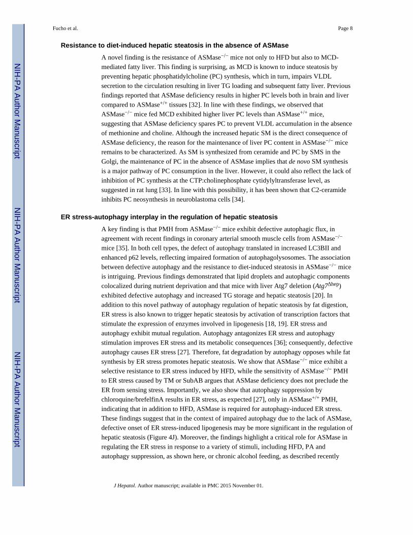
ASMase−/− mice are resistant to HFD-induced ER stress
Since ER stress is a critical mechanism that promotes lipogenesis and fatty liver [18, 19], we
next analyzed the sensitivity of ASMase−/− mice to HFD-induced ER stress. Unlike
Fucho et al. Page 4
J Hepatol. Author manuscript; available in PMC 2015 November 01.
NIH
-PA
Author M
anuscriptN
IH-P
A A
uthor Manuscript
NIH
-PA
Author M
anuscript
ASMase+/+ mice, ASMase−/− mice were resistant to HFD-mediated increased mRNA
expression of Atf4, Pdi, Edem, BiP and IRE1α phosphorylation (Figure 2A-D) as well as
uXBP-1, sXBP-1 and ATF6α (Supplementary Figure 3). These findings suggest that
ASMase is required for the induction of the 3 arms of the UPR by HFD. Resistance was
specific for HFD as PMH from ASMase+/+ and ASMase−/− mice were equally sensitive to
cleavage of GRP78/BiP by SubAB (Figure 2E), a bacterial toxin that rapidly degrades
GRP78 causing ER stress [24]. For both cell types, SubAB increased expression of Pdi and
Edem at 6h post-treatment (Figure 2F, G). Moreover, in vivo tunicamycin (TM)
administration caused enhanced expression of Atf4 and Pdi (Figure 2H, I) as well as TG
accumulation (Figure 2J) and steatosis as determined by Oil-Red staining (not shown) to a
similar degree in ASMase+/+ and ASMase−/− mice. Similar to HFD, palmitic acid (PA)
induced ER stress in PMH from ASMase+/+ mice but not ASMase−/− mice (Supplementary
Figure 4). These results indicate that the ER of ASMase−/− mice is functional and that the
resistance of ASMase−/− mice to ER stress is specific for HFD. Moreover, the findings
indicate that ER stress causes hepatic steatosis in both types of mice.
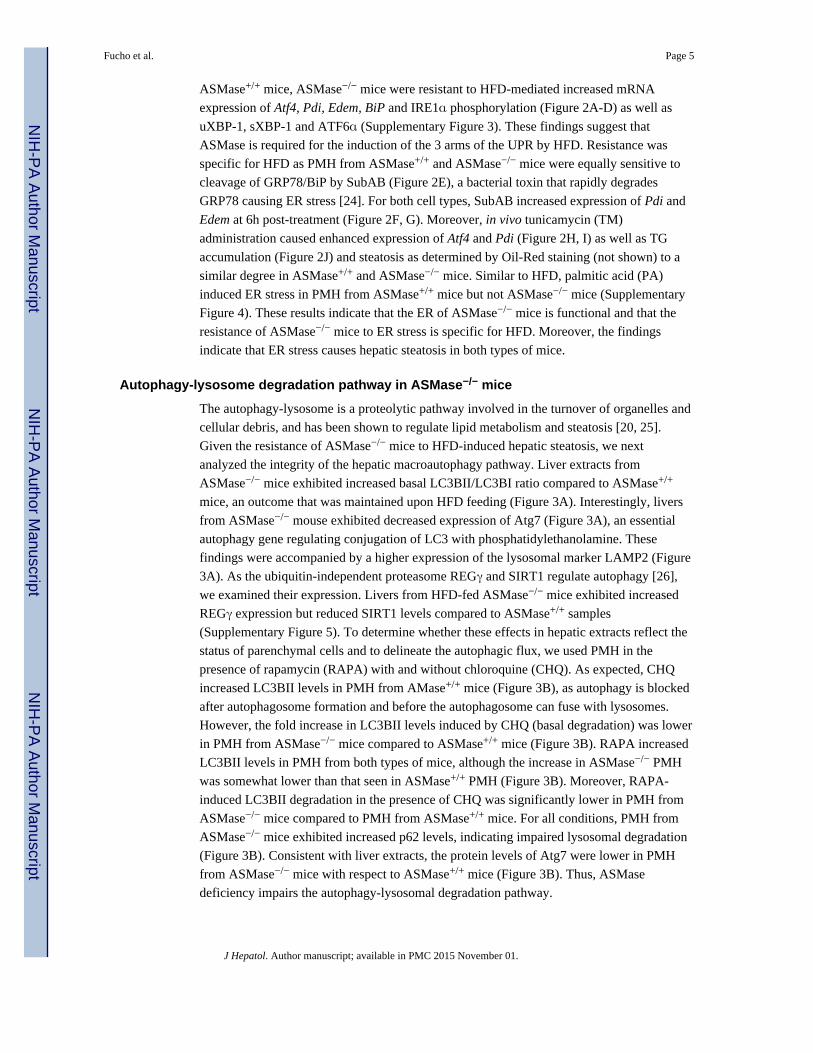
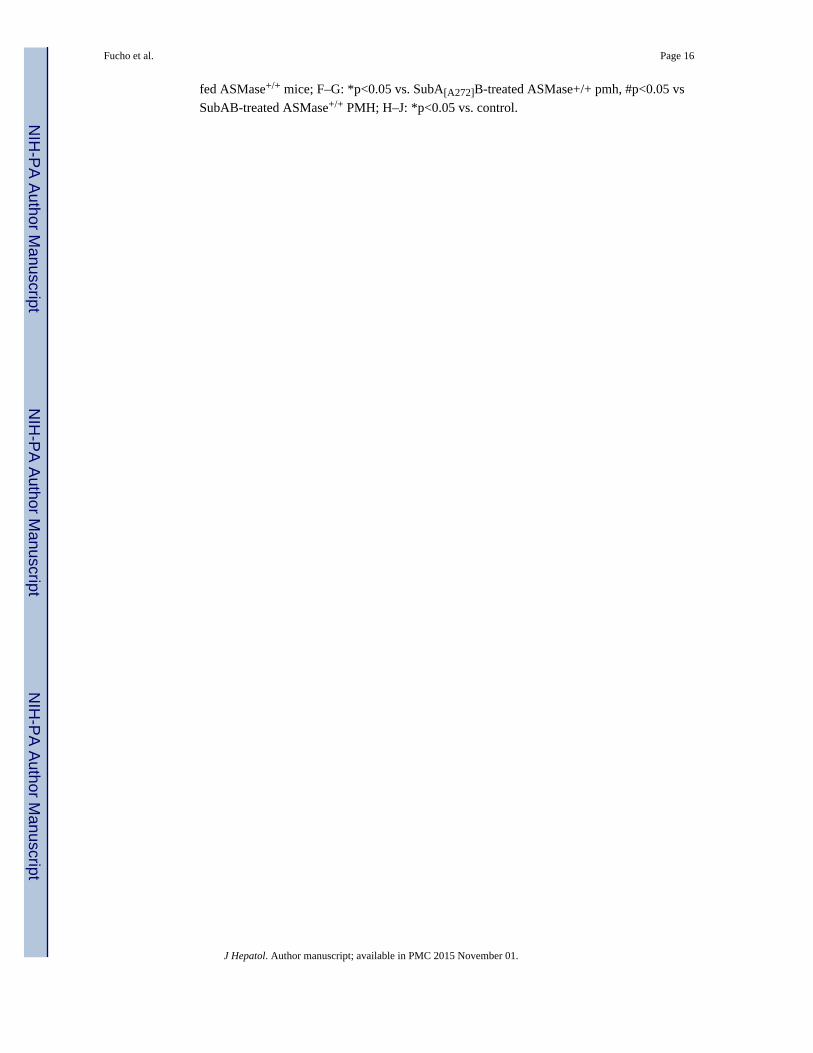
Autophagy-lysosome degradation pathway in ASMase−/− mice
The autophagy-lysosome is a proteolytic pathway involved in the turnover of organelles and
cellular debris, and has been shown to regulate lipid metabolism and steatosis [20, 25].
Given the resistance of ASMase−/− mice to HFD-induced hepatic steatosis, we next
analyzed the integrity of the hepatic macroautophagy pathway. Liver extracts from
ASMase−/− mice exhibited increased basal LC3BII/LC3BI ratio compared to ASMase+/+
mice, an outcome that was maintained upon HFD feeding (Figure 3A). Interestingly, livers
from ASMase−/− mouse exhibited decreased expression of Atg7 (Figure 3A), an essential
autophagy gene regulating conjugation of LC3 with phosphatidylethanolamine. These
findings were accompanied by a higher expression of the lysosomal marker LAMP2 (Figure
3A). As the ubiquitin-independent proteasome REGγ and SIRT1 regulate autophagy [26],
we examined their expression. Livers from HFD-fed ASMase−/− mice exhibited increased
REGγ expression but reduced SIRT1 levels compared to ASMase+/+ samples
(Supplementary Figure 5). To determine whether these effects in hepatic extracts reflect the
status of parenchymal cells and to delineate the autophagic flux, we used PMH in the
presence of rapamycin (RAPA) with and without chloroquine (CHQ). As expected, CHQ
increased LC3BII levels in PMH from AMase+/+ mice (Figure 3B), as autophagy is blocked
after autophagosome formation and before the autophagosome can fuse with lysosomes.
However, the fold increase in LC3BII levels induced by CHQ (basal degradation) was lower
in PMH from ASMase−/− mice compared to ASMase+/+ mice (Figure 3B). RAPA increased
LC3BII levels in PMH from both types of mice, although the increase in ASMase−/− PMH
was somewhat lower than that seen in ASMase+/+ PMH (Figure 3B). Moreover, RAPA-
induced LC3BII degradation in the presence of CHQ was significantly lower in PMH from
ASMase−/− mice compared to PMH from ASMase+/+ mice. For all conditions, PMH from
ASMase−/− mice exhibited increased p62 levels, indicating impaired lysosomal degradation
(Figure 3B). Consistent with liver extracts, the protein levels of Atg7 were lower in PMH
from ASMase−/− mice with respect to ASMase+/+ mice (Figure 3B). Thus, ASMase
deficiency impairs the autophagy-lysosomal degradation pathway.
Fucho et al. Page 5
J Hepatol. Author manuscript; available in PMC 2015 November 01.
NIH
-PA
Author M
anuscriptN
IH-P
A A
uthor Manuscript
NIH
-PA
Author M
anuscript
Impaired autophagy-induced ER stress requires ASMase
In addition to the regulation of lipid metabolism and steatosis, autophagy suppression has
been shown to cause ER stress [27]. This finding implies that the onset of hepatic steatosis
due to defective autophagy could reflect the combination of decreased fat degradation by
autophagy and increased fat synthesis by ER stress. Thus, we next examined the
susceptibility of ASMase−/− mice to impaired autophagy-mediated ER stress. Chloroquine
and brefeldinA increased the expression of ER stress markers in PMH from ASMase+/+
mice (Figure 2 K–N), in agreement with previous findings [27]. However, this response was
blunted in PMH from ASMase−/− mice. Chloroquine/brefeldin increased p62 levels and
expression of LC3BII in both types of PMH (Figure 3C). Thus, similar to HFD, ASMase is
required for ER stress caused by autophagy suppression.
Lysosomal cholesterol and resistance to LMP and cell death increase in ASMase−/− mice
We next analyzed the distribution of the increased hepatic cholesterol levels observed in
ASMase−/− mice (Figure 1C, 1D). Confocal imaging of ASMase−/− PMH stained with
Lysotracker and filipin revealed increased lysosomal mass and a significant increase in
lysosomal cholesterol content compared to ASMase+/+ PMH (Figure 3D). No colocalization
of cholesterol with mitochondria was found, in agreement with previous results [28]. This
outcome paralleled the increase of cholesterol in isolated lysosomes and was reproduced in
PMH from wild type mice fed a high cholesterol diet (HCD) (Supplementary Figure 6).
Moreover, autophagy inhibition by chloroquine in ASMase+/+ PMH increased total
cholesterol levels but, unlike ASMase−/− PMH, the increase in free cholesterol did not
accumulate in lysosomes (Supplementary Figure 7), in agreement with previous findings
[20]. To examine the functional impact of increased lysosomal cholesterol in LMP, we
challenged PMH with O-methyl-serine-dodecylamide hydrochloride (MSDH), a known
lysosomotropic detergent that induces cell death following LMP [29]. MSDH dose-
dependently killed ASMase+/+ PMH (Figure 3E). Importantly, ASMase−/− hepatocytes were
resistant to MSDH-induced cell death (Figure 3E) and these effects were reproduced in
PMH from mice fed HCD (Supplementary Figure 8). The resistance of ASMase−/− PMH or
PMH from wild type mice fed HCD to MSDH correlated with decreased caspase-3
activation (Figure 3F, Supplementary Figure 8). Decreasing cholesterol content by the
oxysterol 25-hydroxycholesterol [29], reversed the resistance of ASMase−/− PMH to
MSDH-induced cell death (Figure 3G). Since LMP has been shown to contribute to PA-
induced lipotoxicity [21], we examined the susceptibility of ASMase−/− PMH to PA.
Consistent with MSDH, PMH from ASMase−/− mice were less sensitive to PA-induced cell
death (Figure 3H). PA increased total cholesterol levels but did not cause lysosomal
cholesterol accumulation (Supplementary Figure 9). To pinpoint whether the resistance to
PA was due to lysosomal cholesterol accumulation, we explored whether U18666A, an
amphiphile that disrupts cholesterol homeostasis and causes lysosomal cholesterol
accumulation [30], mimics the resistance to PA in PMH from wild type mice. U18666A
reproduced the lysosomal cholesterol accumulation, and more importantly, the resistance to
PA observed in ASMase−/− mice (Figure 3I, 3J). In agreement with previous results [29],
U18666A increased p62 and LC3BII levels (Figure 3K) amd similar effects were observed
upon MSDH treatment (not shown). Consistent with these findings in PMH, ASMase−/−
mice exhibited reduced liver injury determined by serum ALT levels in response to HFD as
Fucho et al. Page 6
J Hepatol. Author manuscript; available in PMC 2015 November 01.
NIH
-PA
Author M
anuscriptN
IH-P
A A
uthor Manuscript
NIH
-PA
Author M
anuscript
well as decreased HFD-mediated susceptibility to LPS (Supplementary Fig 10). Thus,
ASMase deficiency increases lysosomal cholesterol, which determines decreased LMP and
resistance to saturated fatty acid-induced lipotoxicity.
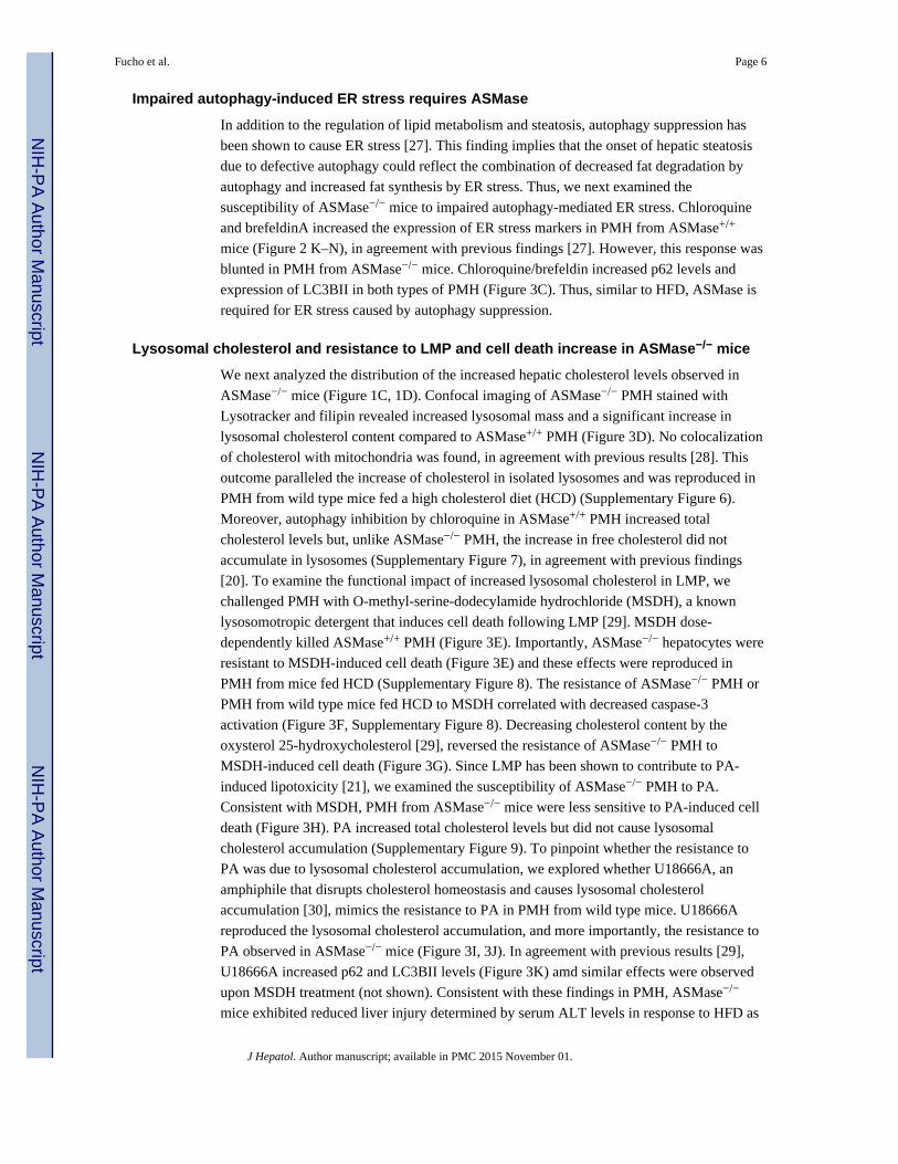
ASMase inhibition protects against HFD-induced NASH
To examine the relevance of the preceding findings on HFD-induced NASH, we explored
the use of amitriptyline, which prevents the proteolytic activation of ASMase [31], to inhibit
in vivo ASMase in wild type mice. HFD stimulated ASMase activity compared to mice fed
control diet (from 5±0.2 to 12±1.6 pmol/min/mg protein) and amitriptyline significantly
inhibited ASMase activation in both cases by 83% and 78%, respectively. Amitriptyline did
not affect food intake but significantly ameliorated the body weight gain in response to HFD
(Supplementary Fig 11) as well as the hepatomegaly and the epidydimal white adipose
weight gain (Supplementary Fig 11). Moreover, amitriptyline normalized the hyperglycemia
induced by HFD (Supplementary Fig 11) and reduced blood glucose levels during glucose
(GTT) and insulin tolerance tests (ITT) following HFD feeding (Supplementary Figure 11).
The beneficial effects of amitriptyline on body weight and glucose tolerance seen in HFD-
fed wild type mice were abrogated in ASMase−/− mice (not shown), indicating the
dependence of amitriptyline on ASMase to regulate body weight and glucose homeostasis.
Interestingly, the effects of HFD feeding in blood glucose homeostasis following GTT and
ITT were aggravated in ASMase−/− mice compared to ASMase+/+ mice despite similar
hepatic insulin signaling in both cases (unpublished observations).
Moreover, amitriptyline abolished HFD-induced hepatic steatosis, reflected by lower Oil-red
staining and the biochemical determination of liver TG and FFA (Figure 4A-D). In contrast
to ASMase−/− mice, ASMase inhibition by amitriptyline reduced both liver SM content (not
shown) and hepatic cholesterol levels in response to HFD (Figure 4C). Importantly,
amitriptyline normalized liver histology and prevented liver injury as determined by the
release of serum ALT (Figure 4E, F). Moreover, fibrosis estimated by Sirius-red staining
(Figure 4G) and expression of markers such as Col1A mRNA (Figure 4H) increased in
HFD-fed control mice and these effects were ameliorated by amitriptyline, in agreement
with previous findings [13]. In addition, although the increase in inflammatory markers,
including MCP-1 mRNA, in control mice fed HFD was prevented by amitriptyline (Figure
4I), there was not significant inflammatory infiltrates at histological examination, indicating
minimal inflammation in mice fed HFD for 16 weeks. As expected HFD increased the
expression of ER stress markers in control mice but not in HFD-fed mice treated with
amitriptyline (Supplementary Fig 12). Thus, these findings indicate that ASMase inhibition
by amitriptyline prevents diet-induced NASH.
DISCUSSION
Here, we report a novel role of ASMase in key events in NASH. Specifically, ASMase
regulates diet-induced steatosis and ER stress, lysosomal cholesterol homeostasis, autophagy
and LMP-mediated apoptosis (Figure 4J). More importantly, ASMase inhibition in wild type
mice prevents HFD induced liver injury, inflammation and fibrosis, characteristic of NASH.
Fucho et al. Page 7
J Hepatol. Author manuscript; available in PMC 2015 November 01.
NIH
-PA
Author M
anuscriptN
IH-P
A A
uthor Manuscript
NIH
-PA
Author M
anuscript
Resistance to diet-induced hepatic steatosis in the absence of ASMase
A novel finding is the resistance of ASMase−/− mice not only to HFD but also to MCD-
mediated fatty liver. This finding is surprising, as MCD is known to induce steatosis by
preventing hepatic phosphatidylcholine (PC) synthesis, which in turn, impairs VLDL
secretion to the circulation resulting in liver TG loading and subsequent fatty liver. Previous
findings reported that ASMase deficiency results in higher PC levels both in brain and liver
compared to ASMase+/+ tissues [32]. In line with these findings, we observed that
ASMase−/− mice fed MCD exhibited higher liver PC levels than ASMase+/+ mice,
suggesting that ASMase deficiency spares PC to prevent VLDL accumulation in the absence
of methionine and choline. Although the increased hepatic SM is the direct consequence of
ASMase deficiency, the reason for the maintenance of liver PC content in ASMase−/− mice
remains to be characterized. As SM is synthesized from ceramide and PC by SMS in the
Golgi, the maintenance of PC in the absence of ASMase implies that de novo SM synthesis
is a major pathway of PC consumption in the liver. However, it could also reflect the lack of
inhibition of PC synthesis at the CTP:cholinephosphate cytidylyltransferase level, as
suggested in rat lung [33]. In line with this possibility, it has been shown that C2-ceramide
inhibits PC neosynthesis in neuroblastoma cells [34].
ER stress-autophagy interplay in the regulation of hepatic steatosis
A key finding is that PMH from ASMase−/− mice exhibit defective autophagic flux, in
agreement with recent findings in coronary arterial smooth muscle cells from ASMase−/−
mice [35]. In both cell types, the defect of autophagy translated in increased LC3BII and
enhanced p62 levels, reflecting impaired formation of autophagolysosomes. The association
between defective autophagy and the resistance to diet-induced steatosis in ASMase−/− mice
is intriguing. Previous findings demonstrated that lipid droplets and autophagic components
colocalized during nutrient deprivation and that mice with liver Atg7 deletion (Atg7Δhep)
exhibited defective autophagy and increased TG storage and hepatic steatosis [20]. In
addition to this novel pathway of autophagy regulation of hepatic steatosis by fat digestion,
ER stress is also known to trigger hepatic steatosis by activation of transcription factors that
stimulate the expression of enzymes involved in lipogenesis [18, 19]. ER stress and
autophagy exhibit mutual regulation. Autophagy antagonizes ER stress and autophagy
stimulation improves ER stress and its metabolic consequences [36]; consequently, defective
autophagy causes ER stress [27]. Therefore, fat degradation by autophagy opposes while fat
synthesis by ER stress promotes hepatic steatosis. We show that ASMase−/− mice exhibit a
selective resistance to ER stress induced by HFD, while the sensitivity of ASMase−/− PMH
to ER stress caused by TM or SubAB argues that ASMase deficiency does not preclude the
ER from sensing stress. Importantly, we also show that autophagy suppression by
chloroquine/brefelfinA results in ER stress, as expected [27], only in ASMase+/+ PMH,
indicating that in addition to HFD, ASMase is required for autophagy-induced ER stress.
These findings suggest that in the context of impaired autophagy due to the lack of ASMase,
defective onset of ER stress-induced lipogenesis may be more significant in the regulation of
hepatic steatosis (Figure 4J). Moreover, the findings highlight a critical role for ASMase in
regulating the ER stress in response to a variety of stimuli, including HFD, PA and
autophagy suppression, as shown here, or chronic alcohol feeding, as described recently
Fucho et al. Page 8
J Hepatol. Author manuscript; available in PMC 2015 November 01.
NIH
-PA
Author M
anuscriptN
IH-P
A A
uthor Manuscript
NIH
-PA
Author M
anuscript

[28]. Diet and genetic obesity have been shown to alter ER lipid composition, which in turn,
regulates SERCA-mediated ER Ca2+ homeostasis [19]. As ASMase per se induces ER stress
by disrupting ER Ca2+ regulation [28], our data suggest that ASMase is required for HFD-
mediated ER Ca2+ disruption and subsequent ER stress. While further understanding is
needed as to how ASMase regulates ER Ca2+ homeostasis, our findings showing hepatic
steatosis resistance despite autophagy impairment are in agreement with recent findings.
Mice with liver-specific deletion of FIP200, a core subunit of the Atg1 complex, were
protected from starvation and HFDinduced hepatic steatosis [37]. Importantly, FIP200
mutant mice exhibited defective diet-induced activation of lipogenic pathways. Consistent
with these findings, ASMase−/− mice are resistant to HFD-induced expression of Srebp1c,
Dgat2, Fas and Acc. Moreover, autophagy disruption by shRNA-mediated suppression of
Atg7 failed to cause hepatic steatosis in lean mice [27], while lower hepatic lipid content has
been reported in Atg7Δhep mice fed HFD [38]. Although the reasons for the discrepancy
between the latter findings [38] and the previous report in Atg7Δhep mice [20] are unknown,
it is clear that that role of autophagy in lipid metabolism and hepatic steatosis is complex
and may be influenced by a number of factors including experimental conditions, genetic
background or age of mice.
Hepatic cholesterol and NASH: contrast between ASMase−/− mice and amitriptylinetreatment in wild type mice
An important aspect of the increase in hepatic cholesterol content in the ASMase−/− mice is
that the bulk of it is free and sequestered in lysosomes. This phenotype in hepatocytes is in
agreement with findings described in macrophages from ASMase−/− mice [39]. The
localization of cholesterol in this compartment reflects the high affinity of SM to bind
cholesterol [40, 41], which decreases the efflux/trafficking of cholesterol out of lysosomes.
This trafficking defect impairs the sterification of cholesterol in the ER by acyl-
CoA:cholesterol acyl transferase, as shown in ASMase−/− macrophages, which can further
contribute to the increase in liver free cholesterol seen in ASMase−/− mice. Interestingly,
enrichment of wild type macrophages with exogenous SM reproduces the increased
lysosomal cholesterol seen in macrophages from ASMase−/− mice due to decreased
cholesterol efflux [39]. A crucial difference that accounts for this contrast is the residual
ASMase activity in both paradigms (0% activity in the ASMase−/− mice vs ~20% activity
after amiltriptyline). The lack of ASMase is known to cause the lysosomal storage disease
Niemann-Pick disease type A, which is characterized by the accumulation of lipids such as
SM, cholesterol and glycosphingolipids in the affected organs, predominantly in brain and
peripheral tissues such as liver [23]. The residual ASMase activity left after amitriptyline
treatment is sufficient to prevent the increment of hepatic SM levels and subsequent increase
in cholesterol seen in ASMase−/− mice with no residual activity. The relation of ASMase
activity with cholesterol homeostasis is likely regulated by the SM content. It has been
shown that SM depletion by exogenous sphingomyelinase blocks while SM enrichment
promotes proteolytic processing of SREBP-2 at site 1, the same site that is regulated by the
levels of sterols, to achieve an optimal ratio of SM to cholesterol [42]. Furthermore, as
cholesterol is increasingly recognized as an important player in NASH [43], the protection
of ASMase−/− mice to diet-induced early-stage NASH seems paradoxical. Perhaps as
important as the extent of cholesterol increase is the subcellular location where cholesterol
Fucho et al. Page 9
J Hepatol. Author manuscript; available in PMC 2015 November 01.
NIH
-PA
Author M
anuscriptN
IH-P
A A
uthor Manuscript
NIH
-PA
Author M
anuscript
accumulates. In this regard, mitochondrial cholesterol plays a relevant role in NASH as it
has been shown to sensitize to TNF/Fas-mediated NASH [22]. Importantly, we show that
the increased hepatic cholesterol levels in the ASMase−/− mice are found in lysosomes but
not in mitochondria. This outcome determines not only the sparing of mitochondrial GSH,
as recently reported [28], but also the resistance of ASMase−/− mice to LMP, which is an
important mechanism of lipotoxicity [21]. The causal role of increased lysosomal
cholesterol in apoptosis resistance is further established in wild type PMH treated with the
amphiphile U18666A and the reversal of apoptosis resistance of ASMase−/− PMH upon
cholesterol extraction by the oxysterol 25-hydroxycholesterol. In addition to the regulation
of intracellular cholesterol distribution and LMP, ASMase is proapoptotic and mediates
death receptor-induced hepatocellular apoptosis [8–10]. In conclusion, ASMase promotes
ER stress, autophagy and LMP, which in turn mediate steatosis and lipotoxicity caused by
HFD and saturated fatty acids, events that are blunted in ASMase null mice (Figure 4J).
Therefore, targeting ASMase may be of benefit in protecting against diet-induced liver
injury and NASH.
Supplementary Material
Refer to Web version on PubMed Central for supplementary material.
Acknowledgments
We want to thank the superb technical assistance of Susana Núñez. We are indebted to Dr. Montserrat Mari forvaluable suggestions and critical reading of the manuscript. The study was performed (in part) in the Center EstherKoplowitz.
Financial support: The work was supported by CIBEREHD, Fundació la Marató de TV3 and grants PI11/0325(META) from the Instituto Salud Carlos III and grants SAF2009-11417, SAF2011-23031, and SAF2012-34831from Plan Nacional de I+D, Spain; and the center grant P50-AA-11999 (Research Center for Liver and PancreaticDiseases, NIAAA/NIH).
Abbreviations
ASMase acid sphingomyelinase
ALDLRDKO ASMase/LDL receptor double knockout
CHQ chloroquine
ER endoplasmic reticulum
FFA free fatty acids
GTT glucose tolerance test
ITT insulin tolerance test
IRβ insulin receptor β subunit
HCD high cholesterol diet
HFD high fat diet
LMP lysosomal membrane permeabilization
Fucho et al. Page 10
J Hepatol. Author manuscript; available in PMC 2015 November 01.
NIH
-PA
Author M
anuscriptN
IH-P
A A
uthor Manuscript
NIH
-PA
Author M
anuscript
MDC monodansylcadaverine
MCD methionine and choline deficient diet
MSDH O-methylserine dodecylamide hydrochloride
PA palmitic acid
PMH primary mouse hepatocytes
RAPA rapamycin
RD regular diet
SubAB AB5 subtilase cytotoxin
SMases sphingomyelinases
SM sphingomyelin
SMS sphingomyelin synthases
SPT serine palmitoyl transferase
TG triglycerides
REFERENCES
1. Ratziu V, Bellentani S, Cortez-Pinto H, Day C, Marchesini G. A position statement on NAFLD/NASH based on the EASL 2009 special conference. J Hepatol. 2009; 53:372–384. [PubMed:20494470]
2. Tilg H and Diehl. AM. Citokines in alcoholic and nonalcoholic steatohepatitis. New England J.Med. 2000; 343:1467–1476. [PubMed: 11078773]
3. Hannun YA, Luberto C. Ceramide in the eukaryotic stress response. Trend. Cell. Biol. 2000; 10:73–80.
4. Kolesnick RN, Kronke M. Regulation of ceramide production and apoptosis. Annu. Rev. Physiol.1998; 60:643–665. [PubMed: 9558480]
5. Morales A, Lee H, Goni FM, Kolesnick R, Fernandez-Checa JC. Sphingolipids and cell death.Apoptosis. 2007; 12:923–939. [PubMed: 17294080]
6. Holland WL, Brozinick JT, Wang LP, Hawkins ED, Sargent KM, Liu Y, Narra K, et al. Inhibitionof ceramide synthesis ameliorates glucocorticoid-, saturated-fat-, and obesity-induced insulinresistance. Cell Metab. 2007; 5:167–179. [PubMed: 17339025]
7. Yang G, Badeanlou L, Bielawski J, Roberts AJ, Hannun YA, Samad F. Central role of ceramidebiosynthesis in body weight regulation, energy metabolism, and the metabolic syndrome. Am. J.Physiol. Endocrinol. Metab. 2009; 297:E211–E224. [PubMed: 19435851]
8. Garcia-Ruiz C, Colell A, Mari M, Morales A, Calvo M, Enrich C, Fernandez-Checa JC. DefectiveTNF-alpha-mediated hepatocellular apoptosis and liver damage in acidic sphingomyelinaseknockout mice. J. Clin. Invest. 2003; 111:197–208. [PubMed: 12531875]
9. Mari M, Colell A, Morales A, Pañeda C, Varela-Nieto I, Garcia-Ruiz C, Fernandez-Checa JC.Acidic sphingomyelinase downregulates the liver-specific methionine adenosyltransferase 1A,contributing to tumor necrosis factor-induced lethal hepatitis. J. Clin. Invest. 2004; 113:895–904.[PubMed: 15067322]
10. Dumitru CA, Gulbins E. TRAIL activates acid sphingomyelinase via a redox mechanism andreleases ceramide to trigger apoptosis. Oncogene. 2006; 25:5612–5625. [PubMed: 16636669]
11. Samad F, Hester KD, Yang G, Hannun YA, Bielawski J. Altered adipose and plasma sphingolipidmetabolism in obesity: a potential mechanism for cardiovascular and metabolic risk. Diabetes.2006; 55:2579–2587. [PubMed: 16936207]
Fucho et al. Page 11
J Hepatol. Author manuscript; available in PMC 2015 November 01.
NIH
-PA
Author M
anuscriptN
IH-P
A A
uthor Manuscript
NIH
-PA
Author M
anuscript
12. Caballero F, Fernandez A, Matias N, Martinez L, Fucho R, Elena M, et al. Specific contribution ofmethionine and choline in nutritional nonalcoholic steatohepatitis: impact on mitochondrial S-adenosyl-L-methionine and glutathione. J. Biol. Chem. 2010; 285:18528–18536. [PubMed:20395294]
13. Moles A, Tarrats N, Morales A, Dominguez M, Bataller R, Caballeria, J et al. Acidicsphingomyelinase controls hepatic stellate cell activation and in vivo liver fibrogenesis. Am. J.Pathol. 2010; 177:1214–1224. [PubMed: 20651240]
14. Moles A, Tarrats N, Fernandez-Checa JC, Mari M. Cathepsins B and D drive hepatic stellate cellproliferation and promote their fibrogenic potential. Hepatology. 2009; 49:1297–1307. [PubMed:19116891]
15. Moles A, Tarrats N, Fernandez-Checa JC, Mari M. Cathepsin B overexpression due to acidsphingomyelinase ablation promotes liver fibrosis in Niemann-Pick disease. J. Biol. Chem. 2012;287:1178–1788. [PubMed: 22102288]
16. Deevska GM, Rozenova KA, Giltiay NV, Chambers MA, White J, Boyanovsky BB, et al. AcidSphingomyelinase Deficiency Prevents Diet-induced Hepatic Triacylglycerol Accumulation andHyperglycemia in Mice. J. Biol. Chem. 2009; 284:8359–8368. [PubMed: 19074137]
17. Osawa Y, Seki E, Kodama Y, Suetsugu A, Miura K, Adachi M, Ito H, et al. Acidsphingomyelinase regulates glucose and lipid metabolism in hepatocytes through AKT activationand AMP-activated protein kinase suppression. FASEB J. 2011; 25:1345–1353. [PubMed:21163861]
18. Ozcan U, Yilmaz E, Ozcan L, Furuhashi M, Vaillancourt E, Smith, R.O, et al. Chemicalchaperones reduce ER stress and restore glucose homeostasis in a mouse model of type 2 diabetes.Science. 2006; 313:1137–1140. [PubMed: 16931765]
19. Fu S, Yang L, Li P, Hofmann O, Dicker L, Hide, W et al. Aberrant lipid metabolism disruptscalcium homeostasis causing liver endoplasmic reticulum stress in obesity. Nature. 2011;473:528–531. [PubMed: 21532591]
20. Singh R, Kaushik S, Wang Y, Xiang Y, Novak I, Komatsu, M et al. Autophagy regulates lipidmetabolism. Nature. 2009; 458:1131–1135. [PubMed: 19339967]
21. Li Z, Berk M, McIntyre TM, Gores GJ, Feldstein AE. The lysosomal-mitochondrial axis in freefatty acid-induced hepatic lipotoxicity. Hepatology. 2008; 47:1495–1503. [PubMed: 18220271]
22. Mari M, Caballero F, Colell A, Morales A, Caballeria J, Fernandez A et al. Mitochondrial freecholesterol loading sensitizes to TNF and Fas-mediated steatohepatitis. Cell Metab. 2006; 4:185–198. [PubMed: 16950136]
23. Schuchman EH. Acid sphingomyelinase, cell membranes and human disease: lessons fromNiemann Pick Disease. FEBS Lett. 2010; 584:1895–1900. [PubMed: 19944693]
24. Paton AW, Beddoe T, Thorpe CM, Whisstock JC, Wilce MC, Rossjoh J, et al. AB5 subtilasecytotoxin inactivates the endoplasmic reticulum chaperone BiP. Nature. 2006; 443:548–552.[PubMed: 17024087]
25. Amir M, Czaja MJ. Autophagy in non-alcoholic stetohepatitis. Exp Rev Gastroenterology Hepatol.2011; 5:159–166.
26. Dong S, Jia C, Zhang S, Fan G, Shan P, Sun L et al. The REGg proteasome regulates hepatic lipidmetabolism through inhibition of autophagy. Cell Metab. 2013; 18:380–391. [PubMed: 24011073]
27. Yang L, Li P, Fu S, Calay ES, Hotamisligil GS. Defective hepatic autophagy in obesity promotesER stress and causes insulin resistance. Cell Metab. 2010; 11:467–478. [PubMed: 20519119]
28. Fernandez A, Matias N, Fucho R, Ribas V, Von Montfort C, Nuño N et al. ASMase is required forchronic alcohol induced hepatic endoplasmic reticulum stress and mitochondrial cholesterolloading. J Hepatol. 2013; 59:805–813. [PubMed: 23707365]
29. Appelqvist H, Nilsson C, Garner B, Brown AJ, Kagedal K, Öllinger K. Attenuation of thelysosomal death pathway by lysosomal cholesterol accumulation. Am J Pathol. 2011; 187:629–639. [PubMed: 21281795]
30. Cenedella RJ. Cholesterol inhibitor U18666A and the role of sterol metabolism and trafficking innumerous pathophysiological processes. Lipids. 2009; 44:477–487. [PubMed: 19440746]
Fucho et al. Page 12
J Hepatol. Author manuscript; available in PMC 2015 November 01.
NIH
-PA
Author M
anuscriptN
IH-P
A A
uthor Manuscript
NIH
-PA
Author M
anuscript
31. Lang PA, Schenck M, Nicolay JP, Kempe DS, Lupescu A, Koka S, et al. Liver cell death andanemia in Wilson disease involve acid sphingomyelinase and ceramide. Nat. Med. 2006; 13:164–170. [PubMed: 17259995]
32. Prinetti et, al. Secondary alterations of sphingolipid metabolism in lysosomal storage disease.Neurochem Res. 2011; 36:1654–1668. [PubMed: 21207141]
33. Mallampalli RK, Mathur SN, Warnock LJ, Salome RG, Hunnighake GW. Betamethasonemodulation of sphingomyelin hydrolysis up-regulates CTP:cholinephosphate cytidylyltransferaseactivity in adult rat lung. Biochem J. 1996; 318:333–341. [PubMed: 8761490]
34. Ramos B, Lahti JM, Claro E, Jackowski S. Prevalence of necrosis in C2-ceramide-inducedcytotoxicity in NB16 neuroblastoma cells. Mol Pharmacol. 2003; 64:502–511. [PubMed:12869656]
35. Li X, Xu M, Pitzer AL, Xia M, Boini KM, Li P, Zhang Y. Control of autophagy maturation by acidsphingomyuelinase in mouse coronary arterial smooth muscle cells: protective role inatherosclerosis. J Mol Med. 2014; 92:473–485. [PubMed: 24463558]
36. Backar-Wikstrom E, Wikstrom JD, Ariav Y, Tirosh B, Kaiser N, Cerasi E, Leibowitz G.Stimulation of autophagy improves endoplasmic reticulum stress-induced diabetes. Diabetes.2013; 62:1227–1237. [PubMed: 23274896]
37. Ma D, Molusky MM, Song J, Hu CR, Fang F, Rui C, et al. Autophagy deficiency by FIP200deletion uncouples hepatic steatosis from liver injury in NAFLD. Mol Endocrinol. 2013; 27:1643–1654. [PubMed: 23960084]
38. Kim KH, Jeong YT, Oh H, Kim SH, Cho JM, Kim YN, et al. Autophagy deficiency leads toprotection from obesity and insulin resistance by inducing Fgf21 as a mitokine. Nat Med. 2013;19:83–92. [PubMed: 23202295]
39. Leventhal AR, Chen W, Tall AR, Tabas I. Acid sphingomyelinase-deficient macrophages havedefective cholesterol trafficking and efflux. J. Biol. Chem. 2001; 276 4496-44983.
40. Slotte JP. Sphingomyelin-cholesterol interactions in biological and model membranes. Chem.Phys. Lipids. 1999; 102:13–27. [PubMed: 11001557]
41. Ridway ND. Interactions between metabolism and intracellular distribution of cholesterol andsphingomyelin. Biochim. Biophys Acta. 2000; 1484:129–141. [PubMed: 10760463]
42. Scheek S, Brown MS, Goldstein JL. Sphingomyelin depletion in cultured cells blocks proteolysisof sterol regulatory element binding proteins at site 1. Proc. Natl. Acad. Sci. USA. 1997;94:11179–11183. [PubMed: 9326582]
43. Van Rooyen DM, Larter CZ, Haigh WG, Yeh MM, Ioannou G, Kuver R, et al. Hepatic freecholesterol accumulates in obese, diabetic mice and causes nonalcoholic steatohepatitis.Gastroenterology. 2011; 141:1393–1403. [PubMed: 21703998]
Fucho et al. Page 13
J Hepatol. Author manuscript; available in PMC 2015 November 01.
NIH
-PA
Author M
anuscriptN
IH-P
A A
uthor Manuscript
NIH
-PA
Author M
anuscript
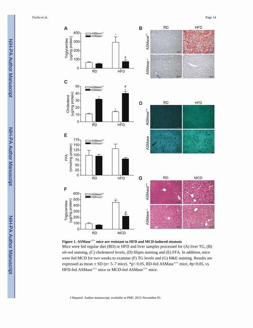
Figure 1. ASMase−/− mice are resistant to HFD and MCD-induced steatosisMice were fed regular diet (RD) or HFD and liver samples processed for (A) liver TG, (B)
oil-red staining, (C) cholesterol levels, (D) filipin staining and (E) FFA. In addition, mice
were fed MCD for two weeks to examine (F) TG levels and (G) H&E staining. Results are
expressed as mean ± SD (n= 5–7 mice). *p< 0.05, RD-fed ASMase+/+ mice, #p<0.05, vs
HFD-fed ASMase+/+ mice or MCD-fed ASMase+/+ mice.
Fucho et al. Page 14
J Hepatol. Author manuscript; available in PMC 2015 November 01.
NIH
-PA
Author M
anuscriptN
IH-P
A A
uthor Manuscript
NIH
-PA
Author M
anuscript
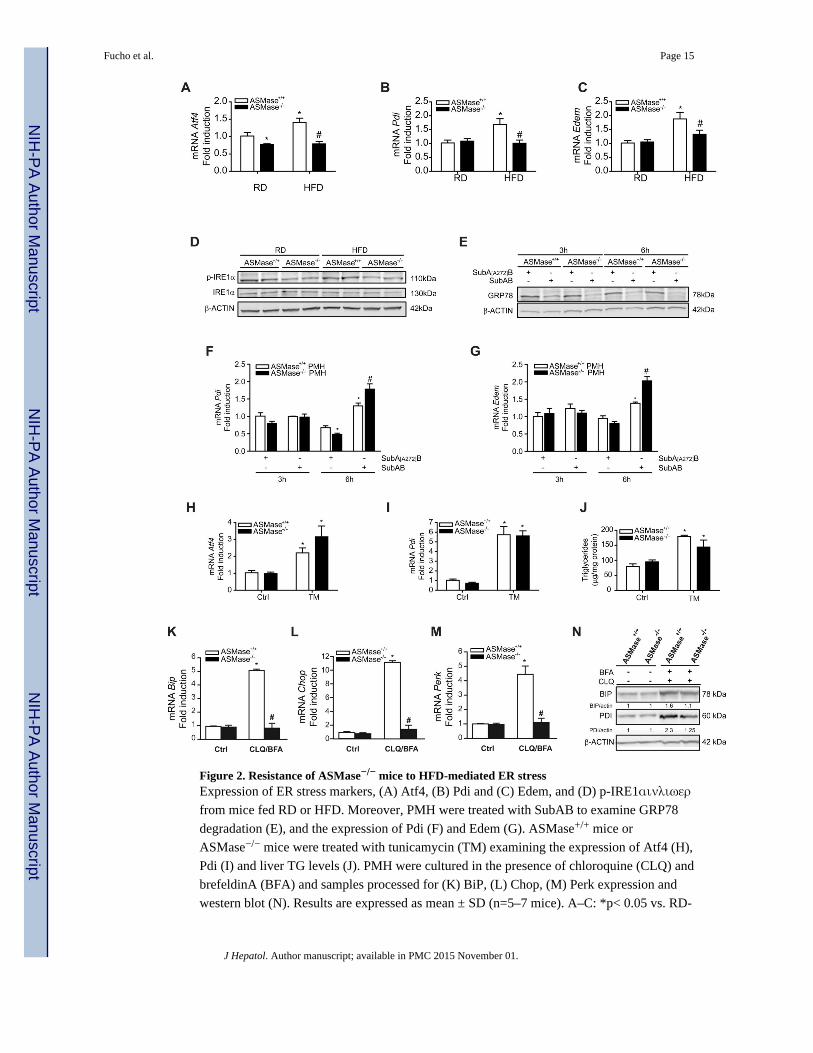
Figure 2. Resistance of ASMase−/− mice to HFD-mediated ER stressExpression of ER stress markers, (A) Atf4, (B) Pdi and (C) Edem, and (D) p-IRE1αινλιωερ
from mice fed RD or HFD. Moreover, PMH were treated with SubAB to examine GRP78
degradation (E), and the expression of Pdi (F) and Edem (G). ASMase+/+ mice or
ASMase−/− mice were treated with tunicamycin (TM) examining the expression of Atf4 (H),
Pdi (I) and liver TG levels (J). PMH were cultured in the presence of chloroquine (CLQ) and
brefeldinA (BFA) and samples processed for (K) BiP, (L) Chop, (M) Perk expression and
western blot (N). Results are expressed as mean ± SD (n=5–7 mice). A–C: *p< 0.05 vs. RD-
Fucho et al. Page 15
J Hepatol. Author manuscript; available in PMC 2015 November 01.
NIH
-PA
Author M
anuscriptN
IH-P
A A
uthor Manuscript
NIH
-PA
Author M
anuscript
fed ASMase+/+ mice; F–G: *p<0.05 vs. SubA[A272]B-treated ASMase+/+ pmh, #p<0.05 vs
SubAB-treated ASMase+/+ PMH; H–J: *p<0.05 vs. control.
Fucho et al. Page 16
J Hepatol. Author manuscript; available in PMC 2015 November 01.
NIH
-PA
Author M
anuscriptN
IH-P
A A
uthor Manuscript
NIH
-PA
Author M
anuscript
Figure 3. Autophagy and LMP in ASMase−/− miceA, Autophagy in liver samples from mice fed RD or HFD. B, Autophagy in PMH
challenged with rapamycin (RAPA), chloroquine (CHQ) or combination RAPA+CHQ (C-
R). C, p62 and LC3BI/II in PMH treated with chloroquine and brefeldinA. D, PMH from
ASMase+/+ and ASMase−/− mice were stained with Lysotracker and filipin. Images shown
are representative of 4–5 individual experiments with similar findings. Cell death (E) and
caspase-3 activity (F) of PMH induced by the lysosomotropic detergent MSDH. G, Effect of
25-hydroxycholesterol in MSDH-induced cell death in PMH. H, PMH challenged with
Fucho et al. Page 17
J Hepatol. Author manuscript; available in PMC 2015 November 01.
NIH
-PA
Author M
anuscriptN
IH-P
A A
uthor Manuscript
NIH
-PA
Author M
anuscript
increasing doses of PA to induce cell death. I, confocal imaging of PMH from wild type
mice treated with U18666A. J, survival of PMH from wild type mice treated with U18666A
challenged with PA. K, p62 and LC3BI/II levels in PMH treated with U18666A with or
without chloroquine. Results are expressed as mean ± SD of 4–6 independent experiments.
*p<0.05 vs. control ASMase+/+ PMH; # p<0.05 vs. ASMase+/+ PMH.
Fucho et al. Page 18
J Hepatol. Author manuscript; available in PMC 2015 November 01.
NIH
-PA
Author M
anuscriptN
IH-P
A A
uthor Manuscript
NIH
-PA
Author M
anuscript
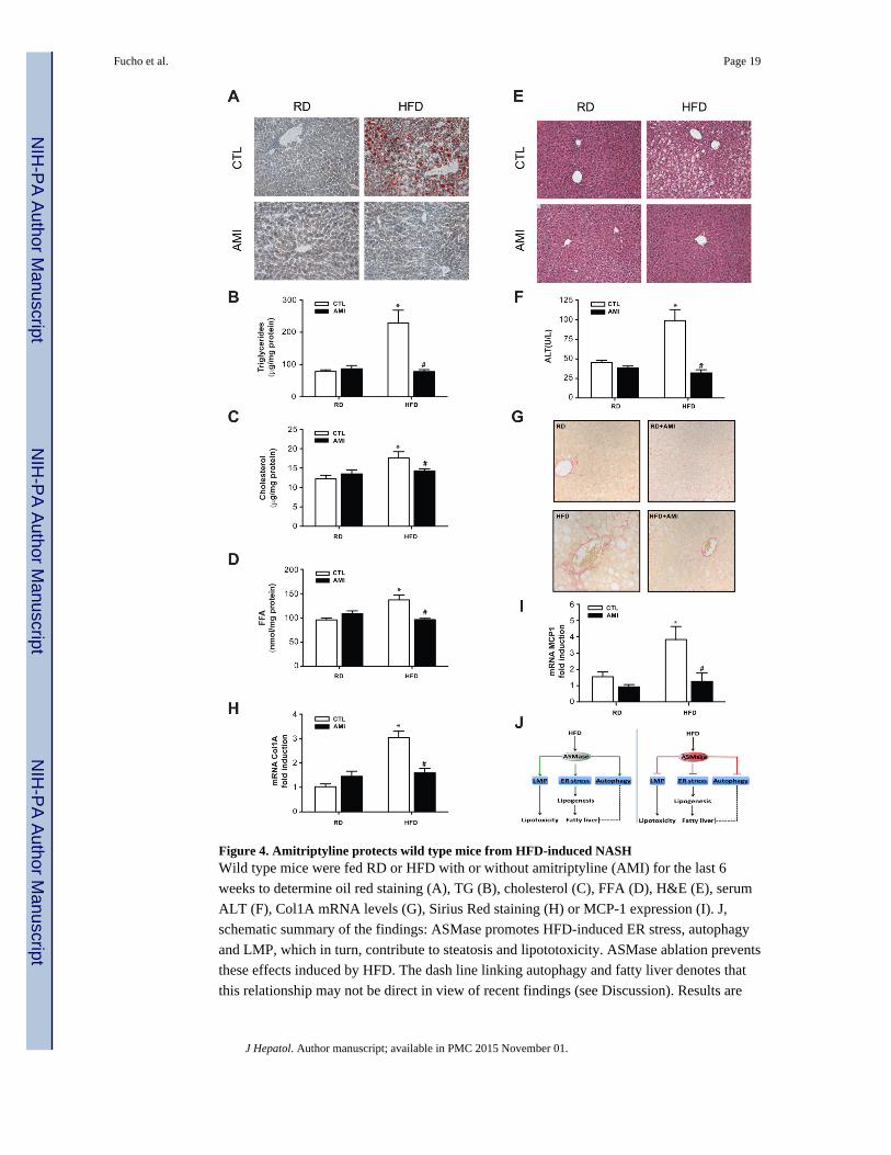
Figure 4. Amitriptyline protects wild type mice from HFD-induced NASHWild type mice were fed RD or HFD with or without amitriptyline (AMI) for the last 6
weeks to determine oil red staining (A), TG (B), cholesterol (C), FFA (D), H&E (E), serum
ALT (F), Col1A mRNA levels (G), Sirius Red staining (H) or MCP-1 expression (I). J,
schematic summary of the findings: ASMase promotes HFD-induced ER stress, autophagy
and LMP, which in turn, contribute to steatosis and lipototoxicity. ASMase ablation prevents
these effects induced by HFD. The dash line linking autophagy and fatty liver denotes that
this relationship may not be direct in view of recent findings (see Discussion). Results are
Fucho et al. Page 19
J Hepatol. Author manuscript; available in PMC 2015 November 01.
NIH
-PA
Author M
anuscriptN
IH-P
A A
uthor Manuscript
NIH
-PA
Author M
anuscript
expressed as mean ± SD of n=10 mice per group. * p<0.05 vs. control mice fed RD;
#p<0.05 vs. control mice fed HFD.
Fucho et al. Page 20
J Hepatol. Author manuscript; available in PMC 2015 November 01.
NIH
-PA
Author M
anuscriptN
IH-P
A A
uthor Manuscript
NIH
-PA
Author M
anuscript