Embed Size (px)
Citation preview

PREXION —A PROVEN BRAND
Specialized in CBCT for 13 yearsA Japanese specialist in cone beam CT technology, PreXion was founded in 2007 as a spin-off from TeraRecon Inc, a leader in advanced medical imaging. Follo-wing the incredibly successful US market launch, PreXion3D EXPLORER is now available in Europe and in a selection of different countries.
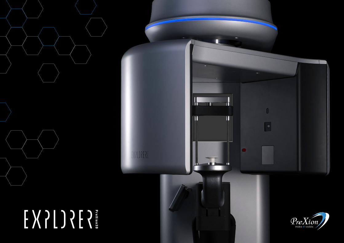
Thanks to Japanese expertise, PreXion presents its latest cone beam innovation: PreXion3D EXPLORER.
The powerful components of the system enable an extraordinary combination of great image detail and low radiation ex posure.
PreXion users can obtain precise 2D and 3D images for reliable diagnostics and digital planning in all dental indications.
Committed to diagnostic excellencePreXion dedicates itself to developing powerful software tools and reliable equip-ment in order to support practitioners in their daily work and clinical development. Thanks to the technological advances made by PreXion, users can achieve high level of sharpness and image detail at low radiation dose.
2
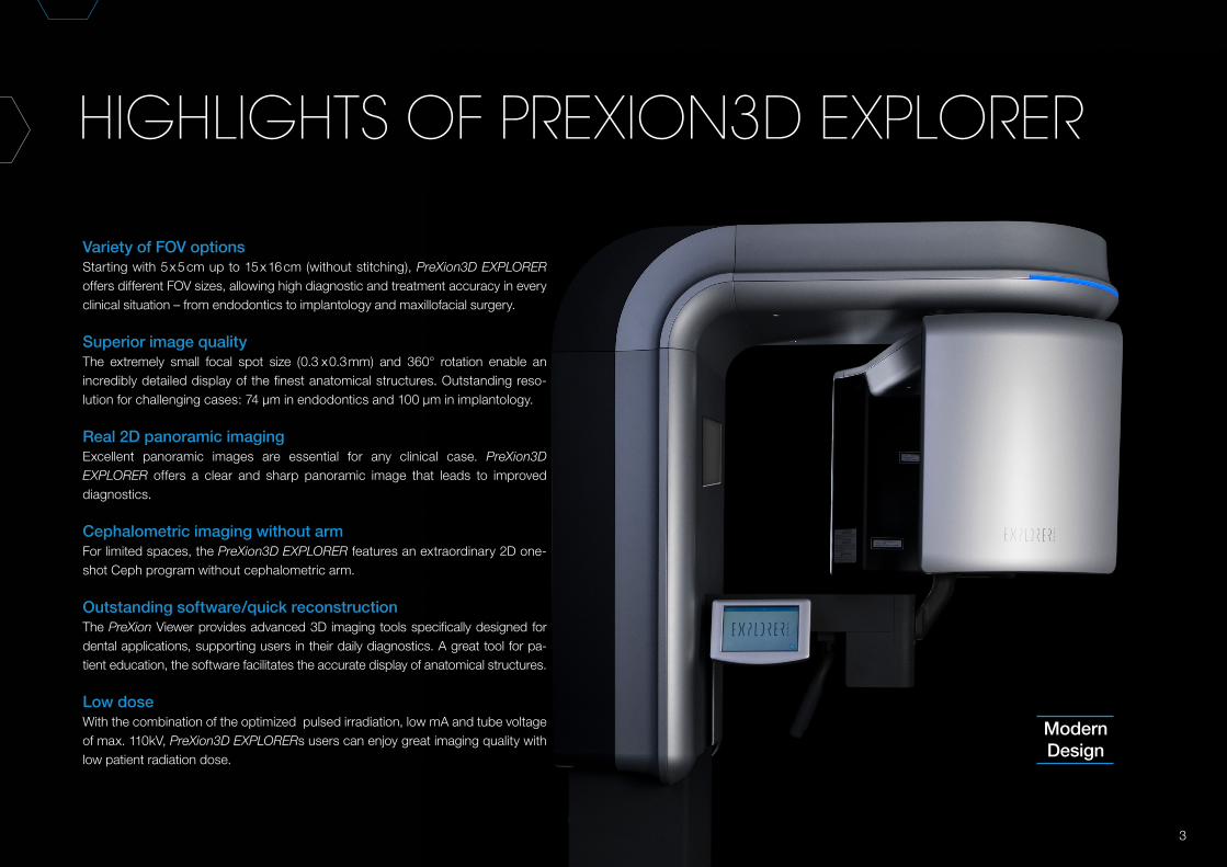
Variety of FOV optionsStarting with 5 x 5 cm up to 15 x 16 cm (without stitching), PreXion3D EXPLORER offers different FOV sizes, allowing high diagnostic and treatment accuracy in every clinical situation – from endodontics to implantology and maxillo facial surgery.
Superior image qualityThe extremely small focal spot size (0.3 x 0.3 mm) and 360° rotation enable an incredibly detailed display of the finest anatomical structures. Out standing reso-lution for challenging cases: 74 μm in endodontics and 100 μm in implantology.
Real 2D panoramic imagingExcellent panoramic images are essential for any clinical case. PreXion3D EXPLORER offers a clear and sharp panoramic image that leads to improved diagnostics.
Cephalometric imaging without armFor limited spaces, the PreXion3D EXPLORER features an extraordinary 2D one-shot Ceph program without cephalometric arm.
Outstanding software/quick reconstructionThe PreXion Viewer provides advanced 3D imaging tools specifically designed for dental applications, supporting users in their daily diagnostics. A great tool for pa-tient education, the software facilitates the accurate display of anatomical structures.
Low doseWith the combination of the optimized pulsed irradiation, low mA and tube voltage of max. 110kV, PreXion3D EXPLORERs users can enjoy great imaging quality with low patient radiation dose.
HIGHLIGHTS OF PREXION3D EXPLORER
ModernDesign
3
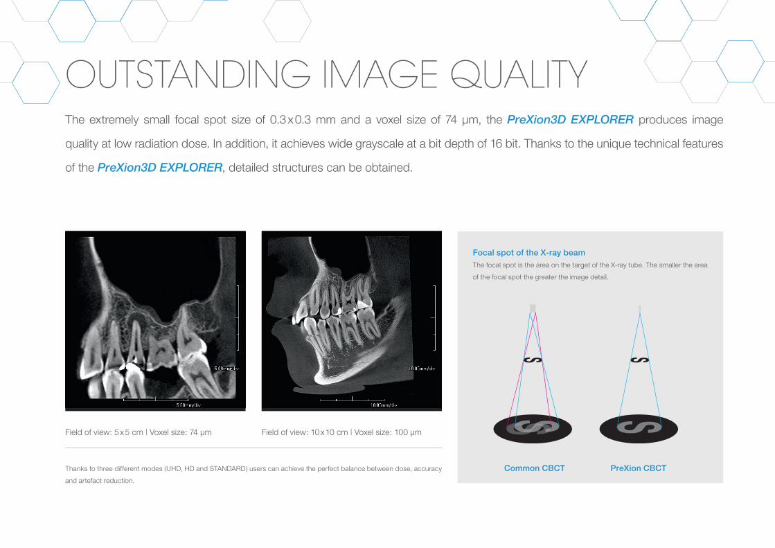
OUTSTANDING IMAGE QUALITY The extremely small focal spot size of 0.3 x 0.3 mm and a voxel size of 74 μm, the PreXion3D EXPLORER produces image
quality at low radiation dose. In addition, it achieves wide grayscale at a bit depth of 16 bit. Thanks to the unique technical features
of the PreXion3D EXPLORER, detailed structures can be obtained.
Field of view: 5 x 5 cm | Voxel size: 74 μm Field of view: 10 x 10 cm | Voxel size: 100 μm
Focal spot of the X-ray beamThe focal spot is the area on the target of the X-ray tube. The smaller the area
of the focal spot the greater the image detail.
Common CBCT PreXion CBCTThanks to three different modes (UHD, HD and STANDARD) users can achieve the perfect balance between dose, accuracy
and artefact reduction.
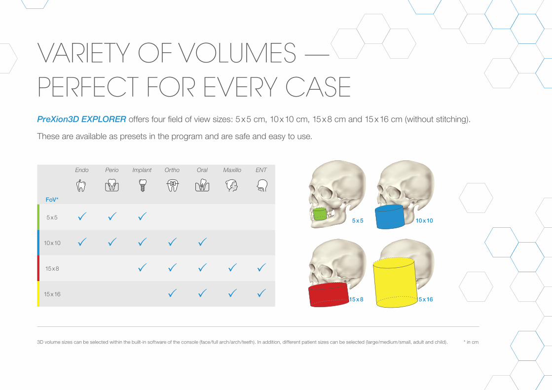
VARIETY OF VOLUMES —PERFECT FOR EVERY CASEPreXion3D EXPLORER offers four field of view sizes: 5 x 5 cm, 10 x 10 cm, 15 x 8 cm and 15 x 16 cm (without stitching).
These are available as presets in the program and are safe and easy to use.
FoV*
Endo Perio Implant Ortho Oral Maxillo ENT
5 x 5
10 x 10
15 x 8
15 x 16
3D volume sizes can be selected within the built-in software of the console (face/full arch/arch/teeth). In addition, different patient sizes can be selected (large/medium/small, adult and child). * in cm
5 x 5
15 x 8 15 x 16
10 x 10
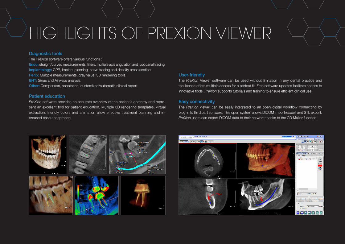
HIGHLIGHTS OF PREXION VIEWERDiagnostic toolsThe PreXion software offers various functions :Endo: straight/curved measurements, filters, multiple axis angulation and root canal tracing.Implantology: CPR, implant planning, nerve tracing and density cross section.Perio: Multiple measurements, gray value, 3D rendering tools.ENT: Sinus and Airways analysis.Other: Comparison, annotation, customized/automatic clinical report.
Patient educationPreXion software provides an accurate overview of the patient’s anatomy and repre-sent an excellent tool for patient education. Multiple 3D rendering templates, virtual extraction, friendly colors and animation allow effective treatment planning and in-creased case acceptance.
User-friendlyThe PreXion Viewer software can be used without limitation in any dental practice and the license offers multiple access for a perfect fit. Free software updates facilitate access to innovative tools. PreXion supports tutorials and training to ensure efficient clinical use.
Easy connectivityThe PreXion viewer can be easily integrated to an open digital workflow connecting by plug-in to third part software. This open system allows DICOM import/export and STL export. PreXion users can export DICOM data to their network thanks to the CD Maker function.
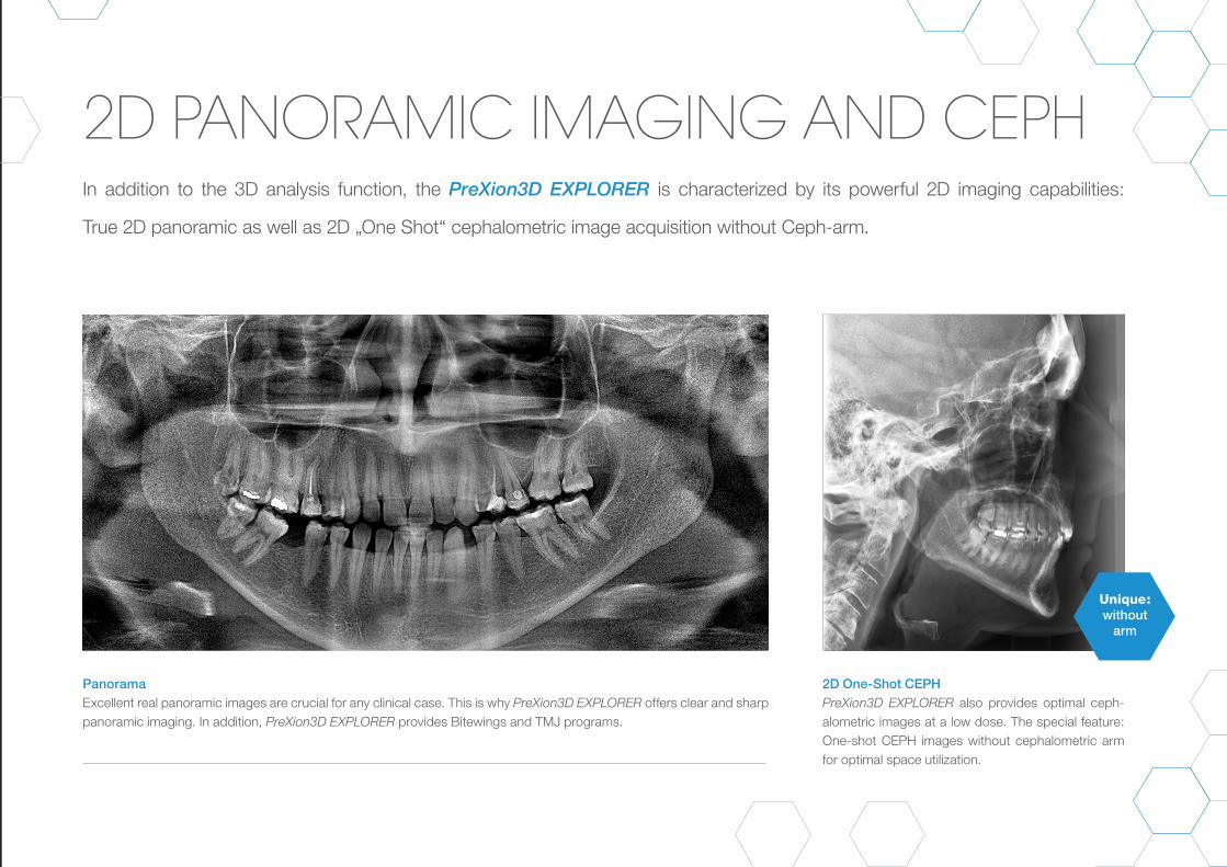
2D PANORAMIC IMAGING AND CEPH
PanoramaExcellent real panoramic images are crucial for any clinical case. This is why PreXion3D EXPLORER offers clear and sharp panoramic imaging. In addition, PreXion3D EXPLORER provides Bitewings and TMJ programs.
2D One-Shot CEPHPreXion3D EXPLORER also provides optimal ceph - a lo metric images at a low dose. The special feature: One-shot CEPH images without cephalometric arm for optimal space utilization.
In addition to the 3D analysis function, the PreXion3D EXPLORER is characterized by its powerful 2D imaging capabilities:
True 2D panoramic as well as 2D „One Shot“ cephalometric image acquisition without Ceph-arm.
Unique:without
arm
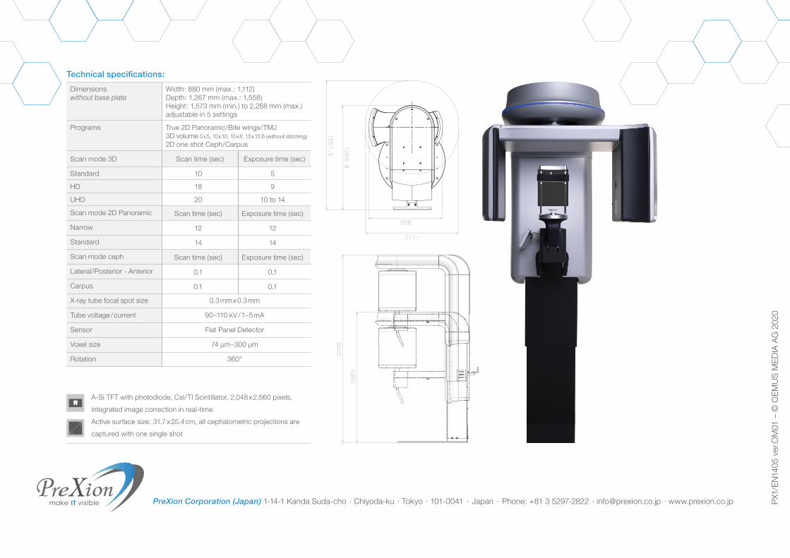
Dimensionswithout base plate
Width: 880 mm (max.: 1,112)Depth: 1,267 mm (max.: 1,558)Height: 1,573 mm (min.) to 2,268 mm (max.)adjustable in 5 settings
Programs True 2D Panoramic/Bite wings/TMJ3D volume 5 x 5, 10 x 10, 15 x 8, 15 x 15.6 (without stitching) 2D one shot Ceph/Carpus
Scan mode 3D Scan time (sec) Exposure time (sec)
Standard 10 5
HD 18 9
UHD 20 10 to 14
Scan mode 2D Panoramic Scan time (sec) Exposure time (sec)
Narrow 12 12
Standard 14 14
Scan mode ceph Scan time (sec) Exposure time (sec)
Lateral/Posterior - Anterior 0.1 0.1
Carpus 0.1 0.1
X-ray tube focal spot size 0.3 mm x 0.3 mm
Tube voltage / current 90–110 kV / 1–5 mA
Sensor Flat Panel Detector
Voxel size 74 μm–300 μm
Rotation 360°
Technical specifications:
PreXion Corporation (Japan) 1-14-1 Kanda Suda-cho · Chiyoda-ku · Tokyo · 101-0041 · Japan · Phone: +81 3 5297-2822 · [email protected] · www.prexion.co.jp
PX1
/EN
1903
ver
.OM
01P
X1/E
N14
05 v
er.O
M01
– ©
OE
MU
S M
ED
IA A
G 2
020
A-Si TFT with photodiode, CsI/TI Scintillator, 2,048 x 2,560 pixels,
integrated image correction in real-time
Active surface size: 31.7 x 25.4 cm, all cephalometric projections are
captured with one single shot