Embed Size (px)
Citation preview
© Copyright 2015
The following content is unaccredited and intended for informational purposes only.
An accredited version is available online at www.mdBriefCase.com / www.AdvancingIn.com until Oct.7, 2015. 1
Program Name: Primary Care Focus on Low Back Pain
Planning Committee:
Julia Alleyne, BHSc, MD, CCFP, Dip Sports Med, FACSM,
MScCH
Carlo Ammendolia, DC, PhD
John Axler, MD, CCFP, FCFP
Hamilton Hall, MD, FRCSC
Janice Harvey, BSc, MD, CCFP, FRCP, Dip Sport Med
Katie Hunter, Msc
Deborah Kopansky-Giles, BPHE, DC, FCCS(C), FICC
Sandra Lincoln, BSc(PT), MSc
Barry Malcolm, MD, FRCSC, MBA
Rhona McGlasson, BScPT, MBA
Shirlee OConnor, RN(EC), BScN, NP-PHC
Jess Rogers, BA
Judie Surridge, RN, BA
Expert committee
Julia Alleyne, BHSc, MD, CCFP, Dip Sports Med, FACSM, MScCH
Hamilton Hall, MD, FRCSC
Barry Malcolm, MD, FRCSC, MBA
Ravinder Ohson, MBBS, CCFP, FCFP
Raja Rampersaud, MD, FRCSC
Accreditation Information:
This version of the program is unaccredited and intended for
informational purposes only. An accredited version is available online
at www.mdBriefCase.com / www.AdvancingIn.com until October 7,
2015.
Sponsor: This case study is supported by an educational grant from: Centre for
Effective Practice.

© Copyright 2015
The following content is unaccredited and intended for informational purposes only.
An accredited version is available online at www.mdBriefCase.com / www.AdvancingIn.com until Oct.7, 2015. 2
Overview Primary Care Focus on Low Back Pain .......................................................................................................... 5
Main .......................................................................................................................................................... 5
Background ............................................................................................................................................... 6
Pre-Test ..................................................................................................................................................... 7
Welcome ................................................................................................................................................... 9
Program Overview .................................................................................................................................... 9
Vignette Selection ................................................................................................................................... 10
Vignette 1: Low Back Pain Status Report .................................................................................................... 10
Main ........................................................................................................................................................ 10
Gaps and Barriers .................................................................................................................................... 10
Test Yourself ........................................................................................................................................ 10
Ontario – Gaps and Barriers ................................................................................................................ 11
Low Back Pain - Common Gaps in Primary Care ................................................................................. 12
Acute Low Back Pain – What are the Current Guidelines? ..................................................................... 13
Case – Ms. Espina ................................................................................................................................ 13
What do the Guidelines Say? .............................................................................................................. 13
Guideline Recommendations .............................................................................................................. 13
Red Flags ............................................................................................................................................. 13
Test Yourself ........................................................................................................................................ 14
Sub- Acute Low Back Pain – What are the Current Guidelines? ............................................................. 14
Case – Mr. Ryggard ............................................................................................................................. 14
What do the Guidelines Say? .............................................................................................................. 14
Guideline Recommendations .............................................................................................................. 15
Test Yourself ........................................................................................................................................ 15
Recurrent or Persistent Low Back Pain – What are the Current Guidelines? ......................................... 15
Case – Ms. Coluna ............................................................................................................................... 15
What do the Guidelines Say? .............................................................................................................. 15
Chronic Low Back Pain- What are the Current Guidelines?.................................................................... 16
............................................................................................................................................................ 16
Case – Mr. Slabinski ............................................................................................................................ 16
What do the Guidelines Say? .............................................................................................................. 16

© Copyright 2015
The following content is unaccredited and intended for informational purposes only.
An accredited version is available online at www.mdBriefCase.com / www.AdvancingIn.com until Oct.7, 2015. 3
Test Yourself ........................................................................................................................................ 17
Introducing the CORE Back Tool ............................................................................................................. 17
Discussion Forum .................................................................................................................................... 19
Resources ................................................................................................................................................ 20
Vignette 2: Clinical Assessment of Low Back Pain ...................................................................................... 20
Main ........................................................................................................................................................ 20
Introduction ............................................................................................................................................ 21
Introduction (cont’d) ........................................................................................................................... 21
Patient History ........................................................................................................................................ 22
Question 1 ........................................................................................................................................... 22
Question 2 ........................................................................................................................................... 23
Test Yourself ........................................................................................................................................ 23
Question 3 ........................................................................................................................................... 23
Test Yourself ........................................................................................................................................ 24
Patient History .................................................................................................................................... 24
Questions 4 and 5 ............................................................................................................................... 25
Patient History (cont’d) ....................................................................................................................... 25
Physical Examination .............................................................................................................................. 26
Physical Examination (cont’d) ............................................................................................................. 26
Physical Examination (cont’d) ............................................................................................................. 27
Physical Examination (cont’d) ............................................................................................................. 28
Physical Examination (cont’d) ............................................................................................................. 29
Physical Examination (cont’d) ............................................................................................................. 30
Physical Examination (cont’d) ............................................................................................................. 32
Physical Examination (cont’d) ............................................................................................................. 33
Physical Examination (cont’d) ............................................................................................................. 34
Physical Examination (cont’d) ............................................................................................................. 36
Case Interpretation ................................................................................................................................. 36
Case Interpretation (cont’d) ............................................................................................................... 37
Discussion Forum .................................................................................................................................... 38
Resources ................................................................................................................................................ 38
Vignette 3: Patient Self-Management ........................................................................................................ 39

© Copyright 2015
The following content is unaccredited and intended for informational purposes only.
An accredited version is available online at www.mdBriefCase.com / www.AdvancingIn.com until Oct.7, 2015. 4
Main ........................................................................................................................................................ 39
Introduction: Definitions and Concepts .................................................................................................. 39
The Continuum of Patient Activation ................................................................................................. 39
What is Patient Self-Management .......................................................................................................... 40
Test Yourself ........................................................................................................................................ 41
Motivational Interviewing ................................................................................................................... 41
Selecting the right Patients for Self-Management ................................................................................. 42
Test Yourself ........................................................................................................................................ 42
Personal Action Planning Tool ................................................................................................................ 43
Goal Setting ............................................................................................................................................. 43
Goal Setting (cont’d) ........................................................................................................................... 44
Overcoming Barriers ............................................................................................................................... 45
Test Yourself ........................................................................................................................................ 46
Applying the Confidence Scale ................................................................................................................ 46
Applying the Confidence Scale (cont’d) .............................................................................................. 47
Setting a Follow-up ............................................................................................................................. 48
3 Key Questions .................................................................................................................................. 49
Key Messages .......................................................................................................................................... 49
Discussion Forum .................................................................................................................................... 50
Resources ................................................................................................................................................ 50
Vignette 4: Clinical Toolkit ......................................................................................................................... 51
Main ........................................................................................................................................................ 51
Overview of the Tool Kit ......................................................................................................................... 51
The CORE Back Tool ................................................................................................................................ 52
The CORE Back Tool ............................................................................................................................ 52
Section A: History ................................................................................................................................ 52
Test Yourself ........................................................................................................................................ 53
Section B: Red Flag Screening ............................................................................................................. 53
Section B: Radiology Criteria ............................................................................................................... 55
Test Yourself ........................................................................................................................................ 55
Section B: Surgical Criteria .................................................................................................................. 56
Section B: Yellow Flags ........................................................................................................................ 57

© Copyright 2015
The following content is unaccredited and intended for informational purposes only.
An accredited version is available online at www.mdBriefCase.com / www.AdvancingIn.com until Oct.7, 2015. 5
Test Yourself ........................................................................................................................................ 57
Section C: Physical Examination .......................................................................................................... 57
Test Yourself ........................................................................................................................................ 58
Section D: Assessment ........................................................................................................................ 58
Section E: Patient Education ............................................................................................................... 59
Test Yourself ........................................................................................................................................ 59
Section F, G, and H. ............................................................................................................................. 60
STarT Back Screening Tool ..................................................................................................................... 60
Opioid Risk Assessment Tool ................................................................................................................. 62
Patient Self Management ....................................................................................................................... 62
Patient Education Selector Guide ........................................................................................................... 62
The Tool Kit ............................................................................................................................................. 63
Case Study .............................................................................................................................................. 64
Discussion Forum .................................................................................................................................... 65
Resources ................................................................................................................................................ 66
For Providers ..................................................................................................................................... 67
For Patients ....................................................................................................................................... 67
Low Back Pain: POST Survey ....................................................................................................................... 67
Thank you! .................................................................................................................................................. 71
Primary Care Focus on Low Back Pain
Main Course Objectives:
1. To describe the current evidence based guidelines for the acute, persistent and recurrent low back pain patient.
2. To conduct an efficient high yield evidence-based history and examination for mechanical low back pain patients.
3. To describe the steps for engaging your patient in a self management strategy for improved sustainability of outcomes.
4. To effectively apply the CORE Back Tool and other relevant clinical tools to case based scenarios in order to integrate course learning.

© Copyright 2015
The following content is unaccredited and intended for informational purposes only.
An accredited version is available online at www.mdBriefCase.com / www.AdvancingIn.com until Oct.7, 2015. 6
Accreditation This program meets the accreditation criteria of The College of Family Physicians of Canada and has
been accredited for up to 4.0 Mainpro-M1 Credits. Physicians in all provinces, including Quebec,
may claim credit for completing this program. In accordance with the requirements outlined in the
College of Family Physicians of Canada's Mainpro® – Accreditation of other CME formats, this online
learning activity must be completed within a 4-week time frame.
This program has been funded by the Ministry of Health and Long Term Care (MOHLTC) as part of
the Let’s Make Healthy Change Happen initiative. For more information, visit
ontario.ca/lowbackpain or www.effectivepractice.org/lowbackpain.
Program available online until: June 1, 2014
Background Primary Care Focus on Low Back Pain is a course designed for primary care providers through the
Province of Ontario with the goal of improving patient outcomes, decreasing unnecessary resource
utilization and increasing capacity for comprehensive primary care. This course will introduce the CORE
Back Tool, which has been designed by engaging primary care providers and specialists in the design
process.
This course will be presented in 4 main vignettes:
Low Back Pain Status Report
Clinical Assessment of Low Back Pain
Patient Self Management
The Clinical Tool Kit This program was designed by the Centre for Effective Practice and acknowledges the course designer,
Dr. Julia Alleyne, who was the educational consultant on the project. The educational curriculum was
developed by faculty members Dr. Julia Alleyne and Dr. Hamilton Hall, in consultation with the
Education Planning Committee and informed by the development of the Provincial Low Back Pain
Toolkit. Included in the toolkit is the new CORE (Clinically Organized Relevant Exam) Back Tool & Guide,
developed under the clinical leadership of Drs. Julia Alleyne, Hamilton Hall and Raja Rampersaud with
the review and advice of the Education Planning Committee and primary care focus groups. All
committee members are listed below. (Note that all committee members have completed conflict of
interest forms, which are available upon request by email: [email protected]).
We strongly encourage organizations and providers to consider hosting or participating in education sessions to provide training on the evidence, skills and approach underpinning this toolkit, prior to using the CORE Tool and complete toolkit. The CORE Tool and Guide are licensed under a Creative Commons Attribution-Noncommercial-NoDerivs 2.5 Canada License. You are free to Share (copy, distribute and transmit the work) given that you provide attribution, do not use the work for commercial purposes and do not alter, transform or build
© Copyright 2015
The following content is unaccredited and intended for informational purposes only.
An accredited version is available online at www.mdBriefCase.com / www.AdvancingIn.com until Oct.7, 2015. 7
upon this work without contacting the Centre for Effective Practice. If you're interested in building upon this work or use the tools for commercial purposes, contact [email protected].
Pre-Test
1. For each of the following rate your level of confidence when assessing a patient with new onset low
back pain: 1 Not at all confident to 5 Very Confident
Screening for Red Flags
Determining criteria for evidence based imaging
Determining criteria for surgical referral
Explaining management strategies to patients
2. How confident are you in assessing and managing a Low Back Pain patient who comes to your office
with new onset symptoms of: 1 Not at all confident to 5 Very Confident
Back Dominant Low Back Pain
Recurrent Back Dominant Pain with referred leg pain
Leg Dominant Pain
Leg Dominant Pain with radicular signs including nerve root irritation
3. What would help you to increase your confidence for any of the above?
4. In the absence of red flags, what are the most common reasons for a referral of a patient with Low
Back Pain and/or Leg Pain to a spinal surgeon?
YES NO N/A
Presence of chronic back pain not responsive to conservative treatment
Presence of persistent radiating leg pain
Presence of altered sensation in affected leg
CT/MRI indicating any disc pathology
CT/MRI indicating clinically relevant nerve root compression
CT/MRI indicating spinal cord compression
Credible source for patient diagnosis and management plan
Clarification of work related modifications and restrictions

© Copyright 2015
The following content is unaccredited and intended for informational purposes only.
An accredited version is available online at www.mdBriefCase.com / www.AdvancingIn.com until Oct.7, 2015. 8
Patient would like second opinion on management
Other, please specify: ______________________
5. Which of the following statements best reflects your approach to the care of patients with new onset
Low Back Pain in the first 4 weeks?
[I do not see low back pain patients in my practice (skip logic)]
Strongly
agree
Somewhat
agree
Neutral Somewhat
disagree
Strongly
disagree
I recommend bed rest for the first
3-7 days and then activity as
advised by a physiotherapist or
chiropractor
I advise my patient to stay as
active as they are able to tolerate
as this will help reduce their
symptoms.
I screen the patient for red flags to
determine appropriate need for
imaging or surgical referrals
I often order a lumbar xray if low
back pain has been present for at
least 3-4 weeks
I may prescribe short-acting
analgesics including opioids for
the first 4-6 weeks if severe pain
symptoms interfere with function
I recommend lumbar supports to
my patients who are required to
lift or stand at work
6. Which clinical tools do you find most helpful in assessment and management of Low Back Pain?
3 Minute Back Exam
Brief Pain Inventory
CORE Back Tool
POCKET Red Flag Tool
Opioid Manager
4 Patterns of Mechanical Low Back Pain (e.g. Saskatchewan Tools)
Clinical Practice Guidelines (e.g. Alberta TOP guidelines)

© Copyright 2015
The following content is unaccredited and intended for informational purposes only.
An accredited version is available online at www.mdBriefCase.com / www.AdvancingIn.com until Oct.7, 2015. 9
Other: (open text comments box) 7. To what extent are you currently using the CORE Back Tool in your practice?
Not at all A little Somewhat To a great extent Not applicable
8. What would help you improve your assessment and management of your Low Back Pain patients?
9. Any other comments about why you are interested in Low Back Pain?
Thank you!
On behalf of the Centre for Effective Practice, we thank you for your feedback.
If you have any other questions or comments about the Low Back Pain Online Module, please
contact Katie Hunter at [email protected].
We encourage you to visit: www.ontario.ca/lowbackpain or
www.effectivepractice.org/lowbackpain
Welcome Video: Welcome Video
Add text under: Dr. Julia Alleyne provides an Introduction to the Low Back Pain Program.
Program Overview Welcome to our latest vignette series: Primary Care Focus on Low Back Pain
This program consists of four vignettes. You may complete as many vignettes as you choose, however, you are encouraged to complete all vignettes in the order they are listed.
This program has been accredited by the College of Family Physicians of Canada for up to 4 Mainpro-M1 credits.
Instructions to Learner
Each vignette can be accessed by clicking on the images found on the vignette selection page.
The vignette index tab within each vignette section will allow you to access the previous vignette, the vignette selection page, or the next vignette.
Once a vignette is completed, participate in the discussion forum within each vignette to receive accreditation.
The main program navigation bar can be accessed from the vignette selection page.
© Copyright 2015
The following content is unaccredited and intended for informational purposes only.
An accredited version is available online at www.mdBriefCase.com / www.AdvancingIn.com until Oct.7, 2015. 10
Vignette Selection 1. Low Back Pain Status Report
o This vignette provides learners with a better understanding of the evidence supporting the assessment and management of patients with low back pain through a number of interactive case scenarios.
2. Clinical Assessment of Low Back Pain o This vignette will prepare the learner to conduct an efficient high yield evidence-based
history and conduct a physical examination identifying the common patterns of low back pain presentations.
3. Patient Self Management o This vignette introduces the key skills required to effectively incorporate Patient Self
Management (PSM) into discussions with patients with low back pain. 4. The Clinical Tool Kit
o This vignette will review each component of the Provincial Toolkit for Low Back Pain, including the newly developed CORE Back Tool & Guide and provide an interactive opportunity to apply the tools to a case scenario.
Note: The Resource Section contains additional material and links to enhance you learning. We encourage you to take advantage of this section.
Vignette 1: Low Back Pain Status Report
Main This vignette provides learners with a better understanding of the evidence supporting the assessment and management of patients with low back pain through a number of interactive case scenarios. Learning objective: Upon completion of this vignette, the learner will be able to:
Describe the current evidence based guidelines for the acute, persistent and recurrent low back pain patient.
Gaps and Barriers Test Yourself What is your most common barrier to implementing best care for your patients? Choose only one.
Patient expectations
Activity management
Patient education
Access to spine surgeons
Access to other specialist
Access to imaging

© Copyright 2015
The following content is unaccredited and intended for informational purposes only.
An accredited version is available online at www.mdBriefCase.com / www.AdvancingIn.com until Oct.7, 2015. 11
Medication management
Self management resources
Access to funded rehab services
Ontario – Gaps and Barriers Approximately 90% of MRI back scans will show abnormalities, but will not impact clinical
decision making (Orthopaedic Expert Panel, 2010).
Abnormal findings from imaging results usually causes a higher intensity of care than evidence
would suggest is necessary.
The majority of low back pain patients will benefit from lifestyle changes, which often leads to
better outcomes for patients than diagnostic tests.
25% of low back pain patients will develop persistent or chronic low back pain and have
traditionally utilized more services in imaging, referral and family practice visits.
Practice Tip: Provincial adoption of evidence-based care for low back pain will lead the way
to improving patient outcomes and system savings from reduction of unnecessary costs.
Figure. Website for Provincial Low Back Pain Strategy (http://www.effectivepractice.org/lowbackpain)

© Copyright 2015
The following content is unaccredited and intended for informational purposes only.
An accredited version is available online at www.mdBriefCase.com / www.AdvancingIn.com until Oct.7, 2015. 12
Learn more about the Provincial Low Back Pain Strategy at ontario.ca/lowbackpain (or in French at
ontario.ca/lombalgie)
Low Back Pain - Common Gaps in Primary Care The literature has identified common barriers to optimal low back pain care that are consistent across
health care systems internationally. These common gaps in care can be divided into Patient, Provider and
System. In the 2010 publication, Managing Low Back Pain in the Primary Care Setting: The Know-Do Gap,
Scott et al. identified 14 studies in primary care settings where clinical practice was compared to guideline
recommendations and consistently found gaps in red flag assessment, diagnostic imaging criteria, activity
advice, medications and treatment recommendations. Here are some of the common issues encountered
by patient, provider and the health care system when dealing with low back pain issues.
Table 1: Common Gaps in Primary Care
Patient Provider System
Lack of understanding for
reason to investigation/
refer
Desire for funding for
physiotherapy
Lack of self-management
strategies
Request for more
medications
Request for time off work
Dealing with complex
chronic low back pain
Patient expectations for
MRI requests & referrals
Psychosocial patient
needs
Lack of patient
educational resources
Work related restrictions
Medication (Opioid
Management)
Poor communication
between patient
providers for care
Lack of coordinated
patient education
material
Lack of web resources
Lack of consensus on
guidelines
Lack of common approach
between providers
assessment and
treatment

© Copyright 2015
The following content is unaccredited and intended for informational purposes only.
An accredited version is available online at www.mdBriefCase.com / www.AdvancingIn.com until Oct.7, 2015. 13
Lack of understanding of
urgent symptoms versus
pain escalation
Acute Low Back Pain – What are the Current Guidelines? Case – Ms. Espina Ms. Espina is a 42 year old Nurse who noticed the gradual but progressive onset of low back pain 2
weeks ago which she thinks might be related to starting some tennis lessons. She has visited your office
today because she is beginning to have some radiating pain into her left buttock and she finds that she
cannot sit for longer than 10 minutes before her pain begins to worsen. She has been using some over-
the-counter medications and does get relief. She has not noticed any loss of sensation or change in her
bladder or bowel control. She is otherwise healthy and has two children aged 12 and 16 years.
What care would you provide for her according to Guideline Recommendations?
What do the Guidelines Say? TOP Alberta, Towards Optimizing Practice, issued Low Back Pain guidelines that were seeded by 7 of the
international guidelines and provide quick and easy synopsis of information for the provider and the
patient. Take a look at the website to help you decide what is best for Ms. Espina.
Guideline Recommendations Practice Tip: The guidelines for management of acute low back pain pertain to the first 4-6 weeks of
symptoms and recommend the following:
History, Physical, Neurological Assessment
Screen for Red and Yellow Flags
No Imaging unless Red Flags are present
Educate, Exercise, Activity Prescription
Self-care Strategies
Consider Analgesics
Red Flags Red flags are risk factors for serious pathology that may indicate the need for investigations, referrals or
management for conditions other than mechanical low back pain. It is prudent to screen for red flags on
all first visits for low back pain and on any follow-up visits where the patient is not responding to
treatment without a reason.
Practice Tip: In remembering the red flags, you may want to use the mnemonic NIFTI.

© Copyright 2015
The following content is unaccredited and intended for informational purposes only.
An accredited version is available online at www.mdBriefCase.com / www.AdvancingIn.com until Oct.7, 2015. 14
N- Neurological I – Infection F- Fracture T- Tumour I - Inflammatory
Test Yourself Which one of the following are examples of red flags for low back pain? Choose only one.
a) Referred pain below the knee with intermittent tingling b) Waking at night with positional pain and morning stiffness c) Urinary retention followed by insensible urinary overflow d) Recurrent bladder infection with stress incontinence
Sub- Acute Low Back Pain – What are the Current Guidelines? Case – Mr. Ryggard Mr. Ryggard is a 39 year old, sedentary office worker, who has come to see you for the 4th visit related
to his low back pain which started 10 weeks ago. He states that he is experiencing “good days and bad
days” and complains of a dull ache across his lumbar area which can vary from a 3-9/10 on a pain scale.
He has recently started some massage therapy which gives him temporary relief.
He has found that he needs regular medication to deal with his pain and is
spending more time lying down when he is not at work. He thinks that some time
off would help improve his pain and discomfort.
What care would you provide for him according to Guideline Recommendations?
What do the Guidelines Say? Sub-acute low back pain occurs when a patient has experienced symptoms of low back pain for 6-12
weeks even if they previously had an episode, which resolved and then some time later re-occurred.
Often, in sub acute low back pain, patient self management is not consistent and the primary care
provider needs to focus on emphasizing the key messages for optimal management.
Practice Tip: Key Messages for Your Patient
Your examination today does not demonstrate that there are any red flags present to indicate serious pathology, but if your symptoms persist for > 6 weeks, schedule a follow-up appointment.
Imaging tests like X- rays, CT scans and MRIs are not helpful for recovery or management of acute or recurring low back pain unless there are signs of serious pathology.
Low back pain is often recurring and recovery can happen without needing to see a healthcare provider. You can learn how to manage low back pain when it happens and use this information to help you recover next time.
You may need pain medication to help you return to your daily activities and initiate exercise more comfortably. It is activity, however, and not the medication that will help you recover more quickly.

© Copyright 2015
The following content is unaccredited and intended for informational purposes only.
An accredited version is available online at www.mdBriefCase.com / www.AdvancingIn.com until Oct.7, 2015. 15
If you are feeling symptoms of sadness or anxiety, this could be related to your condition and could impact your recovery: schedule a follow-up appointment.
Guideline Recommendations The New Zealand Yellow Flags Guidelines describe the psychosocial risk factors for development of
chronic pain in low back pain patients. Traditionally, the identification of chronic low back pain has been
delayed and treatment has been less effective. Within the current guidelines, it is recommended that
early identification of risk factors for chronicity should be built into the assessment and management of
low back pain in the acute and sub-acute stages so that appropriate counselling, medication and
education can be initiated to improve outcomes.
There are four key areas to assess:
o Belief that Back pain is harmful o Fear-avoidance of activity o Tendency to low mood or withdrawal o Reliance on passive treatment for recovery
Download the Alberta TOP Yellow Flags Summary (PDF)
Test Yourself Which of the following would not be a “yellow flag” question for patient interview?
a) How do you manage your pain in day-to-day activities? b) Have you found that your mood has changed as you are dealing with low back pain? c) Would you describe your work environment as positive or negative for your back condition? d) Do you have any dependents that you are responsible for?
Recurrent or Persistent Low Back Pain – What are the Current Guidelines? Case – Ms. Coluna Ms. Coluna complains of a 10 month history of low back pain which is described as initially having
recurrent “ups and downs” with some pain-free days but now complains of constant pain for the last 3
months. She is frustrated and has tried physiotherapy, chiropractic care and massage. She states that
she had tried each type of treatment for at least 2-3 visits but her pain remains unresolved. She has
been given a home exercise program but is confused because sometimes it hurts her and sometimes it
helps her so she has stopped doing it. She would like to try a new type of “electrical traction” therapy
that a neighbour said had helped her.
What care would you provide for him according to Guideline Recommendations?
What do the Guidelines Say? This stage of back pain relates to as early as 6 weeks and up to 12 months since the episode onset. This
group of patients is more complex and often challenging to manage in family practice. The frustration of

© Copyright 2015
The following content is unaccredited and intended for informational purposes only.
An accredited version is available online at www.mdBriefCase.com / www.AdvancingIn.com until Oct.7, 2015. 16
both patient and provider can lead to ordering of unnecessary diagnostic tests or referrals in an attempt
to find pathology to explain the persistence. Yet, in the absence of red flags despite escalating pain and
decreasing function, there is no indication for investigation or referral. Often, we feel that a lack of
treatment response at this stage can be reason for investigation but, more correctly, this would only be
the case if there was failure to respond to evidence based compliant conservative care of at least 12
weeks.
The 2007 American College of Physicians and the American Pain Society issued a publication entitled
“Diagnosis and Treatment of Low Back Pain: A Joint Clinical Practice Guideline”. The table available here
from this guideline, provides an overview of evidence-based active rehabilitation treatment. The
evidence for treatment effectiveness is still emerging for some modalities but this guide is correct in
principle that “active” care being exercise, patient education and activity management has yielded much
better outcomes than “passive “ care such as prolonged modalities, lumbar supports or traction.
Here are two excellent guidelines for assessing appropriate goal-specific therapy. Think of the last
patient who asked you about a particular therapy to try, now take a look through the resources and
see if it is evidence-based.
NHS: National Institute for Health and Clinical Excellence (2009). Low Back Pain: Early
Management of persistent non-specific low back pain.
Diagnosis and Treatment of Low Back Pain: A Joint Clinical Practice Guideline from the
American College of Physicians and the American Pain Society (2007)
Chronic Low Back Pain- What are the Current Guidelines?
Case – Mr. Slabinski Mr. Slabinski has been dealing with low back pain for 12 months. He has learned to modify his activities
and he chooses to garden to maintain his activity. He takes daily acetaminophen and will sometimes
combine with ibuprofen for increased effectiveness. He uses occasional massage for reduction of
intermittent muscle tension and wants to avoid any medications that might be addictive. He has visited
your office to learn more about possible medication options.
What care would you provide for him according to Guideline Recommendations?
What do the Guidelines Say? Chronic Low Back pain does not mean Chronic Pain Syndrome.
The difference is that Chronic Low Back Pain will have a mechanical component and is localized to the
lumbar patterns of pain but the nature of pain is more frequent, higher intensity and there are seldom
episodes of full relief for any length of time. These patients require medications, self-management and
exercise to maintain their quality of life as they can become discouraged and depressed.

© Copyright 2015
The following content is unaccredited and intended for informational purposes only.
An accredited version is available online at www.mdBriefCase.com / www.AdvancingIn.com until Oct.7, 2015. 17
Chronic Pain Syndrome, on the other hand, is a psychosocial disorder that occurs in some patients with
chronic non-cancer pain in which symptoms of the pain can incapacitate the patient and pain signals can
be altered through the brain affecting sleep patterns, sensitivity and mood stability.
Chronic Low Back Pain patients strive for stability in their pain and functional capacity but they can
experience exacerbations and onset of new low back pain symptoms. Therefore, never forget to screen
for red or yellow flags when symptoms become unstable in any way.
Download the Patient Education Handout from the Alberta TOP Guideline (PDF)
Test Yourself Which of the following medication regimes for pain management would be supported by the current
guidelines? Choose one.
a) Daily low dose muscle relaxants to a maximum of three doses per day during the first 4 weeks of an acute low back pain episode.
b) Trial of gabapentin medication if pain is escalating and not well controlled with non-opioid analgesics
c) Trial of short acting opioids in patient with acute onset radicular leg pain who cannot work due to constant and high intensity pain
d) Topical diclofenac applied three times a day to low back area in patient who is unable to tolerate systemic non-steroidal medications due to recent gastric ulcer and has low grade but recurrent symptoms of back pain.
Introducing the CORE Back Tool The Centre for Effective Practice undertook an evaluated process to determine appropriate clinical tools
for primary care assessment and management of Low Back Pain. This included the use of an integrated
knowledge translation process to collaborate with providers by doing an inventory of current tools,
employing a needs assessment through focus groups, consulting an expert group, review with a
curriculum education committee and employing focus groups for usability testing.

© Copyright 2015
The following content is unaccredited and intended for informational purposes only.
An accredited version is available online at www.mdBriefCase.com / www.AdvancingIn.com until Oct.7, 2015. 18
The key questions that the primary care focus groups were asked were:
o Can this tool be realistically implemented in Real Time for primary care? o Is this tool relevant for primary care? o Will this tool assist clinical decision making for improved patient outcomes?

© Copyright 2015
The following content is unaccredited and intended for informational purposes only.
An accredited version is available online at www.mdBriefCase.com / www.AdvancingIn.com until Oct.7, 2015. 19
The results of the process was the development of the Clinically Oriented Relevant Exam (CORE) Low
Back Pain Tool in response to the needs identified by primary care providers to integrate multiple
existing tools and evidence into a comprehensive single tool for use in practice. The tool includes
sections on: high yield history, red flags, yellow flags, radiology and surgical referral criteria, physical
assessment, patient education and management.
We will be referring and integrating the tool into our clinical teaching in the remaining few vignettes so
that you are comfortable implementing this into your practice.
Download the CORE Back Tool (PDF)
Discussion Forum 1. In patients with acute low back pain, would you prescribe muscle relaxants? If so, what dose and
for how long? If not, why not?
2. How do you reassure patients that investigations and especially MRI is not needed for best
practice when many other conditions are managed with lots of investigations?

© Copyright 2015
The following content is unaccredited and intended for informational purposes only.
An accredited version is available online at www.mdBriefCase.com / www.AdvancingIn.com until Oct.7, 2015. 20
Resources Guidelines
Toward Optimized Practice (TOP): Guideline for the Evidence Informed Primary Care Management of
Low Back Pain.
NHS: National Institute for Health and Clinical Excellence (2009). Low Back Pain: Early Management of
persistent non-specific low back pain.
Diagnosis and Treatment of Low Back Pain: A Joint Clinical Practice Guideline from the American College
of Physicians and the American Pain Society (2007).
Patient Handouts
Chronic Pain Patient Education Handout (Alberta TOP Guideline)
Tools
Clinically Oriented Relevant Exam (CORE) Low Back Pain Tool
DN4 Questionnaire
Opioid Manager
Learn more about the Provincial Low Back Pain Strategy at ontario.ca/lowbackpain or
http://www.effectivepractice.org/lowbackpain.
Vignette 2: Clinical Assessment of Low Back Pain Main This vignette will prepare the learner to conduct an efficient high yield evidence-based history and conduct a physical examination identifying the common patterns of low back pain presentations.
Learning objective: Upon completion of the vignette, the learner will be able to:
Conduct an efficient high yield evidence-based history and examination for mechanical low back pain patients.

© Copyright 2015
The following content is unaccredited and intended for informational purposes only.
An accredited version is available online at www.mdBriefCase.com / www.AdvancingIn.com until Oct.7, 2015. 21
Introduction
In 1987, the Quebec Task Force on Spinal Disorders proposed a diagnostic classification to help make
clinical decisions, evaluate quality of care, assess prognosis, and conduct research. The conclusions of
initial literature review and research stated that “distinct patterns of reliable clinical findings are the
only logical basis for back pain categorization and subsequent treatment.”
There has been much international research and publication supporting an approach to low back pain
where mechanical pain patterns are differentiated into four patterns; two back dominant and two leg
dominant. The leg pain is differentiated as being referred pain without neurological signs in the back
dominant cases or radicular pain with signs of nerve root involvement in the leg dominant cases where
there may be irritant pain or conduction loss of sensory or motor function.
More than 90% of back pain seen in family practice is the result of minor alterations in spinal mechanics.
It is rarely the result of malignancy, infection, systemic illness or major trauma. Most back pain is
mechanical, that is pain directly related to movement or position. The pain arises from a structural
element or elements within the spine. The precipitating abnormality is so minor and structurally
insignificant that, in the overwhelming majority of cases, it cannot be precisely identified. This vignette
will present a clinical approach to identifying the mechanical patterns of low back pain based on history,
physical examination and evaluation of treatment response.
The vignette will prepare the learner to:
1. Conduct an efficient high yield evidence-based history for low back pain patients. 2. To conduct the physical examination identifying the common patterns of low back pain
presentations
References:
Atlas SJ, Deyo RA, Patrick DL, Convery K, Keller RB, Singer DE. The Quebec Task Force classification for
Spinal Disorders and the severity, treatment, and outcomes of sciatica and lumbar spinal stenosis , BMC
Musculoskelet Disord. 2012 Nov 28;13:236
Current Clinical Care, January 2013, Educational Supplement, Back Pain Management Reprinted with permission from http://www.healthplexus.net/back-pain/editorial. All practicing Canadian Physicians can subscribe for free at http://www.healthplexus.net/physician.
Introduction (cont’d)
Dr. Hamilton Hall, Spine Surgeon, has devoted his career to the refinement of mechanical back pain
patterns and the identification of the key history and physical findings that define them. In this video
segment, he will explain each of the patterns and how they present in the clinical scenario.
Left Panel Right Panel

© Copyright 2015
The following content is unaccredited and intended for informational purposes only.
An accredited version is available online at www.mdBriefCase.com / www.AdvancingIn.com until Oct.7, 2015. 22
Section D: Core Tool INSERT VIDEO: Description of the 4 patterns:
Dr. Hamilton Hall ( 4 min)
Patient History Question 1 Although you may open the patient visit with questions related to the purpose of the visit and the
patient’s experience with their symptoms, you should quickly move into a few high yield questions that
allow you to identify if the patient symptoms are consistent with mechanical low back pain or if
screening questions for red flags are needed.
Question 1: “Where is your pain the worst?”
You mission is to determine if the pain is back or leg dominant. This is a key factor in pattern
recognition as it immediately identifies the back dominant patients who are least likely to benefit from
referrals and investigations and the leg dominant patients who may require more intervention. Often
we mistakenly use the intensity of pain as the triggering factor for ordering imaging or making a referral
to a specialist. This question shifts our focus appropriately to the location of the dominant region of
pain.
Back Dominant symptoms are worse in the low back, buttocks, coccyx, groin and/or around the outer
aspects of the hips.
Leg Dominant symptoms are worse around and below the inferior gluteal fold in the lowest portion of
the buttock and in the thigh, calf, and/or foot.
One common point of confusion is the location of the leg pain. Referred back dominant pain can spread
all the way to the foot while radicular leg dominant pain may stop in the thigh. Whether the pain
extends above or below the knee is not relevant. The determining factor is whether the pain is most
troublesome or intense in the back, buttocks and around the trunk or whether it is predominant in any
part of the leg.
Patients frequently have pain in both the back and leg; but with careful questioning, it is possible to
determine which site predominates. This can be challenging, but distinguishing the site of dominant pain
is essential for pattern recognition.
This question can be further explained to the patient in the following manner to emphasize the need to
determine the worst pain area.
Examples:
“I know you have pain in both your back and your leg, but which pain is the most disabling, the most
severe or the most worrisome.”
“I realize that your goal is to get rid of all your pain, but I can start with only one area, so if it can be only
one, which area of pain, back or leg, would you choose to get rid of?”

© Copyright 2015
The following content is unaccredited and intended for informational purposes only.
An accredited version is available online at www.mdBriefCase.com / www.AdvancingIn.com until Oct.7, 2015. 23
“I have a back pain pill and a leg pain pill and I can only give you one. Which pill do you want?”
Question 2 Question 2: “Is your pain constant or intermittent?”
Many patients will describe their pain as constant because they are highly aware of it and it is interfering
with their usual activities. Sometimes patients perceive intermittent pain to be less important and they
want to emphasize to the clinician the degree of discomfort that they are experiencing. It is always
important to acknowledge the patient’s pain with empathy and understanding.
But an accurate assessment of the character of the pain is essential and the global classification of the
pain as “constant” is not sufficient. This question must be very clear and specific. It is best asked in two
parts:
A) “Is there ever a time during the day or a position or movement you can use when your pain stops, even for a brief moment and even though it may quickly return?”
B) “When your pain stops, does it disappear completely; is it totally gone?”
The importance of this question is related to determining if the pain is likely mechanical or if it could be
a red flag to serious pathology. Truly intermittent back dominant pain is never the result of spinal
malignancy, fracture or an infection. Therefore, although sinister pathologies are rare, constant pain
should trigger the clinician to ask the “red flag” questions.
Test Yourself
Which of the following questions would help you determine whether your patient’s mechanical
pain is intermittent or constant?
a) If you are taking your narcotic pain medication consistently, is your pain relieved? b) When you sleep at night, do you wake with pain? c) Is there ever a particular position or activity that takes your pain away? d) Does acupuncture treatment take away your pain?
Question 3
Question 3: “Does bending forward make your typical pain worse?”
This deliberately direct question helps the clinician narrow the probable pattern quickly to Pattern 1 or
Pattern 3. Pattern 1 is back dominant, aggravated with flexion and can be constant or intermittent.
Pattern 3 is leg dominant, worse in flexion and always constant.
In a more general way the question can be phrased this way:
© Copyright 2015
The following content is unaccredited and intended for informational purposes only.
An accredited version is available online at www.mdBriefCase.com / www.AdvancingIn.com until Oct.7, 2015. 24
“What movements or positions aggravate your pain?”
Test Yourself Which of the following activities would you not describe as a flexion position?
A) Lying on stomach B) Sitting C) Snow Shovelling D) Sit-ups
Patient History In summary, with these three questions you can quickly identify which is the likely mechanical pattern
that your patient is experiencing.
Question 1
If their pain is back dominant, then you are looking at Pattern 1 or 2.
If their pain is leg dominant you are looking at Pattern 3 or 4.
Question 2
If their pain is back dominant and constant then you are looking at Pattern 1. Any constant back dominant pain requires you to review the red flags to ensure there is no serious pathology.
If their pain is back dominant and intermittent then you are looking at Pattern 1 or 2
If their pain is leg dominant and constant they you are looking at Pattern 3. This is sciatica.
If their pain is leg dominant and intermittent then you are looking at Pattern 4. This is usually neurogenic claudication.
Question 3
If their pain is aggravated with flexion, then you are looking at Pattern 1 or 3, depending on whether the symptoms are back or leg dominant.
If their pain is not aggravated by flexion you are looking at Pattern 2 or 4, depending on whether the symptoms are back or leg dominant.
Insert: “slides for 4 patterns” on an automatic progression with approximately 2 seconds per
animation.
Download printable overview of 4 patterns (PDF)
Practice Tip: When the questions do not create a pattern then you are dealing with non-spinal or non-mechanical pain. Always ask questions 4 and 5 even if no other red flags are raised to ensure that you do not miss a
Cauda Equina Syndrome, the only genuine surgical emergency or systemic inflammatory disease, the
commonest of the non-mechanical red flag conditions. They can be combined with a history of
mechanical low back pain.

© Copyright 2015
The following content is unaccredited and intended for informational purposes only.
An accredited version is available online at www.mdBriefCase.com / www.AdvancingIn.com until Oct.7, 2015. 25
Questions 4 and 5 The first three questions establish a probable pattern. The next two questions address the only urgent
surgical red flag and the most common non-mechanical cause of low back pain.
Question 4: “Have you had any unexpected changes in your bowel or bladder function since this
episode of your back/leg pain began?”
Urinary retention followed by insensible overflow and unrecognized fecal incontinence are indicative of
an Acute Cauda Equina Syndrome, a surgical emergency. Asking the question in this way avoids
confusion with long standing and unrelated urinary or GI problems.
Question 5: “If you are 45 years old or younger, are you experiencing stiffness in your back lasting
longer than 30 minutes after you get out of bed?”
This question is aimed at identifying younger patients who have a higher likelihood of an inflammatory
systemic arthritic condition. If the patient’s morning stiffness, an inability to move easily through their
daily activities, is prolonged for more than 30 minutes then it would be prudent to ask other identifying
questions for inflammatory arthritis before proceeding with a mechanical back pain diagnosis. This can
lead to changes in medical management, investigations and referrals.
Feature Inflammatory Non-Inflammatory
Joint pain With activity and at rest With activity
Joint swelling Soft tissue Bony
Local erythema Sometimes Absent
Local warmth Frequent Absent
Morning stiffness >30 minutes <30 minutes
Systematic symptoms Common, especially fatigue Absent
Source: Medical Services Commission of British Columbia, Guidelines and Protocols Advisory
Committee. Rheumatoid Arthritis: Diagnosis, Management and Monitoring. C 2012 [cited 2013 April
30]. Available from http://www.bcguidelines.ca/.
Patient History (cont’d) Listen to Dr. Hall speak about the High Yield history to reinforce the rationale for using these questions
and practical tips on how to ask the questions effectively.
Left Screen Right Screen
Section A (History) of CORE Back Too INSERT VIDEO: High Yield History (3:07)

© Copyright 2015
The following content is unaccredited and intended for informational purposes only.
An accredited version is available online at www.mdBriefCase.com / www.AdvancingIn.com until Oct.7, 2015. 26
Dr. Hall will now interview a patient in a clinical situation using the high yield history questions.
Left Screen Right Screen
Section A (History) of CORE Back Tool INSERT VIDEO: Back Pain History (3:45)
Physical Examination The diagnosis of a mechanical pattern of low back pain is made with the high yield history questions.
The physical examination supports or refutes the proposed pattern. A concordant physical examination
is confirmation of the history. The components are selected in response to the patient’s history and the
clinicians need to eliminate red flags.
The CORE Tool organizes the key components of the clinical examination into positions of standing,
sitting, supine lying and prone lying. The examination can be conducted in any order that the clinician or
patient prefers and can be adapted to the positions of comfort for the patient.
Insert CORE Tool: Section C
The examination begins with observation. Assess the patient’s preferred positions of comfort, gait,
activity level and general demeanour to ensure that they correspond with the history. Looking at the
back note any discolouration, scars, deformities or protective postures.
Palpation of the spine in the mechanical syndromes is not diagnostic and can be misleading. The
anatomy of the spine and its complex neuromuscular components do not allow local palpation to
identify disc pathology, joint irritation or mobility, muscle or ligament strain or sprain.
Physical Examination (cont’d) Standing
A) Movement
In standing, have the patient bend forward (flexion) and backward (extension). Ask the patient if they
have any typical pain while standing erect and then ask what bending forward and bending backward
does to the typical pain. Do the movements aggravate or ease their pain. The range of movement is not
diagnostic but may predict pain severity and functional level. Pain on side flexion and rotation are not
specific to any mechanical pattern and do not contribute to pattern recognition.
B) Neurology
Motor testing includes L4, L5 and S1 L4 and L5 Heel Walking (Ankle dorsiflexors)

© Copyright 2015
The following content is unaccredited and intended for informational purposes only.
An accredited version is available online at www.mdBriefCase.com / www.AdvancingIn.com until Oct.7, 2015. 27
In the low back examination this is usually a test for L5 since it is much more frequently involved
than L4 (40% to 10% respectively). The patient walks on their heels for 5 steps and the clinician
observes the quality of the movement to look for muscle fatigue causing the foot to drop.
S1 –Toe Walking, Raises on Toes (Ankle plantar flexors) The patient walks on their toes for 5 steps and the clinician observes the quality of the movement to
look for muscle fatigue causing the heel to drop to the floor.
L5 Tredelenburg (Hip abductors) The patient has a positive test if, when standing on one leg, the pelvis sinks on the opposite side. The
test is for the leg on which the patient is standing and a normal response produces the same pelvic
position for both legs.
Reference: Current Clinical Care, January 2013, Educational Supplement, Back Pain Management. Reprinted with permission from http://www.healthplexus.net/back-pain/editorial. All practicing Canadian Physicians can subscribe for free at http://www.healthplexus.net/physician.
Physical Examination (cont’d)
Left Screen Right Screen
CORE Tool : Section C: Standing INSERT VIDEO: Standing Examination. ( 2:00)
Clinical Pearls
© Copyright 2015
The following content is unaccredited and intended for informational purposes only.
An accredited version is available online at www.mdBriefCase.com / www.AdvancingIn.com until Oct.7, 2015. 28
In assessing mechanical patterns the most important aspect of lumbar movement in flexion and
extension is:
1) Measuring the degrees of movement when pain occurs
Incorrect: The measurement of degrees associated with pain does not determine the mechanical
pattern. Whether a patient has pain at 30 degrees of flexion or 60 degrees of flexion does not change
the pattern although it may provide insight to the severity of the pain and the need for enhanced pain
management.
2) Identifying the movements which aggravate or ease pain symptoms
Correct: The purpose of movement observation in flexion and extension is to determine if there is a
directional preference for the production or reduction of the patient’s typical pain. This information
confirms the mechanical pattern and suggests which management strategies will be required.
3) Measuring the range of movement by estimating percentage of range achieved
Incorrect: Many clinicians document that a patient was able to demonstrate half range or one-quarter
range on assessment. This information can be useful for monitoring progress but it will not assist the
clinician with determining the mechanical pattern.
4) Observing the paraspinal spasm and pelvic alignment associated with movement
Incorrect: Muscular spasm and body mal-alignment is often the result of compensation secondary to
pain or strain. This observation may be useful for monitoring progress but it will not assist you
determining the mechanical pattern.
Physical Examination (cont’d) Sitting Neurology
In the sitting position, you can test the patient’s deep tendon reflexes and motor power.
To test the reflexes, the patient’s lower leg and foot must be free to move. To test muscular power the
examiner needs to gain sufficient mechanical advantage to overcome the patient’s normal strength.
For patients for whom it is really too painful to sit in a chair for examination, these tests can all be
adapted to the lying position.
L3 and L4

© Copyright 2015
The following content is unaccredited and intended for informational purposes only.
An accredited version is available online at www.mdBriefCase.com / www.AdvancingIn.com until Oct.7, 2015. 29
Patellar deep tendon reflex o This is tested easily with a reflex hammer tap to the patellar tendon below the knee
with the patient seated and lower leg hanging free.
Quadriceps power o This is tested with patient seated and the knee held in an extended position against
downward pressure from the examiner using both strength and body weight.
L4 and L5 (primarily a test for L5)
Ankle dorsiflexion power o This is tested with the patient seated, foot on floor, and the forefoot held in an elevated
position against downward pressure from the examiner using both strength and body weight.
L5
Extensor hallucis longus power
This is tested with the patient seated, foot on floor and the great toe held in an elevated position against downward pressure from the examiner.
S1
o Flexor hallucis longus power o This is tested with the patient seated, foot on floor, and the great toe held curled as the
examiner tried to straighten it. o Ankle deep tendon reflex
o This can be tested in sitting by striking the Achilles tendon. This can also be tested very easily in the kneeling position on the chair seat with the ankle off the seat edge.
Upper Motor Neuron Test
o Conducting an upper motor neuron test is mandatory to ensure that any neurological symptoms or signs are not emanating from the spinal cord. The presence of any upper motor finding negates a mechanical lumbar pattern.
o The plantar response (Babinski reflex), can be performed in sitting or lying. A positive test is elevation of the great toe and fanning of the other toes.
o Clonus can also be used and more than 5 repetitive beats is generally considered abnormal.
Physical Examination (cont’d)
Left Screen Right Screen
Core Tool : Section C: Sitting INSERT VIDEO: Sitting Examination. ( 2:00)

© Copyright 2015
The following content is unaccredited and intended for informational purposes only.
An accredited version is available online at www.mdBriefCase.com / www.AdvancingIn.com until Oct.7, 2015. 30
Clinical Pearls
Interpreting tendon reflex response Interpreting muscle power testing
0 = no response; always abnormal
1+ = a slight but definitely present
response; may or may not be normal **
2+ = a brisk response; normal
3+ = a very brisk response; may or may
not be normal **
4+ = a tap elicits a repeating reflex
(clonus); always abnormal
0 – No contraction
1 – Flicker of contraction, no movement
2 – Full range of motion without gravity
3 – Full range of motion with gravity
4 – Full range of motion , minimal
resistance
5 – Full range of motion, maximal resistance
** The interpretation of a 1+ or 3+ as normal is determined clinically by the patient’s overall reflex response and any associated findings such as concurrent myotomal or conduction abnormalities.
Note: True myotomal weakness is a smooth giving way as the patient is trying to resist but unable. Immediate giving way or cogwheel release may be pain inhibition or have non-organic origins.
Reference: Clinical Methods: The History, Physical, and Laboratory Examinations. 3rd edition.Walker HK,
Hall WD, Hurst JW, editors. Boston: Butterworths; 1990.
Physical Examination (cont’d) Supine
A) Movement and other tests
The hip joint should be checked passively to ensure that low back pain is not actually originating from
the hip, usually from an osteoarthritic joint. Passive flexion and internal rotation will reproduce the
patient’s typical pain. In addition, you may check the peripheral pulses or perform an abdominal
examination when suggested by history.
B) Neurology
© Copyright 2015
The following content is unaccredited and intended for informational purposes only.
An accredited version is available online at www.mdBriefCase.com / www.AdvancingIn.com until Oct.7, 2015. 31
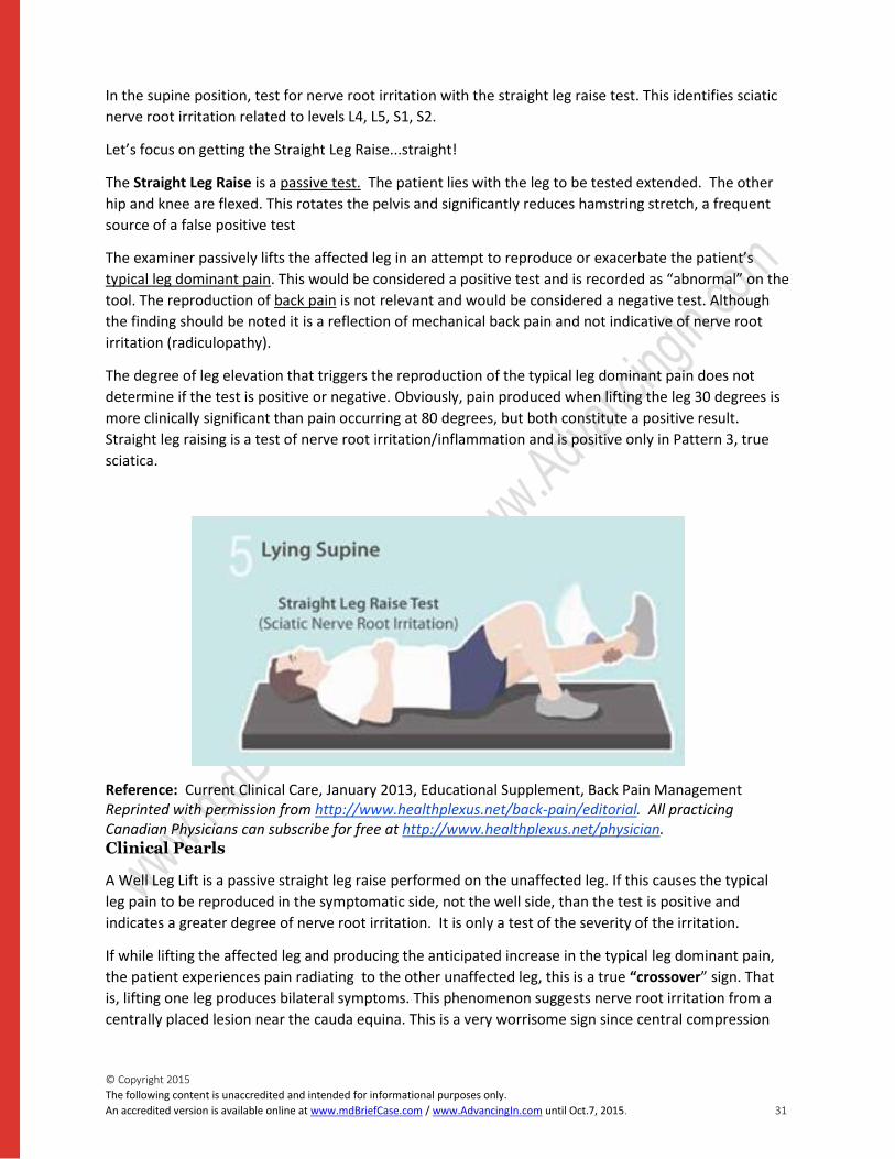
In the supine position, test for nerve root irritation with the straight leg raise test. This identifies sciatic
nerve root irritation related to levels L4, L5, S1, S2.
Let’s focus on getting the Straight Leg Raise...straight!
The Straight Leg Raise is a passive test. The patient lies with the leg to be tested extended. The other
hip and knee are flexed. This rotates the pelvis and significantly reduces hamstring stretch, a frequent
source of a false positive test
The examiner passively lifts the affected leg in an attempt to reproduce or exacerbate the patient’s
typical leg dominant pain. This would be considered a positive test and is recorded as “abnormal” on the
tool. The reproduction of back pain is not relevant and would be considered a negative test. Although
the finding should be noted it is a reflection of mechanical back pain and not indicative of nerve root
irritation (radiculopathy).
The degree of leg elevation that triggers the reproduction of the typical leg dominant pain does not
determine if the test is positive or negative. Obviously, pain produced when lifting the leg 30 degrees is
more clinically significant than pain occurring at 80 degrees, but both constitute a positive result.
Straight leg raising is a test of nerve root irritation/inflammation and is positive only in Pattern 3, true
sciatica.
Reference: Current Clinical Care, January 2013, Educational Supplement, Back Pain Management Reprinted with permission from http://www.healthplexus.net/back-pain/editorial. All practicing Canadian Physicians can subscribe for free at http://www.healthplexus.net/physician. Clinical Pearls
A Well Leg Lift is a passive straight leg raise performed on the unaffected leg. If this causes the typical
leg pain to be reproduced in the symptomatic side, not the well side, than the test is positive and
indicates a greater degree of nerve root irritation. It is only a test of the severity of the irritation.
If while lifting the affected leg and producing the anticipated increase in the typical leg dominant pain,
the patient experiences pain radiating to the other unaffected leg, this is a true “crossover” sign. That
is, lifting one leg produces bilateral symptoms. This phenomenon suggests nerve root irritation from a
centrally placed lesion near the cauda equina. This is a very worrisome sign since central compression

© Copyright 2015
The following content is unaccredited and intended for informational purposes only.
An accredited version is available online at www.mdBriefCase.com / www.AdvancingIn.com until Oct.7, 2015. 32
can affect the lower sacral roots (S2, 3, 4) leading to an Acute Cauda Equina Syndrome, a surgical
emergency.
Physical Examination (cont’d)
Left Screen Right Screen
Core Tool : Section C: Supine INSERT VIDEO: Supine Examination. ( 1:30)
Clinical Pearls
Which of the following statements is true?
A) The straight leg raise is initiated by the patient to 70 degrees and then the clinician passively moves the leg to its limit.
Incorrect: The straight leg raise is a fully passive test. Actively raising the leg requires the patient to
exert effort and muscle contraction in the low back, abdomen and thigh that distort the results. A
patient with genuine acute sciatica will not be able to initiate the movement.
B) The straight leg raise can be done in a sitting position as long as it is done passively.
Correct: The lying position is often preferred since the spine is stable and controlled therefore
eliminating potential posture changes that might trigger mechanical pain. However if it is done
passively, and correctly interpreted as a reproduction of the typical leg dominant pain, it is appropriate
to conduct the test in sitting. Many clinician will do both and compare result for consistency. This is only
valid if the opposite hip and knee are flexed in the supine position to place the pelvis in the same
rotation found in sitting.
C) If the history suggests leg pain but the straight leg raise is completely negative, even to 90 degrees, the clinician must dorsiflex the foot passively in the elevated position to provoke a potential sciatic irritation further.
Incorrect: The history is the key. If the patient has a history of leg dominant pain and if the history is
accurate, the SLR must be positive. If there is no pain on full straight leg raising then you must carefully
review the history. The patient may have back dominant pain with substantial referred (not radicular)
leg pain.
Once the typical leg pain has been exacerbated the pattern is confirmed. Hurting the patient again with
ankle dorsiflexion, pressure in the popliteal fossa or by flexing the hip and knee to 90 degrees and then
extending the knee is unnecessary and unkind.
Very rarely the acute root irritation in Pattern 3 may be transient. In these unusual cases the patient will
always have a recent onset profound neurological deficit.
© Copyright 2015
The following content is unaccredited and intended for informational purposes only.
An accredited version is available online at www.mdBriefCase.com / www.AdvancingIn.com until Oct.7, 2015. 33
D) It is not helpful to do a well leg raise to determine the degree of nerve root irritation for pain management strategies.
Incorrect: It is a helpful comparison to do with patients and if positive, it does indicate a higher degree
of nerve root irritation often requiring more pain medication and frequent changes of position. The test
will be positive only when the sciatica in the affected leg is sufficient to prevent the patient from even
fully extending the knee or limits the SLR to a few degrees of elevation.
Physical Examination (cont’d) Prone A) Neurology
As primary care clinicians, we don’t often use the prone position for examination but in the low back
patient this position is very useful for determining the mechanical back pain syndrome and for
completing the neurological examination to ensure that we don’t miss a key sensory loss sign. If you feel
that your patient’s mobility limits them getting into the prone position, you may want to use a step stool
by the examination bed or even have a yoga mat in the office so that you can have them prone on the
floor since this is often a position for exercise or recovery.
In the prone position, the examination includes:
o femoral nerve stretch test (L3 and 4), o power test for the gluteus maximus (S1) o saddle sensation testing (S2, 3, 4) o prone passive back extension movement test.
The femoral nerve stretch is similar in principle to the straight leg raise test. It is a passive test that
stretches the femoral nerve (L2, 3, 4) to determine if there are nerve root irritation symptoms present.
The examiner lifts the patient’s extended leg into further extension to try and reproduce their typical leg
dominant pain which would be in the anterior distal thigh. Extending the lumbar spine frequently
produces low back pain. This is a mechanical response and, while it may be quite painful, is not a
positive test. This test is optional and may be reserved for the patient whose dominant symptom is
constant anterior thigh pain. As an alternative to prone position, it can be done with the patient in side
lying position also.

© Copyright 2015
The following content is unaccredited and intended for informational purposes only.
An accredited version is available online at www.mdBriefCase.com / www.AdvancingIn.com until Oct.7, 2015. 34
Reference: Current Clinical Care, January 2013, Educational Supplement, Back Pain Management Reprinted with permission from http://www.healthplexus.net/back-pain/editorial. All practicing Canadian Physicians can subscribe for free at http://www.healthplexus.net/physician.
This test is most accurate when the knee is extended so that the quad muscle is relaxed to avoid a
false positive.
S1
Hip Extension: In the prone position, the patient repeatedly tenses and relaxes their buttocks. With
palpation the examiner will note loss of power or muscle tone on the affected side within 5 or 6
repetitions.
S2, 3, 4
Saddle Sensation: Every low back exam must include a check for saddle sensation. Sensation in the
perineum is subtended by the same sacral nerves that supply the bladder and bowel .This is essential to
ensure that you never miss a patient with an Acute Cauda Equina Syndrome.
o The best high-yield test to ensure that the sacral segments are intact is to test light touch in the midline at the top of the gluteal fold which is easily accessible in prone lying below the belt line.
o If sensation is altered and you suspect a sacral nerve abnormality, carefully review the bowel and bladder function, check for a crossover sign, test sensation in the rest of the perineal area and perhaps perform a rectal examination to determine rectal sphincter tone. Urgent referral may be indicated.
Physical Examination (cont’d)
B) Movement
Prone Passive Back Extension
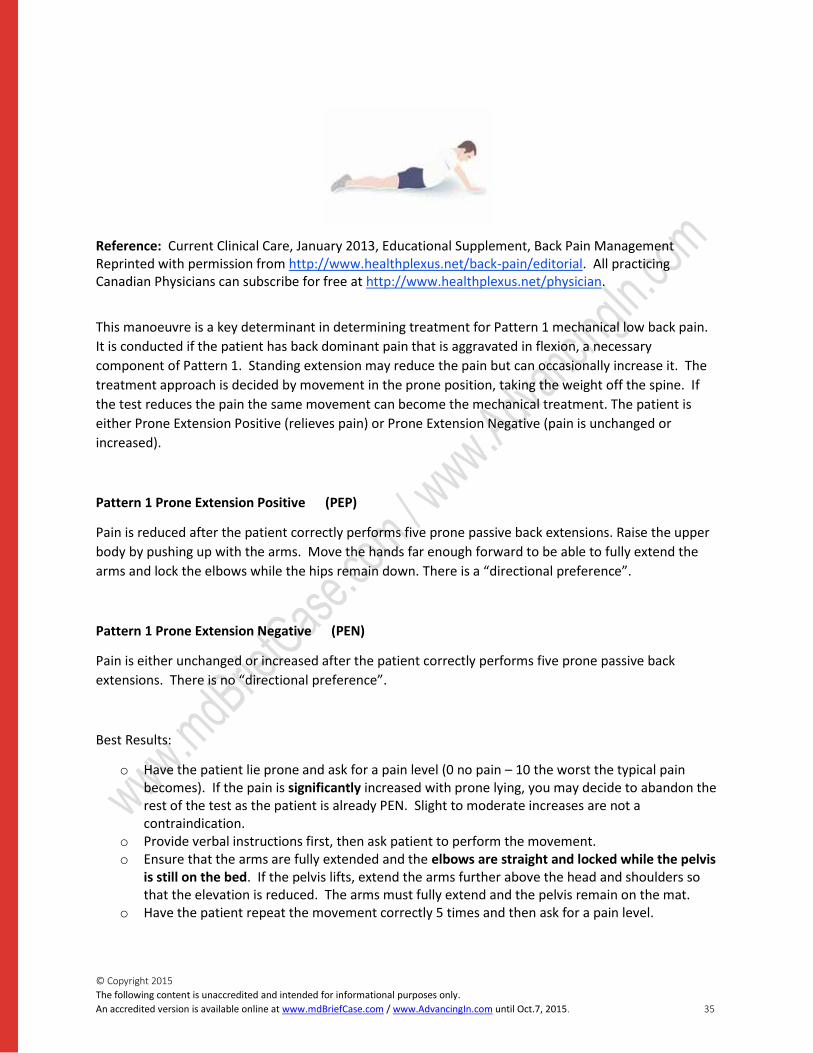
© Copyright 2015
The following content is unaccredited and intended for informational purposes only.
An accredited version is available online at www.mdBriefCase.com / www.AdvancingIn.com until Oct.7, 2015. 35
Reference: Current Clinical Care, January 2013, Educational Supplement, Back Pain Management Reprinted with permission from http://www.healthplexus.net/back-pain/editorial. All practicing Canadian Physicians can subscribe for free at http://www.healthplexus.net/physician.
This manoeuvre is a key determinant in determining treatment for Pattern 1 mechanical low back pain.
It is conducted if the patient has back dominant pain that is aggravated in flexion, a necessary
component of Pattern 1. Standing extension may reduce the pain but can occasionally increase it. The
treatment approach is decided by movement in the prone position, taking the weight off the spine. If
the test reduces the pain the same movement can become the mechanical treatment. The patient is
either Prone Extension Positive (relieves pain) or Prone Extension Negative (pain is unchanged or
increased).
Pattern 1 Prone Extension Positive (PEP)
Pain is reduced after the patient correctly performs five prone passive back extensions. Raise the upper
body by pushing up with the arms. Move the hands far enough forward to be able to fully extend the
arms and lock the elbows while the hips remain down. There is a “directional preference”.
Pattern 1 Prone Extension Negative (PEN)
Pain is either unchanged or increased after the patient correctly performs five prone passive back
extensions. There is no “directional preference”.
Best Results:
o Have the patient lie prone and ask for a pain level (0 no pain – 10 the worst the typical pain becomes). If the pain is significantly increased with prone lying, you may decide to abandon the rest of the test as the patient is already PEN. Slight to moderate increases are not a contraindication.
o Provide verbal instructions first, then ask patient to perform the movement. o Ensure that the arms are fully extended and the elbows are straight and locked while the pelvis
is still on the bed. If the pelvis lifts, extend the arms further above the head and shoulders so that the elevation is reduced. The arms must fully extend and the pelvis remain on the mat.
o Have the patient repeat the movement correctly 5 times and then ask for a pain level.

© Copyright 2015
The following content is unaccredited and intended for informational purposes only.
An accredited version is available online at www.mdBriefCase.com / www.AdvancingIn.com until Oct.7, 2015. 36
Physical Examination (cont’d)
Left Screen Right Screen
Core Tool : Section C: Prone INSERT VIDEO: Prone Examination. ( 2:30)
Clinical Pearls: Which if the following back dominant scenarios would fit a mechanical pattern of Pattern 1 PEN?
a) Flexion aggravated, standing extension partially relieved, prone extension relieved fully
b) Flexion aggravated, standing extension fully relieved, straight leg raise reproduces back pain
c) Extension aggravated, standing flexion relieved, prone extension aggravated
d) Extension aggravated in standing and in prone lying, flexion aggravated in standing but not aggravated as much as with extension
Case Interpretation
Let’s summarize our clinical findings on this patient. Please mark True or False by each of the following
statements:
Question Answer
The patient described his pain as intermittent, back dominant with buttock
pain
Answer: True. The patient described back pain and buttock pain and this is
classified as back rather than leg dominant.
T
The patient described his pain as intermittent, leg dominant pain with back
pain
Answer: False. Leg dominant pain is located at the gluteal fold or below and
is rated as being more intense and present more frequently than any back
pain that is experienced. This was not true with this patient.
F
The patient described his pain as being worse with flexion activities.
Answer: True, the patient described increased discomfort with sitting and
bending. On physical examination, he had more pain when bending forward
and while standing inflexion. Even if a patient continues to have pain with
T

© Copyright 2015
The following content is unaccredited and intended for informational purposes only.
An accredited version is available online at www.mdBriefCase.com / www.AdvancingIn.com until Oct.7, 2015. 37
standing extension, you are looking for which movement aggravates their
pain and which produces some relief.
The patient described his pain as being worse with extension activities.
Answer: False. The patient described his pain as being worse with flexion
activities. You may want to replay the history and standing examination
video sequences.
F
The patient examination demonstrated neurological deficits on sacral
sensation testing.
Answer: False. The patient has normal upper motor, lower motor and
sensory responses on examination in the sitting and supine video sequences.
F
The patient’s straight leg test was positive reproducing back pain.
Answer: False. A positive straight leg raise must reproduce the patient’s
typical radiating leg pain not just back pain. This test is conducted passively
with the opposite leg resting in flexion to ease any hamstring tightness.
F
The patient pain was relieved with prone extension movement.
Answer: True. In the prone lying position, the patient performed repeated
spine extension movement which alleviated his low back pain. This is called
prone extension positive (PEP).
T
Case Interpretation (cont’d)
Please download and complete the CORE tool on this patient. This will give you a chance to interpret the
examination and anticipate your management strategy.
Download the CORE Tool to be completed (PDF)
Note: this tool has been formatted to be completed electronically.
Competing the Core Tool
Left Panel Right Panel
Completed CORE Back Tool For Vignette 2 INSERT VIDEO: Completed Core Tool video
with Hamilton Hall (3 min 15 seconds)

© Copyright 2015
The following content is unaccredited and intended for informational purposes only.
An accredited version is available online at www.mdBriefCase.com / www.AdvancingIn.com until Oct.7, 2015. 38
Discussion Forum 1. The 5 high yield questions are used for identifying mechanical patterns of back pain. Are there
other questions you find useful for advising management strategies?
2. How would you adapt the physical examination to an older person with limited mobility?
Resources Videos Description of the 4 patterns of Low Back Pain Description of the High Yield History Questions Taking a High Yield History with a Patient Part 1: Conducting a Physical Examination - Standing Part 2: Conducting a Physical Examination - Sitting Part 3: Conducting a Physical Examination - Lying Supine Part 4: Conducting a Physical Examination - Lying Prone Application of the CORE Back Tool to a Patient Case
Articles
Atlas SJ, Deyo RA, Patrick DL, Convery K, Keller RB, Singer DE. The Quebec Task Force classification for Spinal Disorders and the severity, treatment, and outcomes of sciatica and lumbar spinal stenosis , BMC Musculoskelet Disord. 2012 Nov 28;13:236
Current Clinical Care, January 2013, Educational Supplement, Back Pain Management Reprinted with permission from http://www.healthplexus.net/back-pain/editorial. All practicing
Canadian Physicians can subscribe for free at http://www.healthplexus.net/physician. Medical Services Commission of British Columbia, Guidelines and Protocols Advisory Committee.
Rheumatoid Arthritis: Diagnosis, Management and Monitoring. C 2012 [cited 2013 April 30]. Available
from http://www.bcguidelines.ca/.
Clinical Methods: The History, Physical, and Laboratory Examinations. 3rd edition.Walker HK, Hall WD,
Hurst JW, editors. Boston: Butterworths; 1990.
Tools CORE Back Tool
Other
© Copyright 2015
The following content is unaccredited and intended for informational purposes only.
An accredited version is available online at www.mdBriefCase.com / www.AdvancingIn.com until Oct.7, 2015. 39
Completed CORE Tool (based on case scenario provided in vignette 2) Slideshow overview of 4 patterns Overview of 4 patterns (PDF)
Vignette 3: Patient Self-Management Main This vignette introduces the key skills required to effectively incorporate Patient Self Management (PSM) into discussions with patients with low back pain.
Upon completion of this vignette, the learner will be able to:
Describe the steps for engaging your patient in a self management strategy for improved sustainability of outcomes.
Introduction: Definitions and Concepts
This lesson introduces the key skills required to introduce Patient Self Management (PSM) into your
practice. The Personal Action Planning (PAP) Clinical Tool will be used as a model for practice
implementation. While this tool can be used for other clinical conditions, the purpose of this vignette is
to apply the theory and clinical tool to low back pain practice.
Patient Self Management (PSM) has been embraced by a number of health organizations throughout
the world, including the American Medical Association (Physician resource guide to patient self-
management support), the Health Council of Canada (Self-management support for Canadians with
chronic health conditions: A focus for primary health care). As well, organizations in Australia, China,
provincial ministries of health in Canada, and the Stanford Chronic Disease Self-Management Program
have all implemented and used Self-Management in practice. Most of the programs have focused on
chronic conditions such as diabetes, mood disorders and cardiovascular disease. However, the current
international low back guidelines all support the use of active self-management with appropriate back
education as a key recommendation in the clinical guidelines.
“Around 90% of the care a person needs to manage a chronic disease must come directly from the
patient. Evidence is growing that self-management interventions, such as self-monitoring and decision
making, lead not only to improvements in health outcomes and health status, but also to increased
patient satisfaction and reductions in hospital and emergency room costs.”
Reference: California Healthcare Foundation (2013). Patient Self-Management. Retrieved 19 March
2013, from: http://www.chcf.org/topics/patient-self-management.
The Continuum of Patient Activation

© Copyright 2015
The following content is unaccredited and intended for informational purposes only.
An accredited version is available online at www.mdBriefCase.com / www.AdvancingIn.com until Oct.7, 2015. 40
Patient Education
Patient Self-Monitoring Patient Self-Management
The process of activating patients to be aware, involved and engaged in their own care is a partnership
where the health care provider and the patient each play a unique role. The health care provider
remains an expert in the diagnosis and prescription of evidence-based treatment. However, the patient
is an expert in knowing what will work in their lives, their fears, their barriers and their experience.
Patient Education is the process of providing medical and health information from the health care
provider to the patient and family with the goal of learning condition specific facts that will frame
treatment recommendations. This is often in the form of a one-way fact sheet whether written, verbal
or electronic. Some patient education interventions will be more interaction utilizing patient risk
assessment, questions and answers or group discussion groups. Patient education can occur in a one-to-
one interaction with a provider, in a group setting in a medical facility or in the community or
individually in a more self-directed manner.
Patient Self-Monitoring is a task(s) that the patient is given by the provider that allows the patient to
gather information about themselves in-between medical visits for the purpose of monitoring treatment
response, symptom changes or emergence of new issues. This information is then shared with the
provider at medical visits so that the provider can have a broader perspective of the condition response
and symptom development before recommending the next steps in the investigation or management
process. Some examples would include log books for recording pain levels with activity, home blood
pressure instruments and food diaries. Self-monitoring can be followed in a health care provider visit, a
telephone call and even web-based systems where both patient and provider have secure and
confidential access.
Can you describe Patient Self Management ?
What is the definition?
Give an Example
What is Patient Self-Management Historically, clinicians treat patients as the subject of care. Care is something 'done to or for them'. A
more recent view treats the patient as a partner in their own care. Patients come with their own
knowledge of self and in many ways are expert in their selves and their conditions. By coupling this
knowledge with the knowledge clinicians have, patients and clinicians can be partners in their care.
Some describe this as a form of collaborative care .
Reference: Bodenheimer et al, Patient Self-management of Chronic Disease in Primary Care, JAMA,
November 20, 2002—Vol 288, No. 19 2469
Patients know themselves, their capabilities and limits. Indeed, they are experts in their own lives!

© Copyright 2015
The following content is unaccredited and intended for informational purposes only.
An accredited version is available online at www.mdBriefCase.com / www.AdvancingIn.com until Oct.7, 2015. 41
“If physicians view themselves as experts whose job is to get patients to behave in ways that reflect that
expertise, both will continue to be frustrated…Once physicians recognize patients as experts on their
own lives, they can add their medical expertise to what patients know about themselves to create a plan
that will help patients achieve their goals.”
Funnell & Anderson JAMA 2000;284:1709
Given the scarcity of clinical resources, the effectiveness of clinicians is greatly enhanced by partnering
with the patient (and their families) to devise a course of action that is created by the patient and
agreed to by the patient. The key to success is to promote:
Patient Self Efficacy
Patient Autonomy
Integration of motivational interviewing
Test Yourself 1. What is Patient Self Management? Choose one.
a) Patient Self Management is another name for patient education where the patient is taught
how to manage their health.
a) Patient Self Management is the individual’s ability to manage their symptoms, treatment,
physical and social consequences and lifestyle changes inherent in living with a health condition.
b) Patient Self Management is synonymous with disease self monitoring as this increases the role
of the patient in their own care.
c) Patient Self Management is a process for the patient to take control and advocate for
themselves without the need for a health care provider.
Motivational Interviewing What is the clinician’s role?
Clinicians provide:
Options for the patient to select a behaviour to change
Facilitation of goal setting by asking questions that allow the patient to develop details for their plan
Assistance with problem-solving barriers that the patient identifies
Interest and support for follow-up
Positive acknowledgement of any measure of success
How is this best achieved?
Motivational Interviewing is a scientifically tested method of counselling developed by Miller and Rollnick
and applied mostly as an intervention strategy in the treatment of lifestyle issues and chronic conditions.
It has been described by Miller and Rollnick as “directive, client-centred counselling style for eliciting
behaviour change”.
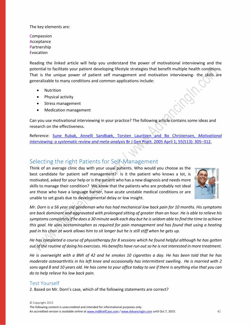
© Copyright 2015
The following content is unaccredited and intended for informational purposes only.
An accredited version is available online at www.mdBriefCase.com / www.AdvancingIn.com until Oct.7, 2015. 42
The key elements are:
Compassion Acceptance Partnership Evocation
Reading the linked article will help you understand the power of motivational interviewing and the
potential to facilitate your patient developing lifestyle strategies that benefit multiple health conditions.
That is the unique power of patient self management and motivation interviewing- the skills are
generalizable to many conditions and common applications include:
Nutrition
Physical activity
Stress management
Medication management
Can you use motivational interviewing in your practice? The following article contains some ideas and
research on the effectiveness.
Reference: Sune Rubak, Annelli Sandbæk, Torsten Lauritzen and Bo Christensen, Motivational
interviewing: a systematic review and meta-analysis Br J Gen Pract. 2005 April 1; 55(513): 305–312.
Selecting the right Patients for Self-Management Think of an average clinic day with your usual patients. Who would you choose as the
best candidate for patient self management? Is it the patient who knows a lot, is
motivated, asked for your help or is the patient who has a new diagnosis and needs more
skills to manage their condition? We know that the patients who are probably not ideal
are those who have a language barrier, have acute unstable medical conditions or are
unable to set goals due to developmental delay or low insight.
Mr. Dorn is a 56 year old gentleman who has had mechanical low back pain for 10 months. His symptoms
are back dominant and aggravated with prolonged sitting of greater than an hour. He is able to relieve his
symptoms completely if he does a 30 minute walk each day but he is seldom able to find the time to achieve
this goal. He uses acetaminophen as required for pain management and has found that using a heating
pad in his chair at work allows him to sit longer but he is still stiff when he gets up.
He has completed a course of physiotherapy for 8 sessions which he found helpful although he has gotten
out of the routine of doing his exercises. His benefits have run out so he is not interested in more treatment.
He is overweight with a BMI of 42 and he smokes 10 cigarettes a day. He has been told that he has
moderate osteoarthritis in his left knee and occasionally has intermittent swelling. He is married with 2
sons aged 8 and 10 years old. He has come to your office today to see if there is anything else that you can
do to help relieve his low back pain.
Test Yourself 2. Based on Mr. Dorn’s case, which of the following statements are correct?

© Copyright 2015
The following content is unaccredited and intended for informational purposes only.
An accredited version is available online at www.mdBriefCase.com / www.AdvancingIn.com until Oct.7, 2015. 43
a) Mr. Dorn is not a good candidate for patient self-management as he is demonstrating a moderate risk of his symptoms becoming chronic
b) Mr. Dorn is not a good candidate for patient self-management because progress will only be made if he can participate in his plan on a daily and consistent basis.
c) Mr. Dorn is not a good candidate for patient self-management as he has multiple health concerns (knee osteoarthritis, smoker and overweight) which will confuse the ability to set a clear and detailed goal.
d) Mr. Dorn is not a good candidate for patient self management because when asked what goal he would like to work on to address his health concern, he answers that he is not ready to work on any behavioural changes.
Personal Action Planning Tool INSERT VIDEO: Patient Self Management Tool
l Personal Action Planning Tool
Goal Setting Patient self management is a process that can be assisted by the guidance of a clinical tool like the
personal action planning tool
The concept of using brevity and patient self management action planning together
evolved because of the need for an efficient and effective process to facilitate patient-
centred goal setting in time-pressured clinical settings. Based on evidence
from multiple theoretical frameworks, including self-management support research,
behavioural psychology, and motivational interviewing, the concept of brief action planning was
first developed by Dr. Steven Cole MD in 2002.
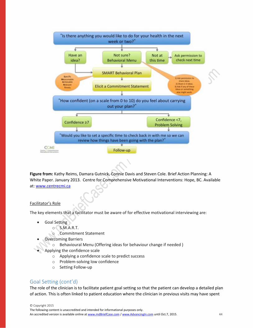
© Copyright 2015
The following content is unaccredited and intended for informational purposes only.
An accredited version is available online at www.mdBriefCase.com / www.AdvancingIn.com until Oct.7, 2015. 44
Figure from: Kathy Reims, Damara Gutnick, Connie Davis and Steven Cole. Brief Action Planning: A
White Paper. January 2013. Centre for Comprehensive Motivational Interventions: Hope, BC. Available
at: www.centrecmi.ca
Facilitator’s Role
The key elements that a facilitator must be aware of for effective motivational interviewing are:
Goal Setting o S.M.A.R.T. o Commitment Statement
Overcoming Barriers o Behavioural Menu (Offering ideas for behaviour change if needed )
Applying the confidence scale o Applying a confidence scale to predict success o Problem-solving low confidence o Setting Follow-up
Goal Setting (cont’d) The role of the clinician is to facilitate patient goal setting so that the patient can develop a detailed plan
of action. This is often linked to patient education where the clinician in previous visits may have spent

© Copyright 2015
The following content is unaccredited and intended for informational purposes only.
An accredited version is available online at www.mdBriefCase.com / www.AdvancingIn.com until Oct.7, 2015. 45
time educating patients on the importance of exercise, stress management, weight management or pain
management.
One framework for goal setting is called SMART Goals – Specific, Measurable, Achievable, Relevant and
Timed.
Specific goals are focused and detailed versus vague or general goals. Goals should be measurable
where outcomes or progress can be easily evaluated and recorded or charted. Goals should be 'within
reach' and achievable; but yet, not too difficult or too easy to attain. Goals should be relevant to the
patient’s interests, health, wellbeing and recovery. Goals should have clear and realistic time frames.
Goals without deadlines or time targets can be overlooked and are at higher risk for failure.
Patients are much more likely to follow through with the action required to achieve these goals when
they participate directly in establishing them; rather than have someone else (e.g. a clinician) establish
goals on their behalf.
SMART goals form the basis for a clear commitment from the patient. After goals have been
established, the clinician or coach elicits a final “commitment statement”. The strength of the
commitment statement predicts success on a personal action plan. We are more likely to believe and do
what we hear ourselves say!
Can you write a detailed goal for yourself so that you can see what it feels like?
Is there anything that you would like to do for your Health? What is it?
How would you like to do it?
Where will you do it?
When would you like to do it?
How often will you do it ?
Can you tell me in your words what you have committed to doing for your health ?
Overcoming Barriers Patients may create barriers for a number of reasons possibly related to their personal circumstances,
their family commitments or their work situations. Socioeconomic factors such as financial instability,
low education level or psychosocial factors can be part of the complexity of dealing with barriers. In
clinical practice, we often interpret barriers as “patient resistance” and we may lack the skills or time to
address them.
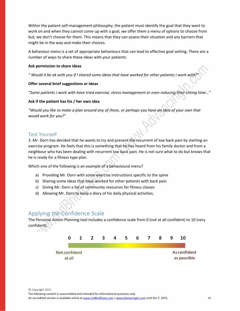
© Copyright 2015
The following content is unaccredited and intended for informational purposes only.
An accredited version is available online at www.mdBriefCase.com / www.AdvancingIn.com until Oct.7, 2015. 46
Within the patient self-management philosophy, the patient must identify the goal that they want to
work on and when they cannot come up with a goal, we offer them a menu of options to choose from
but; we don’t choose for them. This means that they can assess their situation and any barriers that
might be in the way and make their choices.
A behaviour menu is a set of appropriate behaviours that can lead to effective goal setting. There are a
number of ways to share these ideas with your patients:
Ask permission to share ideas
“ Would it be ok with you if I shared some ideas that have worked for other patients I work with?”
Offer several brief suggestions or ideas
“Some patients I work with have tried exercise, stress management or even reducing their sitting time…”
Ask if the patient has his / her own idea
“Would you like to make a plan around any of these, or perhaps you have an idea of your own that
would work for you?”
Test Yourself 3. Mr. Dorn has decided that he wants to try and prevent the recurrent of low back pain by starting an
exercise program. He feels that this is something that he has heard from his family doctor and from a
neighbour who has been dealing with recurrent low back pain. He is not sure what to do but knows that
he is ready for a fitness type plan.
Which one of the following is an example of a behavioural menu?
a) Providing Mr. Dorn with some exercise instructions specific to the spine
b) Sharing some ideas that have worked for other patients with back pain
c) Giving Mr. Dorn a list of community resources for fitness classes
d) Allowing Mr. Dorn to keep a diary of his daily physical activities.
Applying the Confidence Scale The Personal Action Planning tool includes a confidence scale from 0 (not at all confident) to 10 (very
confident).

© Copyright 2015
The following content is unaccredited and intended for informational purposes only.
An accredited version is available online at www.mdBriefCase.com / www.AdvancingIn.com until Oct.7, 2015. 47
It is important to understand how confident the patient is in being able to achieve the goals they have
set. Confidence is a predictor of success and those who are confident believe that they can achieve their
goals. This is called self-efficacy. Higher self-efficacy is associated with healthier behaviours and better
outcomes. The number 7/10 or higher is assessed as Confident.
On the other hand, if a patient has low self-confidence, below 7, we must respect the right of the
individual to not change and acknowledge that ambivalence regarding change. This is an important part
of the process and the rating may indicate that the goal and plan are not yet suitable or even that the
individual is not yet ready.
Bandura, 1983; Lorig et al, Med Care 2001; Bodenheimer review, CHCF 2005; Bodenheimer, Pt Ed Couns
2009.
Applying the Confidence Scale (cont’d) If patients have low confidence (less than 7) then one needs to use Problem Solving skills to engage the
patient further. (See figure at right.)
Figure from: Kathy Reims, Damara Gutnick, Connie Davis and Steven Cole. Brief Action Planning: A White
Paper. January 2013. Centre for Comprehensive Motivational Interventions: Hope, BC. Available at:
www.centreCMI.ca
This may include using a behavioural menu or resetting the goal.
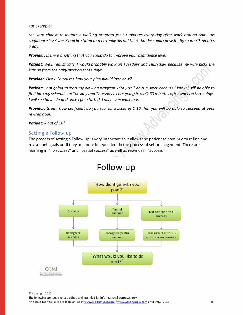
© Copyright 2015
The following content is unaccredited and intended for informational purposes only.
An accredited version is available online at www.mdBriefCase.com / www.AdvancingIn.com until Oct.7, 2015. 48
For example:
Mr Dorn choose to initiate a walking program for 30 minutes every day after work around 6pm. His
confidence level was 3 and he stated that he really did not think that he could consistently spare 30 minutes
a day.
Provider: Is there anything that you could do to improve your confidence level?
Patient: Well, realistically, I would probably walk on Tuesdays and Thursdays because my wife picks the
kids up from the babysitter on those days.
Provider: Okay. So tell me how your plan would look now?
Patient: I am going to start my walking program with just 2 days a week because I know I will be able to
fit it into my schedule on Tuesday and Thursdays. I am going to walk 30 minutes after work on those days.
I will see how I do and once I get started, I may even walk more.
Provider: Great, how confident do you feel on a scale of 0-10 that you will be able to succeed at your
revised goal.
Patient: 8 out of 10!
Setting a Follow-up The process of setting a Follow-up is very important as it allows the patient to continue to refine and
revise their goals until they are more independent in the process of self-management. There are
learning in “no success” and “partial success” as well as rewards in “success”

© Copyright 2015
The following content is unaccredited and intended for informational purposes only.
An accredited version is available online at www.mdBriefCase.com / www.AdvancingIn.com until Oct.7, 2015. 49
Figure from: Kathy Reims, Damara Gutnick, Connie Davis and Steven Cole. Brief Action Planning: A White
Paper. January 2013. Centre for Comprehensive Motivational Interventions: Hope, BC. Available at:
www.centreCMI.ca.
3 Key Questions In the CORE Back tool we abbreviate the process to 3 key questions:
Key Messages INSERT VIDEO: Patient Self Management Application with Patient
Now that you have learned some patient self management skills, try it with your next patient and see
how easily you can work it into your practice. On average, the process takes 2-4 minutes, that’s all!
Key Messages:
The patient drives the process and the clinician facilitates
Motivation Interviewing principles are the foundation
Very successful in chronic conditions for sustained behavioural outcomes

© Copyright 2015
The following content is unaccredited and intended for informational purposes only.
An accredited version is available online at www.mdBriefCase.com / www.AdvancingIn.com until Oct.7, 2015. 50
Emerging literature in Low Back Pain suggests that Patient Education is key to the ability for a patient to self manage
Discussion Forum 1. How would you approach a patient with low back pain who is interested in exercise but
cannot consistently find the required time?
2. How would you approach a patient with low back pain who has come up with a goal
specific plan but identifies their confidence level as a 3 out of 10?
Resources Tools
Personal Action Planning for Patient Self Management Tool.
Videos
List all videos included in the module
Articles
Bodenheimer et al, Patient Self-management of Chronic Disease in Primary
Care, JAMA, November 20, 2002—Vol 288, No. 19 2469
California Healthcare Foundation (2013). Patient Self-Management. Retrieved 19 March 2013, from: http://www.chcf.org/topics/patient-self-management. Current Clinical Care, January 2013, Educational Supplement, Back Pain Management. Reprinted with permission from http://www.healthplexus.net/back-pain/editorial. All practicing Canadian Physicians can subscribe for free at http://www.healthplexus.net/physician. Du S, Yuan C, Xiao X, Chu J, Qiu Y, Qian H. Self-management programs for chronic musculoskeletal pain
conditions: a systematic review and meta-analysis. Patient Educ Couns. 2011 Dec;85(3):e299-310.
J H Barlow, J Sturt and H Hearnshaw. Self-management interventions for people with chronic conditions
in primary care: Examples from arthritis, asthma and diabetes. Health Education Journal: 61(4) 2002; 365-
378.

© Copyright 2015
The following content is unaccredited and intended for informational purposes only.
An accredited version is available online at www.mdBriefCase.com / www.AdvancingIn.com until Oct.7, 2015. 51
Kathy Reims, Damara Gutnick, Connie Davis and Steven Cole. Brief Action Planning: A White Paper.
January 2013. Centre for Comprehensive Motivational Interventions: Hope, BC. Available at:
www.centreCMI.ca
Sune Rubak, Annelli Sandbæk, Torsten Lauritzen and Bo Christensen, Motivational interviewing: a
systematic review and meta-analysis Br J Gen Pract. 2005 April 1; 55(513): 305–312.
Wilk V, Palmer HD, Stosic RG, McLachlan AJ. Evidence and practice in the self-management of low back pain: findings
from an Australian internet-based survey. Clin J Pain. 2010 Jul-Aug;26(6):533-40.
Vignette 4: Clinical Toolkit
Main This vignette will review each component of the Provincial Toolkit for Low Back Pain, including the newly developed CORE Back Tool & Guide and provide an interactive opportunity to apply the tools to a case scenario.
Upon completion of the vignette, learners will be able to:
Effectively apply the CORE Back Tool and other relevant clinical tools to a case based scenario in order to integrate course learning.
Overview of the Tool Kit The Toolkit for Low Back Pain consists of the CORE Back Tool and accompanying Guide plus 4 Supporting
Tools and 9 Reference Tools. The purpose of the toolkit is to provide the primary care provider with one
point-of-care tool, CORE, and then additional supporting tools for more in-depth screening or
management and an array of reference tools to customize your patient education options. Clinical tools
have been proven to be useful for the implementation of evidence based information into clinical
practice. All of the tools have been informed by primary care focus groups and tested through in-person
education sessions with primary care providers to ensure that they are relevant, useful and applicable to
primary care patient visits.

© Copyright 2015
The following content is unaccredited and intended for informational purposes only.
An accredited version is available online at www.mdBriefCase.com / www.AdvancingIn.com until Oct.7, 2015. 52
Figure 1. Overview of Provincial Toolkit
The CORE Back Tool The CORE Back Tool The CORE tool will guide the primary care clinician in recognizing common mechanical back pain
syndromes while screening for other conditions where further investigations and referrals may be
required. The CORE Guide is an easy-to-follow explanation of each sections of the tool so the clinician
can understand the rationale and the key messages, as well as when this information is pertinent to
clinical practice and what resources support the recommendations.
The CORE tool is designed for primary care clinical decision-making and not as a detailed rehabilitation
treatment tool. It is expected that the primary care clinician will be able to deliver education and
exercise for patient self-management but if goal-specific rehabilitation is required then a referral to a
therapist may be indicated.
Download the CORE Tool (PDF)
Download the CORE Guide (PDF)
[INSERT JULIA’S VIDEO INTRODUCING THE CORE TOOL]
Section A: History The clinical history is focused on 5 questions. Each question helps the provider determine if their
presentation is one of mechanical back pain (90% of patient presentations) or a more serious pathology
where investigation and referrals are required.
Toolkit
ClinicalTools(NEW!)• ClinicallyOrganizedRelevantExam
(CORE)BackTool• COREBackTool:AGuide
Suppor ngTools• OpioidRiskTool• Pa entEduca onInventory• PersonalAc onPlanningforPa ent
SelfManagement• TheKeeleStarTBackScreeningTool
ReferenceTools(foraddi onalsupport):FORPATIENTS
• BackBook• GeneralRecommenda onsforMaintaininga
HealthyBack:Pa entInforma on• BackPainExercisesforall4pa erns
• Pa ern#1Pa entEduca on• Pa ern#2Pa entEduca on• Pa ern#3Pa entEduca on• Pa ern#4Pa entEduca on
• SoYourBackHurts…• WhatyoushouldknowaboutAcutePain• WhatyoushouldknowaboutChronicPainFORPROVIDERS
• AcuteandSubacuteLowBackPain(LBP)–PharmacologicalAlterna ves
• AcuteandSubacuteLowBackPain(LBP)–TopicalandHerbalProducts
• EvidenceSummaryforManagementofNon-specificChronicLowBackPain(POCKET)
• OpioidManager• SwitchingOpioids

© Copyright 2015
The following content is unaccredited and intended for informational purposes only.
An accredited version is available online at www.mdBriefCase.com / www.AdvancingIn.com until Oct.7, 2015. 53
For more information on how to ask these questions, please see Vignette 2 and/or the videos in the
resources section.
Figure 2. Section A (History) of CORE Back Tool
Test Yourself Which of the following questions would lead you to the red flags for screening?
1) Where is your pain the worst? 2) Is your pain intermittent or constant? 3) Do you have tingling in your legs? 4) Does bending backward increase your pain?
Section B: Red Flag Screening
Question 4 identifies the key question for Cauda Equina, a surgical emergency and Question 5, identifies
inflammatory back pain as a systemic rheumatologic condition which is the most common of the non-
mechanical type of low back pain accounting for 5% of all back pain.
Practice Tip:
Question 4 (Have you had any unexpected accidents with your bowel or bladder function since this
episode of your low back/leg pain started?) and
Question 5 (If age of onset < 45 years, are you experiencing morning stiffness in your back > 30
minutes?) relate directly to red flag screening.

© Copyright 2015
The following content is unaccredited and intended for informational purposes only.
An accredited version is available online at www.mdBriefCase.com / www.AdvancingIn.com until Oct.7, 2015. 54
For a reminder of how to ask your 5 high yield questions, re-visit section A of the CORE back tool.
The Red Flag box is an organized method of cueing the practitioner to questions to identify serious
pathology if the patient does not easily fall into a pattern of mechanical back pain or if the patient is not
responsive to evidence-based treatment.
N-I-F-T-I is the acronym for:
Neurological
Infection
Fracture
Tumour
Inflammatory
Lastly, if there are no red flags, be sure to tick the box (no red flags) indicating that you have considered
the possibilities as this will be a good record for future and a medical-legal requirement since this is
guideline-recommended as a usual standard of care.
Figure 3. Section B (Red Flags) of CORE Back Tool

© Copyright 2015
The following content is unaccredited and intended for informational purposes only.
An accredited version is available online at www.mdBriefCase.com / www.AdvancingIn.com until Oct.7, 2015. 55
Section B: Radiology Criteria
Figure 4. Section B (Radiology Criteria) of CORE Back Tool
Most radiology reports will show abnormalities that are related to normal aging of the spinal structures
but are not often the cause of specific low back symptoms. Many patients are pain free with high levels
of function yet, if imaged, the results would indicate degenerative changes including disc bulging, canal
narrowing (stenosis) and osteophyte development. These degenerative changes documented in a report
can often confuse the patient and the clinician and change of the focus of clinical management to
disability instead of symptom control and increased function.
“Diagnostic imaging is indicated for patients with low back pain only if they have severe progressive
neurologic deficits or signs or symptoms that suggest a serious or specific underlying condition. In other
patients, evidence indicates that routine imaging is not associated with clinically meaningful benefits but
can lead to harms. Implementing a selective approach to low back imaging, as suggested by the
American College of Physicians and American Pain Society guideline on low back pain, would provide
better care to patients, improve outcomes, and reduce costs. “
Read More:
Chou,R.; Qaseem,A.; Owens,D.K.; Shekelle,P.; Clinical Guidelines Committee of the American College of
Physicians; Diagnostic imaging for low back pain: advice for high-value health care from the American
College of Physicians, Ann.Intern.Med., 2011, 154, 3, 181-189
Test Yourself Which of the following patients would you order an MRI on if they failed to respond to evidence-based
conservative treatment?
a) Ms. Ado has back dominant constant pain that is aggravated by flexion and is not relieved in prone lying extension. She is taking intermittent analgesia and states that exercise is difficult to do.
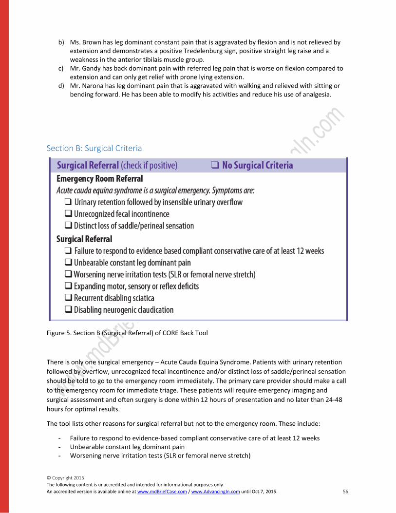
© Copyright 2015
The following content is unaccredited and intended for informational purposes only.
An accredited version is available online at www.mdBriefCase.com / www.AdvancingIn.com until Oct.7, 2015. 56
b) Ms. Brown has leg dominant constant pain that is aggravated by flexion and is not relieved by extension and demonstrates a positive Tredelenburg sign, positive straight leg raise and a weakness in the anterior tibilais muscle group.
c) Mr. Gandy has back dominant pain with referred leg pain that is worse on flexion compared to extension and can only get relief with prone lying extension.
d) Mr. Narona has leg dominant pain that is aggravated with walking and relieved with sitting or bending forward. He has been able to modify his activities and reduce his use of analgesia.
Section B: Surgical Criteria
Figure 5. Section B (Surgical Referral) of CORE Back Tool
There is only one surgical emergency – Acute Cauda Equina Syndrome. Patients with urinary retention
followed by overflow, unrecognized fecal incontinence and/or distinct loss of saddle/perineal sensation
should be told to go to the emergency room immediately. The primary care provider should make a call
to the emergency room for immediate triage. These patients will require emergency imaging and
surgical assessment and often surgery is done within 12 hours of presentation and no later than 24-48
hours for optimal results.
The tool lists other reasons for surgical referral but not to the emergency room. These include:
- Failure to respond to evidence-based compliant conservative care of at least 12 weeks - Unbearable constant leg dominant pain - Worsening nerve irritation tests (SLR or femoral nerve stretch)

© Copyright 2015
The following content is unaccredited and intended for informational purposes only.
An accredited version is available online at www.mdBriefCase.com / www.AdvancingIn.com until Oct.7, 2015. 57
- Expanding motor, sensory or reflex deficits - Recurrent disabling sciatica - Disabling neurogenic claudication
Section B: Yellow Flags Insert image
Figure 6. Section B (Barriers/Yellow Flags) of CORE Back Tool
Yellow flags are indications of psychosocial risk factor for the development of chronic low back pain. The
yellow flags are not a psychiatric diagnosis but do indicate if the patient is reliant on passive treatment,
avoids activities and movement for fear of pain, demonstrates some social withdrawal or low mood or
lacks a supportive work and home environment. If the yellow flags are positive and the patient has not
responded to treatment after 6 weeks, then the clinician may choose to use the StarT Back Tool to
identify if the patient has a low, medium or high risk of developing chronic pain in order to choose the
most appropriate treatment strategy at the earliest possible time for intervention.
Review the New Zealand Yellow Flags Guidelines for some examples of questions you may wish to ask
your patients to help you identify the Yellow Flags.
Test Yourself Ms. Jonah has had back dominant low back pain for 3 weeks. She states that she is fearful of exercise as
it many hurt her back more. She is tearful in the office and states that she would like to go back to work
but worries that she will not be able to do her duties. Which of the following options would you try with
her?
a) Refer her to a psychologist for cognitive behavioural therapy. b) Give her some relaxation tapes to reduce her worry. c) Provide her with education on “hurt versus harm” to improve her level of activity. d) Refer her to a psychiatrist as she may have clinical signs of depression.
Section C: Physical Examination The history leads the clinician to develop their differential diagnosis based on their patient’s symptoms.
The examination is conducted to support or refute your clinical diagnosis (history) and each test should
be interpreted within your clinical thinking. The body diagrams can be shaded according to the patient
history so that the clinician accurately records the areas of pain and can compare them in future visits.
Figure 7. Section C (Physical Examination) of CORE Back Tool
The physical examination section is laid out as a guideline for organizing your examination. The
examination components are divided into the patient’s positions of standing, sitting, kneeling, and
prone and supine lying so that the examiner can easily adjust the examination to the patient’s positions.

© Copyright 2015
The following content is unaccredited and intended for informational purposes only.
An accredited version is available online at www.mdBriefCase.com / www.AdvancingIn.com until Oct.7, 2015. 58
However, if you patient cannot sit and you wish to do the full examination in the lying position, then you
can adjust your tests to the that position. The highlighted examination components are the minimum
requirements for best practice to ensure that the clinician does not miss an important element. The
other tests can be conducted to clarify a clinical picture or provide the clinician with a comprehensive
approach to assessment.
The chart for documentation allows the clinician to record if a test is abnormal or normal but also to
document the findings.
Test Yourself How would you record the following clinical test and patient response?
On passive straight leg raise, the patient complains of reproducible low back pain at 45 degrees.
a)
Test Normal Abnormal Comments
Straight Leg Raise X Low Back Pain at 45 degrees
b)
Test Normal Abnormal Comments
Straight Leg Raise X Pain at 45 degrees
c)
Test Normal Abnormal Comments
Passive Hip Flexion X Low Back Pain at 45 Degrees
d)
Test Normal Abnormal Comments
Passive Hip Rotation X Hip Pain at 45 Degrees
For more information on physical assessment, please see related physical exam videos from vignette
2.
Section D: Assessment Insert image: “Section D”
Figure 8. Section D (Assessment) of CORE Back Tool

© Copyright 2015
The following content is unaccredited and intended for informational purposes only.
An accredited version is available online at www.mdBriefCase.com / www.AdvancingIn.com until Oct.7, 2015. 59
This section would be like your “A” in S-O-A-P charting format. In this section, you will commit to a
pattern of mechanical back pain or identify that your patient has non-spine related pain or spine pain
that does not fit a mechanical pattern. Occasionally, a patient may have more than one pattern
occurring at the same time. For example, you may have a patient with Pattern 4 Neurogenic claudication
evidenced by the fact that their leg pain is worse with walking and relieved with flexion. However, with
prolonged sitting flexion, they also develop back pain and have symptoms consistent with Pattern 1.
The use of pattern numbers is often helpful for clinicians to identify and discuss the mechanics of
treatment, however you may wish to describe the symptom dominance and aggravating positions to the
patient.
Section E: Patient Education Insert image: “Section E”
Figure 9. Section E (Patient Education) of CORE Back Tool
During your assessment, you may be able to advise the patient on the best positions for recovery and
scheduled rest by demonstrating or explaining the positions. The following articles clearly outline the
management of low back pain through positions and exercise.
The latest in Back Pain Management: Managing Back Dominant Pain
The latest in Back Pain Management: Managing Leg Dominant Pain
For Pattern 1, back dominant pain aggravated with flexion and relieved with extension, a “sloppy push-
up” or prone extension position will relieve pain. This patient will improve their sitting tolerance with an
extension roll in the curve of their lumbar spine.
For Pattern 2, back dominant pain aggravated with extension, this syndrome is less common but will
relieve quickly with sitting in a flexed position with hands on the ankles and then placing hands on the
knees to assist to return to neutral.
For Pattern 3, leg dominant pain aggravated by flexion, this patient will likely have to be lying for relief
and then may be able to tolerate the “z” position with legs elevated on a chair. They often will get worse
if left in this position for too long so their “dose” of the position has to be determined by the response
of their pain to the position.
Pattern 4, leg dominant pain aggravated by extension will respond to sitting or bending forward and
leaning on a shopping cart or podium. Activity modification and core stability is the best conservative
treatment for this group.
Test Yourself Based on reading the 2 articles in The latest in Back Pain Management, what would you suggest for the
following patient?

© Copyright 2015
The following content is unaccredited and intended for informational purposes only.
An accredited version is available online at www.mdBriefCase.com / www.AdvancingIn.com until Oct.7, 2015. 60
Ms. Sloka is a 43 year old office worker with a 3 week history of low back pain with occasional radiation
to her left buttock and posterior thigh. Forward flexion aggravates her pain to a 5/10 and standing
extension does reduce her pain but only to a 2/10. Prone lying extension relieves her pain to 0/10 after
2 repetitions. Her neurological examination is normal.
a) Take her off work so that she does not aggravate her condition by sitting.
b) Start a core stability program to increase her control and overall strength for activity.
c) Advise an x-ray since she is having leg pain and if normal, advise her to walk more for pain relief.
d) Advise her to do the prone extension movements frequently through the day and to reduce her
sitting and to sit with a lumbar roll.
Section F, G, and H. Section F is an abbreviated Patient Self Management tool and this has been taught in Vignette 3.
Figure 10. Section F (Goal Setting & Patient Self Management) of CORE Back Tool
Section G is an opportunity to document your treatment plan and use any applicable boxes. You may
wish to access the reference tools for medication management or patient education materials.
Figure 11. Section G (Recommendations) of CORE Back Tool
Section H is to document advice on follow-up and chart any additional information.
Figure 12. Section H (Follow-up) of CORE Back Tool
The patient Key Messages is a checklist of patient-friendly phrases that reinforce the guideline-based
key messages for optimal management.
Figure 13. “Key Messages for Your Patient” from CORE Back Tool
STarT Back Screening Tool
The Keele STarT Back Screening Tool (SBST) is a brief validated tool, designed to screen primary care
patients with low back pain for prognostic indicators that are relevant to initial decision making. This
tool is suitable when the Yellow Flags are positive and the patient has not responded to care in the first
4-6 weeks of a low back pain episode.

© Copyright 2015
The following content is unaccredited and intended for informational purposes only.
An accredited version is available online at www.mdBriefCase.com / www.AdvancingIn.com until Oct.7, 2015. 61
Figure 14. The Keele StarT Back Screening Tool
This tool allocates patients to different treatment pathways based on their prognosis (low, medium, or
high risk of poor outcome). This model is aimed at identifying the patients who require more
comprehensive psychosocial intervention earlier in the development of their symptoms to avoid or
reduce the impact of persistent disabling symptoms and improve patient outcomes.
Figure 15. Scoring System from The Keele StarT Back Screening Tool
Low Risk Medium Risk High Risk
Optimal Treatment Psychoeducation on
Hurt versus Harm,
Activity Promotion,
and Pain
Management
Cognitive Behavioural
Therapy for
management of
anxiety and mood
symptoms
Multidisciplinary
chronic pain program
for integration of
psychological and
physical therapies.
Download Keele StarT Back Screening Tool (PDF)
Read more: Keele University. StarT Back Screening Tool Website. Available at:
http://www.keele.ac.uk/sbst/

© Copyright 2015
The following content is unaccredited and intended for informational purposes only.
An accredited version is available online at www.mdBriefCase.com / www.AdvancingIn.com until Oct.7, 2015. 62
Hill et al. A Primary Care Back Pain Screening Tool: Identifying Patient Subgroups for Initial Treatment.
Arthritis & Rheumatism (Arthritis Care & Research). Vol. 59, No. 5, May 15, 2008, pp 632-641.
Opioid Risk Assessment Tool The Opioid Manager is a comprehensive tool that has been developed for the Canadian Guideline for
Safe and Effective Use of Opioids for Chronic Non-Cancer Pain. Within this tool is the Opioid Risk
Assessment tool that allows a clinician to identify if their patient has an increased of addiction with
Opioid use. This is a safe and effective way of determining best medication choices and informing the
patient of risk of dependency.
Primary care providers may have already obtained some of the required patient information through
their cumulative patient profile and some questions need to be specifically answered with current
information. This tool is best administered through a discussison with the patient and not as a self-
administered patient questionnaire. If a clinician decides to proceed with opioid prescription, then the
full Opioid manager is available with dosage guidelines, patient contracts and recommendations on
short and long term use.
Download Opioid Risk Tool (PDF)
Patient Self Management The Personal Action Planning tool incorporates goal setting and action planning to engage the patient as
a partner in their recovery to play an active role in deciding what lifestyle behaviours to attempt to
improve. This is best applied in the case of Low Back Pain when patient education on key messages and
evidence-based treatment has occurred.
The vignette on Patient Self Management addresses the theory and practice of this tool. The CORE Back
Tool has a 3-question starter for initiating discussion and reminding the clinician of the components for
success. The supporting tool, Personal Action Planning, has a more detailed format where the patient’s
decisions and plan can be documented and followed up on future visits. This is a sample of a patient self
management tool; there are many others with similar core values that clinicians may already be
successfully using.
Download Personal Action Planning Tool (PDF)
For more information on patient self management, please see related patient self-management videos
from vignette 3.
If you haven’t yet completed vignette 3, this would be a good time to plan to review the vignette.
Module 3 contains an in-depth learning experience on patient self management. Upon completion of
this vignette, the learner will be able to describe the steps for engaging your patient in a self
management strategy for improved sustainability of outcomes.
Estimated time for completion: 45-60 minutes.
Patient Education Selector Guide Patient education is recognized as a key component of successful management of low back pain.
However, every patient is different in what type of material is best suited to their comprehension,

© Copyright 2015
The following content is unaccredited and intended for informational purposes only.
An accredited version is available online at www.mdBriefCase.com / www.AdvancingIn.com until Oct.7, 2015. 63
learning style and memory capacity. Some patients prefer material that contains pictures and simple
phrases while others like descriptive paragraphs that take a reader through many concepts. For this
reason, we have identified the common low back patient education material that is currently used most
often in Canadian health care settings and we will let you and your patient select the appropriate
patient education format. In the Patient Education Selector Guide, you will find a brief overview of the
materials as well as a link to the sites where they are available.
The Saskatchewan Ministry of Health adopted the four patterns of mechanical low back pain in 2010
and developed patient education handouts for each pattern that provides photographs and descriptions
of the advised exercises and positions. Patients find the hand-outs clear and easy to understand. In
addition to the pattern specific exercises, the Saskatchewan site also provides a general information
sheet including posture, exercise and lifestyle changes
The Back Book is a publication from the United Kingdom that has been adopted by the Workplace Safety
and Insurance Board (WSIB) for Ontario. It was initially published in 1996 and the 8th edition was
released in 2011. Since March of 2012, it has been distributed by the WSIB Ontario to all new low back
pain claimants upon injury to increase patient awareness of principles for recovery and for the worker to
discuss with their health care professional. This 20 page small booklet is written in simple and
straightforward language and covers the essential key messages related to pain management, anxiety
and muscle tension, role of activity and prevention of chronic pain.
“So Your Back Hurts” was released by the Institute for Work and Health in 2009. This 28 page booklet is
aimed at acute low back pain and provides the patient with information on modifying activity and work
to maintain movement.
Alberta has released Low Back Pain guidelines through their Towards Optimizing Practice (TOP) website.
These guidelines are seeded in the international guidelines and have been packaged into Patient
Handouts on acute and chronic low back pain. The key messages have been incorporated and they are
presented in one-page formats.
Download Patient Education Inventory Tool (PDF)
The Tool Kit The image below illustrates the links between various sections of the CORE Back Tool and related
supporting and reference tools, available for additional information.

© Copyright 2015
The following content is unaccredited and intended for informational purposes only.
An accredited version is available online at www.mdBriefCase.com / www.AdvancingIn.com until Oct.7, 2015. 64
Figure 16. Ontario Ministry of Health, Low Back Pain Strategy, Clinical Tool Kit
Case Study Now that you are familiar with the tools, we would like to give you the opportunity to apply the
principles to a real patient case. Given the case details, your job will be to fill out the CORE tool on this
patient. Once you have completed this, you will be able to download a CORE Tool completed by Drs
Alleyne and Hamilton to compare your answers.
Download the CORE Tool to be completed (PDF)
Note: this tool has been formatted to be completed electronically.
Mr. Bob Stekel, 61 years old, has already had several episodes of low back pain over the last 10 years.
Several months ago he noticed that in addition to his back pain, his legs were starting to hurt
occasionally but now his leg pain seems more frequent. The pain, which occurs mainly in his left
posterior thighs, begins whenever he walks for longer than a few minutes—the farther he goes, the
more his legs ache, forcing him to sit down. Within a few minutes, his leg pain disappears completely
and he can start walking again. His pain is the least in the morning and he does not feel any stiffness,
numbness or tingling.
He has noticed that he can’t manage his pain and it is different than before so he has come to see you
for advice. He states that he was walking in the grocery store when he noticed that his pain became very
sharp and spread down his “good leg”. He had to grasp the cart and lean forward to complete his
shopping. He has not been able to figure out what triggered his pain and he visited a massage therapist
to relieve his spasm which only gave him temporary relief.

© Copyright 2015
The following content is unaccredited and intended for informational purposes only.
An accredited version is available online at www.mdBriefCase.com / www.AdvancingIn.com until Oct.7, 2015. 65
Bob has noticed that he has put on 15 pounds of additional weigh in the last few months and he is
concerned that this may lead to other health problems because both his parents had high blood
pressure and Bob is a smoker. He has cut down to 1 pack a day but has not been able to reduce any
further. He has had a few episodes of loss of control of his bladder but he thinks this may be due to his
prostate which was enlarged on his last check-up with his doctor.
He had an MRI and X-ray about 4 years ago that described his spine as degenerative with stenosis and
some bulging discs.
On examination, forward flexion produces some mild low back discomfort but no leg pain. Extension
produces some low back pain that radiates into the left leg when it is repeated or prolonged.
Neurological examination for DTR, myotomes and dermatomes are normal. Straight Leg raise produces
mild back pain. Femoral nerve stretch produces low back pain. Prone lying extension position is difficult
to maintain as Bob becomes very uncomfortable with low back pain spreading into the buttock. Upper
motor neuron testing is normal.
“My back pain is a nuisance, but it’s the leg pain that keeps me from doing the things I want to do. I
can’t go shopping with my wife without stopping every few minutes to sit and rest. When I am not active
my legs feel fine, but as soon as I get going I am in trouble again. Should I go to the Emergency room if it
happens again? ”
What does the CORE tool look like for Bob? Please complete the CORE tool based on the case scenario
above.
Download the CORE Tool to be completed (PDF)
Discussion Forum Now that you’ve completed your CORE tool, download an example of the CORE tool completed by Drs
Alleyne and Hall to compare your answers.
Completed CORE Back Tool (PDF)
1. Are there any elements in these tools, which a patient could complete while in the waiting
room?
2. Of the resources listed in the patient education, select your favourite. What are the benefits and
barriers of that tool?

© Copyright 2015
The following content is unaccredited and intended for informational purposes only.
An accredited version is available online at www.mdBriefCase.com / www.AdvancingIn.com until Oct.7, 2015. 66
Resources
Provincial Toolkit for Primary Care Providers
Overview of Toolkit
Clinically Organized Relevant Exam (CORE) Back Tool
CORE Back Guide
Opioid Risk Tool
Personal Action Planning Tool
Patient Education Inventory Tool
StarT Back Screening Tool
Video
Introducing the Core Back Tool
Guidelines
Canadian Guideline for Safe and Effective Use of Opioids for Chronic Non-Cancer Pain
Articles
Chou,R.; Qaseem,A.; Owens,D.K.; Shekelle,P.; Clinical Guidelines Committee of the American College of
Physicians; Diagnostic imaging for low back pain: advice for high-value health care from the American
College of Physicians, Ann.Intern.Med., 2011, 154, 3, 181-189
Hill et al. A Primary Care Back Pain Screening Tool: Identifying Patient Subgroups for Initial Treatment.
Arthritis & Rheumatism (Arthritis Care & Research). Vol. 59, No. 5, May 15, 2008, pp 632-641.
Other
Completed Correct CORE Tool (based on case scenario provided in vignette 4)

© Copyright 2015
The following content is unaccredited and intended for informational purposes only.
An accredited version is available online at www.mdBriefCase.com / www.AdvancingIn.com until Oct.7, 2015. 67
Reference Tools for Additional
Support
Low Back Pain: POST Survey POST-Survey: 1 month after completion of the pre-test
1. For each of the following rate your level of confidence when assessing a patient with new onset low
back pain:
Not at all confident
Not confident
Neutral Confident Very Confident
Screening for Red Flags Determining criteria for evidence based imaging
Determining criteria for surgical referral
Explaining management strategies to patients
2. How confident are you in assessing and managing a Low Back Pain patient who comes to your office
with new onset symptoms of:
Not at all
Confident
Not
Confident
Neutral Confident Very
Confident
For Providers
Pharmacy Tables: St. Michael’s Hospital,
Department of Family and Community
Medicine:
Acute and Subacute Low Back Pain
(LBP) - Pharmacological Alternatives
Acute and Subacute Low Back Pain
(LBP) - Topical and Herbal Products
Evidence Summary for Management of
Non-specific Chronic Low Back
Pain (POCKET)
Opioid Manager Switching Opioids
Form (also available in French)
For Patients
Back Book (UK) (available for online viewing only
or for purchase at: www.tsoshop.co.uk)
Back Exercises for All 4 Patterns (Saskatchewan):
Pattern #1 Patient Education
Pattern #2 Patient Education
Pattern #3 Patient Education
Pattern #4 Patient Education
General Recommendations for Maintaining a
Healthy Back: Patient Information (Saskatchewan)
So Your Back Hurts… (POCKET)
What You Should Kknow About Acute Pain (TOP)
What You Should Know About Chronic Pain (TOP)

© Copyright 2015
The following content is unaccredited and intended for informational purposes only.
An accredited version is available online at www.mdBriefCase.com / www.AdvancingIn.com until Oct.7, 2015. 68
Back Dominant Low Back Pain
Recurrent Back Dominant Pain with
referred leg pain
Leg Dominant Pain
Leg Dominant Pain with radicular signs
including nerve root irritation
3. What would help you to increase your confidence for any of the above?
4. In the absence of red flags, what are the most common reasons for a referral of a patient with Low
Back Pain and/or Leg Pain to a spinal surgeon?
YES NO N/A
Presence of chronic back pain not responsive to conservative treatment
Presence of persistent radiating leg pain
Presence of altered sensation in affected leg
CT/MRI indicating any disc pathology
CT/MRI indicating clinically relevant nerve root compression
CT/MRI indicating spinal cord compression
Credible source for patient diagnosis and management plan
Clarification of work related modifications and restrictions
Patient would like second opinion on management
Other, please specify: ______________________
4b. Are there any other common reasons for a referral of a patient with Low Back Pain and/or Leg Pain
to a spinal surgeon?
5. Do you see low back pain patients in your practice?
Yes/No
5b. Which of the following statements best reflects your approach to the care of patients with new
onset Low Back Pain in the first 4 weeks?

© Copyright 2015
The following content is unaccredited and intended for informational purposes only.
An accredited version is available online at www.mdBriefCase.com / www.AdvancingIn.com until Oct.7, 2015. 69
Strongly
agree
Somewhat
agree
Neutral Somewhat
disagree
Strongly
disagree
I recommend bed rest for the first
3-7 days and then activity as
advised by a physiotherapist or
chiropractor
I advise my patient to stay as
active as they are able to tolerate
as this will help reduce their
symptoms.
I screen the patient for red flags to
determine appropriate need for
imaging or surgical referrals
I often order a lumbar xray if low
back pain has been present for at
least 3-4 weeks
I may prescribe short-acting
analgesics including opioids for
the first 4-6 weeks if severe pain
symptoms interfere with function
I recommend lumbar supports to
my patients who are required to
lift or stand at work
6. Which clinical tools do you find most helpful in assessment and management of Low Back Pain?
3 Minute Back Exam
Brief Pain Inventory
CORE Back Tool
POCKET Red Flag Tool
Opioid Manager
4 Patterns of Mechanical Low Back Pain (e.g. Saskatchewan Tools)
Clinical Practice Guidelines (e.g. Alberta TOP guidelines)
Other: (open text comments box) 7. To what extent are you currently using the CORE Back Tool in your practice?
Not at all A little Somewhat To a great extent Not applicable

© Copyright 2015
The following content is unaccredited and intended for informational purposes only.
An accredited version is available online at www.mdBriefCase.com / www.AdvancingIn.com until Oct.7, 2015. 70
8. What would help you improve your assessment and management of your Low Back Pain patients?
9. What type of patients do you most often use the CORE Back tool with?
First visit
Recurrent low back pain episodes,
Chronic management.
I do not use the CORE Back Tool
10. How likely are you to recommend the CORE Back Tool to a colleague?
0 1 2 3 4 5 6 7 8 9 10
Not at all likely Very likely
11. To help us better understand the applicability of the CORE Back Tool in Primary Care please give us
your perspective:
To no extent 1
2 3 4 To a great extent 5
Is the CORE Back Tool relevant to primary care?
Will the CORE Back Tool assist clinical decision making for improved patient outcomes?
Can this tool be realistically implemented in real time for primary care?
12. Any Comments on the CORE Back Tool specifically?
13. Have the online module(s) impacted your approach to Low Back Pain (knowledge, skills and/or
attitudes)?
Yes
No
14. How likely are you to recommend the online module(s) to a colleague?
0 1 2 3 4 5 6 7 8 9 10
Not at all likely Very likely
15. Did you implement changes in your practice as a result of the online module(s)?
Yes/No

© Copyright 2015
The following content is unaccredited and intended for informational purposes only.
An accredited version is available online at www.mdBriefCase.com / www.AdvancingIn.com until Oct.7, 2015. 71
16. If yes, what specific change(s) did you implement?
17. If you have not changed your practice as a result of this program, did you find the program to be
irrelevant to your practice?
Yes/No
If ‘yes’, please explain.
18. Have you encountered specific barriers to implementing change?
Yes/No
If ‘yes’, please explain.
19. Any other comments:
Thank you! On behalf of the Centre for Effective Practice, we thank you for your feedback.
If you have any other questions or comments about the Low Back Pain Online Module, please
contact Katie Hunter at [email protected].
We encourage you to visit: www.ontario.ca/lowbackpain or
www.effectivepractice.org/lowbackpain