Embed Size (px)
Citation preview

Primary or Basic Tissue. Introduction
A tissue is composed of similar cells that are specialized to
perform a common function (s).
Four adult primary types of tissues form the "fabric" of the
human organism:
1. Epithelial tissues (ET; covering/lining);
2. Connective tissues (CT; support);
3. Muscle tissues (MT; movement);
4. Nervous tissues (NT; control).
Are derived from three embryonic germ layers:
1. Ectoderm (outside) gives rise to ET and NT;
2. Mesoderm (middle) gives rise to ET, CT and MT;
3. Endoderm (inside) gives rise to ET.

The Four Basic Tissues
Epithelium
Lining or glandular, less or no intercellular space,
junctions, avascular
Connective tissue
Has intercellular space, vascularized
Muscle tissue
Elongated cells specialized for contraction (smooth,
skeletal, cardiac)
Nervous tissue
Elongated cells specialized to receive & send signals
(neurons)
Glial supportive cells

Stomach as an Organ Within Digestive System

Epithelium
Epi → above
Thelium → nipple like
Most of the epithelial organs of the body are derived from
Ectoderm and Endoderm germ layers
Few organs containing epithelia that arise from Mesoderm
e.g.
Kidney, male and female reproductive system, lining of
vessels and body cavities

Common features of epithelia
Cells are connected to one another providing a lining for a surface
or a hollow organ or tube
Sit on a layer of fine filaments, called a "basal lamina".
Form a boundary between the external environment and the
remainder of the organ.
Control movement of substances into and out of that organ.

General Structural Characteristics1. Cellularity: ETs show tightly packed sheet (s) of cells with
little intercellular material between them.2. Polarity:
a. Always have a free surface (apical surface) which opens to the outside or to an internal space (lumen);
b. Free surface may possess modifications → microvilli, cilia
3. Basement Membrane:"basal surface" anchored to underlying CT by a basement
membrane4. Specialized contacts include:
a. Tight junctions (zipper-like junctions that prevent intercellular leakage)
b. Desmosomes (hold adjacent cells).c. Gap junctions
5. Avascularity: a. no blood vessels.b. nourished with nutrients by diffusion.
6. Regeneration: high regeneration capacity, due to rapid cell division

General Structural Characteristics
7. Locations: ETs cover us and line us:
a. Coverings:
body (i.e. epidermis)
b. Linings:
Internal spaces (i.e. lumen of the intestine),
line body cavities (i.e. Visceral & parietal
membranes),
line ducts of exocrine glands (i.e. sweat
glands).
Functions:
a. Protection (i.e. epidermis)
b. Absorption (i.e. lining of intestine)
c. Secretion (i.e. glands)
d. Excretion (i.e. G.I.T and kidney)
e. Filtration. (i.e. lining of kidney capillaries)
f. Sensory perception ( i.e. skin, G.I.T)


Epithelium
Definition
Type of tissue formed by collection of cells in the form of layer (s),
with or without very little intercellular space, whose main function
is to cover and protect body surfaces but can also form ducts and
glands or be specialized for secretion, excretion, absorption and
lubrication.
Classified according to the;
1. NUMBER OF CELL LAYERS
Simple epithelium; composed of a single layer of cells
Stratified epithelium; contains several layers.
2. SHAPE OF THE APICAL CELLS.
Squamous, (flat cells → scale-like, or leaf like)
Cuboidal ( cube-shaped )
Columnar ( tall, elongated ) .
To correctly identify the type of epithelium, requires three words
(e.g., simple columnar epithelium, stratified squamous epithelium,
etc.)


Key cell structures at each surface
Microvilli, Cilia,
Stereocilia
Specialized
junctions
(Cellular interdigitations)
Basal lamina + receptors


Epithelial Cell Types -
Nomenclature
SIMPLE – 1 cell layer thick
STRATIFIED – 2 or more cell layers thick
Squamous – cell width > height
(i.e., flat)
Cuboidal – width/depth/height ~same
Columnar – cell height >> width

Simple squamous epithelium
Squama → flat, leaf like or
scale like
A single layer of flat cells
Surface view, tile-like pattern
of closely adherent cells,
polygonal in outline ( eggs in
a fry pan )
Flat ( circular nucleus)

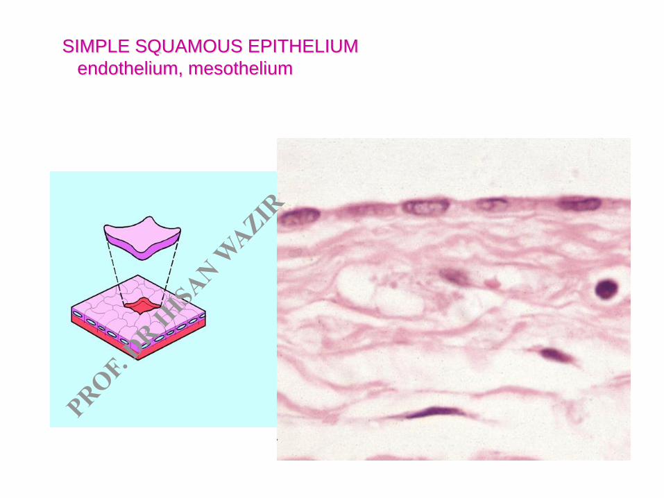
SIMPLE SQUAMOUS EPITHELIUM
endothelium, mesothelium

• In sections perpendicular to the plane of the epithelium →
• Cell are thin and fusiform or rectangular


Simple squamous epithelium

Simple cuboidal epithelium
In surface view,mosaic of
polygonal cell,
Outlines much smaller than
those of sq. epth.
Rounded nuclei in the centre
e.g Thyroid, ducts of various
glands, kidney tubules

SIMPLE CUBOIDAL EPITHELIUM
proximal and distal renal tubules


Duct


Simple cuboidal epithelium


Figure 4.4a
Simple cuboidal epithelium


Simple columnar epithelium
Cells have rectangular outlines
with their long axis
perpendicular to the basal
lamina
Height of the cells is more than
their width,
Tall and slender and, hence,
columnar in form,
Their nuclei tend to be aligned
at the same level ( usually
close to the base )
e.g. digestive tract, uterus,

Striated borderat apical surface
Basement membrane
A simple
columnar
epithelium
may have
more than
1 type cell
within it.


... with cilia


Simple columnar epithelium


STRATIFIED EPITHELIUM
Stratified Squamous epithelium
Cells on the basal lamina are
rounded (may be elongated)
Above this layer are irregularly
polyhedral cells
Becoming increasingly flattened
toward the surface,
Superficial layers they are thin
squamous cells.
Two types
Keratinized and Nonkeratinized
e.g. skin, esophagus,


Stratified squamous epithelium

Stratified squamous epithelium

Stratified Cuboidal Epithelium
Found only (typically) in the
ducts of sweat glands in the
adult and consists two layers of
cuboidal cells.
Cells of superficial layer are
usually smaller as seen in
cross section than those of the
basal layer.
Other locations (mammary
glands salivary glands
pancreas)


Figure 4.4b
Stratified Cuboidal Epithelium


Stratified Cuboidal Epithelium
Two (or more layers) of cells;
Secretes sweat; ovarian hormones & produces sperm
Found in sweat gland ducts; ovarian follicles & seminiferous
tubules

specialized epithelium = Seminiferous tubules (Germinal Epithelium)

Stratified columnar epithelium
Uncommon type of epithelium
Superficial cells are columnar and basal cells are cuboidal
One or more rows of polygonal cells may be lying between basal cells and the columnar cells
e.g. conjunctiva, pharynx, large excretory ducts of some glands
Ciliated stratified columnar epithelium → soft palate, larynx


Stratified columnar Epithelium

Pseudostratified columnar epithelium
All cells are in contact with the basal
lamina but all of them do not reach the
surface
Some cells are broad at their base but
narrow upward
Taller cells extend through the entire
thickness of epithelium, widest near the
free surface and narrow at basal lamina,
Nuclei are in wider part of cells of both
shapes,

Nuclei aligned at two levels in
the epith: creating a false
impression of cell stratification
hence,→ Pseudostratified epith:
e.g. urethra of male and in the
excretory duct of parotid gland,
Ciliated; Pseudostratified
columnar epithelium e.g. trachea
Pseudostratified columnar epithelium


Pseudostratified columnar epithelium

Transitional epithelium “Urothelium"Found in urinary tract
Appearance, in histological
sections, varies greatly depending
on its degree of distension
Empty, contracted bladder, it has
many cell layers ( usually 5 – 7)
Base have a cuboidal or low
columnar shape cells
Above, polyhedral cells, superficial
cells are very much larger, with a
characteristic rounded free surface
(dome shaped appearance)

Apical surface may appear “domed” (empty)

Transitional epithelium

Transitional Epithelium
empty bladder full bladder
from renal pelvis to
neck of urethra.


When the urinary bladder is full
(apical surface appear flattened)

Figure 4.4c
Transitional epithelium

specialized epithelium = Olfactory Epithelium